Embed Size (px)
Citation preview
Gallbladder Cancer
By Jake Lenington
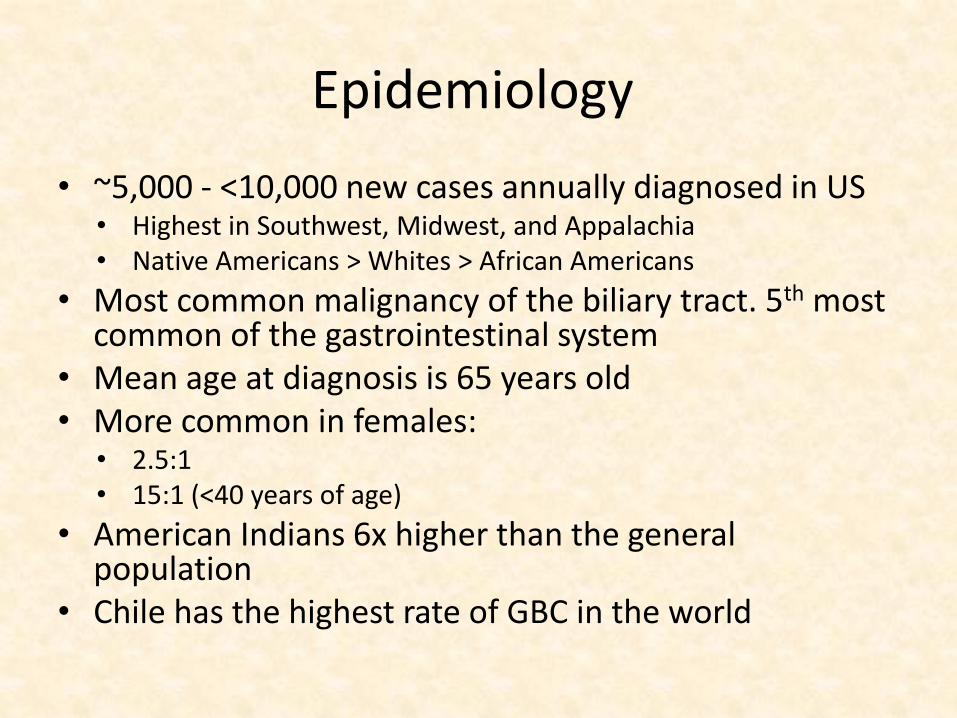
Epidemiology
• ~5,000 - <10,000 new cases annually diagnosed in US • Highest in Southwest, Midwest, and Appalachia • Native Americans > Whites > African Americans
• Most common malignancy of the biliary tract. 5th most common of the gastrointestinal system
• Mean age at diagnosis is 65 years old • More common in females:
• 2.5:1 • 15:1 (<40 years of age)
• American Indians 6x higher than the general population
• Chile has the highest rate of GBC in the world
Risk Factors
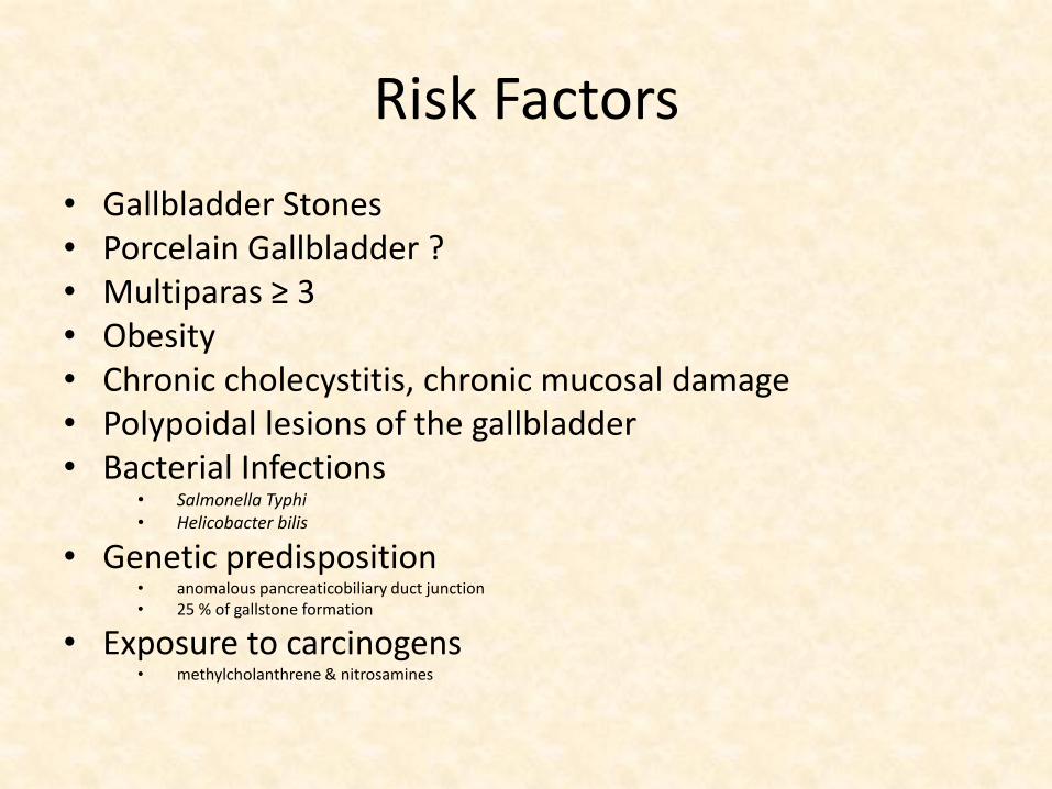
• Gallbladder Stones • Porcelain Gallbladder ? • Multiparas ≥ 3 • Obesity • Chronic cholecystitis, chronic mucosal damage • Polypoidal lesions of the gallbladder • Bacterial Infections
• Salmonella Typhi • Helicobacter bilis
• Genetic predisposition • anomalous pancreaticobiliary duct junction • 25 % of gallstone formation
• Exposure to carcinogens • methylcholanthrene & nitrosamines
Etiology – Cholelithiasis & Cholecystits
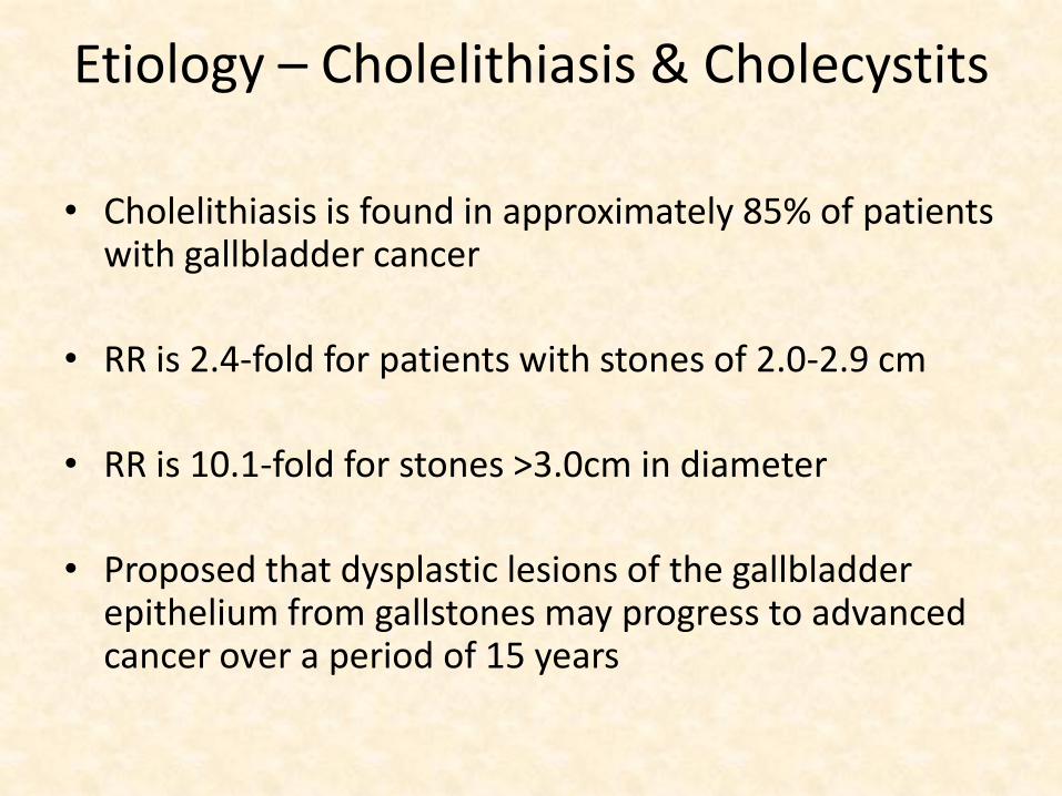
• Cholelithiasis is found in approximately 85% of patients with gallbladder cancer
• RR is 2.4-fold for patients with stones of 2.0-2.9 cm
• RR is 10.1-fold for stones >3.0cm in diameter
• Proposed that dysplastic lesions of the gallbladder epithelium from gallstones may progress to advanced cancer over a period of 15 years
Basis for cancer development in the setting of cholelithiasis
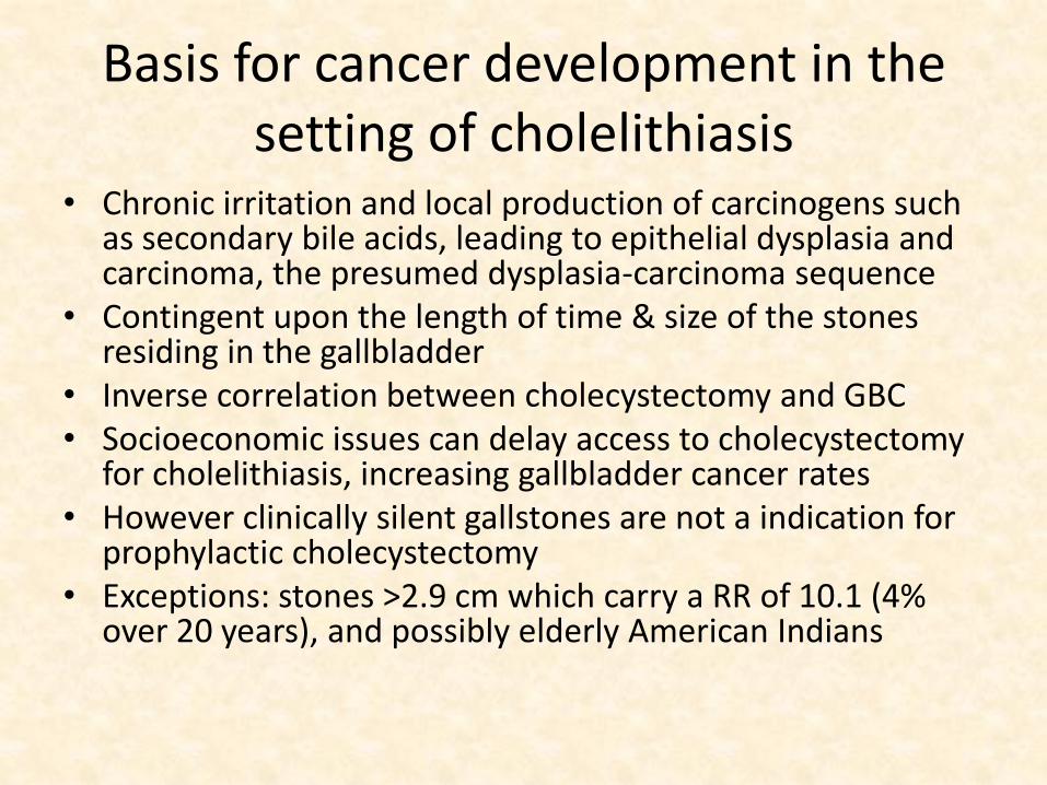
• Chronic irritation and local production of carcinogens such as secondary bile acids, leading to epithelial dysplasia and carcinoma, the presumed dysplasia-carcinoma sequence
• Contingent upon the length of time & size of the stones residing in the gallbladder
• Inverse correlation between cholecystectomy and GBC • Socioeconomic issues can delay access to cholecystectomy
for cholelithiasis, increasing gallbladder cancer rates • However clinically silent gallstones are not a indication for
prophylactic cholecystectomy • Exceptions: stones >2.9 cm which carry a RR of 10.1 (4%
over 20 years), and possibly elderly American Indians
Etiology continued – Anomalous Pancreaticobiliary Duct Junction
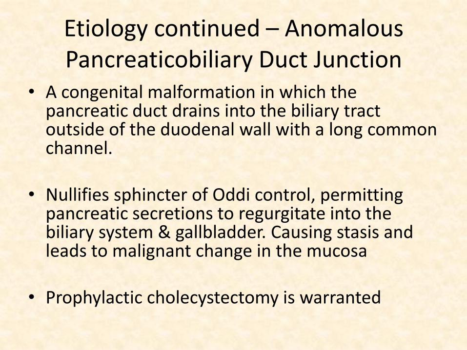
• A congenital malformation in which the pancreatic duct drains into the biliary tract outside of the duodenal wall with a long common channel.
• Nullifies sphincter of Oddi control, permitting
pancreatic secretions to regurgitate into the biliary system & gallbladder. Causing stasis and leads to malignant change in the mucosa
• Prophylactic cholecystectomy is warranted
Anomalous Pancreaticobiliary Duct Junction
• http://www.ajronline.org/content/180/1/173/F3.expansion.html
• http://download.imaging.consult.com/ic/images/S1933033207761876/gr7a-midi.jpg
• http://upload.wikimedia.org/wikipedia/commons/1/15/Gray1100.png
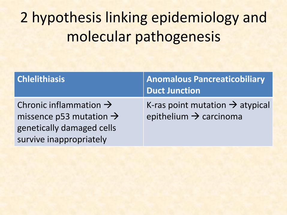
2 hypothesis linking epidemiology and molecular pathogenesis
Chlelithiasis Anomalous Pancreaticobiliary Duct Junction
Chronic inflammation missence p53 mutation genetically damaged cells survive inappropriately
K-ras point mutation atypical epithelium carcinoma
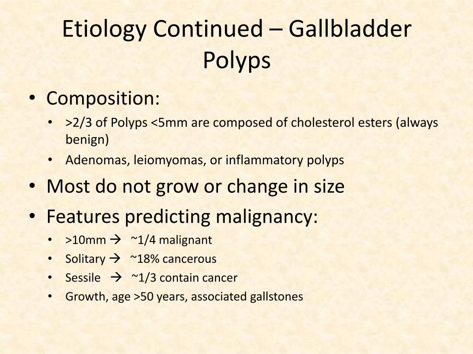
Etiology Continued – Gallbladder Polyps
• Composition: • >2/3 of Polyps <5mm are composed of cholesterol esters (always
benign)
• Adenomas, leiomyomas, or inflammatory polyps
• Most do not grow or change in size
• Features predicting malignancy: • >10mm ~1/4 malignant
• Solitary ~18% cancerous
• Sessile ~1/3 contain cancer
• Growth, age >50 years, associated gallstones
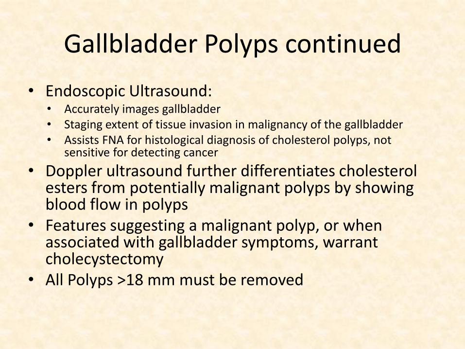
Gallbladder Polyps continued
• Endoscopic Ultrasound: • Accurately images gallbladder • Staging extent of tissue invasion in malignancy of the gallbladder • Assists FNA for histological diagnosis of cholesterol polyps, not
sensitive for detecting cancer
• Doppler ultrasound further differentiates cholesterol esters from potentially malignant polyps by showing blood flow in polyps
• Features suggesting a malignant polyp, or when associated with gallbladder symptoms, warrant cholecystectomy
• All Polyps >18 mm must be removed
Etiology continued – Porcelain Gallbladder
• Term originated from the description of a gallbladder wall extensively infiltrated with and replaced by calcium. A process resulting in a gallbladder with a fragile, brittle consistency and a blue discoloration
• The reported incidence of GBC arising from a porcelain gallbladder has ranged from 12-61%
Porcelain Gallbladder – Origin of the bad rep
• Association of calcium deposition in the wall and GBC was 1st proposed in 1951
• 1959: Bellevue Hospital pathology reports of cholecystectomy specimens from 1922-1956; 16 calcified gallbladders, 2 with carcinoma
• 1962: Argentina; 78 GBC & 26 calcified gallbladders. 16 had both calcified wall and cancer. Hence 16/26 incidence of gallbladder cancer in porcelain gallbladder
2 Pathologic types of gallbladder wall calcification
• Described by Sir William Osler, in the 1925 edition of Principles and Practice of Medicine
1) Diffuse intramural calcification
• Classic Porcelain gallbladder
2) Selective mucosal calcification
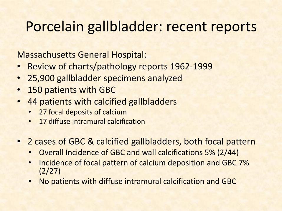
Porcelain gallbladder: recent reports
Massachusetts General Hospital: • Review of charts/pathology reports 1962-1999 • 25,900 gallbladder specimens analyzed • 150 patients with GBC • 44 patients with calcified gallbladders
• 27 focal deposits of calcium • 17 diffuse intramural calcification
• 2 cases of GBC & calcified gallbladders, both focal pattern • Overall Incidence of GBC and wall calcifications 5% (2/44) • Incidence of focal pattern of calcium deposition and GBC 7%
(2/27) • No patients with diffuse intramural calcification and GBC
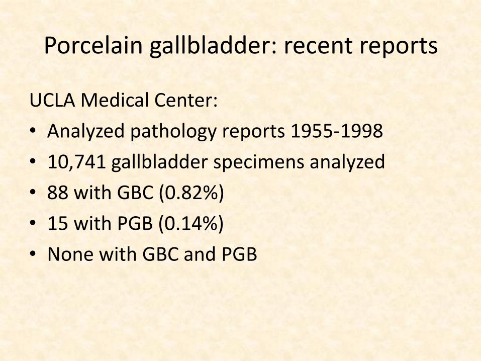
Porcelain gallbladder: recent reports
UCLA Medical Center:
• Analyzed pathology reports 1955-1998
• 10,741 gallbladder specimens analyzed
• 88 with GBC (0.82%)
• 15 with PGB (0.14%)
• None with GBC and PGB
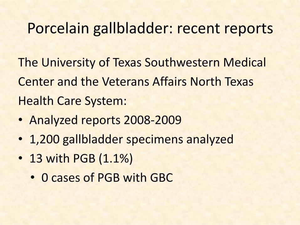
Porcelain gallbladder: recent reports
The University of Texas Southwestern Medical
Center and the Veterans Affairs North Texas
Health Care System:
• Analyzed reports 2008-2009
• 1,200 gallbladder specimens analyzed
• 13 with PGB (1.1%)
• 0 cases of PGB with GBC
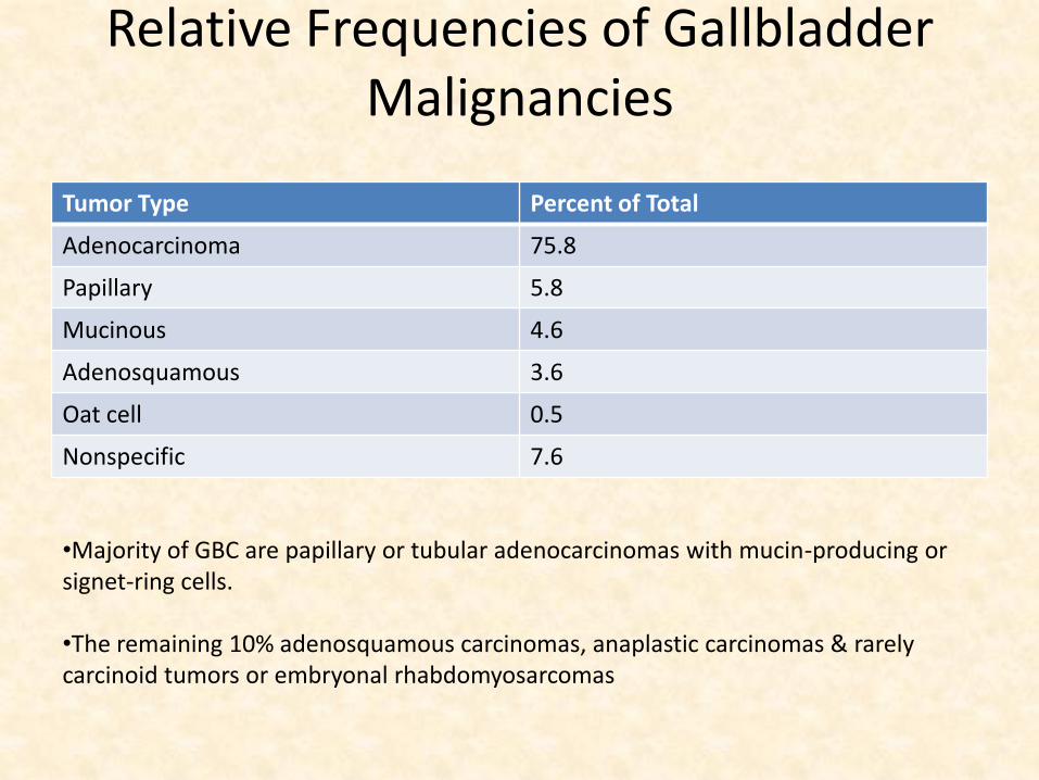
Relative Frequencies of Gallbladder Malignancies
Tumor Type Percent of Total
Adenocarcinoma 75.8
Papillary 5.8
Mucinous 4.6
Adenosquamous 3.6
Oat cell 0.5
Nonspecific 7.6
•Majority of GBC are papillary or tubular adenocarcinomas with mucin-producing or signet-ring cells. •The remaining 10% adenosquamous carcinomas, anaplastic carcinomas & rarely carcinoid tumors or embryonal rhabdomyosarcomas
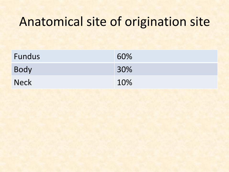
Anatomical site of origination site
Fundus 60%
Body 30%
Neck 10%
Clinical Presentation and GBC discovery
• Similar signs and symptoms as seen in cholelithiasis & cholecystitis • Nonspecific right upper quadrant abdominal pain and tenderness
• Primary GBC is incidentally found in 0.3-3% of laparoscopic cholecystectomies performed for cholelithiasis • Frequency dependent on regional prevalence
• Late, when tumor has invaded surrounding structures or metastasized. Most common • Anorexia and jaundice are indicative of advanced disease
General Histology
From outside inwards: Four layers, serous, perimuscular, muscular, and mucosa. 1) Serous coat: Nothing but the derivations of the peritoneal layer, thus all the
places of the gall bladder covered with the peritoneum are under this, and covers them partially. It is actually not absolutely complete, and covers only the entire fundus and also some of the portions of the body as well as the neck.
1) The perimuscular layer: Connective tissue, along with the arteries, veins, nerves
and the lymphatics. 1) Muscular layer: Smooth muscle fibers. Space between muscle which
are filled with the reticular, elastic, or collagenous fibres and fibroblasts.
4) Mucous membrane: Devoid of the muscularis mucosae. Single columnar epithelium. No glands with the exception of some mucus glands at the neck
www.gall-bladder.info/histology.html
AJCC Cancer Staging Manual, Seventh Edition (2010)
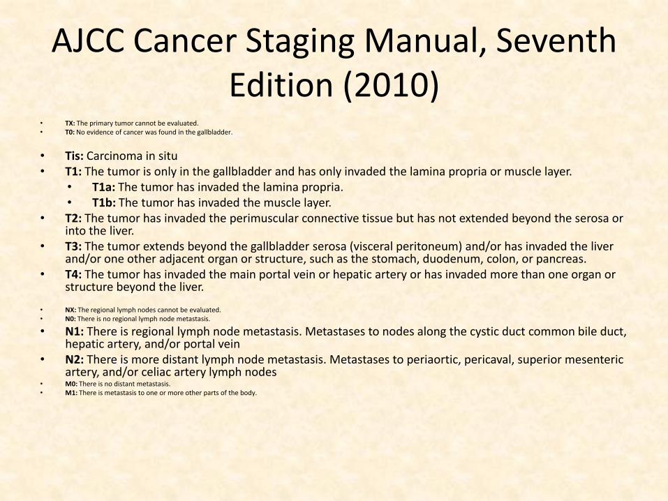
• TX: The primary tumor cannot be evaluated. • T0: No evidence of cancer was found in the gallbladder.
• Tis: Carcinoma in situ • T1: The tumor is only in the gallbladder and has only invaded the lamina propria or muscle layer.
• T1a: The tumor has invaded the lamina propria. • T1b: The tumor has invaded the muscle layer.
• T2: The tumor has invaded the perimuscular connective tissue but has not extended beyond the serosa or into the liver.
• T3: The tumor extends beyond the gallbladder serosa (visceral peritoneum) and/or has invaded the liver and/or one other adjacent organ or structure, such as the stomach, duodenum, colon, or pancreas.
• T4: The tumor has invaded the main portal vein or hepatic artery or has invaded more than one organ or structure beyond the liver.
• NX: The regional lymph nodes cannot be evaluated. • N0: There is no regional lymph node metastasis.
• N1: There is regional lymph node metastasis. Metastases to nodes along the cystic duct common bile duct, hepatic artery, and/or portal vein
• N2: There is more distant lymph node metastasis. Metastases to periaortic, pericaval, superior mesenteric artery, and/or celiac artery lymph nodes
• M0: There is no distant metastasis. • M1: There is metastasis to one or more other parts of the body.
AJCC Cancer Staging Manual, Seventh Edition (2010)
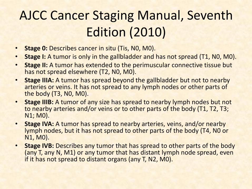
• Stage 0: Describes cancer in situ (Tis, N0, M0). • Stage I: A tumor is only in the gallbladder and has not spread (T1, N0, M0). • Stage II: A tumor has extended to the perimuscular connective tissue but
has not spread elsewhere (T2, N0, M0). • Stage IIIA: A tumor has spread beyond the gallbladder but not to nearby
arteries or veins. It has not spread to any lymph nodes or other parts of the body (T3, N0, M0).
• Stage IIIB: A tumor of any size has spread to nearby lymph nodes but not to nearby arteries and/or veins or to other parts of the body (T1, T2, T3; N1; M0).
• Stage IVA: A tumor has spread to nearby arteries, veins, and/or nearby lymph nodes, but it has not spread to other parts of the body (T4, N0 or N1, M0).
• Stage IVB: Describes any tumor that has spread to other parts of the body (any T, any N, M1) or any tumor that has distant lymph node spread, even if it has not spread to distant organs (any T, N2, M0).
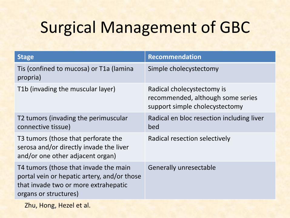
Surgical Management of GBC
Stage Recommendation
Tis (confined to mucosa) or T1a (lamina propria)
Simple cholecystectomy
T1b (invading the muscular layer) Radical cholecystectomy is recommended, although some series support simple cholecystectomy
T2 tumors (invading the perimuscular connective tissue)
Radical en bloc resection including liver bed
T3 tumors (those that perforate the serosa and/or directly invade the liver and/or one other adjacent organ)
Radical resection selectively
T4 tumors (those that invade the main portal vein or hepatic artery, and/or those that invade two or more extrahepatic organs or structures)
Generally unresectable
Zhu, Hong, Hezel et al.

Surgical Treatment
• Surgical resection of the primary tumor is the only potentially curative therapy
• However only 25-50% of patients are resectable at presentation
Chemotherapy in GBC
• Adjuvant chemotherapy (alone) following surgery with curative intent
• Several small studies suggesting some benefit from adjuvant chemotherapy
• However there are no randomized phase III clinical trial data to support a standard adjuvant regimen (NCCN)
Chemotherapy for locally advanced or metastatic GBC that is unresectable
• Median survival of patients with advanced disease is in the range of only a few months
• NCCN Chemotheraputic Options: • Gemcitabine/cisplatin combination therapy (category 1)
• Fluoropyrimidine-based or other gemcitabine-based chemotherapy regimen
Chemotherapy for locally advanced or metastatic GBC that is unresectable
• Eckel and Schmid: Pooled analysis of chemotherapy in biliary tract cancer
• 104 trails analyzed, 1985-2006
• Total of 2,810 patients treated
• Subgroup analysis focused on cytotoxic agents: • Fluoropryimidines
• Gemcitabine
• Platinum compounds
Chemotherapy for locally advanced or metastatic GBC that is unresectable
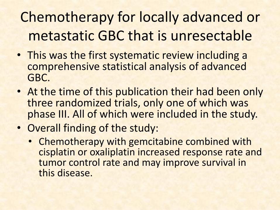
• This was the first systematic review including a comprehensive statistical analysis of advanced GBC.
• At the time of this publication their had been only three randomized trials, only one of which was phase III. All of which were included in the study.
• Overall finding of the study: • Chemotherapy with gemcitabine combined with
cisplatin or oxaliplatin increased response rate and tumor control rate and may improve survival in this disease.
Chemotherapy for locally advanced or metastatic GBC that is unresectable
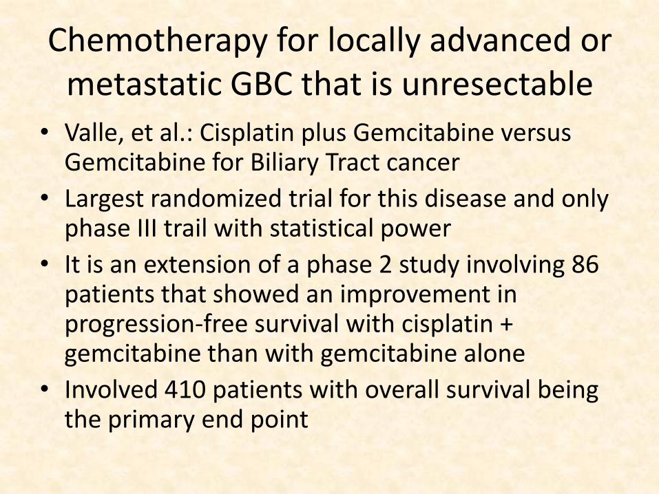
• Valle, et al.: Cisplatin plus Gemcitabine versus Gemcitabine for Biliary Tract cancer
• Largest randomized trial for this disease and only phase III trail with statistical power
• It is an extension of a phase 2 study involving 86 patients that showed an improvement in progression-free survival with cisplatin + gemcitabine than with gemcitabine alone
• Involved 410 patients with overall survival being the primary end point
Chemotherapy for locally advanced or metastatic GBC that is unresectable
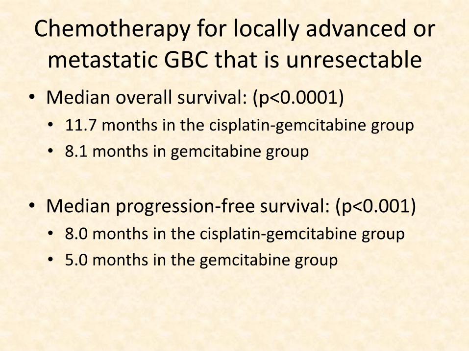
• Median overall survival: (p<0.0001)
• 11.7 months in the cisplatin-gemcitabine group
• 8.1 months in gemcitabine group
• Median progression-free survival: (p<0.001)
• 8.0 months in the cisplatin-gemcitabine group
• 5.0 months in the gemcitabine group
Adjuvant Therapy in GBC
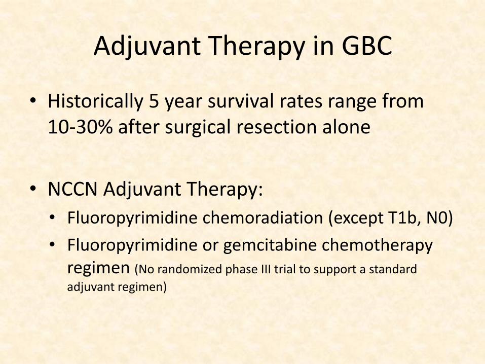
• Historically 5 year survival rates range from 10-30% after surgical resection alone
• NCCN Adjuvant Therapy:
• Fluoropyrimidine chemoradiation (except T1b, N0)
• Fluoropyrimidine or gemcitabine chemotherapy regimen (No randomized phase III trial to support a standard
adjuvant regimen)
Adjuvant Therapy in GBC
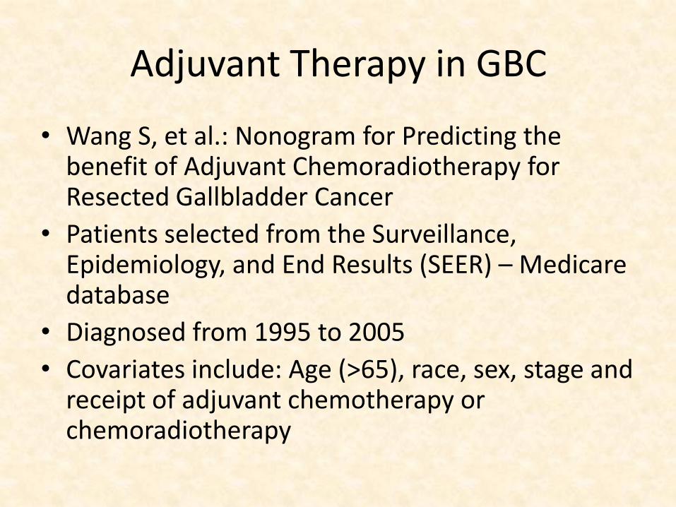
• Wang S, et al.: Nonogram for Predicting the benefit of Adjuvant Chemoradiotherapy for Resected Gallbladder Cancer
• Patients selected from the Surveillance, Epidemiology, and End Results (SEER) – Medicare database
• Diagnosed from 1995 to 2005
• Covariates include: Age (>65), race, sex, stage and receipt of adjuvant chemotherapy or chemoradiotherapy
Adjuvant Therapy in GBC
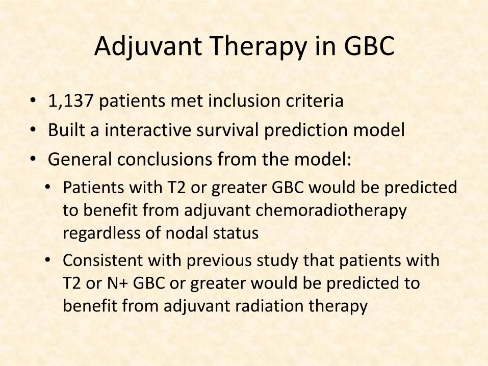
• 1,137 patients met inclusion criteria
• Built a interactive survival prediction model
• General conclusions from the model:
• Patients with T2 or greater GBC would be predicted to benefit from adjuvant chemoradiotherapy regardless of nodal status
• Consistent with previous study that patients with T2 or N+ GBC or greater would be predicted to benefit from adjuvant radiation therapy
Adjuvant Therapy in GBC
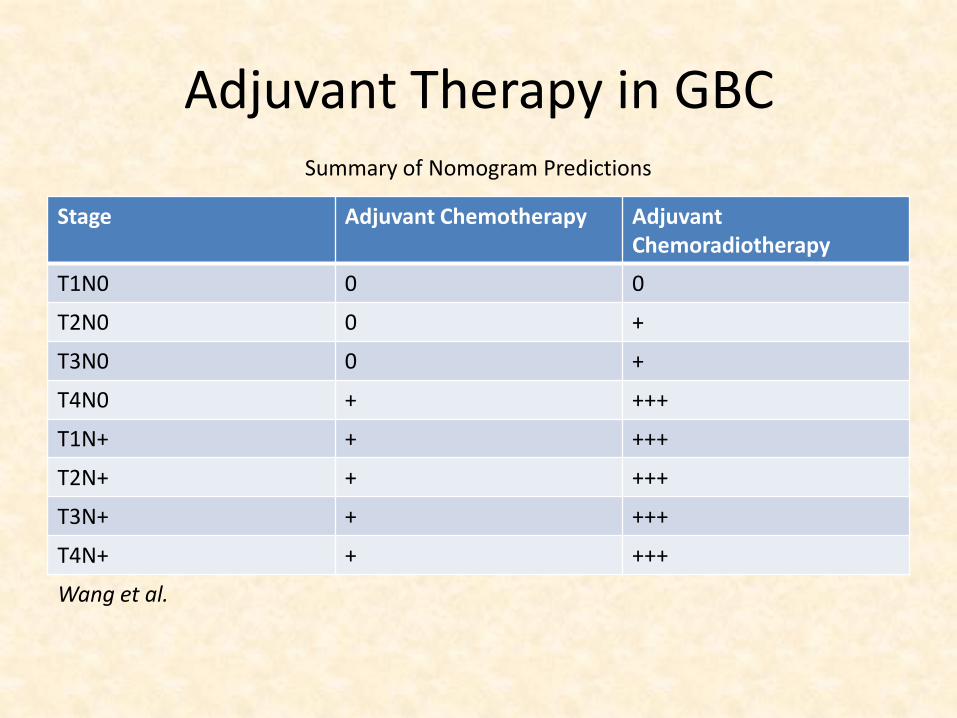
Stage Adjuvant Chemotherapy Adjuvant Chemoradiotherapy
T1N0 0 0
T2N0 0 +
T3N0 0 +
T4N0 + +++
T1N+ + +++
T2N+ + +++
T3N+ + +++
T4N+ + +++
Wang et al.
Summary of Nomogram Predictions
Adjuvant chemoradiotherapy in GBC
• Duke University medical center:
• 22 patients with primary nonmetastatic GBC treated with radiation therapy after surgical resection. 18 received 5-fluorouracil chemotherapy (primarily T3/4 and/or N+)
• 5 year survival was 37%. Median survival was 1.9 years
Adjuvant chemoradiotherapy in GBC
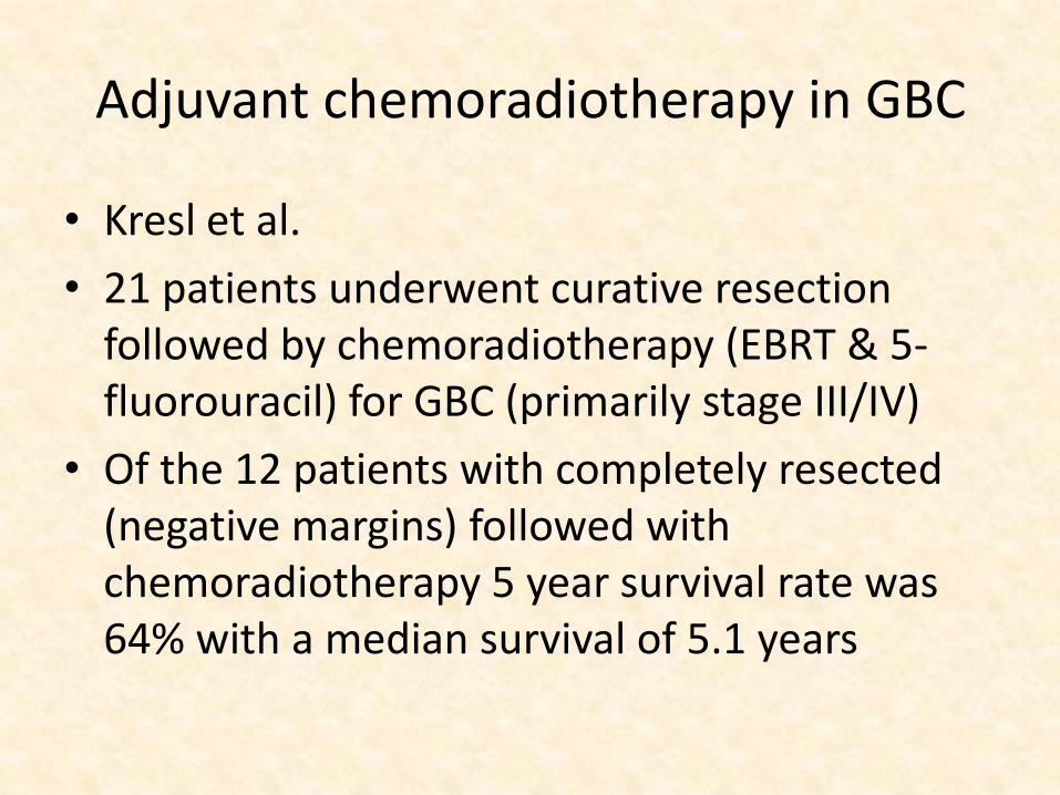
• Kresl et al.
• 21 patients underwent curative resection followed by chemoradiotherapy (EBRT & 5-fluorouracil) for GBC (primarily stage III/IV)
• Of the 12 patients with completely resected (negative margins) followed with chemoradiotherapy 5 year survival rate was 64% with a median survival of 5.1 years
Adjuvant chemoradiotherapy in GBC
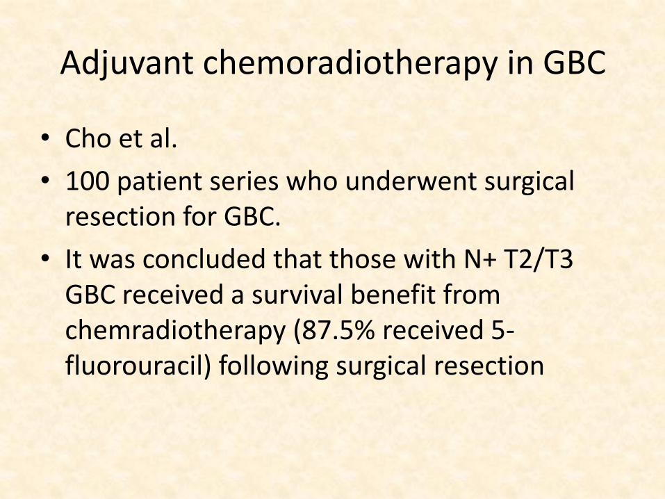
• Cho et al.
• 100 patient series who underwent surgical resection for GBC.
• It was concluded that those with N+ T2/T3 GBC received a survival benefit from chemradiotherapy (87.5% received 5-fluorouracil) following surgical resection
Adjuvant Radiation therapy in GBC
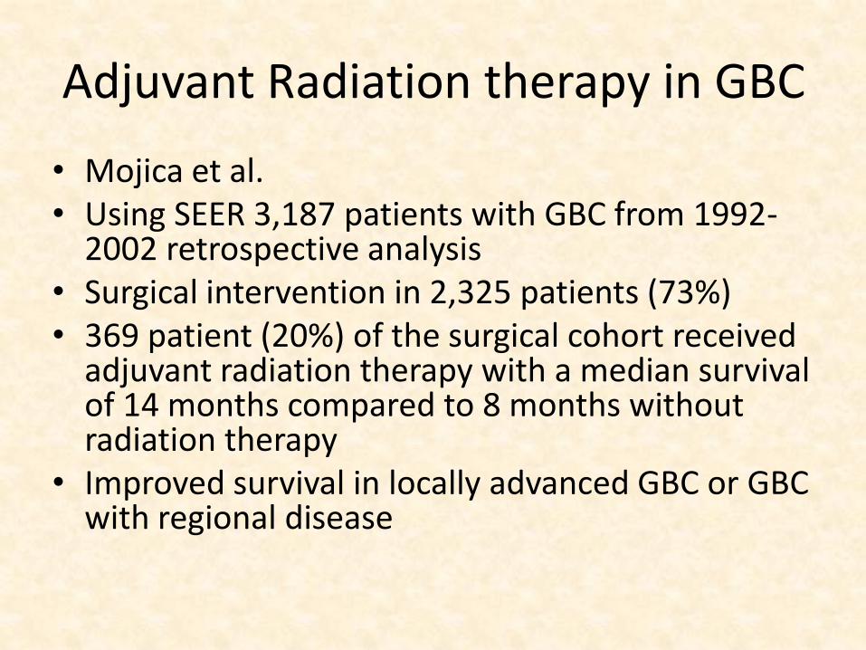
• Mojica et al. • Using SEER 3,187 patients with GBC from 1992-
2002 retrospective analysis • Surgical intervention in 2,325 patients (73%) • 369 patient (20%) of the surgical cohort received
adjuvant radiation therapy with a median survival of 14 months compared to 8 months without radiation therapy
• Improved survival in locally advanced GBC or GBC with regional disease
References
1) Cassier P, Thevenet C, Walter T, et al.: Outcome of patients receiving chemotherapy for advanced biliary tract or gallbladder carcinoma. European Journal of Gastroenteology & Hepatology 2010;22:1111-1117
2) Cho S, Kim S, Park SJ, et al.: Adjuvant chemoradiation therapy in gallbladder cancer. Journal of Surgical Oncology 2010;102:87-93
3) Czito B, Hurwitz H, Clough R, et al.: Adjuvant external –beam radiotherapy with concurrent chemotherapy after resection of primary gallbladder carcinoma: A 23-Year experience. Int. J. Radiation Oncology Biol. Phys 2005;64:1030-1034
4) Eckel F, Schmid RM.: Chemotherapy In advance biliary tract carcinoma: a pooled analysis of clinical trials. British Journal of Cancer 2007;96:896-902
5) Khan Z, Livingston E, Huerta S.: Reassessing the need for prophylactic surgery in patients with porcelain gallbladder. Archives Of Surgery 2011;146:1143-1147
6) Kim MJ, Oh DY, Lee SH, et al.: Gemcitabine-based versusfluoropyrimidine-based chemotherapy with or without platinum in unresectable biliary tract cancer: a retrospective study. BMC Cancer 2008;8:1471-2407
7) Kresl J, Schild S, Henning G, et al.: Adjuvant external beam radiation therapy with concurrent chemotherapy in the management of gallbladder carcinoma. Int. J. Radiation Oncology Biol. Phys. 2002;52:167-175

References
8) Mojica P, Smith D, Ellenhorn J.: Adjuvant radiation therapy is associated with improved survival for gallbladder carcinoma with regional metastatic disease. Journal of Surgical Oncology 2007;96:8-13
9) Shaffer E.: Gallbladder cancer. Gastroenterology & Hepatology 2008;4:737-741 10) Sheth S, Bedford A, Chopra S.: Primary gallbladder cancer: recognition of risk factors and the role
of prophylactic cholecystectomy. The American Journal Of Gastroenterology 2000;95:1402-1410 11) Stephen A, Berger D.: Carcinoma in the porcelain gallbladder: A relationship revisited. Surgery
2001;129:699-703 12) Valle J, Wasan H, Palmer D, et al.: Cisplatin plus gemcitabine versus gemcitabine for biliary tract
cancer. The New England Journal of Medicine 2010;362:1273-81 13) Verslype C, Prenen H, Van-Cutsem E.: The role of chemotherapy in biliary tract carcinoma. HPB
2008;10:164-167 14) Wang S, Lemieux A, Kalpathy-Cramer J, et al.: Nomogram for predicting the benefit of adjuvant
chemoradiotherapy for resected gallbladder cancer. Journal Of Clinical Oncology 2011;Published ahead of Print on November 7
15) Zhu A, Hong T, Hezel A, et al.: Current management of gallbladder carcinoma. The Oncologist Hepatobiliary 2010;15:168-181
16) Access Medicine. Carcinomas of the Gallbladder and biliary tract. The MD Anderson Manual of Medical Oncology, Chapter 19. Hepatobilliary Malignancies
17) NCCN Guidelines Version, Inc 2011. Seventh Edition


![Chronic Cholecystitis which Mimics Gallbladder Cancer: a ......malignant gallbladder disorders from benign ones [1-3]. We describe a case of chronic cholecystitis that showed focal](https://img.pdfslide.net/doc/110x75/5e9edb35d364e168286b9adc/chronic-cholecystitis-which-mimics-gallbladder-cancer-a-malignant-gallbladder.jpg)