Embed Size (px)
Citation preview
Gastrointestinal Bleeding
Rajeev Jain, M.D.
GI Bleeding
• Clinical Presentation
• Acute Upper GI Bleed
• Acute Lower GI Bleed
Core Principles in GI Bleeding Management
• Assessment and stabilization of hemodynamic status
• Determine the source of bleeding
• Stop active bleeding
• Treatment of underlying abnormality
• Prevent recurrent bleeding
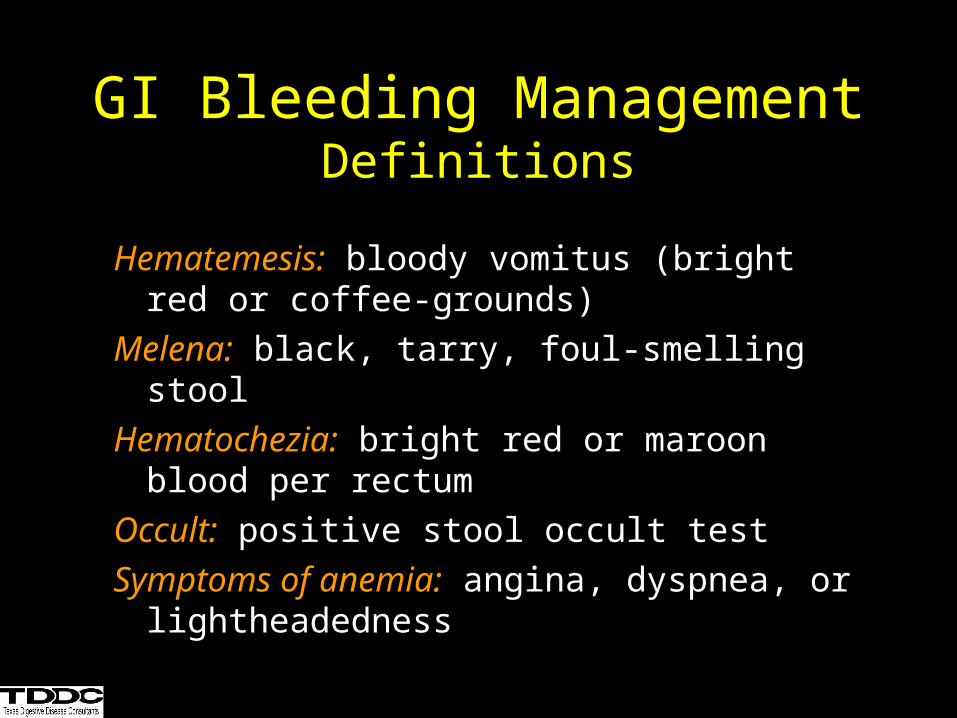
GI Bleeding ManagementDefinitions
Hematemesis: bloody vomitus (bright red or coffee-grounds)
Melena: black, tarry, foul-smelling stool
Hematochezia: bright red or maroon blood per rectum
Occult: positive stool occult test
Symptoms of anemia: angina, dyspnea, or lightheadedness
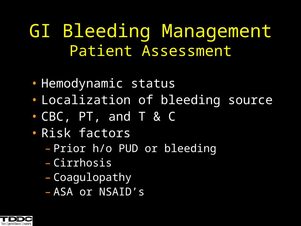
GI Bleeding ManagementPatient Assessment
• Hemodynamic status• Localization of bleeding source• CBC, PT, and T & C• Risk factors
– Prior h/o PUD or bleeding– Cirrhosis– Coagulopathy– ASA or NSAID’s
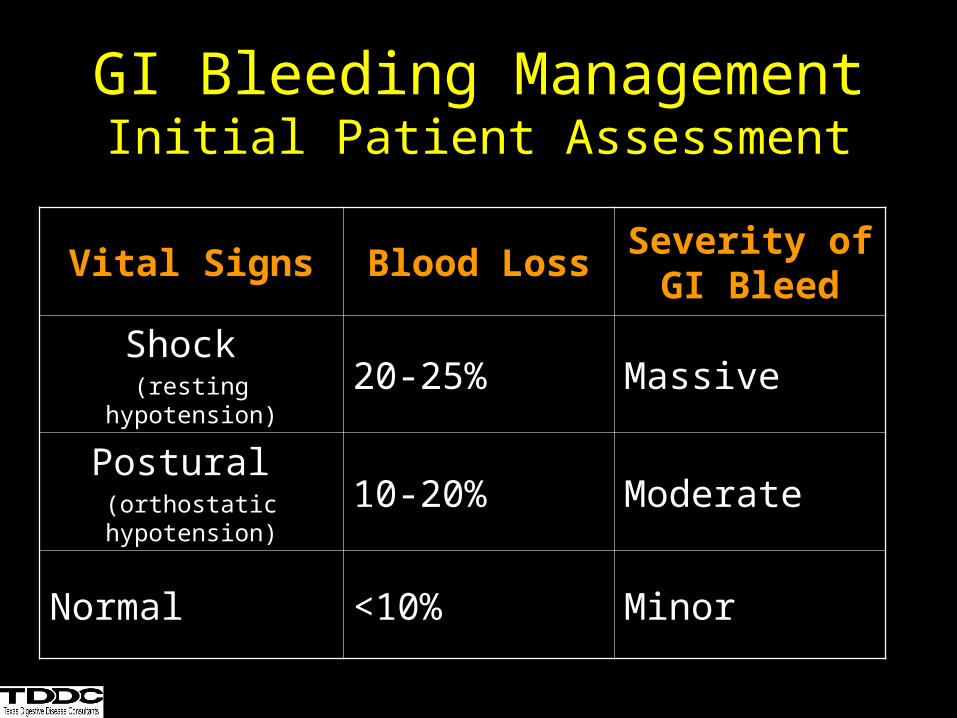
GI Bleeding ManagementInitial Patient Assessment
Vital Signs Blood LossSeverity of GI
Bleed
Shock (resting hypotension)
20-25% Massive
Postural (orthostatic hypotension)
10-20% Moderate
Normal <10% Minor
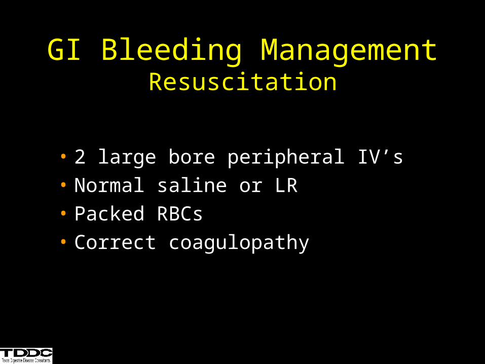
GI Bleeding ManagementResuscitation
• 2 large bore peripheral IV’s
• Normal saline or LR
• Packed RBCs
• Correct coagulopathy
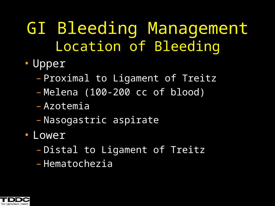
GI Bleeding ManagementLocation of Bleeding
• Upper– Proximal to Ligament of Treitz– Melena (100-200 cc of blood)– Azotemia– Nasogastric aspirate
• Lower– Distal to Ligament of Treitz– Hematochezia
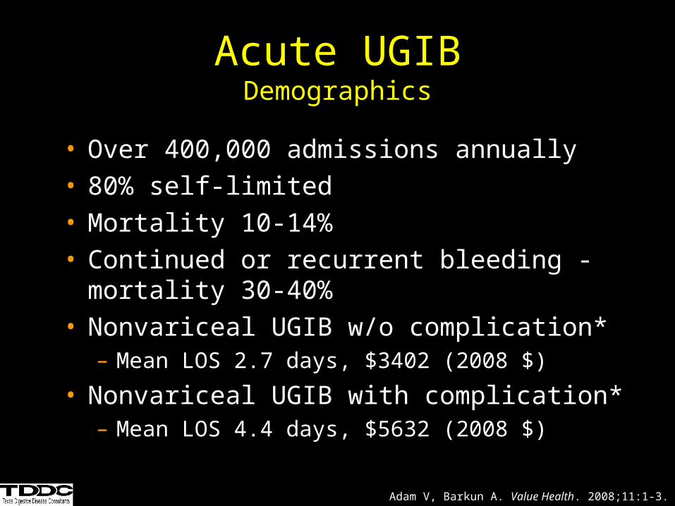
Acute UGIBDemographics
• Over 400,000 admissions annually• 80% self-limited• Mortality 10-14%• Continued or recurrent bleeding - mortality 30-
40%• Nonvariceal UGIB w/o complication*
– Mean LOS 2.7 days, $3402 (2008 $)
• Nonvariceal UGIB with complication*– Mean LOS 4.4 days, $5632 (2008 $)
Adam V, Barkun A. Value Health. 2008;11:1-3.
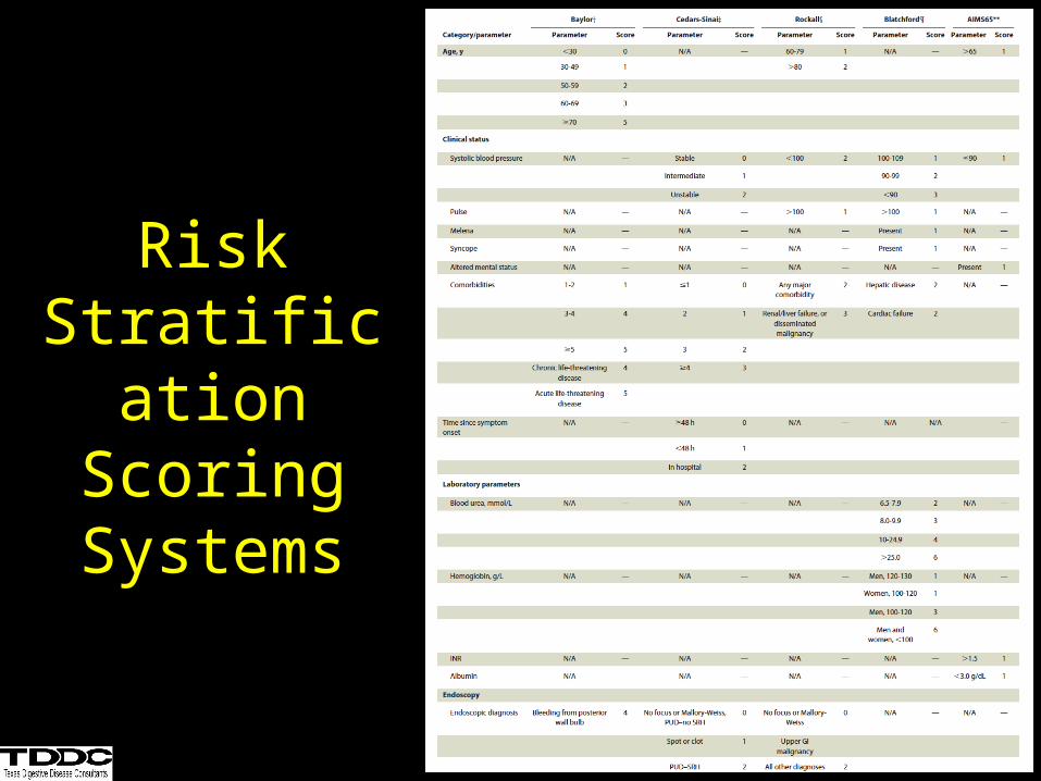
Risk Stratification
Scoring Systems
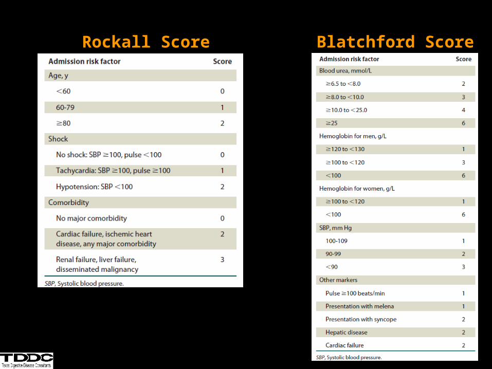
Blatchford ScoreRockall Score
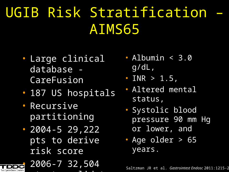
UGIB Risk Stratification – AIMS65
• Albumin < 3.0 g/dL, • INR > 1.5, • Altered mental
status, • Systolic blood
pressure 90 mm Hg or lower, and
• Age older > 65 years.
Saltzman JR et al. Gastrointest Endosc 2011:1215-22.
• Large clinical database - CareFusion
• 187 US hospitals• Recursive partitioning • 2004-5 29,222 pts to
derive risk score• 2006-7 32,504 pts to
validate
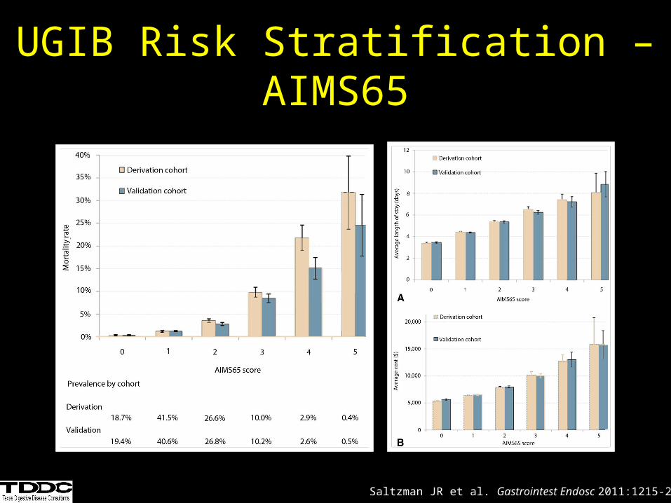
UGIB Risk Stratification – AIMS65
Saltzman JR et al. Gastrointest Endosc 2011:1215-22.
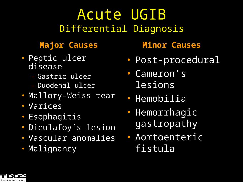
• Peptic ulcer disease– Gastric ulcer– Duodenal ulcer
• Mallory-Weiss tear• Varices• Esophagitis• Dieulafoy’s lesion• Vascular anomalies• Malignancy
• Post-procedural• Cameron’s lesions• Hemobilia• Hemorrhagic
gastropathy• Aortoenteric fistula
Acute UGIBDifferential Diagnosis
Major Causes Minor Causes
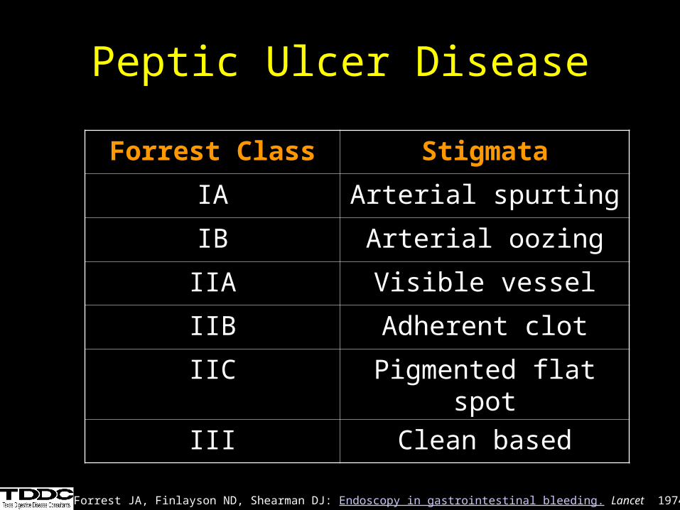
Peptic Ulcer Disease
Forrest Class Stigmata
IA Arterial spurting
IB Arterial oozing
IIA Visible vessel
IIB Adherent clot
IIC Pigmented flat spot
III Clean based
Forrest JA, Finlayson ND, Shearman DJ: Endoscopy in gastrointestinal bleeding. Lancet 1974; 2:394-7
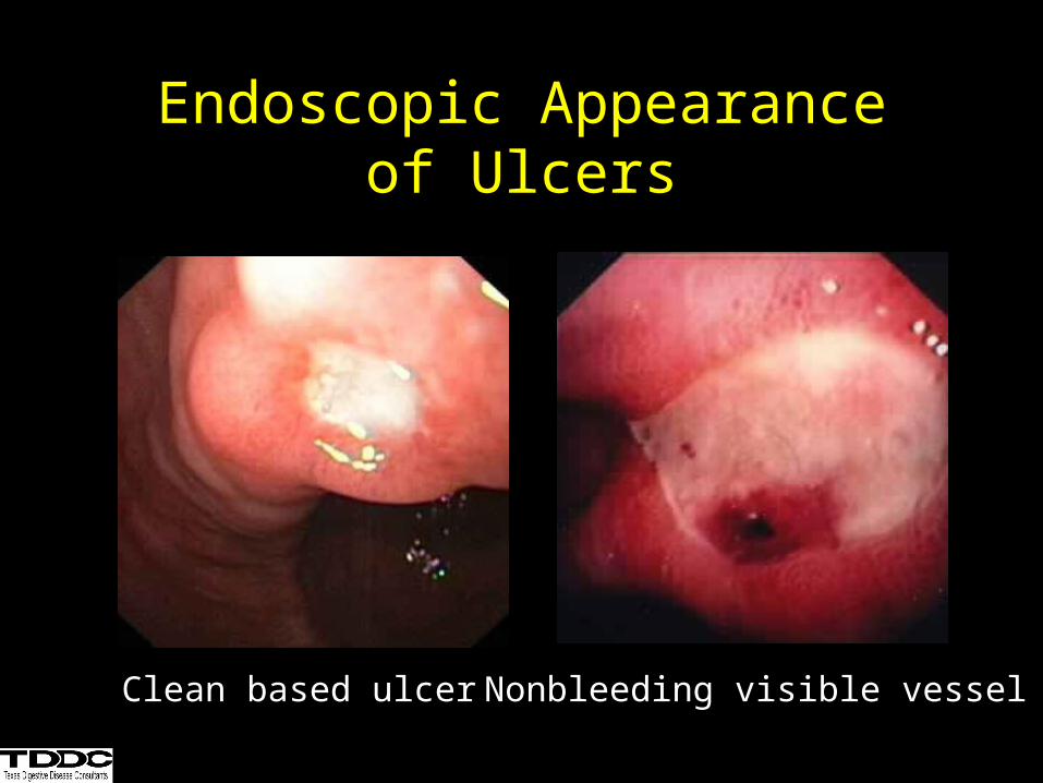
Endoscopic Appearanceof Ulcers
Clean based ulcer Nonbleeding visible vessel
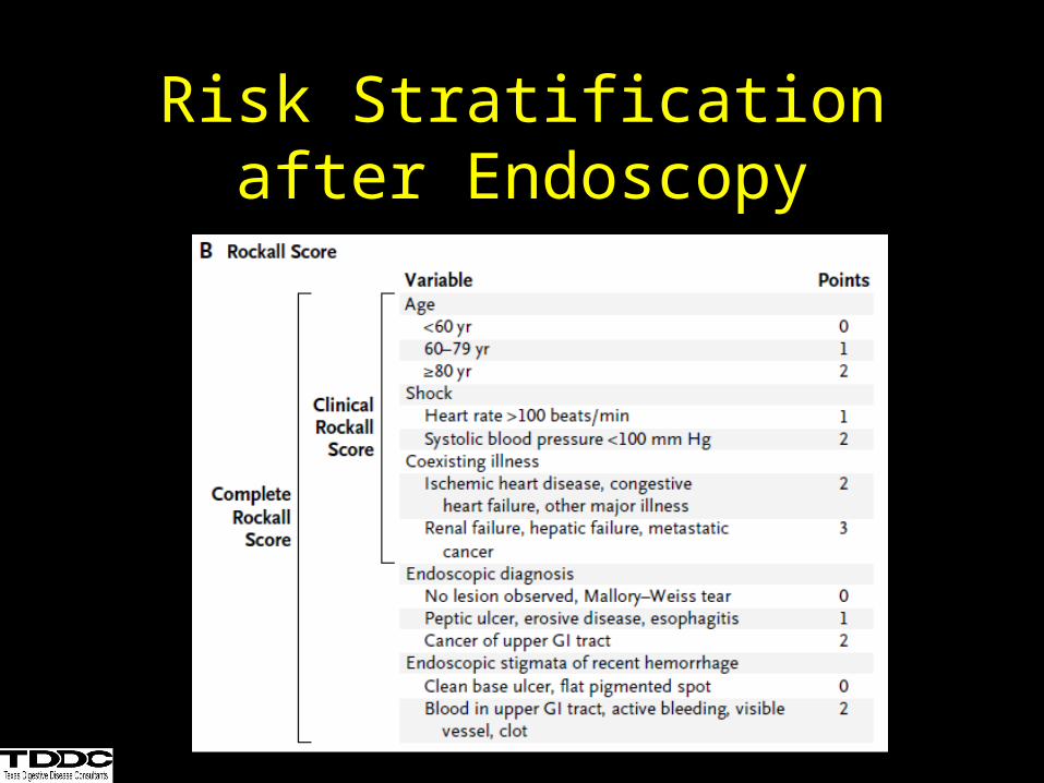
Risk Stratification after Endoscopy
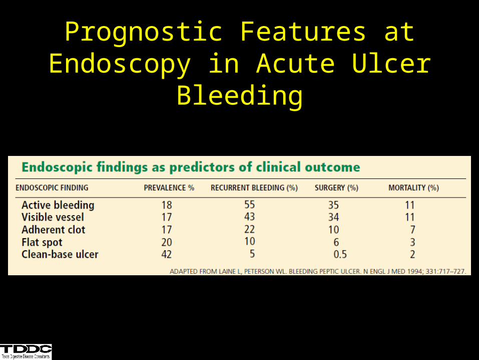
Prognostic Features at Endoscopy in Acute Ulcer Bleeding
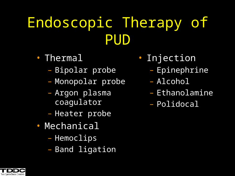
• Thermal– Bipolar probe– Monopolar probe– Argon plasma
coagulator– Heater probe
• Mechanical– Hemoclips– Band ligation
• Injection– Epinephrine– Alcohol– Ethanolamine– Polidocal
Endoscopic Therapy of PUD
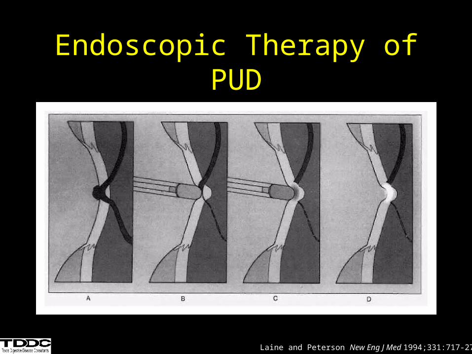
Endoscopic Therapy of PUD
Laine and Peterson New Eng J Med 1994;331:717-27.
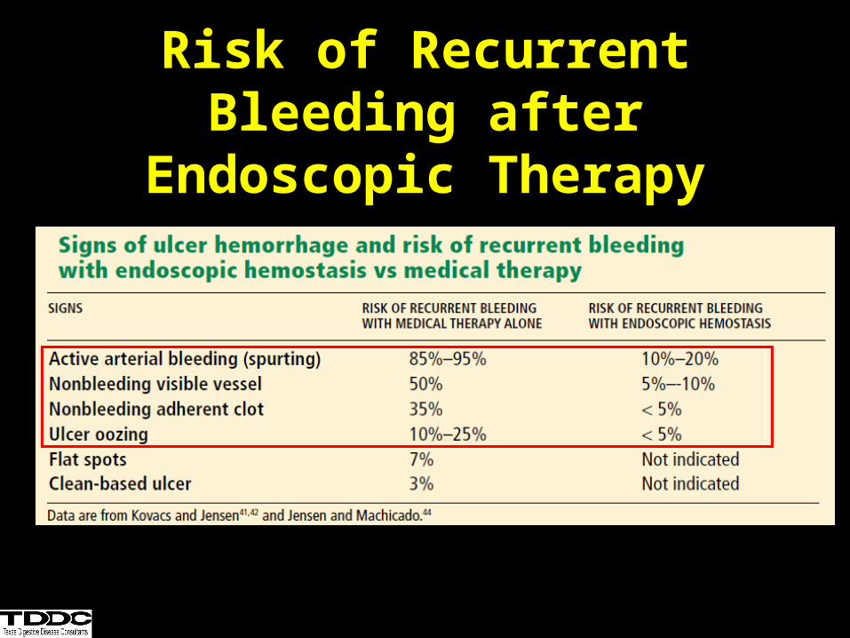
Risk of Recurrent Bleeding after Endoscopic Therapy
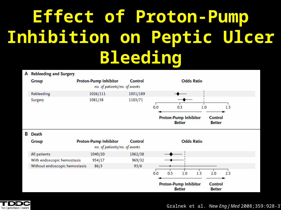
Effect of Proton-Pump Inhibition on Peptic Ulcer Bleeding
Gralnek et al. New Eng J Med 2008;359:928-37.
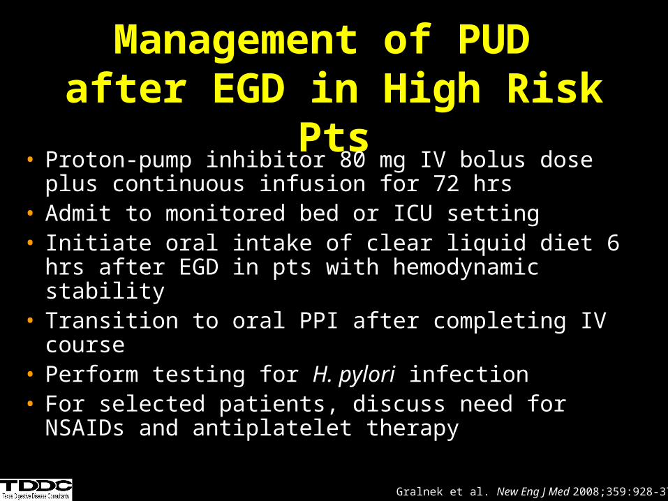
Management of PUD after EGD in High Risk Pts
• Proton-pump inhibitor 80 mg IV bolus dose plus continuous infusion for 72 hrs
• Admit to monitored bed or ICU setting• Initiate oral intake of clear liquid diet 6 hrs after EGD
in pts with hemodynamic stability• Transition to oral PPI after completing IV course• Perform testing for H. pylori infection• For selected patients, discuss need for NSAIDs and
antiplatelet therapy
Gralnek et al. New Eng J Med 2008;359:928-37.
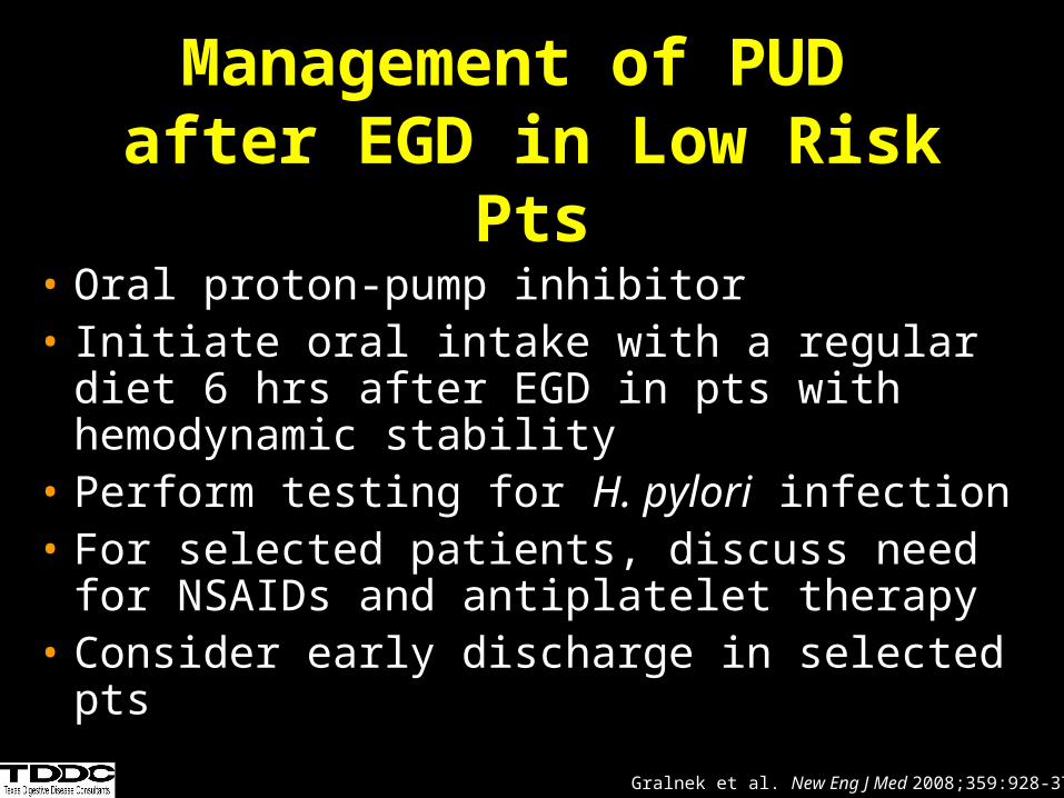
Management of PUD after EGD in Low Risk Pts
• Oral proton-pump inhibitor• Initiate oral intake with a regular diet 6 hrs
after EGD in pts with hemodynamic stability• Perform testing for H. pylori infection• For selected patients, discuss need for
NSAIDs and antiplatelet therapy• Consider early discharge in selected pts
Gralnek et al. New Eng J Med 2008;359:928-37.
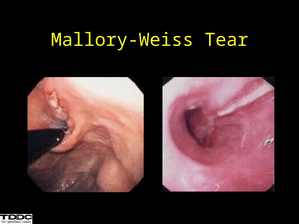
Mallory-Weiss Tear
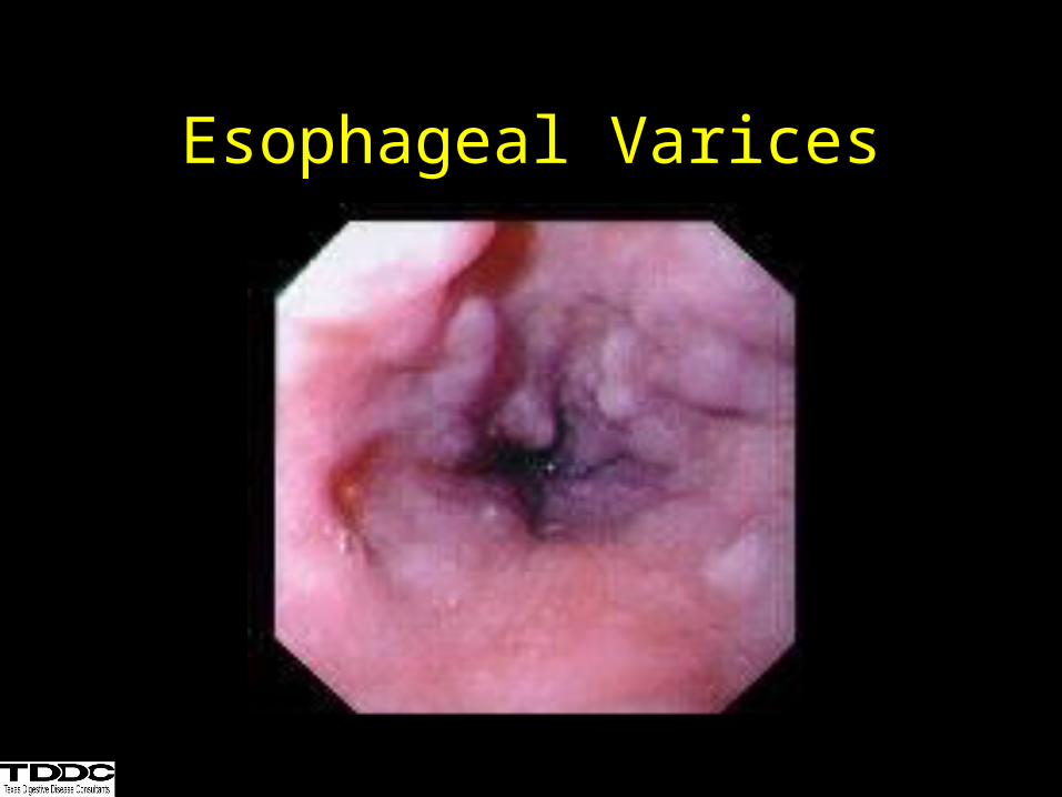
Esophageal Varices
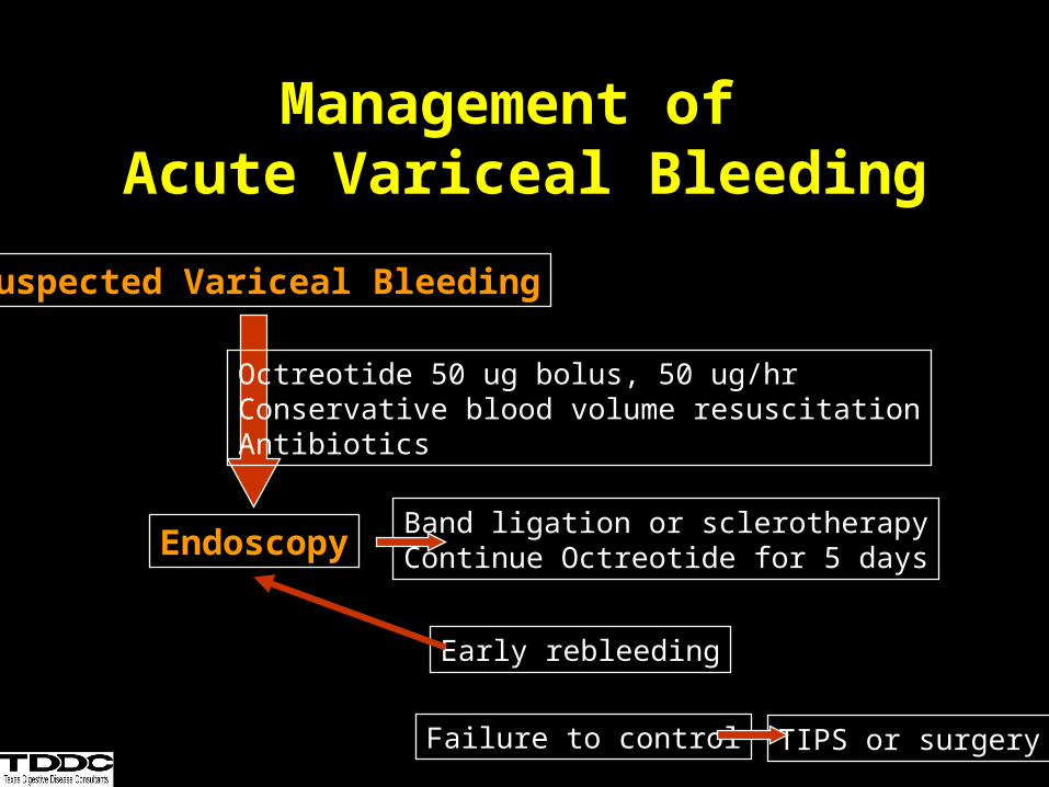
Management of Acute Variceal Bleeding
Suspected Variceal Bleeding
EndoscopyBand ligation or sclerotherapyContinue Octreotide for 5 days
Early rebleeding
Failure to control TIPS or surgery
Octreotide 50 ug bolus, 50 ug/hrConservative blood volume resuscitationAntibiotics
Antibiotic Prophylaxis in GI Bleeding in Cirrhotic Patients
• Fluoroquinolones or amoxicillin + clavulinic acid
• Meta-analysis 1
– Decrease rates of infection• SBP, bacteremia
– Increased short-term survival
• RCT 2
– Reduction in early rebleeding
1.Bernard et al.Hepatology. 29(6):1655-61.1999.2.Hou et al. Hepatology. 39(3):746-53.2004.
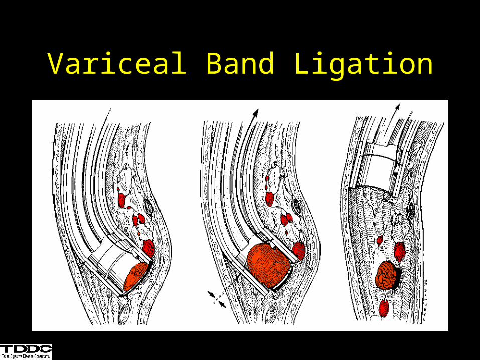
Variceal Band Ligation
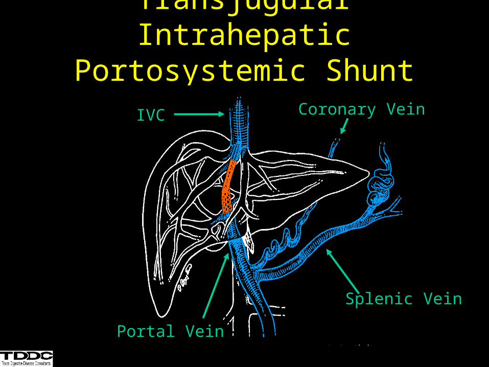
Transjugular Intrahepatic Portosystemic Shunt (TIPS)
IVC
Portal Vein
Splenic Vein
Coronary Vein
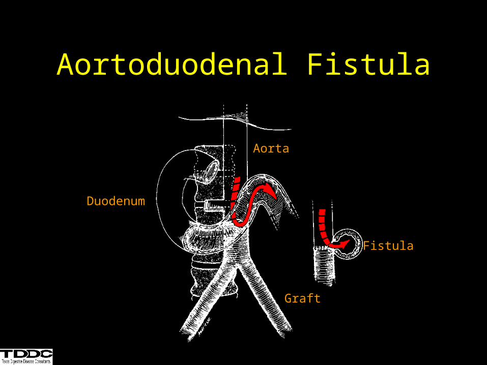
Aortoduodenal Fistula
Aorta
Duodenum
Graft
Fistula
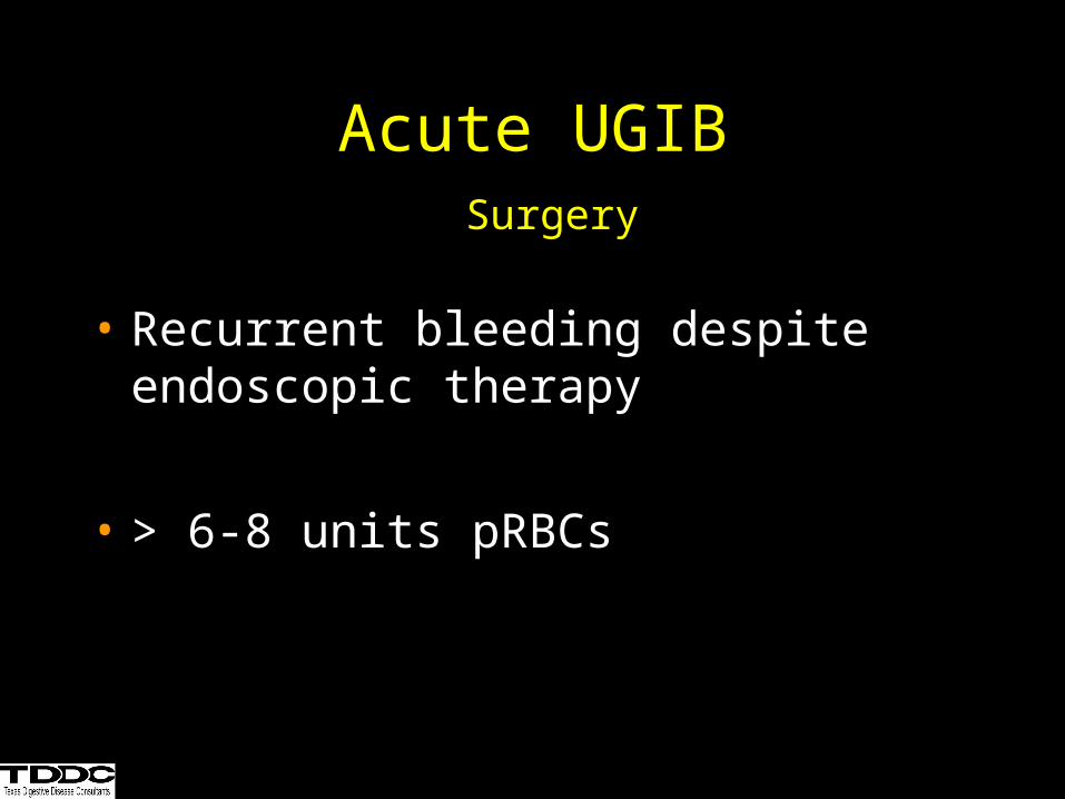
Acute UGIB Surgery
• Recurrent bleeding despite endoscopic therapy
• > 6-8 units pRBCs
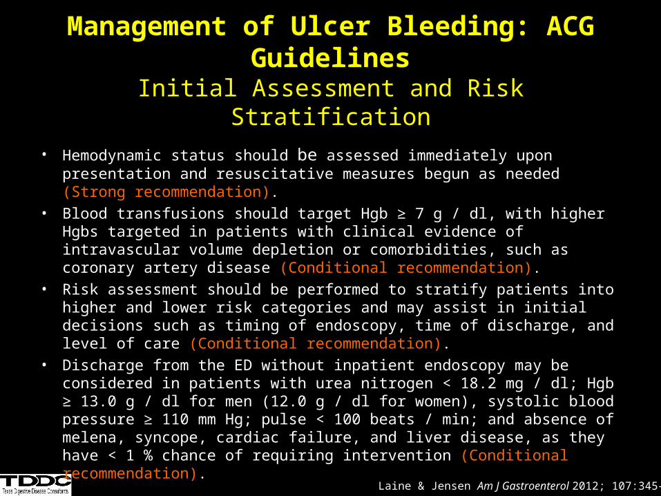
Management of Ulcer Bleeding: ACG Guidelines
Initial Assessment and Risk Stratification
• Hemodynamic status should be assessed immediately upon presentation and resuscitative measures begun as needed (Strong recommendation).
• Blood transfusions should target Hgb ≥ 7 g / dl, with higher Hgbs targeted in patients with clinical evidence of intravascular volume depletion or comorbidities, such as coronary artery disease (Conditional recommendation).
• Risk assessment should be performed to stratify patients into higher and lower risk categories and may assist in initial decisions such as timing of endoscopy, time of discharge, and level of care (Conditional recommendation).
• Discharge from the ED without inpatient endoscopy may be considered in patients with urea nitrogen < 18.2 mg / dl; Hgb ≥ 13.0 g / dl for men (12.0 g / dl for women), systolic blood pressure ≥ 110 mm Hg; pulse < 100 beats / min; and absence of melena, syncope, cardiac failure, and liver disease, as they have < 1 % chance of requiring intervention (Conditional recommendation).
Laine & Jensen Am J Gastroenterol 2012; 107:345–360
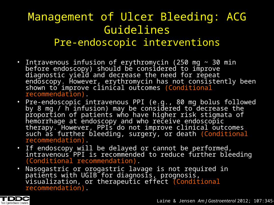
Management of Ulcer Bleeding: ACG GuidelinesPre-endoscopic interventions
• Intravenous infusion of erythromycin (250 mg ~ 30 min before endoscopy) should be considered to improve diagnostic yield and decrease the need for repeat endoscopy. However, erythromycin has not consistently been shown to improve clinical outcomes (Conditional recommendation).
• Pre-endoscopic intravenous PPI (e.g., 80 mg bolus followed by 8 mg / h infusion) may be considered to decrease the proportion of patients who have higher risk stigmata of hemorrhage at endoscopy and who receive endoscopic therapy. However, PPIs do not improve clinical outcomes such as further bleeding, surgery, or death (Conditional recommendation).
• If endoscopy will be delayed or cannot be performed, intravenous PPI is recommended to reduce further bleeding (Conditional recommendation).
• Nasogastric or orogastric lavage is not required in patients with UGIB for diagnosis, prognosis, visualization, or therapeutic effect (Conditional recommendation).
Laine & Jensen Am J Gastroenterol 2012; 107:345–360
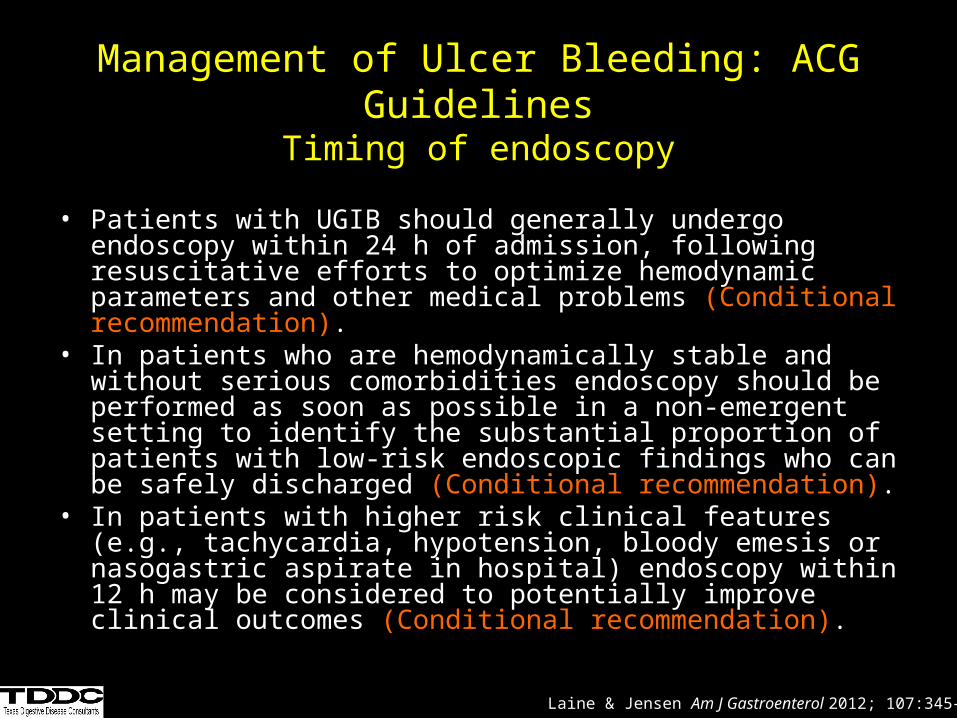
Management of Ulcer Bleeding: ACG GuidelinesTiming of endoscopy
• Patients with UGIB should generally undergo endoscopy within 24 h of admission, following resuscitative efforts to optimize hemodynamic parameters and other medical problems (Conditional recommendation).
• In patients who are hemodynamically stable and without serious comorbidities endoscopy should be performed as soon as possible in a non-emergent setting to identify the substantial proportion of patients with low-risk endoscopic findings who can be safely discharged (Conditional recommendation).
• In patients with higher risk clinical features (e.g., tachycardia, hypotension, bloody emesis or nasogastric aspirate in hospital) endoscopy within 12 h may be considered to potentially improve clinical outcomes (Conditional recommendation).
Laine & Jensen Am J Gastroenterol 2012; 107:345–360
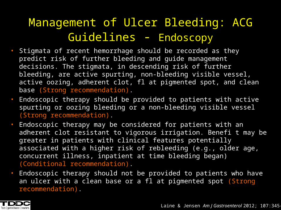
Management of Ulcer Bleeding: ACG Guidelines - Endoscopy
• Stigmata of recent hemorrhage should be recorded as they predict risk of further bleeding and guide management decisions. The stigmata, in descending risk of further bleeding, are active spurting, non-bleeding visible vessel, active oozing, adherent clot, fl at pigmented spot, and clean base (Strong recommendation).
• Endoscopic therapy should be provided to patients with active spurting or oozing bleeding or a non-bleeding visible vessel (Strong recommendation).
• Endoscopic therapy may be considered for patients with an adherent clot resistant to vigorous irrigation. Benefi t may be greater in patients with clinical features potentially associated with a higher risk of rebleeding (e.g., older age, concurrent illness, inpatient at time bleeding began) (Conditional recommendation).
• Endoscopic therapy should not be provided to patients who have an ulcer with a clean base or a fl at pigmented spot (Strong recommendation).
Laine & Jensen Am J Gastroenterol 2012; 107:345–360

Management of Ulcer Bleeding: ACG Guidelines - Endoscopy
• Epinephrine therapy should not be used alone. If used, it should be combined with a second modality (Strong recommendation).
• Thermal therapy with bipolar electrocoagulation or heater probe and injection of sclerosant (e.g., absolute alcohol) are recommended because they reduce further bleeding, need for surgery, and mortality (Strong recommendation).
• Clips are recommended because they appear to decrease further bleeding and need for surgery. However, comparisons of clips vs. other therapies yield variable results and currently used clips have not been well studied (Conditional recommendation).
• For the subset of patients with actively bleeding ulcers, thermal therapy or epinephrine plus a second modality may be preferred over clips or sclerosant alone to achieve initial hemostasis (Conditional recommendation).
Laine & Jensen Am J Gastroenterol 2012; 107:345–360
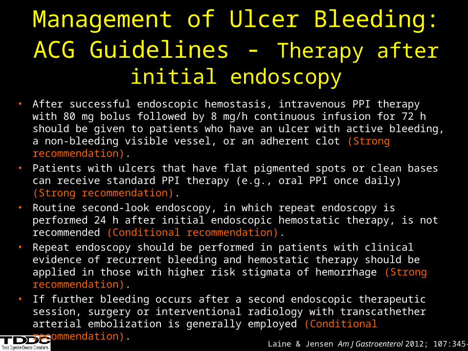
Management of Ulcer Bleeding: ACG Guidelines - Therapy after initial endoscopy
• After successful endoscopic hemostasis, intravenous PPI therapy with 80 mg bolus followed by 8 mg/h continuous infusion for 72 h should be given to patients who have an ulcer with active bleeding, a non-bleeding visible vessel, or an adherent clot (Strong recommendation).
• Patients with ulcers that have flat pigmented spots or clean bases can receive standard PPI therapy (e.g., oral PPI once daily) (Strong recommendation).
• Routine second-look endoscopy, in which repeat endoscopy is performed 24 h after initial endoscopic hemostatic therapy, is not recommended (Conditional recommendation).
• Repeat endoscopy should be performed in patients with clinical evidence of recurrent bleeding and hemostatic therapy should be applied in those with higher risk stigmata of hemorrhage (Strong recommendation).
• If further bleeding occurs after a second endoscopic therapeutic session, surgery or interventional radiology with transcathether arterial embolization is generally employed (Conditional recommendation).
Laine & Jensen Am J Gastroenterol 2012; 107:345–360
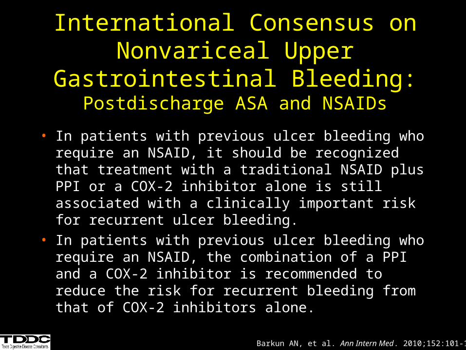
International Consensus on Nonvariceal Upper Gastrointestinal Bleeding:
Postdischarge ASA and NSAIDs
• In patients with previous ulcer bleeding who require an NSAID, it should be recognized that treatment with a traditional NSAID plus PPI or a COX-2 inhibitor alone is still associated with a clinically important risk for recurrent ulcer bleeding.
• In patients with previous ulcer bleeding who require an NSAID, the combination of a PPI and a COX-2 inhibitor is recommended to reduce the risk for recurrent bleeding from that of COX-2 inhibitors alone.
Barkun AN, et al. Ann Intern Med. 2010;152:101-113.
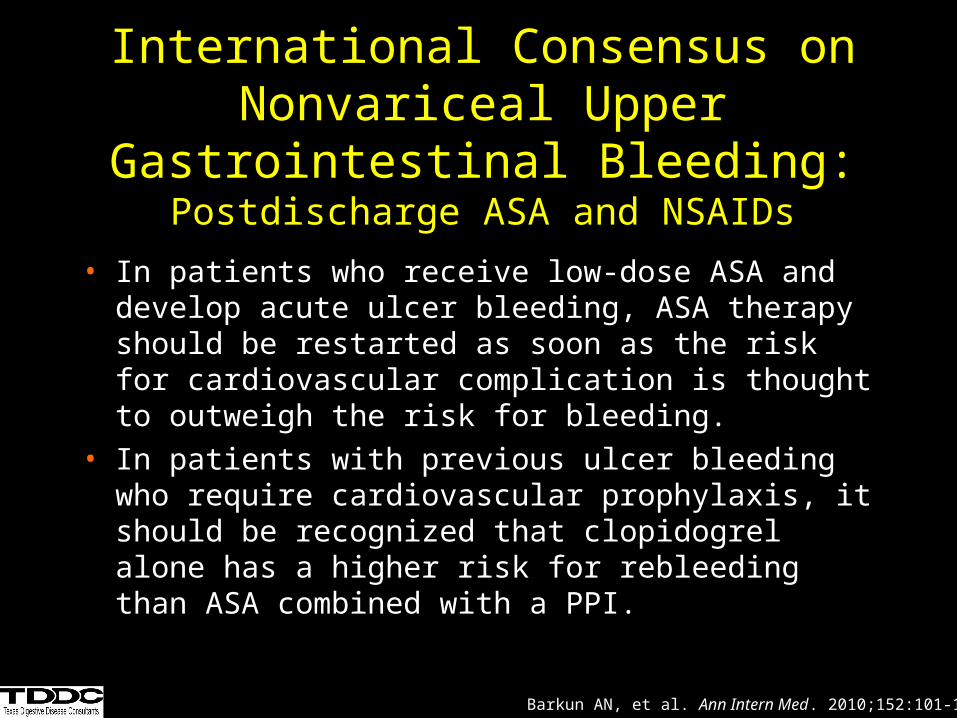
International Consensus on Nonvariceal Upper Gastrointestinal Bleeding:
Postdischarge ASA and NSAIDs
• In patients who receive low-dose ASA and develop acute ulcer bleeding, ASA therapy should be restarted as soon as the risk for cardiovascular complication is thought to outweigh the risk for bleeding.
• In patients with previous ulcer bleeding who require cardiovascular prophylaxis, it should be recognized that clopidogrel alone has a higher risk for rebleeding than ASA combined with a PPI.
Barkun AN, et al. Ann Intern Med. 2010;152:101-113.
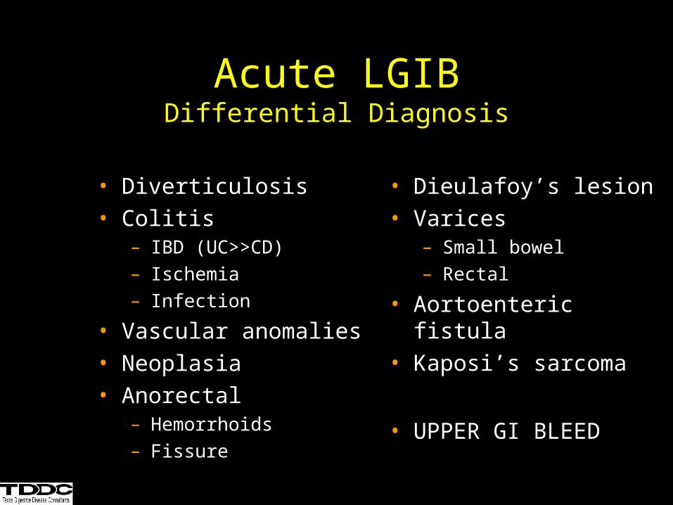
• Diverticulosis• Colitis
– IBD (UC>>CD)– Ischemia– Infection
• Vascular anomalies• Neoplasia• Anorectal
– Hemorrhoids– Fissure
• Dieulafoy’s lesion• Varices
– Small bowel– Rectal
• Aortoenteric fistula• Kaposi’s sarcoma
• UPPER GI BLEED
Acute LGIBDifferential Diagnosis
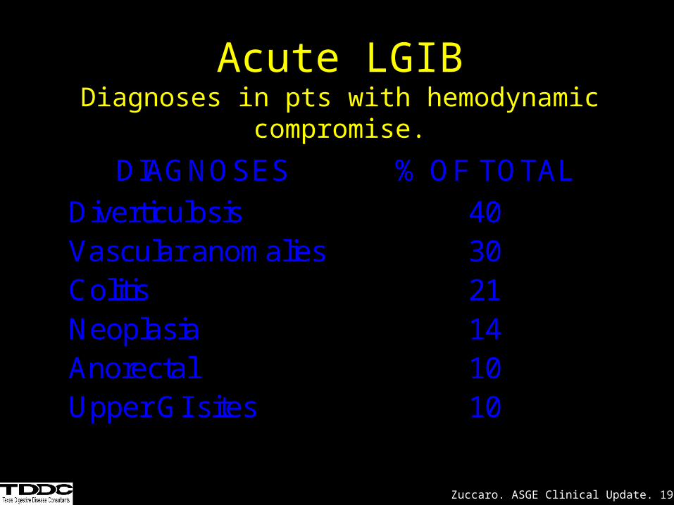
DIAGNOSES % OF TOTAL
Diverticulosis 40Vascular anomalies 30Colitis 21Neoplasia 14Anorectal 10Upper GI sites 10
Acute LGIBDiagnoses in pts with hemodynamic compromise.
Zuccaro. ASGE Clinical Update. 1999.
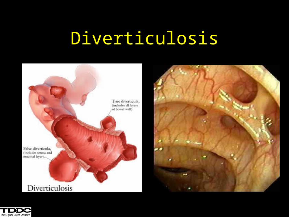
Diverticulosis
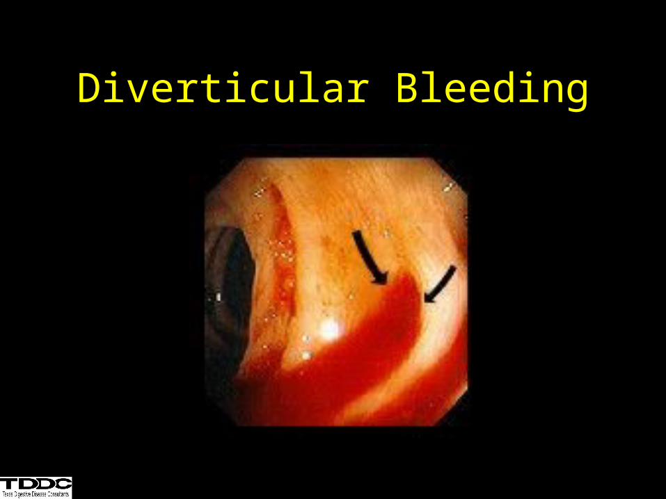
Diverticular Bleeding
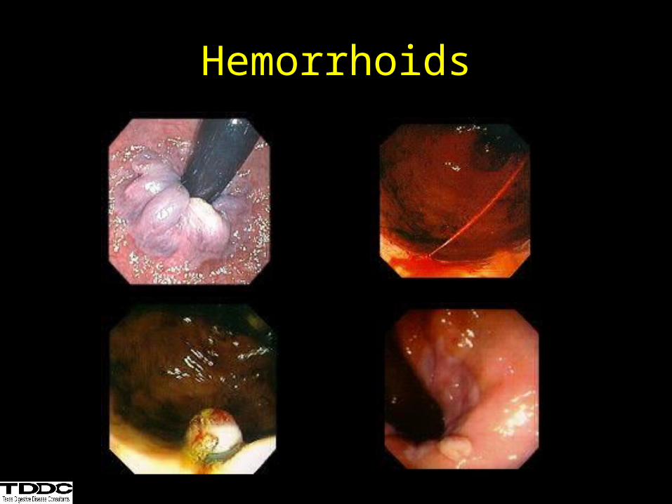
Hemorrhoids
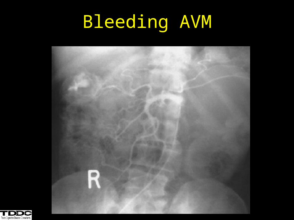
Bleeding AVM
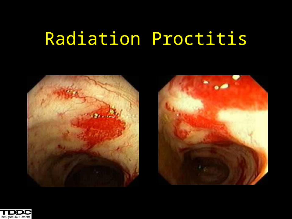
Radiation Proctitis
• Incidence 0.3 - 3.0 %• Etiology Incomplete obliteration of
the vitelline duct.• Pathology50% ileal, 50% gastric,
pancreatic, colonic mucosa• Complications
– Painless bleeding (children, currant jelly)– Intussusception
Acute LGIBMeckel’s Diverticulum
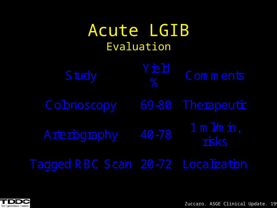
Study Yield
% Comments
Colonoscopy 69-80 Therapeutic
Arteriography 40-78 1 ml/min,
risks
Tagged RBC Scan 20-72 Localization
Acute LGIBEvaluation
Zuccaro. ASGE Clinical Update. 1999.
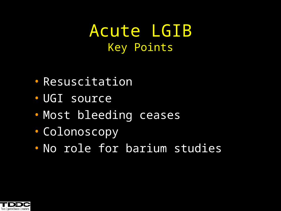
• Resuscitation
• UGI source
• Most bleeding ceases
• Colonoscopy
• No role for barium studies
Acute LGIBKey Points
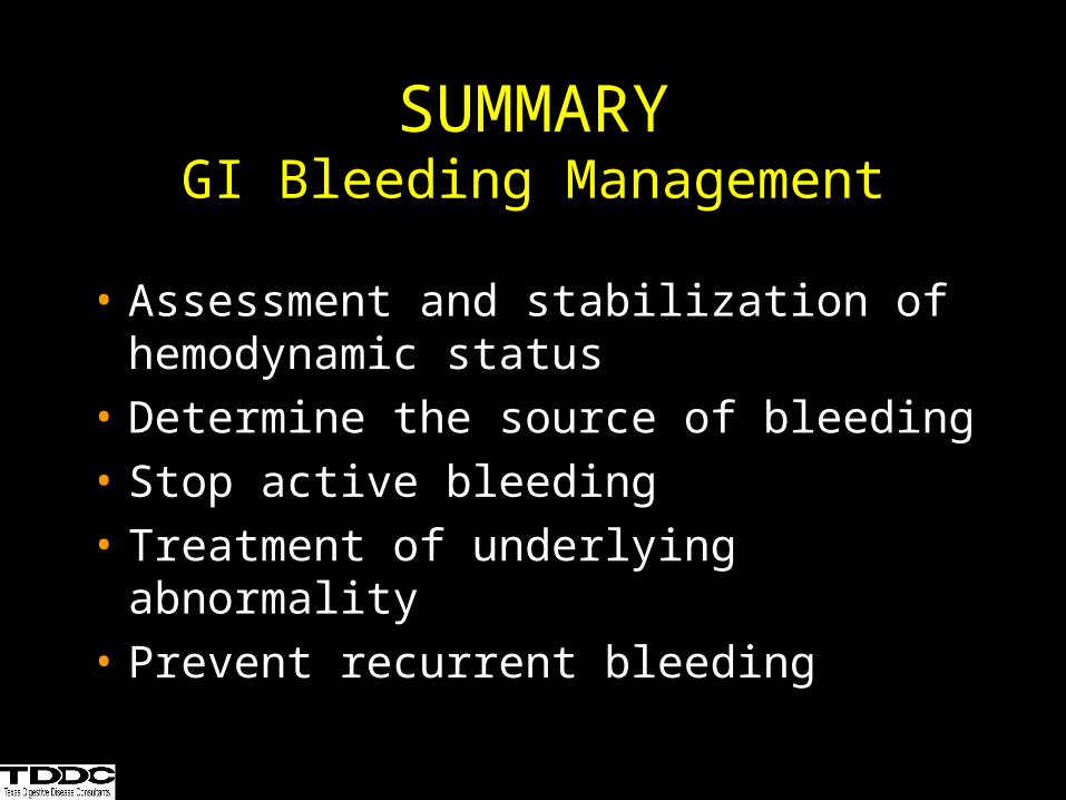
SUMMARYGI Bleeding Management
• Assessment and stabilization of hemodynamic status
• Determine the source of bleeding
• Stop active bleeding
• Treatment of underlying abnormality
• Prevent recurrent bleeding