Embed Size (px)
Citation preview
Genetic Characterization of Viral Nervous Necrosis Infects
Tilapia (Oreochromis sp.) in Indonesia
Uun Yanuhar1, a)*
Novia Christi2, (b)
and Diana Arfiati1, (c)
1Faculty of Fisheries and Marine Science, Brawijaya University, Jl. Veteran, Malang, 65145, East
Java, Indonesia 2Fish Quarantine and Inspection Agency, Jl. Raya Bandar Udara Ir. H. Juanda No. 23 - Sidoarjo
61254 – East Java, Indonesia
a)
Corresponding author: [email protected] b)
[email protected] c)[email protected]
Abstract
Tilapia (Oreochromis sp.) is the most important cultured fish in the 21 century and Indonesia is
the country's second largest tilapia producer after China. It make Indonesia should be more
concerned with any possible attack diseases that can infect both from the class of parasites,
fungi, bacteria, and viruses especially Virus Nervous Necrosis (VNN). The existence of VNN in
tilapia is one of the problems that must be handled. In the present research, a natural outbreak of
VNN in tilapia (Oreochromis sp.) farmed in mid-Java in Indonesia was investigated using
molecular genetic characterization. Research conducted using Reverse transcriptase PCR (RT-
PCR) methods, sequencing analysis, and phylogenetic analysis. the construction of phylogenetic
trees is based on the neighbor-joining method using software MEGA6. The samples used are a
tilapia juvenile size 5-7 cm, with or no clinical symptoms, and taken at random in the cultivation
of tilapia in Central Java. It showed that the tilapia juvenile infected VNN show changes and
damage to the organs and tissues. It can be seen from the examination of clinical symptoms,
anatomic pathology and histopathology. DNA of the tilapia infected by VNN was 294 bp on
both the brain and the eyes organ. Phylogenetic analysis results showed that VNN from infected
tilapia was the same as snapper and grouper and has the closest kinship with genotype Red-
spotted grouper nervous necrosis virus (RGNNV), which is about 99%. It can be stated that
Betanodavirus in freshwater fish potential is derived from Betanodavirus in seawater fish.
Keywords: Viral nervous necrosis, tilapia, genetic expression, phylogenetic
1. INTRODUCTION
Tilapia (Oreochromis sp.) is the most important cultured fish in the 21 century [1]. It has
been introduced into over 90 countries, and increasingly used worldwide in aquaculture (Bigarré et
al., 2009), because it is ease to grow and reproduce in a wide range of environmental conditions
[3].The aquaculture production of tilapias was about 2.5 million tons annually,and they serve as a
primary protein source in the developing world [4]. The production of tilapia in ponds in Indonesia
in 2013 reached 324.984 tons, with an production increase reach 43.18% per year [5]. This result
puts Indonesia as the country's second largest tilapia producer after China. With so many
manufactured products, making Indonesia should be more concerned with any possible attack
diseases that can infect both from the class of parasites, fungi, bacteria, and viruses.
Viral Nervous Necrosis (VNN), or Viral Encephalopathy and Retinopathy (VER) is the
family Nodaviridae that attracted the attention of fish health workers during the last decade. The
174Copyright © 2018, the Authors. Published by Atlantis Press. This is an open access article under the CC BY-NC license (http://creativecommons.org/licenses/by-nc/4.0/).
1st International Conference in One Health (ICOH 2017)Advances in Health Sciences Research (AHSR), volume 5
virus character is a non-enveloped, icosahedral, single-stranded positive-sense RNA virus. It
comprises of two segments: a gene encoding an RNA-dependent RNA polymerase (RNA1) and the
capsid protein (RNA2) [6][7]. Phylogenetic analysis show that fish Noda viruses have been
classified into 4 major genotypes: designated tiger puffer nervous necrosis virus (TPNNV), striped
jack nervous necrosis virus (SJNNV), berfin flounder nervous necrosis virus (BFNNV), and red-
spotted grouper nervous necrosis virus (RGNNV). This phylogenetic was based on similarities in
the partial RNA2 sequences encoding the C-terminal halves of the coat proteins[8]. Virus is
recorded from over 40 fish species covering different geographic locations [9].The disease was first
obtained from larvae of Japanese parrot fish (Oplegnathus jasciatus), barramundi (Lates
calcarifer), turbot (Scophthalmus maximus), in Japan [10], Australia [11], Norway [12],
respectively.
In Indonesia,VNN disease events was first reported in 1997, in Banyuwangi, East Java,on
the cultivation of barramundi (Lates calcaliver). It spread to Bali in 1998, and expanded to grouper
hatchery in Bali that caused mass deaths of 100%. This VNN were grouped in SJNNV [13]. This
disease have resulted in mass deaths of marine aquaculture fish, particularly the larval and juvenile
stages [14][15].
VNN attacks can be transmitted vertically from an infected mother VNN [9] or horizontally
through infected fish [16][17] as well as from the rest of the fish (trash fish) or molluscs used as
feedin the aquaculture [18]. VNN infected fish show the symptoms such as abnormal swimming
behavior (circling or whirling), floats above the stomach, decreased appetite and body color gel
[15].The virus concentrate in the retina, brain, and spinal cord of infected fish. Infected fish
demonstrate erratic swimming patterns and the neurological abnormalities, including cellular
necrosis andvacuolization in the retina and the system of central nervous [19]. Some studies
suggest that the VNN was not only attack the sea waterfish species,but also attack the freshwater
fish species like an ornamental fish, guppy, Poicelia reticulata[20], Acipenser sp [21], fish medaka
(Oryzias latipes) [19], largemouth black bass Micropterus salmoides[22], and tilapia, Oreochromis
niloticus (L.)[23][24].The genetic characterization confirmed that VNN infecting freshwater fish
species was belong to the RGNNV genotype [19][25][24] and the SJNNV genotype [19].
Related to the possibility of genetic variation onVNN infected tilapia, it is important to
know the virus variants by performing DNA sequencing VNN, followed by phylogenetic analysis,
so as to know the kinship between VNN variants found intilapia. In the present research, a natural
outbreak of VNN in tilapia (Oreochromis sp.) farmed in mid-Java in Indonesia was investigated
using molecular genetic characterization.
2. METHODS
Fish Sampling
The samples used are a tilapia juvenile size5-7cm, with orno clinical symptoms, and taken
at random in the cultivation of tilapiain Central Java in mid-December 2014.The number of
samples was105pieces. All sampling is conducted anobservationof clinicalsymptoms, thena sample
of the brain and the eyes were collected for analysis of virus (PCR). Tilapia juvenile of the same
size were used as the control fish
Reverse transcriptase PCR (RT-PCR)
The process of reverse-transcriptase PCR was performed using the Quick Access - AMV®
RT-PCR (Promega) followed by nested PCR using Taq® Go Green Master Mix (Promega) with
175
Advances in Health Sciences Research (AHSR), volume 5
the same primer and marker from Kapa Universal Ladder. The reaction process conducted by the
protocol of each kit, to a final concentration of each primer 2.5 µM. RT-PCR reactions Nested PCR
was performed using a thermal cycler (GeneAmp® PCR System 9700, Applied Biosystems).
Specific F2 primer (5'- GTC AGT CGT GTG TCG CAT CT-3 '), R3 (5'- CGA GTC ACG AAC
GGT GAA GA-3'), NF2 (5 '-GTT CCC TGT ACA ACG ATT CC-3 '), and NR3 (5'-GGA GAC
TTT GGG GCT GCT CA -3') [26] were used to amplify regions of the coat protein varies
Betanodavirus. RT-PCR cycle conditions are: first incubation at 45 °C for 30 min followed by
reverse transcriptase inactivation at 94 °C for 2 min; then 40 cycles consisting of 94 °C for 30s,
60 °C for 30s, and 72 °C for 45s; and 1 cycle of 72 °C for 10 min. Nested PCR cycle conditions
was the same as RT-PCR cycle conditions without reverse-transcriptase process. Products have
been amplified and then analyzed for using an electrophoresis in 1.6% agarose gel stained with
SYBR after save (Invitrogen).
Sequencing Analysis
Amplicons on agel purified was conducted using the QIAQuick®PurificationKit,according
to the manufacturer's instructions. Results of purification were sequenced using
BigDye®Terminator Cycle Sequencing (Applied Biosystems), according to the manufacturer's
instructions. Sequencing cycle conditions were as follows 96°C, 0°C and 60°Cfor 10s, 5s and for
4min, respectively for 25 cycles. Purification of the product cycle sequencing was conducted using
ethanol precipitation/EDTA. Denaturation carried out at a temperature of 95°C for 5 min by adding
Hi-Di formamide1:1 into the product purification. Capillary electrophoresis is conducted by a
sequencer Applied Biosystem3130 DNA Analyzer.
Phylogenetic Analysis
Results sequences in the sample was aligned by the contained data base in Gene Bank
(NCBI) using the ClustalW software [27]. Phylogenetic tree constructed by ClustalW and then
displayed with Tree View [28]. Phylogenetic tree describing the relationships among species was
determined using a method of Neighbour-Joining (NJ tree) [29]. Reliability NJ tree using the boot
strap method Felsenstein was concluded by 1000 replication.
3. RESULT AND DISCUSSION
RT-PCR and sequencing
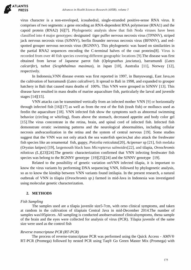
RT-PCR amplification with specific primers shows the specific band at 294 bp on agarose
gel (Fig. 1a). The PCR products were then sequenced at 275-280 nucleotide bases as shown in
Figure 1b.
Sequences results were compared to genotype VNN based on data from GeneBank that
show that VNN from tilapia juvenile has a little difference with the nucleotide base pairs isolates
for comparison, which isolates AY690596.1 (RGNNV) with values aligned score of 97. For isolate
of EU236149.1(TPNNV), EU236147.1(BFNNV), and AB056572.1(SJNNV), each having a value
of aligned score 69, 78, and 68, respectively. It can be state that the VNN isolates from tilapi
ajuvenile has a high homologous with VNN of genotype RGNNV.Similar research regarding mass
mortality in tilapia larvae infected VNN in Thailand als oshowed the high homologous and closely
related to genotype RGNNV [24], from a reading o three base pairs (triplet) which is the genetic
code (codon).
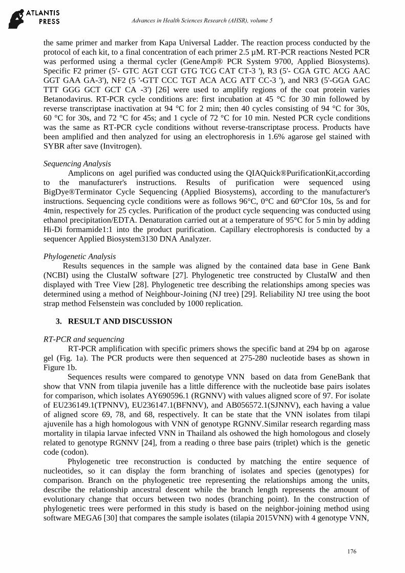
Phylogenetic tree reconstruction is conducted by matching the entire sequence of
nucleotides, so it can display the form branching of isolates and species (genotypes) for
comparison. Branch on the phylogenetic tree representing the relationships among the units,
describe the relationship ancestral descent while the branch length represents the amount of
evolutionary change that occurs between two nodes (branching point). In the construction of
phylogenetic trees were performed in this study is based on the neighbor-joining method using
software MEGA6 [30] that compares the sample isolates (tilapia 2015VNN) with 4 genotype VNN,
176
Advances in Health Sciences Research (AHSR), volume 5
namely barfin Flounder Nervous Necrosis Virus (BFNNV), Striped Jack Nervous Necrosis Virus
(SJNNV), Tiger Puffer Nervous Necrosis Virus (TPNNV) and Red-spotted Grouper Nervous
Necrosis Virus (RGNNV). Isolates tilapia 2015VNN has the closest kinship with Red spotted
isolates grouper nervous necrosis virus (RGNNV), which is about 99%. So to say that the VNN
infected tilapia juvenile derived from genotyping RGNNV, as shown in Fig. 2.
(a) (b)
Figure1. (a) Detection of VNN gene from tilapiaby RT-PCR amplification.
Lanes M. DNA ladder, 1 & 3: amplified product from total nucleic acids extracted from brain of
tilapia; 2 & 4: eye; amplified product from total nucleic acids extracted from brain; 5: Positive
Control; (b) Sequence result
Figure 2. Phylogenetic tree constructed by protein gene sequences base of Tilapia-2015-VNN and other
published isolates of betanodavirus. The tree was designed using Neighbor-Joining methods
Tilapia used in this study came from fresh water while the VNN is a disease that infects many
species of saltwater fish. It showed that VNN has the ability to infect across species and adapt to a
new host species [19]. A specific host on the most viral infections are usually controlled by the
interaction with a viral surface protein receptors on the surface of target cells precisely [31], so
long as the tilapia have receptors that can bind to the attachment site (s) virus particles, then
certainly virus will entry into host cells (tilapia), because it does not directly host cell have
facilitated the virus to enter the cell [32]. The tilapia have such receptors HSC70, as well as
groupers [33], which specifically alleged that the virus can perform attachment and involved in an
interaction that could cause the virus to enter the body tilapia. Interactions between viruses and
cellular receptors is a dynamic process chain that allows the entry of the virus into the cell [34][35].
177
Advances in Health Sciences Research (AHSR), volume 5
Based on sequencing probes followed by BLAST, it shows that the VNN that infect tilapia
is similar VNN infecting both snapper and grouper. After matching to theGeneBank data,isolates of
tilapia 2015VNN (VNN fromtilapia) has a nucleotide sequence that is similar to T156700 isolates
of identity snapper with a max value of 100% (data not shown), whereas fo rEA-150102-IL isolates
from grouper with max identity value of 99% (data not shown). It indicate that tilapia isolates of
2015VNN has a nucleotide sequence that is almost same and there is a little difference of base pairs
of nucleotides. Phylogenetic tree is reconstructed by matching the entire sequence of nucleotides so
that it can display the form branching of isolates and species comparison. The results of the
phylogenetic analysis showed that the VNN infected tilapia derived from genotype RGNNV.
RGNNV is one of genotypes VNN, who lived in the temperature range of 25-30°C, where the
condition of Indonesia is at that temperature range.
There are the homologous in both VNN infecting fresh water fish and VNN infecting sea
water fish. Betanodavirus isolated from Epinephelus taurine (taurine Epinephelus Nervous
Necrosis Virus=ETNNV) has high homolouges with virus isolated from freshwater fish Poecili
areticula (Guppy Nervous Necrosisvirus=GNNV) [20]. Virus isolated fromMicropterus salmoides
has high homologous with RGNNV [22]. In this study, VNN infected tilapia has homologous with
RGNNV. It can be stated that Betanodavirus in freshwater fish potential is derived from
Betanodavirus in sea water fish.
4. CONCLUSION
The existence of VNN in tilapia is one of the problems that must be handled. This study
results show that VNN infected tilapia has homologous with RGNNV. This research finding has
enlarged the recognized susceptible hosts of the VNN with freshwater fish. Further studies, it is
needed to investigate the epidemiology and the mode of transmission to know why VNN are found
in both freshwater and marine fishes in Indonesia.
ACKNOWLEDGMENTS
This work was supported by the Ministry of Research, Technology and Higher Education,
Republic of Indonesia; through the of LeadingResearch of University (PUPT) with contract no:
033/SP2H/LT/DRPM/II/2016.
REFERENCES
[1] M. T. Ridha, Aquac. Res., 37 ( 2), 172–179 (2006).
[2] L. Bigarré, J. Cabon, M. Baud, M. Heimann, A. Body, F. Lieffrig, and J. Castric, J. Fish Dis.
32, 667–673 (2009).
[3] G. G. Tsadik and A. N. Bart, Aquaculture, 272 (1–4),380–388 (2007).
[4] FAO, “FAO Fisheries & Aquaculture - Cultured Aquatic Species Information Programme -
Oreochromis niloticus (Linnaeus, 1758 ), Available:
http://www.fao.org/fishery/culturedspecies/Oreochromis_niloticus/en#tcNA008C.
[Accessed: 04-Jun-2015].
[5] A. Widiarti, “Indonesia tilapia production and trade,” in International Conference on
Tilapia, 2015.
[6] K. Mori, T. Nakai, K. Muroga, M. Aarimoto, K. Mushiake, and I. Furusawa, Virology 187,
368–371 (1992).
[7] B. L. Munday, J. Kwang, and N. Moody, J. Fish Dis. 25 (3), 127–142 (2002).
[8] T. Nishizawa, M. Furuhashi, T. Nagai, T. Nakai, and K. Muroga, Appl. Envir. Microbiol. 63
(4), 1633–1636, (1997).
178
Advances in Health Sciences Research (AHSR), volume 5
[9] I. S. Azad, K. P. Jithendran, M. S. Shekhar, A. R. Thirunavukkarasu, and L. D. de la Pena,
Aquaculture 255, 39–47 (2006).
[10] K. Yoshikoshi and K. Inoue, J. Fish Dis. 13 (1), 69–77 (1990).
[11] J. S. Glazebrook, M. P. Heasman, and S. W. Beer, J. Fish Dis. 13 (3), 245–249 (1990).
[12] B. Bloch, K. Gravningen, and J. Larsen, “Dis. Aquat. Organ. 10 (1984), 65–70 (1991).
[13] Zafran, I. Koesharyani, F. Johnny, K. Yuasa, T. Harada, and K. Hatai, Fish Pathol. 32 (2),
95–96 (2000).
[14] U. Yanuhar, E. Gusman, and D. Arfiati, Adv. Environ. Biol. 6 (1), 388–396 (2012).
[15] R. Yuwanita, U. Yanuhar, and Hardoko, Adv. Environ. Biol.7 (6), 1074–1081 (2013).
[16] J. Ransangan and B. O. Manin, Veterinary Microbiology. Elsevier, 2010.
[17] P. Hick, G. Schipp, J. Bosmans, J. Humphrey, and R. Whittington, Aquaculture 319 (1–2),
41–52 (2011).
[18] D. K. Gomez, K. Mori, Y. Okinaka, T. Nakai, and S. C. Park, Aquaculture 302 (3–4), 58–
163 (2010).
[19] R. Furusawa, Y. Okinaka, and T. Nakai, J. Gen. Virol.87 (Pt 8), 2333–9 (2006).
[20] A. Hegde, H. C. Teh, T. J. Lam, and Y. M. Sin, Arch. Virol. 148 (3), 575–86, (2003).
[21] F. Athanassopoulou, C. Billinis, and T. Prapas, Dis. Aquat. Organ. 60 (3), 247–52 (2004).
[22] G. Bovo, a. Gustinelli, F. Quaglio, F. Gobbo, V. Panzarin, a. Fusaro, F. Mutinelli, M.
Caffara, and M. L. Fioravanti, Dis. Aquat. Organ. 96, 45–54 (2011).
[23] L. Bigarré, J. Cabon, M. Baud, M. Heimann, A. Body, F. Lieffrig, and J. Castric, J. Fish Dis.
32 (8), 667–73 (2009).
[24] J. Keawcharoen, S. Techangamsuwan, A. Ponpornpisit, E. D. Lombardini, T. Patchimasiri,
and N. Pirarat, J. Fish Dis. 38 (1), 49–54 (2015).
[25] C. Maltese and G. Bovo, Ittiopatologia4, 93–146 (2007).
[26] OIE, “Access online: OIE - World Organisation for Animal Health,” Manual of Diagnostic
Tests for Aquatic Animals, 2013. Available: http://www.oie.int/international-standard-
setting/aquatic-manual/access-online/. [Accessed: 01-Jul-2015].
[27] J. D. Thompson, D. G. Higgins, and T. J. Gibson, Nucleic Acids Res. 22 (22) 4673–80
(1994).
[28] R. D. Page, Comput. Appl. Biosci. 12 (4), 357–8 (1996).
[29] N. Saitou and M. Nei, Mol. Biol. Evol. 4 (4), 406–425 (1987).
[30] K. Tamura, G. Stecher, D. Peterson, A. Filipski, and S. Kumar, Mol. Biol. Evol. 30(12),
2725–9 (2013).
[31] E. Baranowski, Science 292 (5519), 1102–1105 (2001).
[32] Y. Ito, Y. Okinaka, K. Mori, T. Sugaya, T. Nishioka, M. Oka, and T. Nakai, Dis. Aquat.
Organ. 79(3), 199–205 (2008).
[33] J. S. Chang and S. C. Chi, J. Virol. 89 (1), 61–70 (2015).
[34] A. M. Haywood, J. Virol. 68(1), 1–5, (1994).
[35] S. J. Schaulies, J. Gen. Virol. 81 (6), 1413–29 (2000).
179
Advances in Health Sciences Research (AHSR), volume 5

![OPEN ACCESS viruses - Semantic Scholar · , viruses [1], the viral etiology of these previously recognized diseases such as infectious pancreatic necrosis, Oregon sockeye disease,](https://img.pdfslide.net/doc/110x75/5e855f018b3d144fe76983d0/open-access-viruses-semantic-scholar-viruses-1-the-viral-etiology-of-these.jpg)