Embed Size (px)
Citation preview
Germ Cell and Stromal Tumors
Germ Cell and Stromal Tumors
of the Ovary
of the Ovary
Fred Ueland, MD
University of Kentucky
Gynecologic Oncology
Fred Ueland, MD
Fred Ueland, MD
University of Kentucky
University of Kentucky
Gynecologic Oncology
Gynecologic Oncology
Clinical Profile and
Clinical Profile and
Classification
Classification
Germ Cell Tumors
Germ Cell Tumors
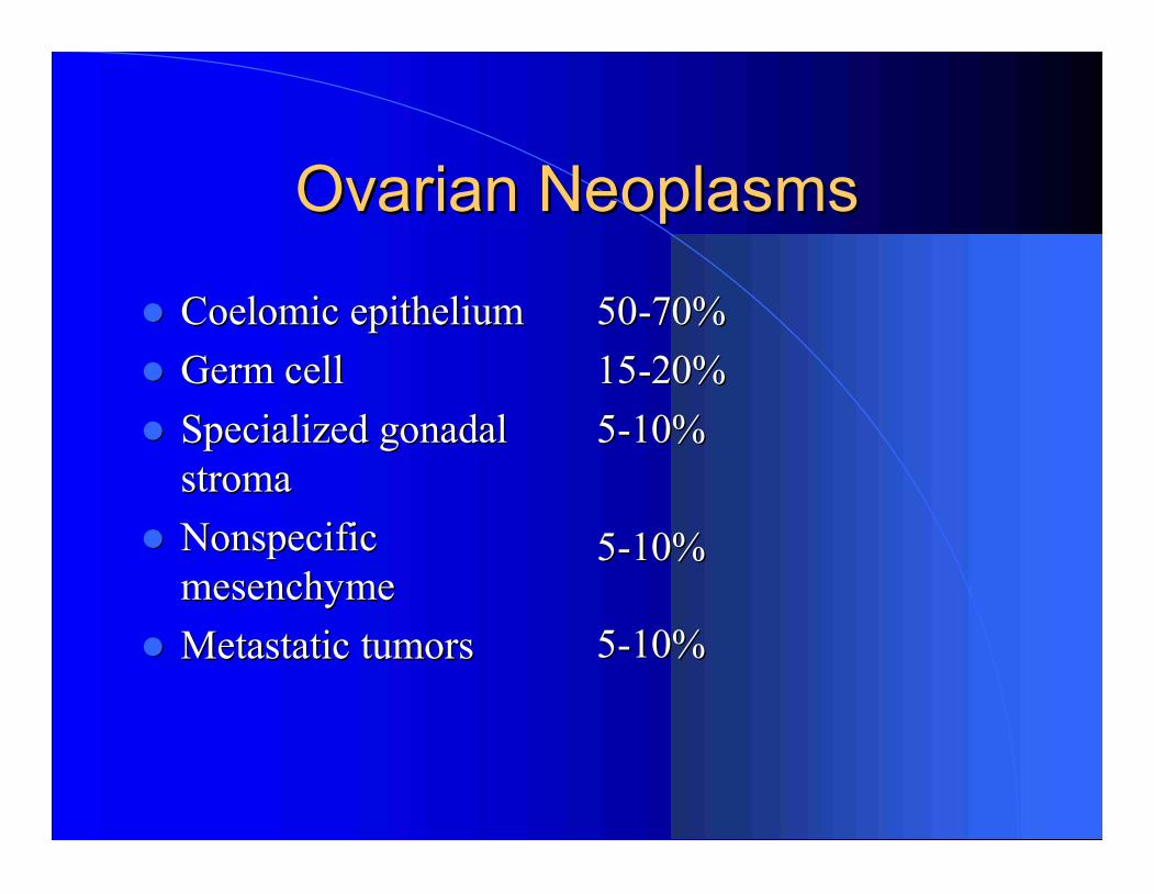
Ovarian Neoplasms
Ovarian Neoplasms
Coelomic epithelium
Coelomic epithelium
Germ cell
Germ cell
Specialized gonadal
Specialized gonadal
stroma
stroma
Nonspecific
Nonspecific
mesenchyme
mesenchyme
Metastatic tumors
Metastatic tumors
50
50
-
-
70%
70%
15
15
-
-
20%
20%
5
5
-
-
10%
10%
5
5
-
-
10%
10%
5
5
-
-
10%
10%
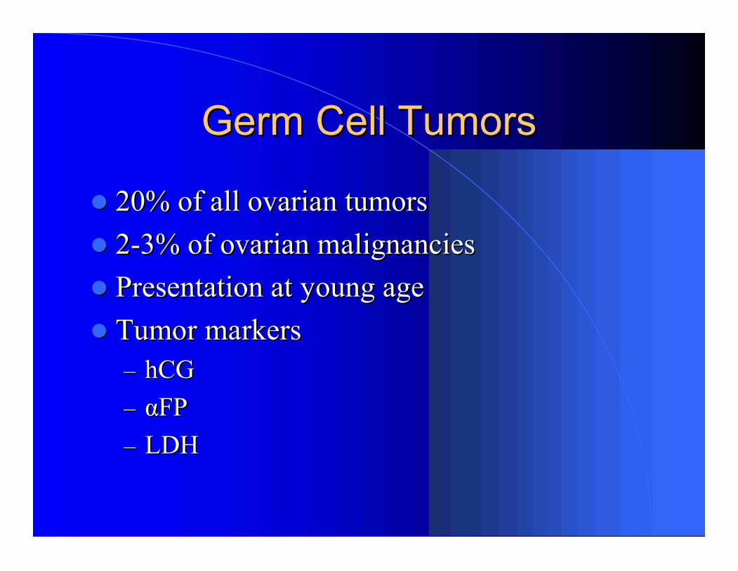
Germ Cell Tumors
Germ Cell Tumors
20% of all ovarian tumors
20% of all ovarian tumors
2
2
-
-
3% of ovarian malignancies
3% of ovarian malignancies
Presentation at young age
Presentation at young age
Tumor markers
Tumor markers
–
–
hCG
hCG
–
–
α
α
FP
FP
–
–
LDH
LDH
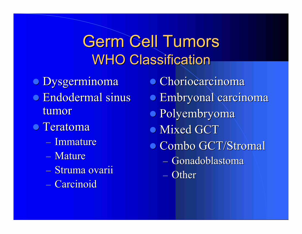
Germ Cell Tumors
Germ Cell Tumors
WHO Classification
WHO Classification
Dysgerminoma
Dysgerminoma
Endodermal sinus
Endodermal sinus
tumor
tumor
Teratoma
Teratoma
–
–
Immature
Immature
–
–
Mature
Mature
–
–
Struma ovarii
Struma ovarii
–
–
Carcinoid
Carcinoid
Choriocarcinoma
Choriocarcinoma
Embryonal carcinoma
Embryonal carcinoma
Polyembryoma
Polyembryoma
Mixed GCT
Mixed GCT
Combo GCT/Stromal
Combo GCT/Stromal
–
–
Gonadoblastoma
Gonadoblastoma
–
–
Other
Other
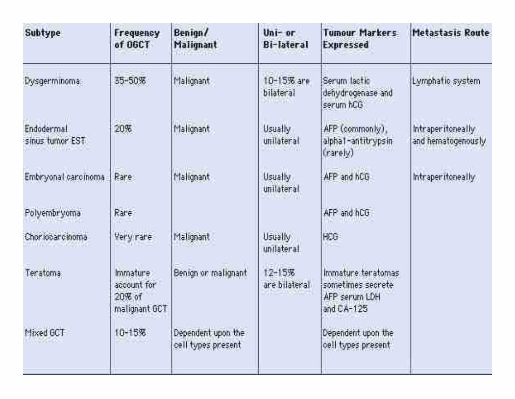
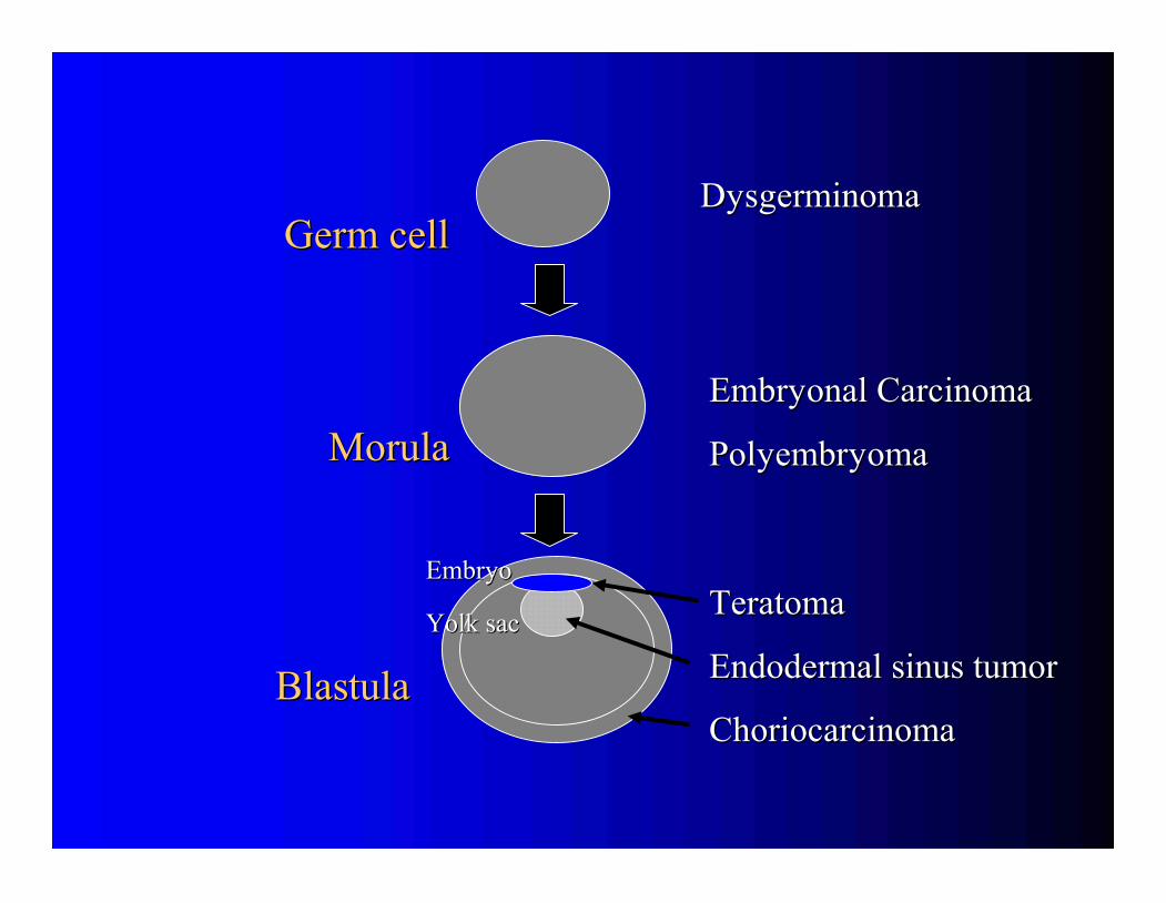
Dysgerminoma
Dysgerminoma
Embryonal Carcinoma
Embryonal Carcinoma
Polyembryoma
Polyembryoma
Teratoma
Teratoma
Endodermal sinus tumor
Endodermal sinus tumor
Choriocarcinoma
Choriocarcinoma
Germ cell
Germ cell
Morula
Morula
Blastula
Blastula
Embryo
Embryo
Yolk sac
Yolk sac
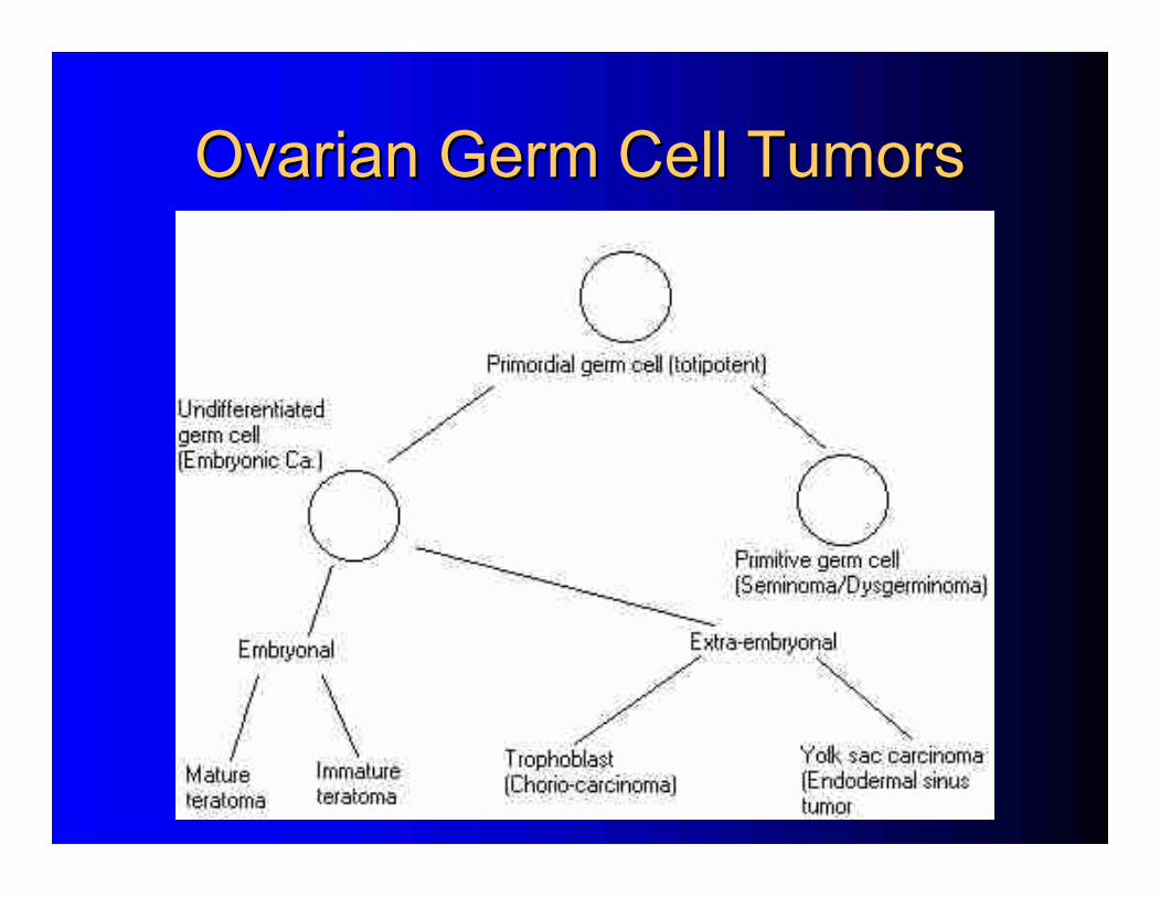
Ovarian Germ Cell Tumors
Ovarian Germ Cell Tumors
Specific Tumor Types
Specific Tumor Types
Germ Cell Tumors
Germ Cell Tumors
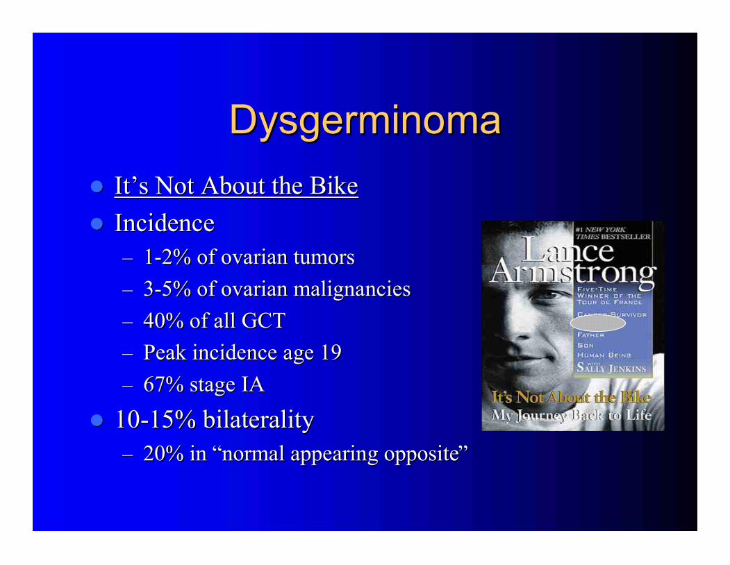
Dysgerminoma
Dysgerminoma
It
It
’
’
s Not About the Bike
s Not About the Bike
Incidence
Incidence
–
–
1
1
-
-
2% of ovarian tumors
2% of ovarian tumors
–
–
3
3
-
-
5% of ovarian malignancies
5% of ovarian malignancies
–
–
40% of all GCT
40% of all GCT
–
–
Peak incidence age 19
Peak incidence age 19
–
–
67% stage IA
67% stage IA
10
10
-
-
15%
15%
bilaterality
bilaterality
–
–
20% in
20% in
“
“
normal appearing opposite
normal appearing opposite
”
”
Dysgerminoma
Dysgerminoma
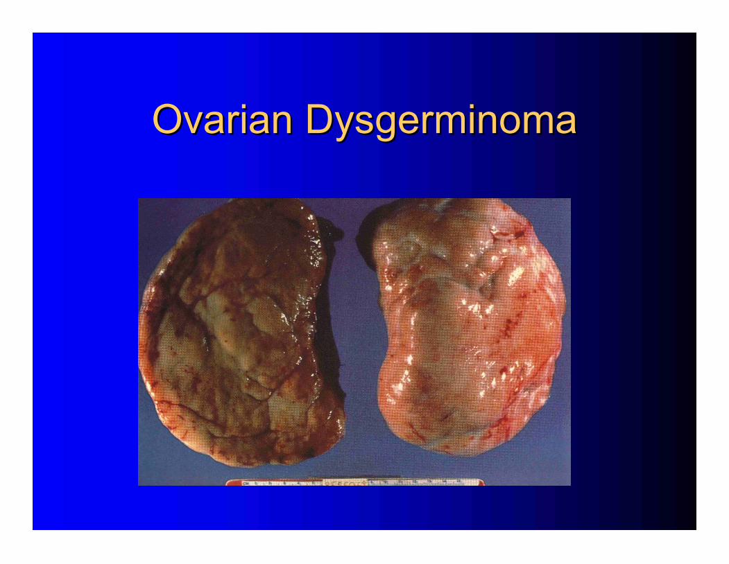
Ovarian Dysgerminoma
Ovarian Dysgerminoma
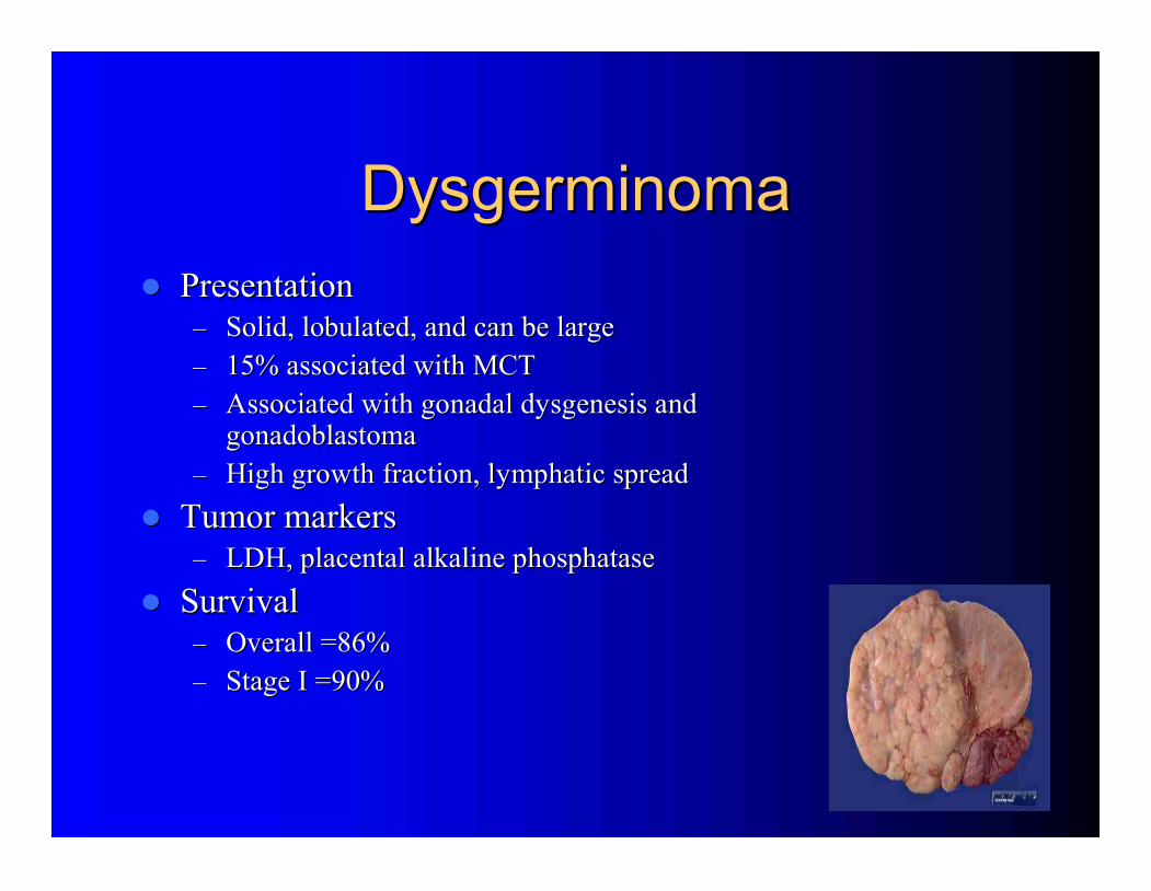
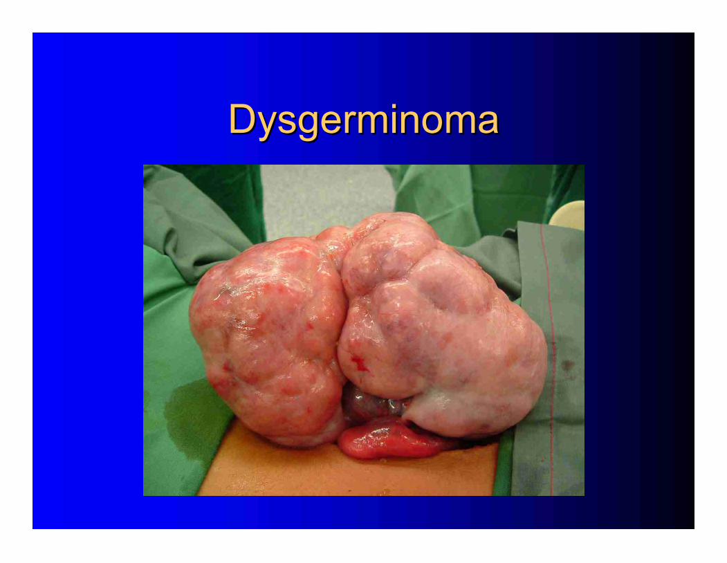
Dysgerminoma
Dysgerminoma
Presentation
Presentation
–
–
Solid, lobulated, and can be large
Solid, lobulated, and can be large
–
–
15% associated with MCT
15% associated with MCT
–
–
Associated with gonadal dysgenesis and
Associated with gonadal dysgenesis and
gonadoblastoma
gonadoblastoma
–
–
High growth fraction, lymphatic spread
High growth fraction, lymphatic spread
Tumor markers
Tumor markers
–
–
LDH,
LDH,
placental alkaline phosphatase
placental alkaline phosphatase
Survival
Survival
–
–
Overall =86%
Overall =86%
–
–
Stage I =90%
Stage I =90%
Dysgerminoma
Dysgerminoma
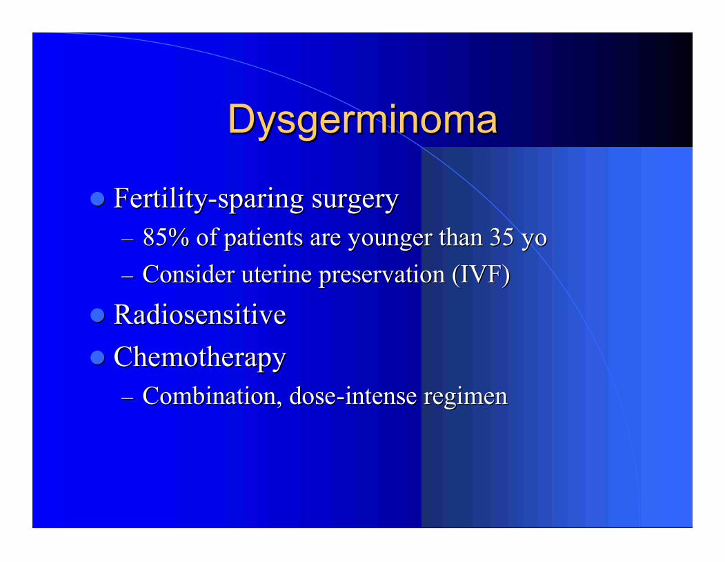
Fertility
Fertility
-
-
sparing surgery
sparing surgery
–
–
85% of patients are younger than 35 yo
85% of patients are younger than 35 yo
–
–
Consider uterine preservation (IVF)
Consider uterine preservation (IVF)
Radiosensitive
Radiosensitive
Chemotherapy
Chemotherapy
–
–
Combination, dose
Combination, dose
-
-
intense regimen
intense regimen
Dysgerminoma
Dysgerminoma
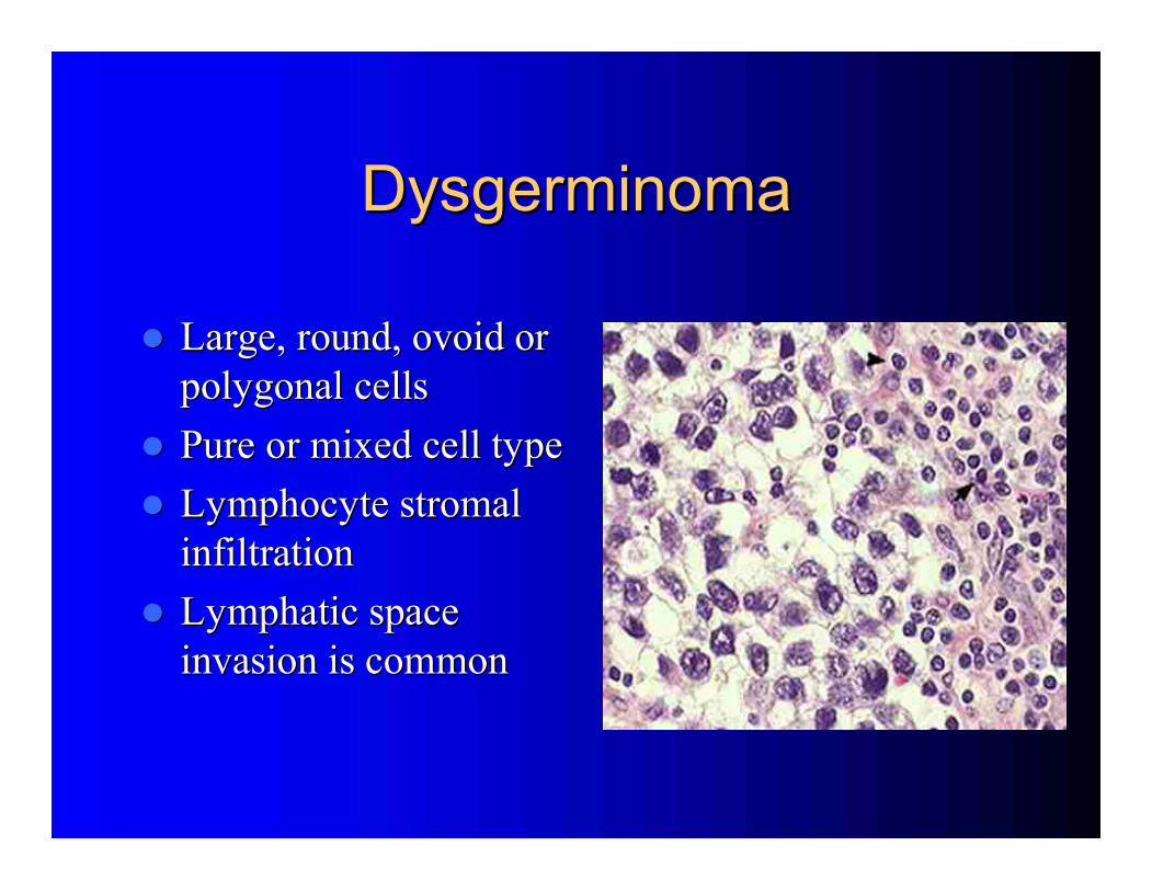
Large, round, ovoid or
Large, round, ovoid or
polygonal cells
polygonal cells
Pure or mixed cell type
Pure or mixed cell type
Lymphocyte stromal
Lymphocyte stromal
infiltration
infiltration
Lymphatic space
Lymphatic space
invasion is common
invasion is common
Dysgerminoma
Dysgerminoma
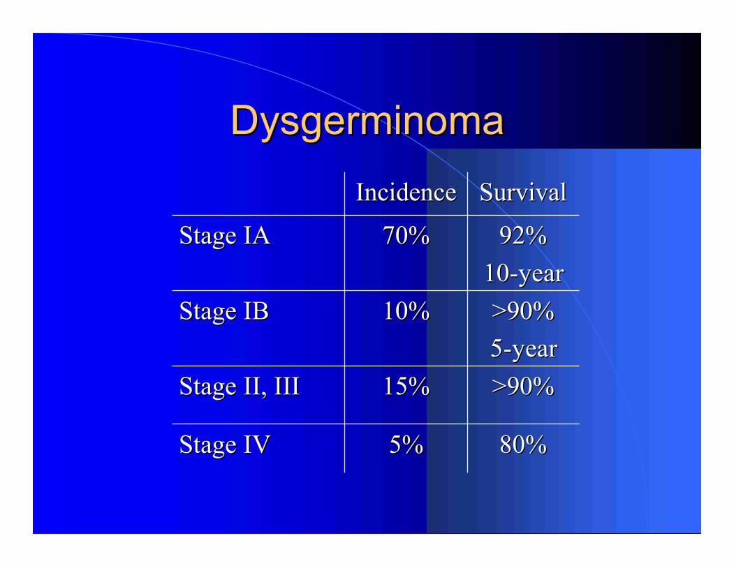
Survival
Survival
Incidence
Incidence
5%
5%
15%
15%
10%
10%
70%
70%
80%
80%
>90%
>90%
>90%
>90%
5
5
-
-
year
year
92%
92%
10
10
-
-
year
year
Stage IV
Stage IV
Stage II, III
Stage II, III
Stage IB
Stage IB
Stage IA
Stage IA
Endodermal Sinus Tumor
Endodermal Sinus Tumor
Presentation
Presentation
–
–
20% of all GCT
20% of all GCT
–
–
Median age 19 yo
Median age 19 yo
–
–
Abdominal pain, large mass
Abdominal pain, large mass
–
–
10
10
-
-
30 cm common
30 cm common
–
–
Very rapid growth, intra
Very rapid growth, intra
-
-
abdominal and hematological spread
abdominal and hematological spread
Tumor marker =
Tumor marker =
α
α
FP,
FP,
α
α
1
1
antitrypsin
antitrypsin
Synonyms
Synonyms
–
–
Yolk sac tumor, Mesonephroma
Yolk sac tumor, Mesonephroma
Survival
Survival
–
–
Overall survival =70%
Overall survival =70%
–
–
Stage I =90%
Stage I =90%
Endodermal Sinus Tumor
Endodermal Sinus Tumor
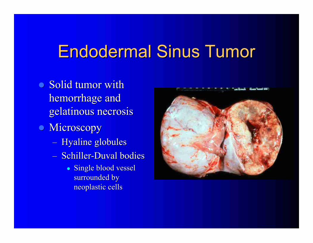
Solid tumor with
Solid tumor with
hemorrhage and
hemorrhage and
gelatinous necrosis
gelatinous necrosis
Microscopy
Microscopy
–
–
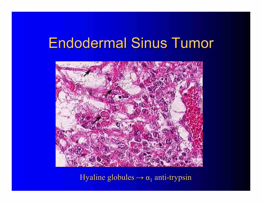
Hyaline globules
Hyaline globules
–
–
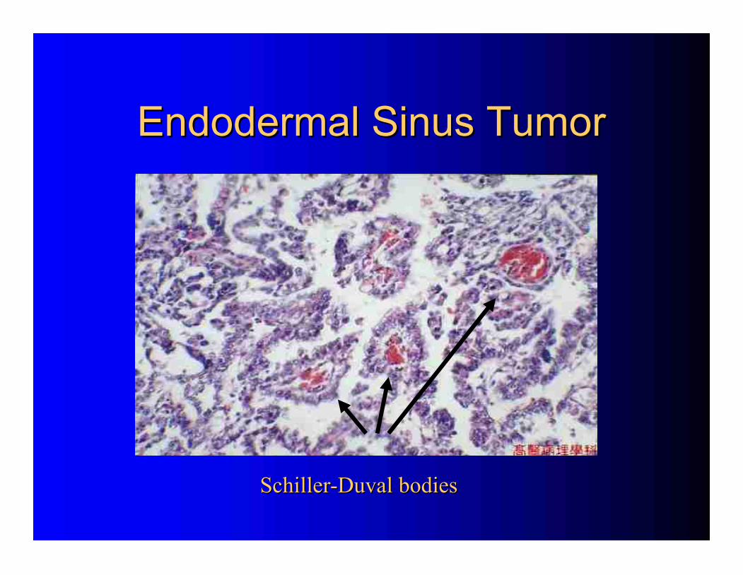
Schiller
Schiller
-
-
Duval bodies
Duval bodies
Single blood vessel
Single blood vessel
surrounded by
surrounded by
neoplastic cells
neoplastic cells
Endodermal Sinus Tumor
Endodermal Sinus Tumor
Hyaline globules
Hyaline globules
→
→
α
α
1
1
anti
anti
-
-
trypsin
trypsin
Endodermal Sinus Tumor
Endodermal Sinus Tumor
Schiller
Schiller
-
-
Duval bodies
Duval bodies

Teratoma Classification
Teratoma Classification
Immature
Immature
Mature
Mature
Specialized
Specialized
–
–
Struma ovarii
Struma ovarii
–
–
Carcinoid
Carcinoid
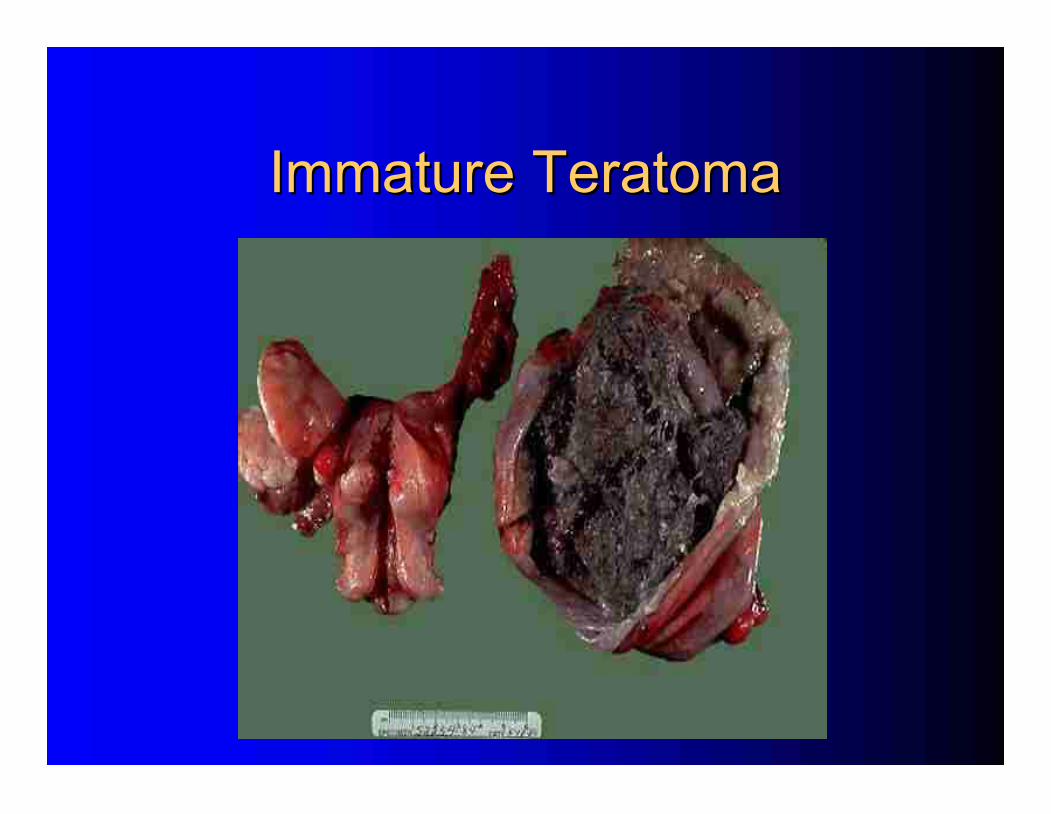
Immature Teratoma
Immature Teratoma
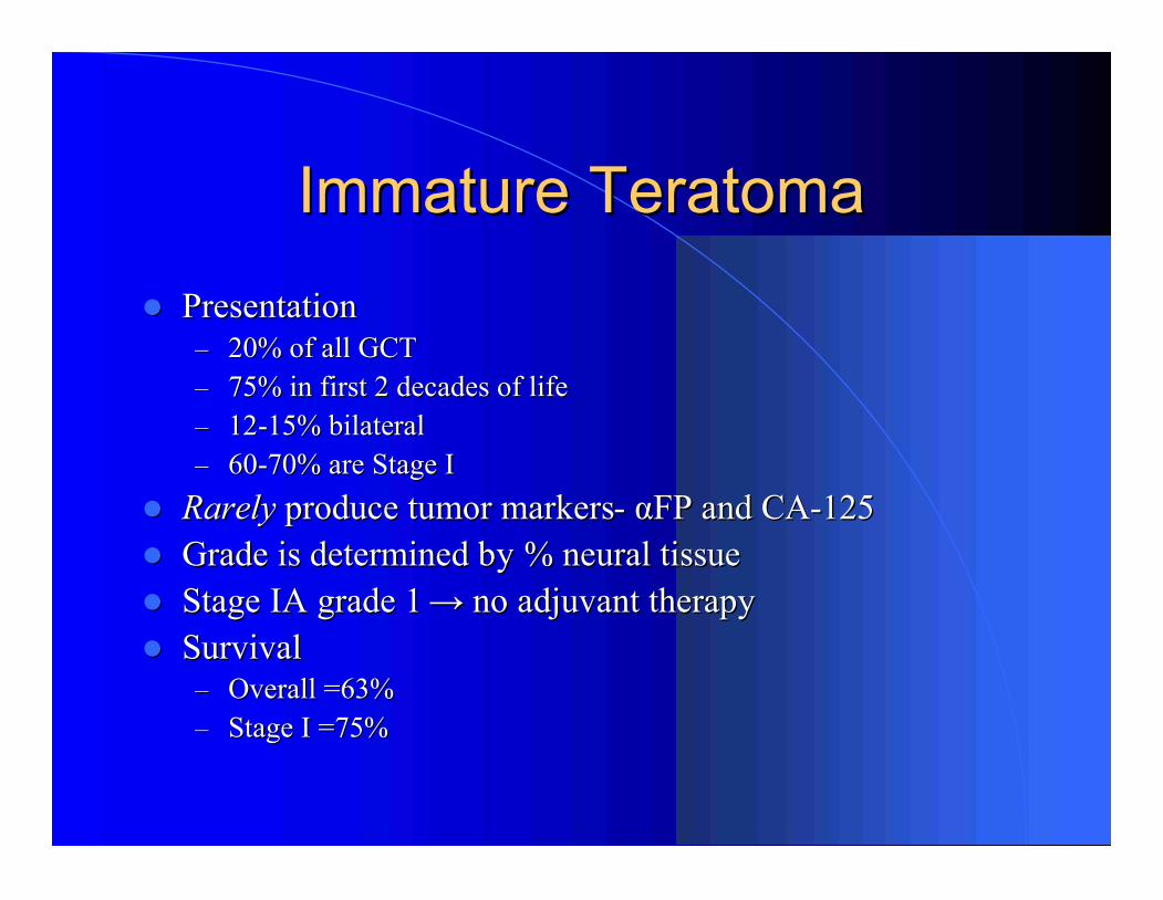
Presentation
Presentation
–
–
20% of all GCT
20% of all GCT
–
–
75% in first 2 decades of life
75% in first 2 decades of life
–
–
12
12
-
-
15% bilateral
15% bilateral
–
–
60
60
-
-
70% are Stage I
70% are Stage I
Rarely
Rarely
produce tumor markers
produce tumor markers
-
-
α
α
FP and CA
FP and CA
-
-
125
125
Grade is determined by % neural tissue
Grade is determined by % neural tissue
Stage IA grade 1
Stage IA grade 1
→
→
no adjuvant therapy
no adjuvant therapy
Survival
Survival
–
–
Overall =63%
Overall =63%
–
–
Stage I =75%
Stage I =75%
Immature Teratoma
Immature Teratoma
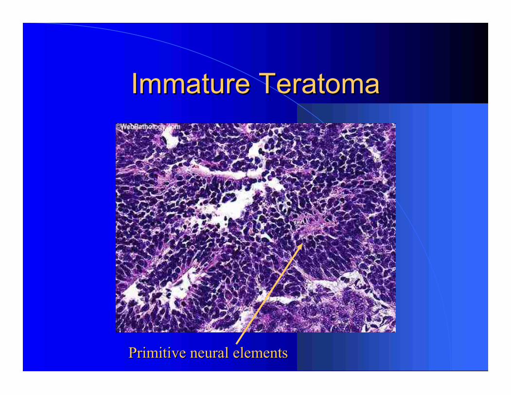
Immature Teratoma
Immature Teratoma
Primitive neural elements
Primitive neural elements
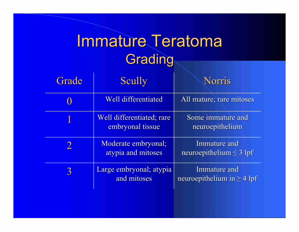
Immature Teratoma
Immature Teratoma
Grading
Grading
Immature and
Immature and
neuroepithelium
neuroepithelium
≤
≤
3 lpf
3 lpf
Moderate embryonal;
Moderate embryonal;
atypia and mitoses
atypia and mitoses
2
2
Immature and
Immature and
neuroepithelium in
neuroepithelium in
≥
≥
4 lpf
4 lpf
Large embryonal; atypia
Large embryonal; atypia
and mitoses
and mitoses
3
3
Some immature and
Some immature and
neuroepithelium
neuroepithelium
Well differentiated; rare
Well differentiated; rare
embryonal tissue
embryonal tissue
1
1
All mature; rare mitoses
All mature; rare mitoses
Well differentiated
Well differentiated
0
0
Norris
Norris
Scully
Scully
Grade
Grade
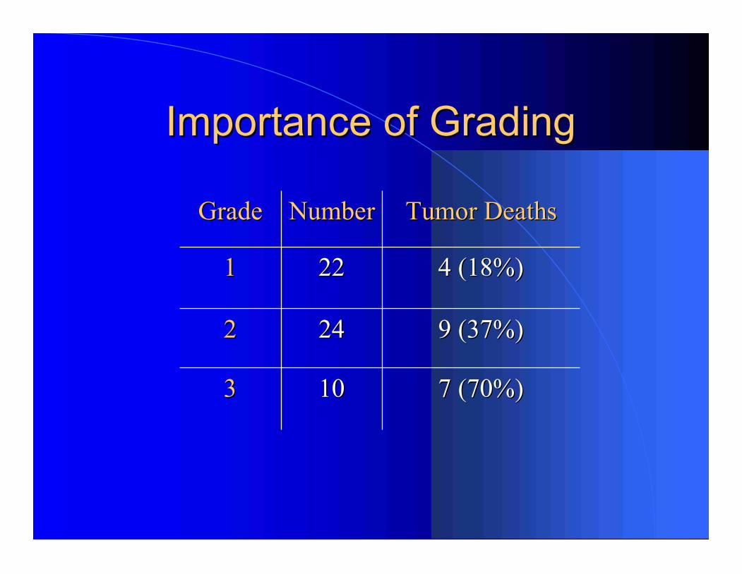
Importance of Grading
Importance of Grading
7 (70%)
7 (70%)
10
10
3
3
9 (37%)
9 (37%)
24
24
2
2
4 (18%)
4 (18%)
22
22
1
1
Tumor Deaths
Tumor Deaths
Number
Number
Grade
Grade
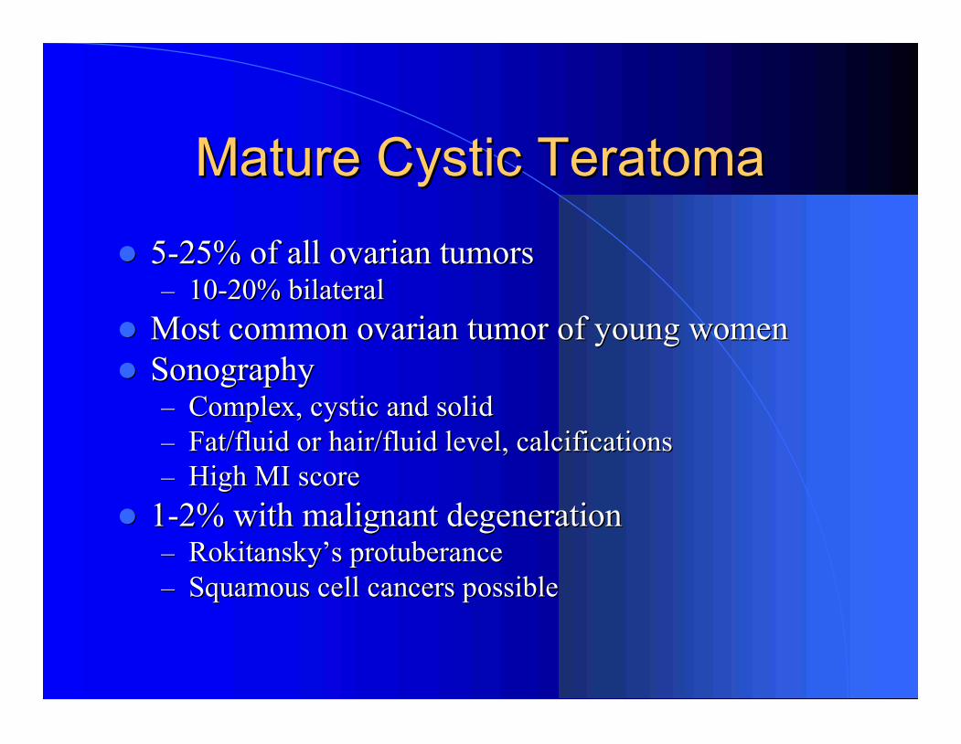
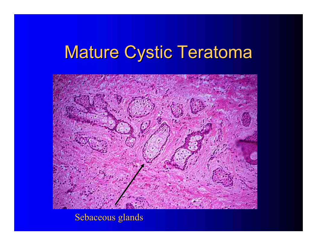
Mature Cystic Teratoma
Mature Cystic Teratoma
5
5
-
-
25% of all ovarian tumors
25% of all ovarian tumors
–
–
10
10
-
-
20% bilateral
20% bilateral
Most common ovarian tumor of young women
Most common ovarian tumor of young women
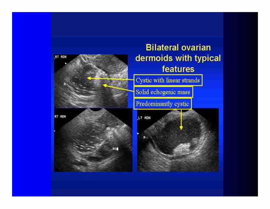
Sonography
Sonography
–
–
Complex, cystic and solid
Complex, cystic and solid
–
–
Fat/fluid or hair/fluid level, calcifications
Fat/fluid or hair/fluid level, calcifications
–
–
High MI score
High MI score
1
1
-
-
2% with malignant degeneration
2% with malignant degeneration
–
–
Rokitansky
Rokitansky
’
’
s
s
protuberance
protuberance
–
–
Squamous cell cancers possible
Squamous cell cancers possible
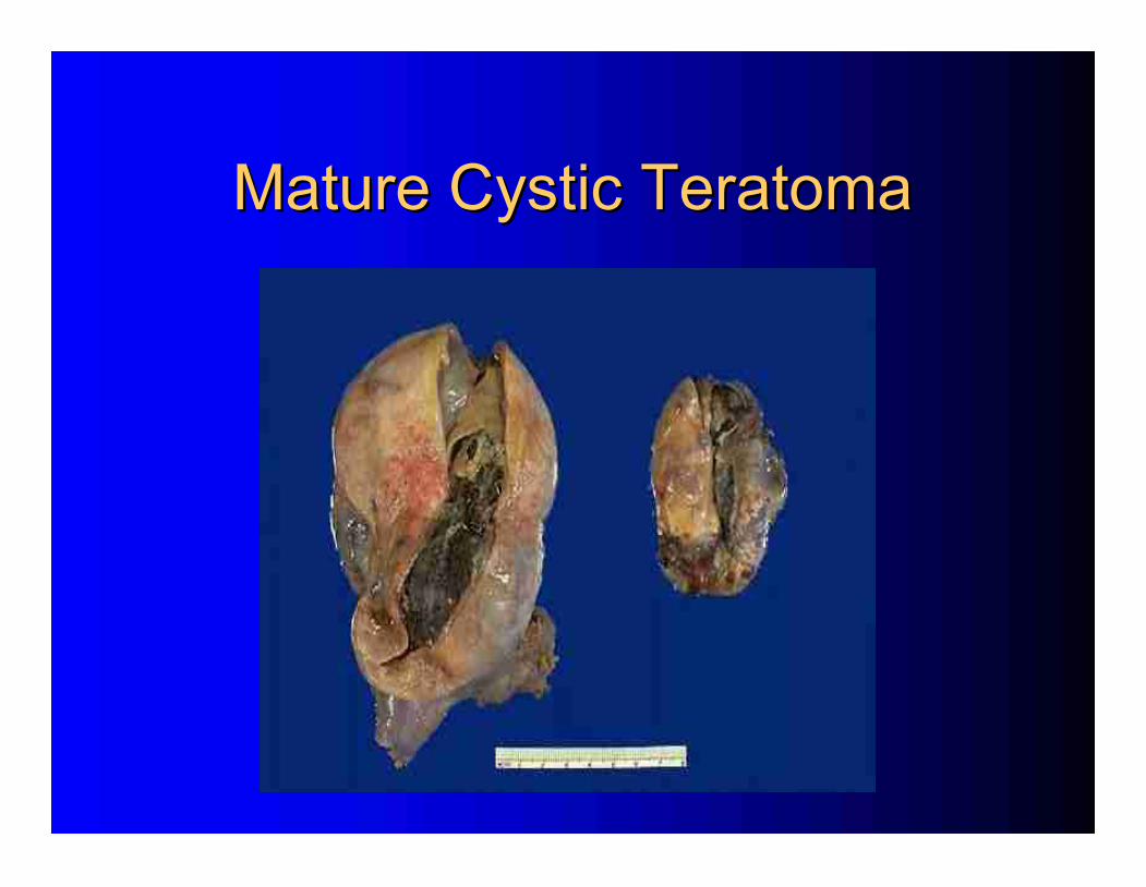
Mature Cystic Teratoma
Mature Cystic Teratoma
Mature Cystic Teratoma
Mature Cystic Teratoma
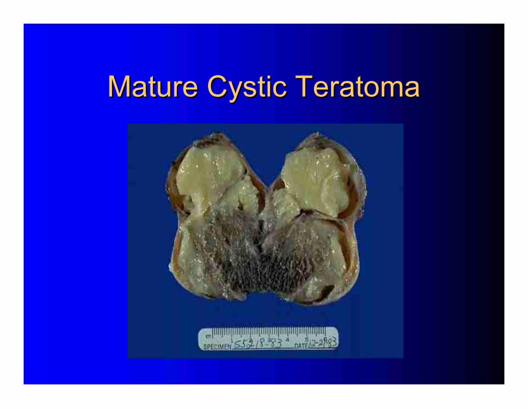
Mature Cystic Teratoma
Mature Cystic Teratoma
Sebaceous glands
Sebaceous glands
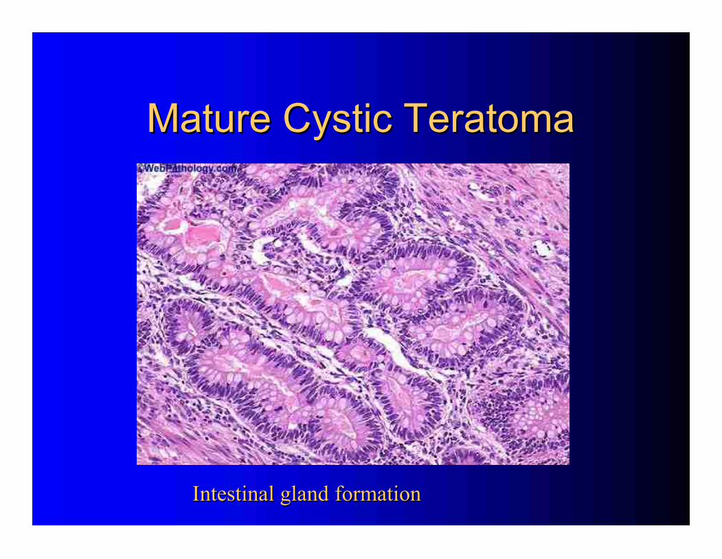
Mature Cystic Teratoma
Mature Cystic Teratoma
Intestinal gland formation
Intestinal gland formation
Specialized Teratomas
Specialized Teratomas
Struma ovarii
Struma ovarii
–
–
2
2
-
-
3% of all teratomas
3% of all teratomas
–
–
25
25
-
-
35% have symptoms of hyperthyroidism
35% have symptoms of hyperthyroidism
–
–
Usually benign, but may undergo malignant transformation
Usually benign, but may undergo malignant transformation
Follicular type
Follicular type
Carcinoid tumors
Carcinoid tumors
–
–
Associated with GI or respiratory epithelium
Associated with GI or respiratory epithelium
–
–
Primary ovarian tumors are rare (N=50)
Primary ovarian tumors are rare (N=50)
–
–
Often PMP
Often PMP
–
–
1/3 have carcinoid syndrome from serotonin
1/3 have carcinoid syndrome from serotonin
–
–
Symptoms resolve with excision
Symptoms resolve with excision
–
–
5
5
-
-
hydroxyindoleacetic acid in urine
hydroxyindoleacetic acid in urine
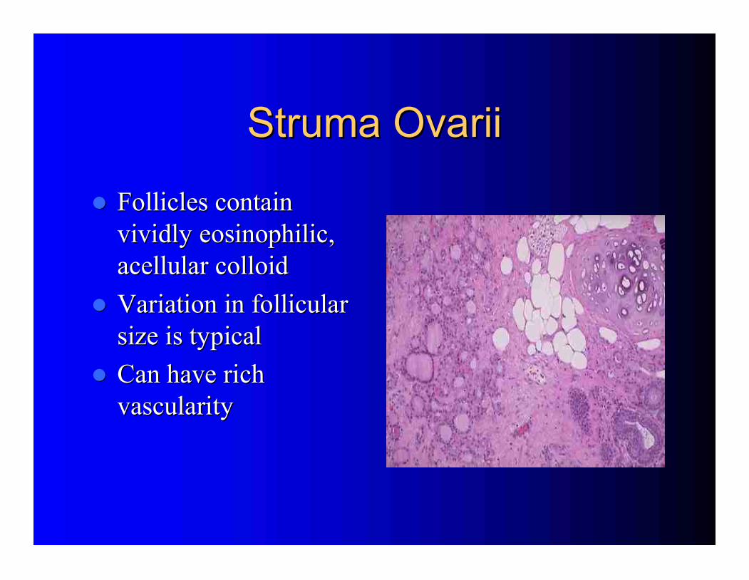
Struma Ovarii
Struma Ovarii
Follicles contain
Follicles contain
vividly eosinophilic,
vividly eosinophilic,
acellular colloid
acellular colloid
Variation in follicular
Variation in follicular
size is typical
size is typical
Can have rich
Can have rich
vascularity
vascularity
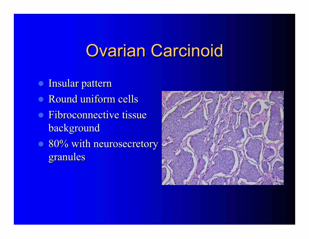
Ovarian Carcinoid
Ovarian Carcinoid
Insular pattern
Insular pattern
Round uniform cells
Round uniform cells
Fibroconnective tissue
Fibroconnective tissue
background
background
80% with neurosecretory
80% with neurosecretory
granules
granules
Choriocarcinoma
Choriocarcinoma
Presentation
Presentation
–
–
Uncommon, aggressive tumor
Uncommon, aggressive tumor
–
–
Often part of mixed GCT
Often part of mixed GCT
–
–
Consider met from gestational chorioCA
Consider met from gestational chorioCA
–
–
Mean age 20 yo, children common
Mean age 20 yo, children common
–
–
Half of premenarchal
Half of premenarchal
→
→
precocious puberty
precocious puberty
Tumor marker
Tumor marker
–
–
hCG
hCG
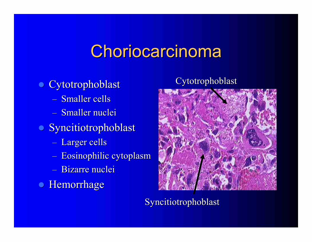
Choriocarcinoma
Choriocarcinoma
Cytotrophoblast
Cytotrophoblast
–
–
Smaller cells
Smaller cells
–
–
Smaller nuclei
Smaller nuclei
Syncitiotrophoblast
Syncitiotrophoblast
–
–
Larger cells
Larger cells
–
–
Eosinophilic cytoplasm
Eosinophilic cytoplasm
–
–
Bizarre nuclei
Bizarre nuclei
Hemorrhage
Hemorrhage
Cytotrophoblast
Cytotrophoblast
Syncitiotrophoblast
Syncitiotrophoblast
Embryonal Carcinoma
Embryonal Carcinoma
Presentation
Presentation
–
–
Mean age < 30 yo
Mean age < 30 yo
–
–
Only 4% of GCT and often part of mixed tumor
Only 4% of GCT and often part of mixed tumor
–
–
60% Stage IA
60% Stage IA
–
–
Poorly differentiated germ cell tumor
Poorly differentiated germ cell tumor
–
–
Aggressive, intra
Aggressive, intra
-
-
abdominal spread and mets common
abdominal spread and mets common
Tumor markers: hCG,
Tumor markers: hCG,
α
α
FP
FP
Survival
Survival
–
–
Overall =40%
Overall =40%
–
–
Stage I =75%
Stage I =75%
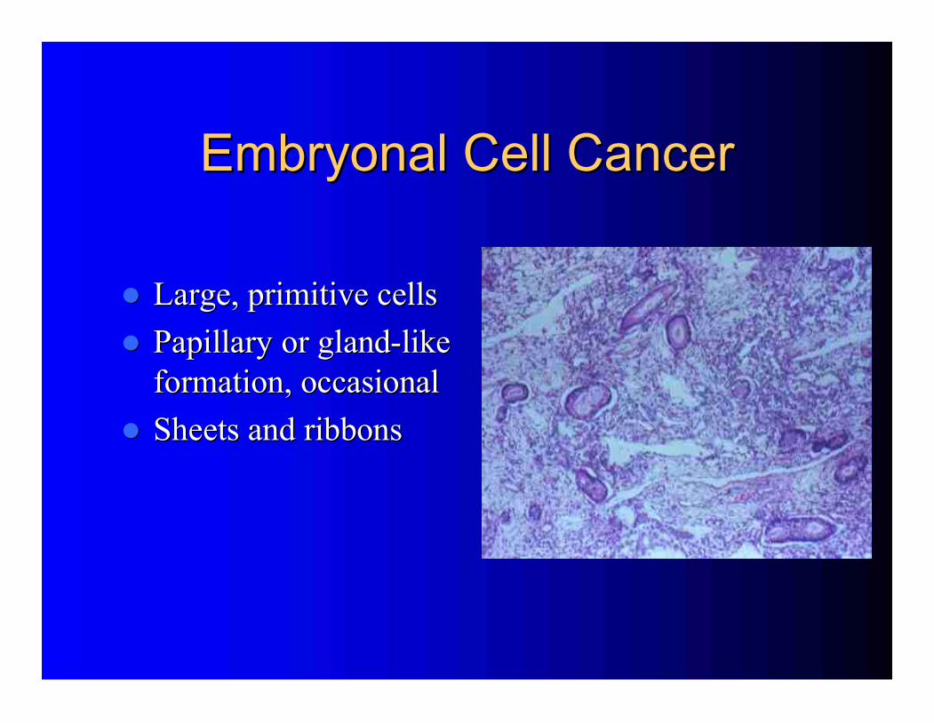
Embryonal Cell Cancer
Embryonal Cell Cancer
Large, primitive cells
Large, primitive cells
Papillary or gland
Papillary or gland
-
-
like
like
formation, occasional
formation, occasional
Sheets and ribbons
Sheets and ribbons
Polyembryoma
Polyembryoma
Best classified as a mixed tumor
Best classified as a mixed tumor
–
–
Never found in pure form
Never found in pure form
–
–
fewer than 50 cases
fewer than 50 cases
–
–
All under age 40
All under age 40
Resembles embryonal carcinoma
Resembles embryonal carcinoma
–
–
Embryo days 13
Embryo days 13
-
-
15
15
Treated like other mixed GCT
Treated like other mixed GCT
Tumor markers:
Tumor markers:
hCG,
hCG,
α
α
FP
FP
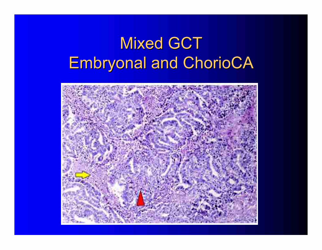
Mixed GCT
Mixed GCT
Embryonal and ChorioCA
Embryonal and ChorioCA
Gonadoblastoma
Gonadoblastoma
Combined Germ Cell / Sex Cord Stromal Tumor
Combined Germ Cell / Sex Cord Stromal Tumor
Presentation
Presentation
–
–
Age 1
Age 1
-
-
38
38
–
–
Small tumors
Small tumors
–
–
Phenotypic
Phenotypic
♀
♀
with virilization
with virilization
90% have Y chromosome
90% have Y chromosome
22% from streak gonads
22% from streak gonads
–
–
Bilaterality 30
Bilaterality 30
-
-
50%
50%
Check chromosomes for dysgenic gonads
Check chromosomes for dysgenic gonads
–
–
BSO if Y
BSO if Y
–
–
If TFS, await puberty before BSO
If TFS, await puberty before BSO

Gonadoblastoma
Gonadoblastoma
Large germ cells, clear
Large germ cells, clear
cytoplasm
cytoplasm
Nests of primordial
Nests of primordial
germ cells surrounded
germ cells surrounded
by specialized stromal
by specialized stromal
cells
cells
Associated sex chord
Associated sex chord
stromal cells
stromal cells
Gonadoblastoma
Gonadoblastoma
Dysgerminoma
Dysgerminoma
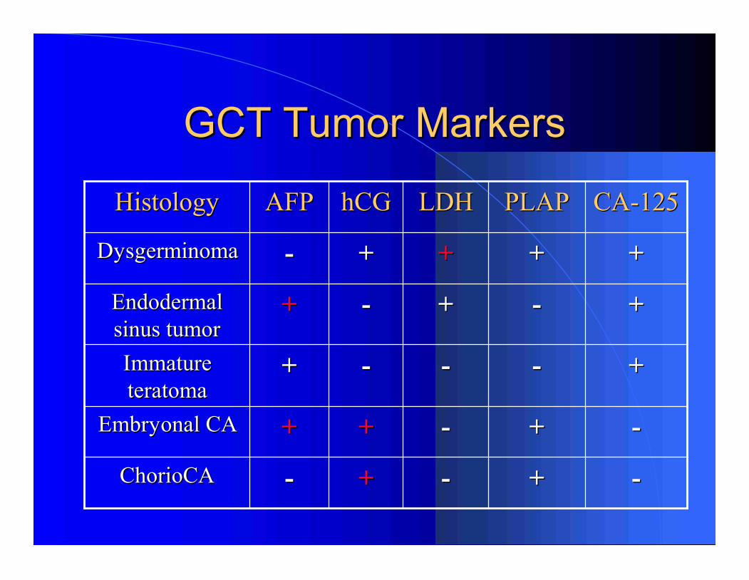
GCT Tumor Markers
GCT Tumor Markers
-
-
+
+
-
-
+
+
-
-
ChorioCA
ChorioCA
-
-
+
+
-
-
+
+
+
+
Embryonal CA
Embryonal CA
+
+
-
-
-
-
-
-
+
+
Immature
Immature
teratoma
teratoma
+
+
-
-
+
+
-
-
+
+
Endodermal
Endodermal
sinus tumor
sinus tumor
+
+
+
+
+
+
+
+
-
-
Dysgerminoma
Dysgerminoma
CA
CA
-
-
125
125
PLAP
PLAP
LDH
LDH
hCG
hCG
AFP
AFP
Histology
Histology
Treatment
Treatment
Germ Cell Tumors
Germ Cell Tumors
Surgery
Surgery
Importance of staging in early disease
Importance of staging in early disease
Fertility
Fertility
-
-
sparing surgery often required
sparing surgery often required
–
–
Can preserve uterus for future IVF, even if
Can preserve uterus for future IVF, even if
BSO
BSO
Debulking improves outcome
Debulking improves outcome
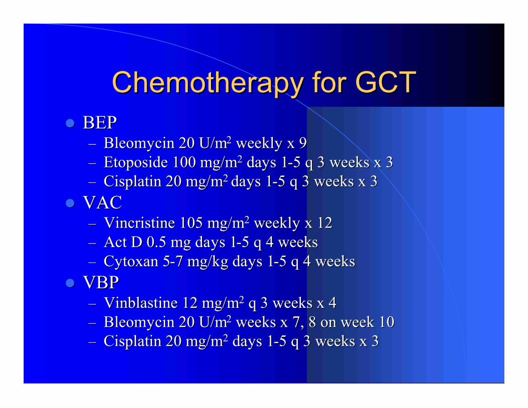
Chemotherapy for GCT
Chemotherapy for GCT
BEP
BEP
–
–
Bleomycin 20 U/m
Bleomycin 20 U/m
2
2
weekly x 9
weekly x 9
–
–
Etoposide 100 mg/m
Etoposide 100 mg/m
2
2
days 1
days 1
-
-
5 q 3 weeks x 3
5 q 3 weeks x 3
–
–
Cisplatin 20 mg/m
Cisplatin 20 mg/m
2
2
days 1
days 1
-
-
5 q 3 weeks x 3
5 q 3 weeks x 3
VAC
VAC
–
–
Vincristine 105 mg/m
Vincristine 105 mg/m
2
2
weekly x 12
weekly x 12
–
–
Act D 0.5 mg days 1
Act D 0.5 mg days 1
-
-
5 q 4 weeks
5 q 4 weeks
–
–
Cytoxan 5
Cytoxan 5
-
-
7 mg/kg days 1
7 mg/kg days 1
-
-
5 q 4 weeks
5 q 4 weeks
VBP
VBP
–
–
Vinblastine 12 mg/m
Vinblastine 12 mg/m
2
2
q 3 weeks x 4
q 3 weeks x 4
–
–
Bleomycin 20 U/m
Bleomycin 20 U/m
2
2
weeks x 7, 8 on week 10
weeks x 7, 8 on week 10
–
–
Cisplatin 20 mg/m
Cisplatin 20 mg/m
2
2
days 1
days 1
-
-
5 q 3 weeks x 3
5 q 3 weeks x 3
Classification
Classification
Stromal Tumors
Stromal Tumors
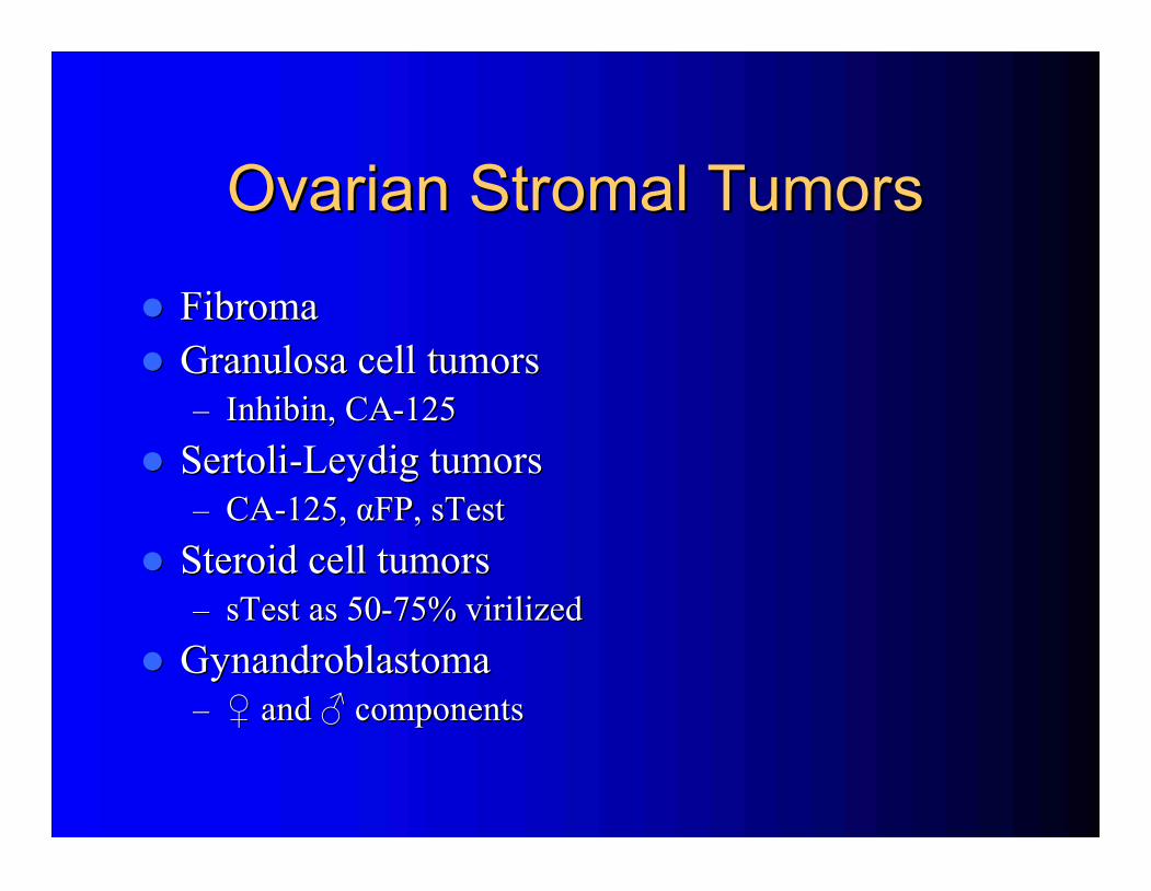
Ovarian Stromal Tumors
Ovarian Stromal Tumors
Fibroma
Fibroma
Granulosa cell tumors
Granulosa cell tumors
–
–
Inhibin, CA
Inhibin, CA
-
-
125
125
Sertoli
Sertoli
-
-
Leydig tumors
Leydig tumors
–
–
CA
CA
-
-
125,
125,
α
α
FP, sTest
FP, sTest
Steroid cell tumors
Steroid cell tumors
–
–
sTest as 50
sTest as 50
-
-
75% virilized
75% virilized
Gynandroblastoma
Gynandroblastoma
–
–
♀
♀
and
and
♂
♂
components
components
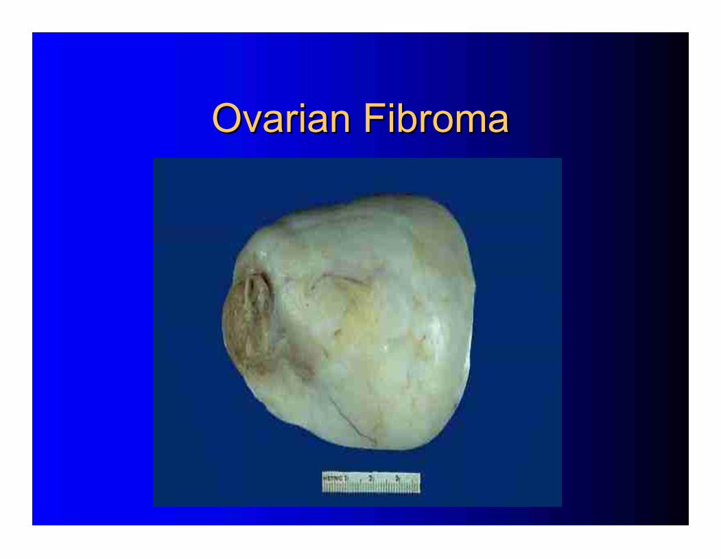
Ovarian Fibroma
Ovarian Fibroma
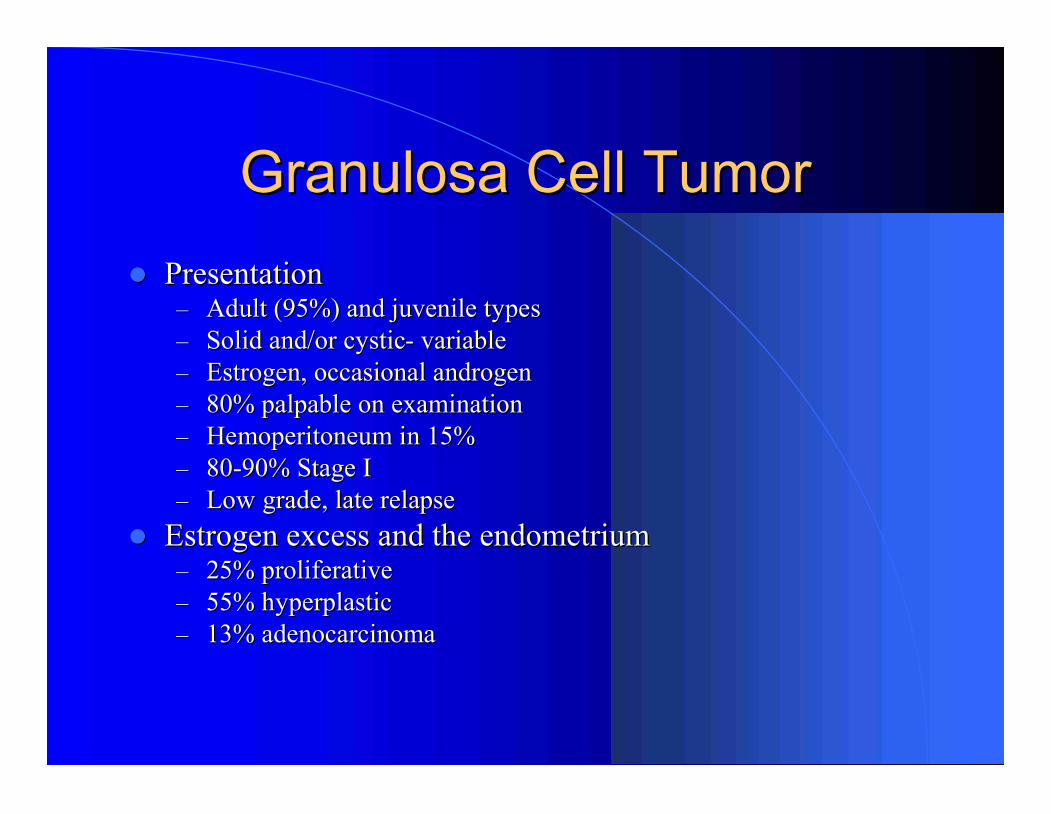
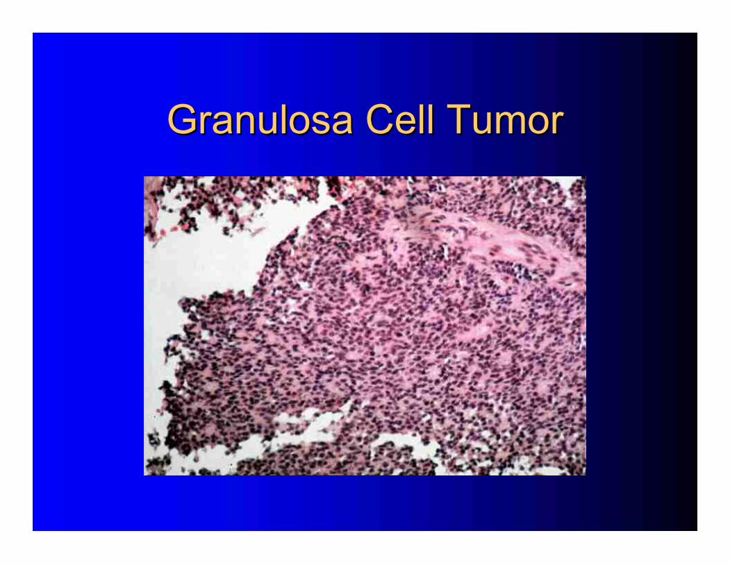
Granulosa Cell Tumor
Granulosa Cell Tumor
Presentation
Presentation
–
–
Adult (95%) and juvenile types
Adult (95%) and juvenile types
–
–
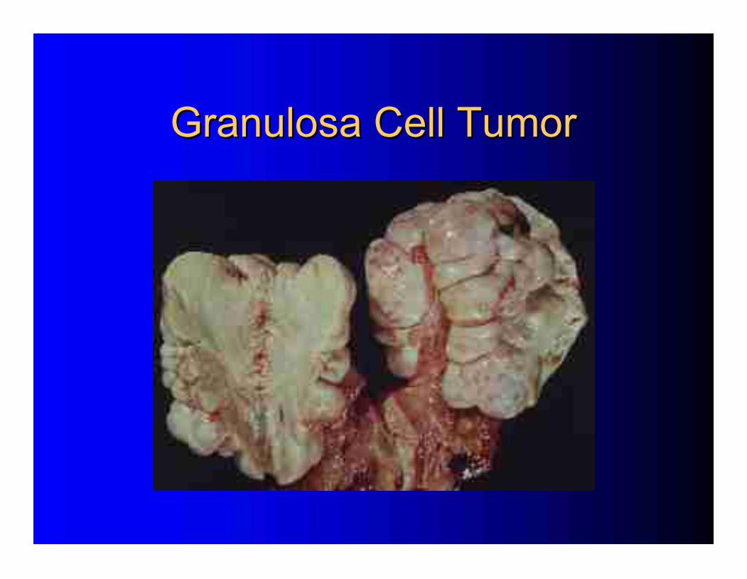
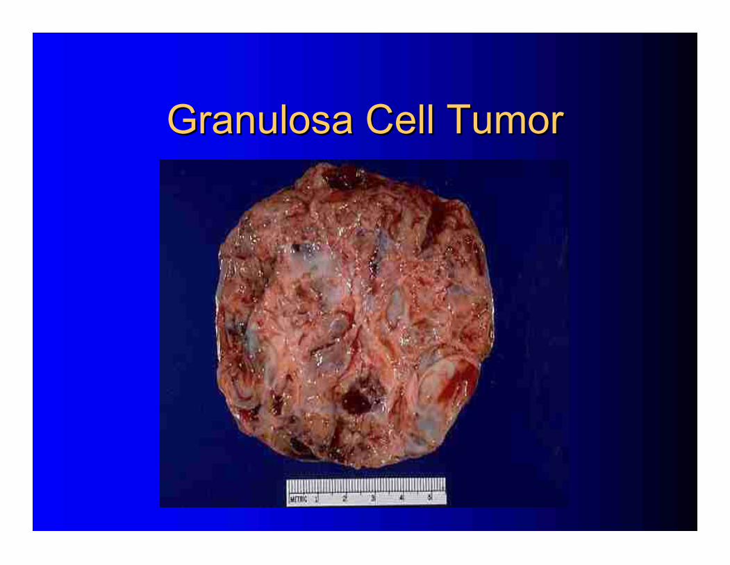
Solid and/or cystic
Solid and/or cystic
-
-
variable
variable
–
–
Estrogen, occasional androgen
Estrogen, occasional androgen
–
–
80% palpable on examination
80% palpable on examination
–
–
Hemoperitoneum in 15%
Hemoperitoneum in 15%
–
–
80
80
-
-
90% Stage I
90% Stage I
–
–
Low grade, late relapse
Low grade, late relapse
Estrogen excess and the endometrium
Estrogen excess and the endometrium
–
–
25% proliferative
25% proliferative
–
–
55% hyperplastic
55% hyperplastic
–
–
13% adenocarcinoma
13% adenocarcinoma
Granulosa Cell Tumor
Granulosa Cell Tumor
Treatment
Treatment
Juvenile
Juvenile
–
–
High cure rate
High cure rate
Adult
Adult
–
–
Resection
Resection
–
–
Chemotherapy
Chemotherapy
BEP
BEP
Carboplatin and Taxol
Carboplatin and Taxol
GnRH analogs
GnRH analogs
Granulosa Cell Tumor
Granulosa Cell Tumor
Granulosa Cell Tumor
Granulosa Cell Tumor
Granulosa Cell Tumor
Granulosa Cell Tumor
Granulosa Cell Tumor
Granulosa Cell Tumor
Granulosa Cell Tumor
Granulosa Cell Tumor
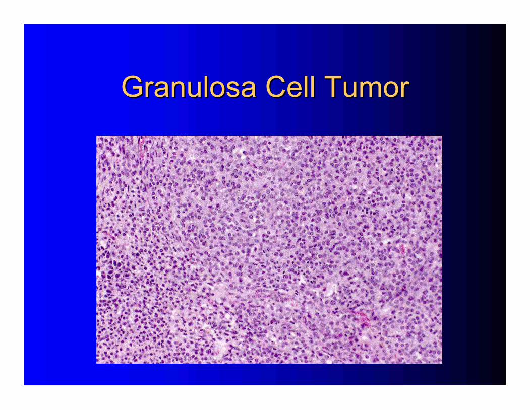
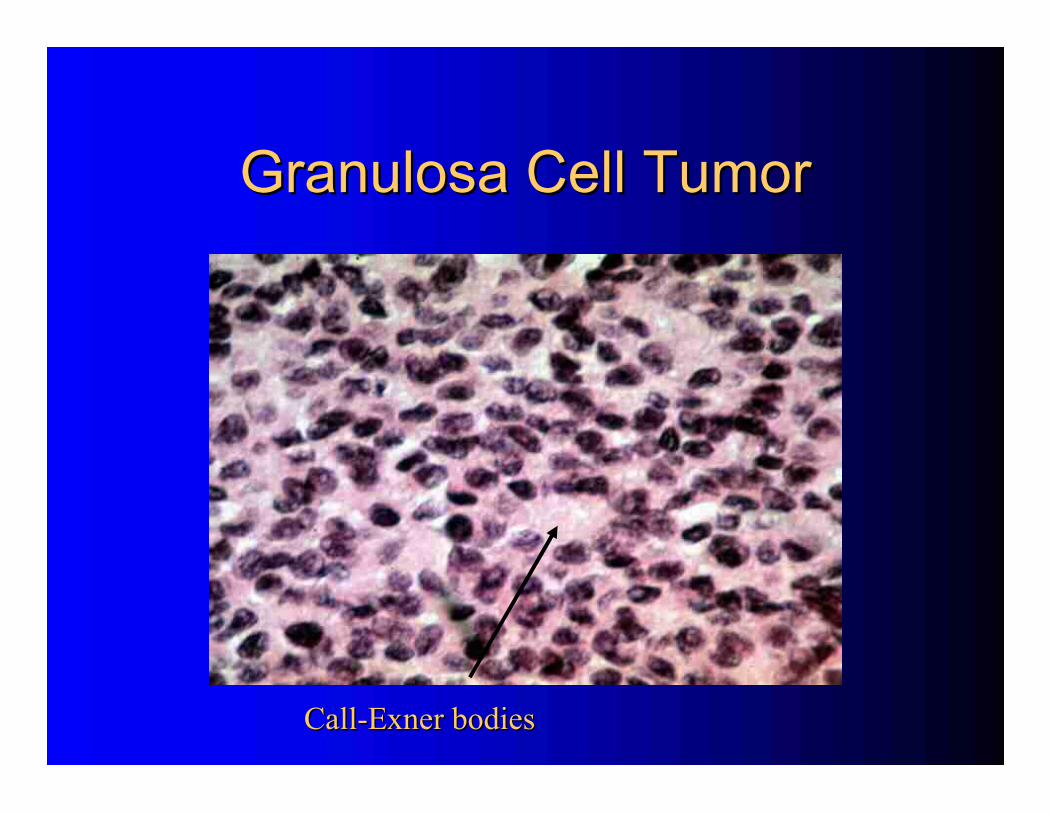
Call
Call
-
-
Exner
Exner
bodies
bodies
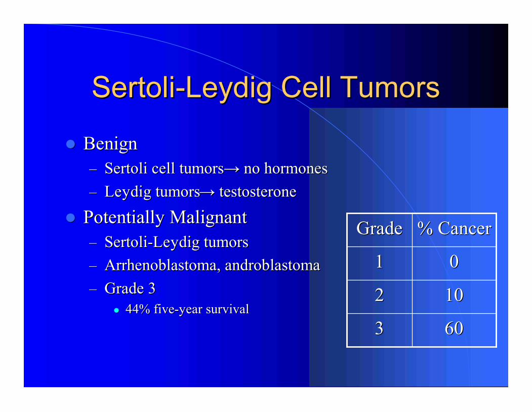
Sertoli
Sertoli
-
-
Leydig Cell Tumors
Leydig Cell Tumors
Benign
Benign
–
–
Sertoli cell tumors
Sertoli cell tumors
→
→
no hormones
no hormones
–
–
Leydig tumors
Leydig tumors
→
→
testosterone
testosterone
Potentially Malignant
Potentially Malignant
–
–
Sertoli
Sertoli
-
-
Leydig tumors
Leydig tumors
–
–
Arrhenoblastoma, androblastoma
Arrhenoblastoma, androblastoma
–
–
Grade 3
Grade 3
44% five
44% five
-
-
year survival
year survival
0
0
1
1
% Cancer
% Cancer
Grade
Grade
60
60
3
3
10
10
2
2
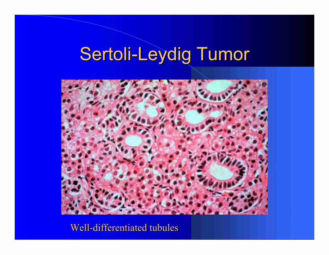
Sertoli
Sertoli
-
-
Leydig Tumor
Leydig Tumor
Well
Well
-
-
differentiated tubules
differentiated tubules
Treatment Summary
Treatment Summary
Germ Cell and Stroma Tumors
Germ Cell and Stroma Tumors
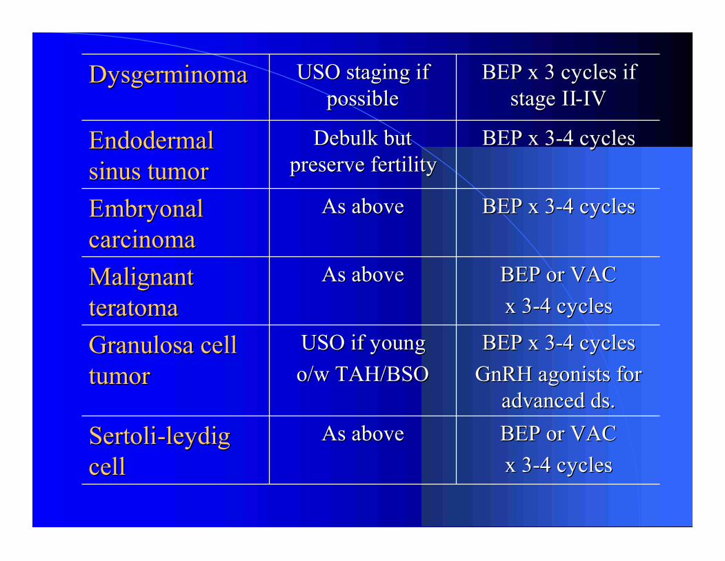
BEP or VAC
BEP or VAC
x 3
x 3
-
-
4 cycles
4 cycles
As above
As above
Sertoli
Sertoli
-
-
leydig
leydig
cell
cell
BEP x 3
BEP x 3
-
-
4 cycles
4 cycles
GnRH agonists for
GnRH agonists for
advanced ds.
advanced ds.
USO if young
USO if young
o/w
o/w
TAH/BSO
TAH/BSO
Granulosa cell
Granulosa cell
tumor
tumor
BEP or VAC
BEP or VAC
x 3
x 3
-
-
4 cycles
4 cycles
As above
As above
Malignant
Malignant
teratoma
teratoma
BEP x 3
BEP x 3
-
-
4 cycles
4 cycles
As above
As above
Embryonal
Embryonal
carcinoma
carcinoma
BEP x 3
BEP x 3
-
-
4 cycles
4 cycles
Debulk
Debulk
but
but
preserve fertility
preserve fertility
Endodermal
Endodermal
sinus tumor
sinus tumor
BEP x 3 cycles if
BEP x 3 cycles if
stage II
stage II
-
-
IV
IV
USO staging if
USO staging if
possible
possible
Dysgerminoma
Dysgerminoma
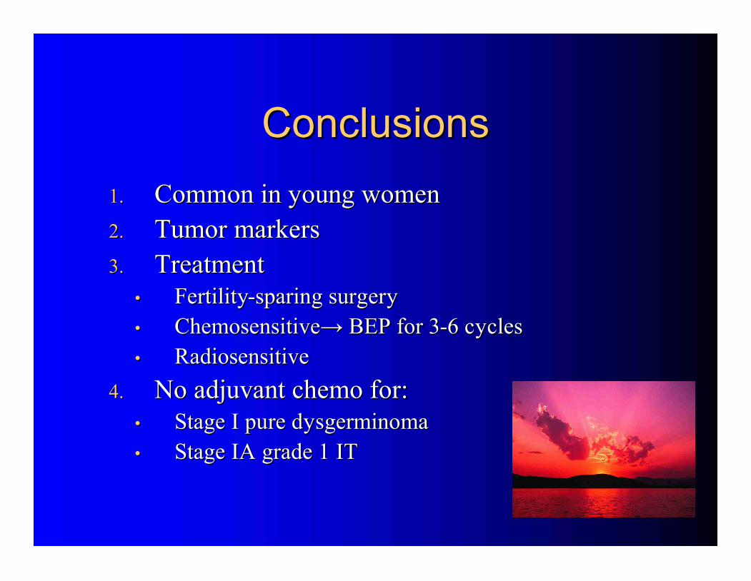
Conclusions
Conclusions
1.
1.
Common in young women
Common in young women
2.
2.
Tumor markers
Tumor markers
3.
3.
Treatment
Treatment
•
•
Fertility
Fertility
-
-
sparing surgery
sparing surgery
•
•
Chemosensitive
Chemosensitive
→
→
BEP for 3
BEP for 3
-
-
6 cycles
6 cycles
•
•
Radiosensitive
Radiosensitive
4.
4.
No adjuvant chemo for:
No adjuvant chemo for:
•
•
Stage I pure dysgerminoma
Stage I pure dysgerminoma
•
•
Stage IA grade 1 IT
Stage IA grade 1 IT