Embed Size (px)
Citation preview
REPORT ON EMERGING TECHNOLOGY
GI endoscopes
cttaflvta
T
at
sdeustiathsctehdtsti
ccstwmteset
The ASGE Technology Committee provides reviews ofexisting, new, or emerging endoscopic technologies thathave an impact on the practice of GI endoscopy. Evidence-based methodology is used, performing a MEDLINE litera-ture search to identify pertinent clinical studies on thetopic and a MAUDE (U.S. Food and Drug AdministrationCenter for Devices and Radiological Health) databasesearch to identify the reported complications of a giventechnology. Both are supplemented by accessing the “re-lated articles” feature of PubMed and by scrutinizingpertinent references cited by the identified studies. Con-trolled clinical trials are emphasized, but in many casesdata from randomized, controlled trials are lacking. Insuch cases, large case series, preliminary clinical studies,and expert opinions are used. Technical data are gatheredfrom traditional and Web-based publications, proprietarypublications, and informal communications with perti-nent vendors.
Technology Status Evaluation Reports are drafted by 1or 2 members of the ASGE Technology Committee, re-viewed and edited by the Committee as a whole, andapproved by the Governing Board of the ASGE. Whenfinancial guidance is indicated, the most recent codingdata and list prices at the time of publication are provided.For this review, the MEDLINE database was searchedthrough September 2010 for articles related to endoscopyby using the key words “gastroscope,” “colonoscope,”“echoendoscope,” “duodenoscope,” “choledochoscope,”“ultraslim endoscope,” “variable stiffness colonoscope,”and “wide-angle colonoscope.”
Technology Status Evaluation Reports are scientific re-views provided solely for educational and informationalpurposes. Technology Status Evaluation Reports are notrules and should not be construed as establishing a legalstandard of care or as encouraging, advocating, requir-ing, or discouraging any particular treatment or paymentfor such treatment.
BACKGROUND
GI endoscopes are devices used for the examinationand treatment of the GI tract. They have evolved fromearly rigid designs with limited capabilities to more sophisti-
Copyright © 2011 by the American Society for Gastrointestinal Endoscopy0016-5107/$36.00
edoi:10.1016/j.gie.2011.01.061
www.giejournal.org
ated flexible instruments with advanced imaging capabili-ies, specialized features for advanced therapeutic interven-ions, and different designs to enable examination of specificreas of the GI tract. This document is an overview of allexible endoscopes. Separate, more detailed technical re-iews are available for echoendoscopes, enteroscopes, ultra-hin endoscopes, choledochoscopes, and high-resolutionnd high-magnification endoscopes.1-5
ECHNICAL CONSIDERATIONS
The basic design of a flexible endoscope is similar forll models and consists of 3 main parts: the control section,he insertion tube, and the connector section.
The control section is held in the left hand and has 2tacked control dials that deflect the instrument tip up/own and left/right. Some endoscopes (eg, some ultrathinndoscopes and choledochoscopes) have only 1 dial forp/down angulation; right/left angulation in these endo-copes is achieved by applying torque to the insertionube of the instrument. The control dials can be lockednto place for prolonged tip deflection. The control sectionlso has separate buttons for suction, air or water insuffla-ion, and image freeze and capture. Some endoscopesave additional programmable buttons for other functionsuch as image printing and video capture. Finally, theontrol section has the entry port for inserting accessorieshrough the channel of the instrument. Some specialtyndoscopes such as duodenoscopes and echoendoscopesave additional features on the control section, which areiscussed separately. Many endoscopes also have controlso activate advanced imaging features, and some colono-copes have a separate dial encircling the control sectionhat allows the user to change the flexibility of part of thensertion tube.
The insertion tube is a flexible shaft attached to theontrol section. The insertion tube contains a workinghannel that allows passage of accessories and enablesuction. Some endoscopes have dual working channelshat allow full suction when an accessory is in the otherorking channel; two accessories can also be passed si-ultaneously in these endoscopes during more complex
herapeutic procedures. Channel diameter varies amongndoscopes, ranging from 1.2 mm for some choledocho-copes to 6 mm for some therapeutic endoscopes. Somendoscopes have an auxiliary water channel that enableshem to be fitted with a foot-controlled water pump for
xtra flushing capabilities. The shaft also contains angula-Volume 74, No. 1 : 2011 GASTROINTESTINAL ENDOSCOPY 1
vtstgado
i
s
i(bEMsncstmild
seamisxlisgicitit
rwtiLlEfno9v
CA
G
riht3ptivlotUtswo
GI endoscopes
tion wires to enable deflection of the instrument tip. Thedegree of angulation of the tip of the insertion tube in anup/down or left/right plane varies among instruments.The tip of the insertion tube of video endoscopes containsa charge-coupled device (CCD) for color image genera-tion, a light guide illumination system, an opening for theair/water channel, a water jet to clear the lens, and anobjective lens. The lens may be oriented for forward-iewing, side-viewing, or oblique-viewing, depending on theype of endoscope. Although less commonly used today,ome fiberoptic endoscopes are still available. The insertionube or shaft of these instruments also contains 1 or 2 lightuide bundles and an image guide bundle. The length, di-meter, and flexibility of the insertion tube vary among en-oscope types and manufacturers (see Tables 1-9, availablenline at www.giejournal.org,).
The connector section attaches the endoscope to anmage processor, light and electrical source, air or CO2
source, and water.
Special features: magnification, high-definition, and enhanced imaging capability
Standard endoscopes magnify the endoscopic image 30to 35 times at baseline. Endoscopes with the ability toincrease magnification are also available. These “zoom”endoscopes optically magnify images up to 150 times bymeans of a movable optical lens in the endoscope tip toobtain a more detailed image of a target lesion whilemaintaining high resolution.5
High-definition (HD) endoscopes generate images with850,000 to more than 1 million pixels compared withimage signals of 100,000 to 400,000 pixels on standardendoscopes.5 They require HD-compatible image proces-ors and monitors to produce a true HD image.
Some endoscopes are equipped with enhanced imag-ng capabilities including narrow-band imaging (NBI)Olympus Medical Systems, Center Valley, Pa) and multi-and imaging (MBI) such as Fujinon Intelligent Colornhancement (Fujinon, Wayne, NJ) and i-SCAN (Pentax,ontvale, NJ). NBI uses filters to illuminate the tissue at
elected wavelengths of blue (415 nm) and green (540m), highlighting vascular detail from surrounding mu-osa. MBI processes the white-light image digitally, recon-tructing it through software rather than a filter to enhancehe appearance of the mucosa. MBI can be combined withagnification. These image enhancement techniques are
ntended to assist in diagnosing and further characterizingesions in the GI tract, but their utility is not yet defined. Aetailed technical review of NBI and MBI is available.6
Image processor/light sourceFiberoptic imaging, which is generally obsolete today
other than for a few specialty endoscopes (eg, some cho-ledochoscopes), is generated by a coherent bundle ofglass fibers that transmit an image from the tip of the
endoscope to the eyepiece. a2 GASTROINTESTINAL ENDOSCOPY Volume 74, No. 1 : 2011
All video endoscopes have a black and white, solid-tate image sensor called a CCD mounted at the tip of thendoscope, which allows an image to be transmitted vian electronic signal to a video processor for display on 1 orore video monitors. This signal is converted to a color
mage by 1 of 2 systems: a red green blue (RGB) sequentialystem or a color CCD system. The RGB system uses aenon lamp to arc white light through a rotating RGB filterocated between the lamp and the light guide; this resultsn bursts of red, green, and blue light to create a visualtrobe effect. When tissue is illuminated, the reflected red,reen, and blue images are sent through a CCD in thenstrument tip and transmitted to the image processor. Theolor CCD system differs in that a micro-mosaic color filters mounted over the CCD chip itself. Whitelight illumina-ion is provided by the xenon lamp, and reflected colormages on the CCD surface are processed by circuitry inhe image processor.
The front panel of the image processor contains theeceiver for the endoscope connector section, air andater pump control buttons, and light adjustment con-
rols. Some image processors incorporate the light sourcento the same chassis, but others separate these devices.ight is provided by a 100- to 300-W xenon lamp. Someight sources also have a 75-W halogen lamp for backup.ndoscopes are compatible only with image processorsrom their manufacturers, and RGB type endoscopes areot compatible with CCD-type image processors. A varietyf image displays and monitors are available. Tables 8 and(available online at www.giejournal.org) list available
ideo processors and US processors.
ATEGORIES OF ENDOSCOPESND INDICATIONS
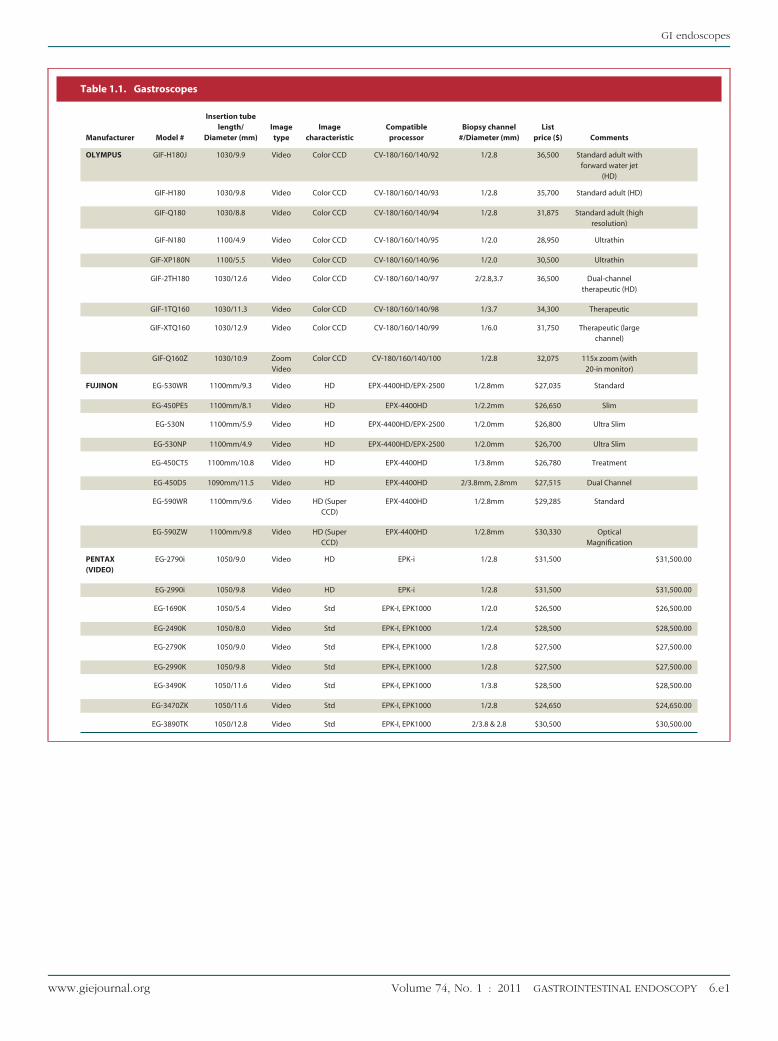
astroscopesGastroscopes are forward-viewing endoscopes with
elatively short insertion tubes designed primarily for usen the esophagus, stomach, and duodenum. Gastroscopesave variable insertion tube lengths (925-1100 mm), inser-ion tube diameters (4.9-12.8 mm), and channel sizes (2.0-.8 mm). Gastroscopes are available in standard adult,ediatric, and therapeutic models. Some therapeutic gas-roscopes have dual working channels for more complexnterventions. Another type of gastroscope is an ultrathinariety in which the insertion tube diameter is 6 mm oress. These endoscopes may be used for unsedated peroralr transnasal esophagoscopy or upper endoscopy or toraverse narrow areas (eg, tight strictures) in the GI tract.ltrathin endoscopes may have 2-way or 4-way tip deflec-
ion, insertion tube diameters between 4.9 and 6 mm, andmall instrument channels (1.5-2 mm).7 They are other-ise similar in design to gastroscopes. Table 1 (availablenline at www.giejournal.org) lists available gastroscopes
nd specifications.www.giejournal.org
ljs
si(hbescda
1m
bdetsfltdtp
atwlmaibetdsas
ofiwv
E
ssGpivtdspvridmhtba
GI endoscopes
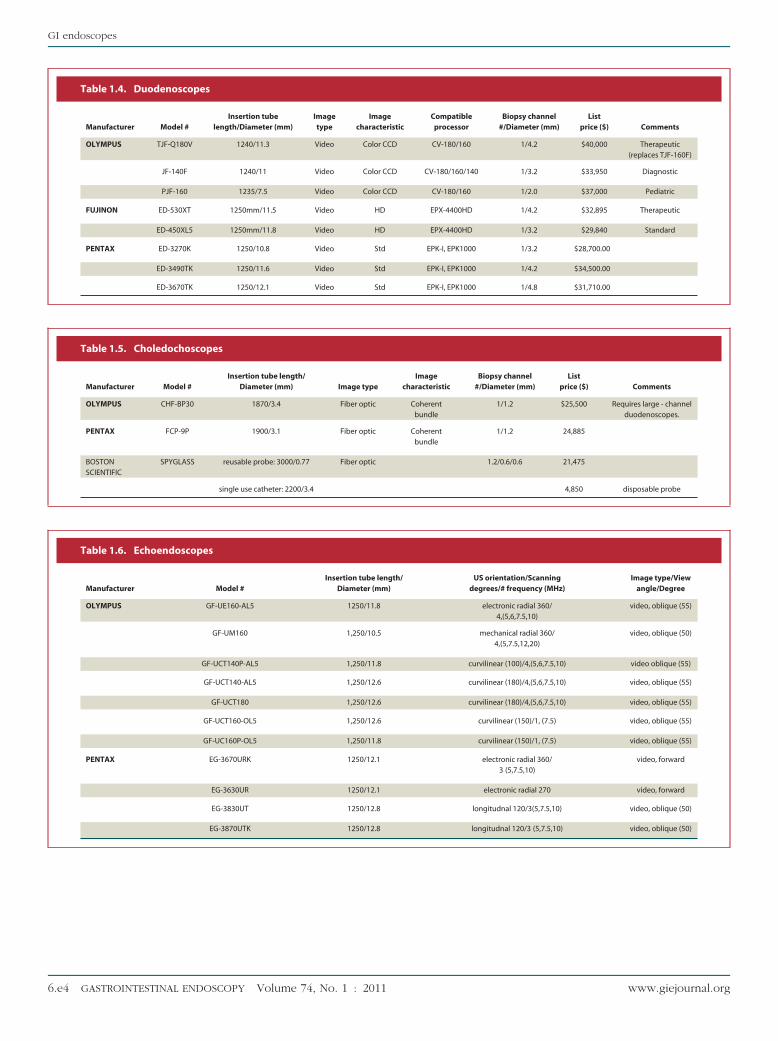
DuodenoscopesDuodenoscopes are side-viewing endoscopes designed
primarily for ERCP. They are available in standard andtherapeutic versions and have variable insertion tubelengths (1235-1250 mm), insertion tube diameters (7.5-12.1 mm), and channel sizes (2.0-4.8 mm). The tip of theduodenoscope has an elevator that raises accessoriespassed through the working channel into the field of view tofacilitate cannulation of the papilla and other interventions.The elevator is operated by the endoscopist via a smalllever on the control section. One available duodeno-scope (V-Scope; Olympus) also has a V-shaped groove atits tip to facilitate locking of guidewires passed throughthe instrument channel. Duodenoscopes with a largeworking channel (�4.2 mm) enable passage of a cho-edochoscope. Table 2 (available online at www.gie-ournal.org) lists available duodenoscopes and theirpecifications.
EnteroscopesEnteroscopes are forward-viewing endoscopes similar
to gastroscopes in design but with a much longer insertiontube used for examination of the duodenum, jejunum,and, in some cases, the ileum. They have working lengthsranging from 1520 to 2200 mm, channel diameters of 2.2mm to 3.8 mm, and insertion tube diameters of 9.2 to 11.6mm.8 Overtubes are sometimes used with these endo-copes to decrease gastric looping and enable deepernsertion. Single-balloon (Olympus) and double-balloonFujinon) enteroscopes are also available. These devicesave a specialized disposable overtube with an inflatablealloon that anchors the instrument in place during short-ning maneuvers. Double-balloon enteroscopes have aecond balloon on the endoscope insertion tube. Enteros-opy can be performed via the oral or anal approach,epending on the indication and lesion site. Table 3 (avail-ble online at www.giejournal.org) lists available entero-
scopes and specifications.
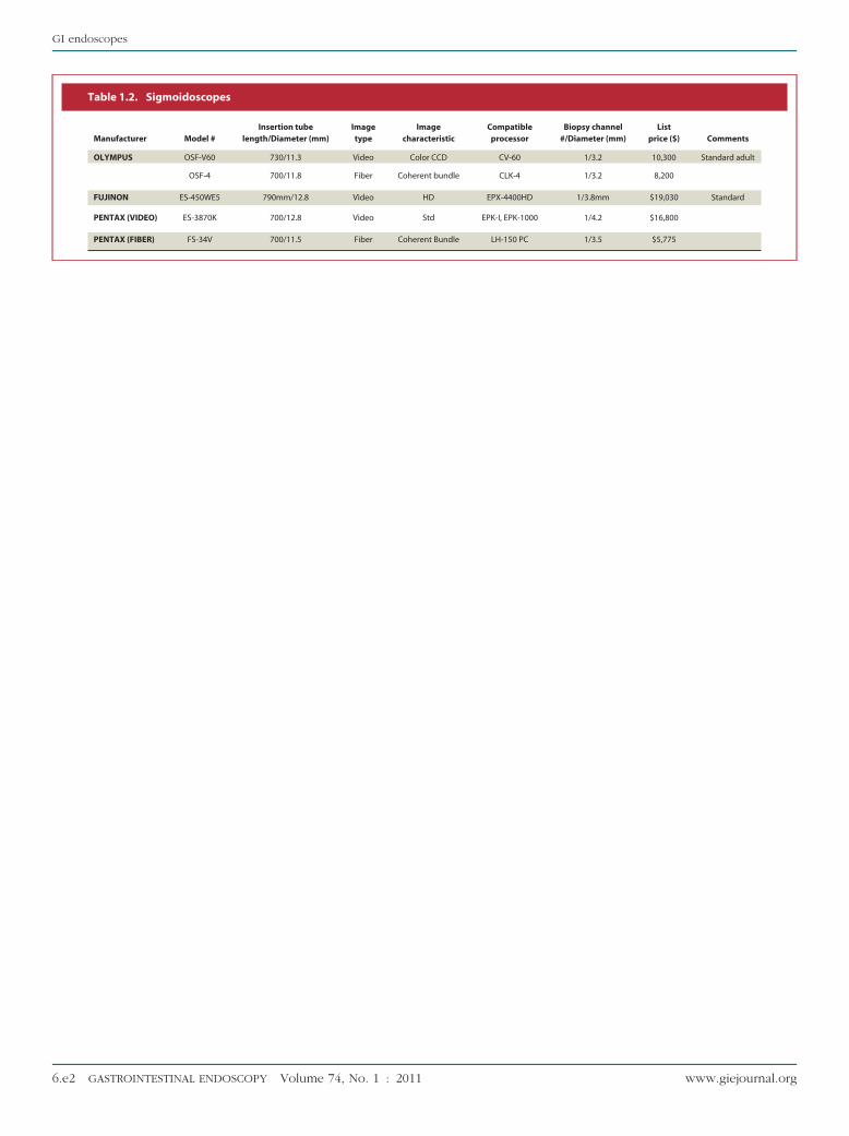
SigmoidoscopesSigmoidoscopes are relatively short forward-viewing
endoscopes designed for examination of the distal colon.They have variable insertion tube lengths (700-790 mm),insertion tube diameters (11.3-12.8 mm), and channelsizes (3.2-4.2 mm). Rigid sigmoidoscopes are used primar-ily by surgeons when evaluating malignancy of the rectumand distal colon or for foreign-body retrieval. Table 4(available online at www.giejournal.org) lists available sig-moidoscopes and their indications.
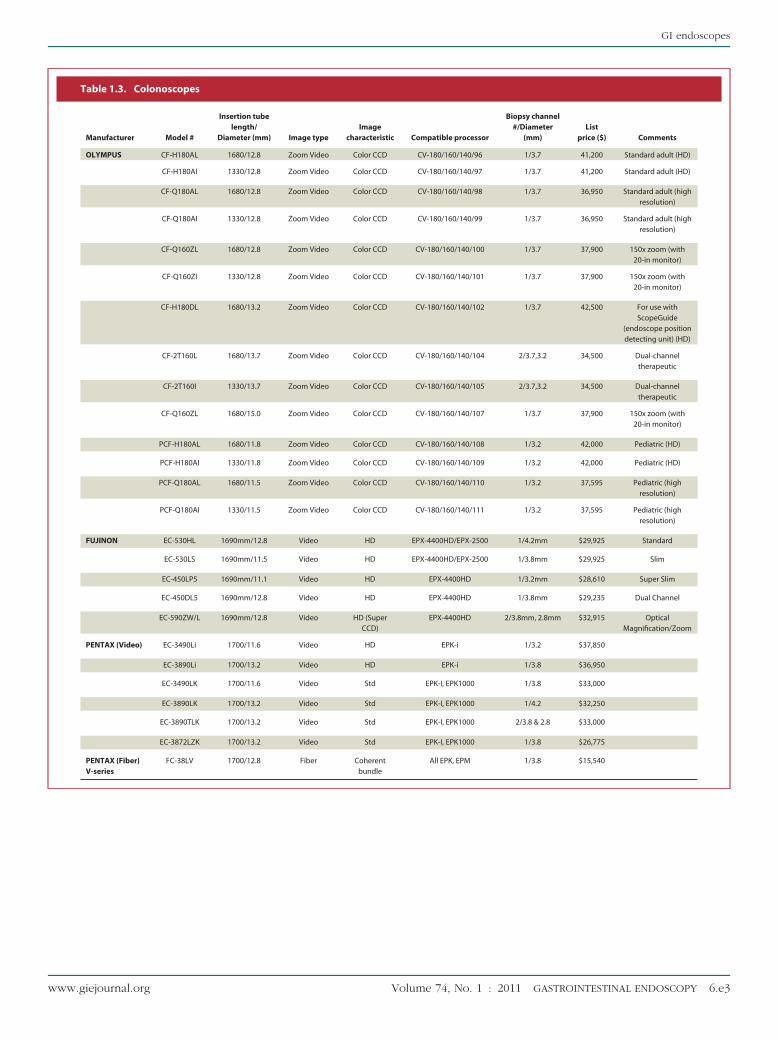
ColonoscopesA colonoscope is a forward-viewing endoscope de-
signed for examination of the entire colon and terminalileum. Colonoscopes are available in pediatric and adult
models and have variable insertion tube lengths (1330- awww.giejournal.org
700 mm), diameters (11.1-15 mm), channel sizes (2.8-4.2m), and channel number (1 or 2).Standard colonoscopes have varying degrees of flexi-
ility throughout the length of the insertion tube.9 Theistal portion of the tube is more flexible to allow thendoscopist to negotiate angulated areas of the colon, andhe more proximal shaft is stiffer to provide columntrength and reduce looping of the bowel. This varyingexibility is achieved by alterations in the resin polymerhat constitutes the outer layer of the endoscope. Theistal 40 cm of the tube is covered with a softer resinhat is gradually replaced with a harder resin moreroximally.10
One company (Olympus) also produces adult and pedi-tric colonoscopes that can be further stiffened as needed byhe endoscopist. These variable-stiffness (VS) colonoscopesere designed to improve ease of insertion by reducing
ooping in more mobile sections of bowel with the ability toaintain flexibility in more fixed sections. They have an
djustable tensioning coil that extends down the length of thensertion tube. An internal wire is attached to the coil and cane stiffened by rotating a dial on the control section of thendoscope. The proximal 40 to 50 cm of the insertion tube ishe only section that has varying stiffness capabilities; theistal 25 cm of the insertion tube and tip are unaffected. Thetiffness control dial can be rotated from position 0 to 3,llowing incremental increase in stiffness of the proximalhaft.10
Another colonoscope modification is a wide-angle fieldf view of 170 degrees versus the standard 140-degreeeld of view (Olympus). Table 5 (available online atww.giejournal.org) lists available colonoscopes and de-ice specifications.
choendoscopesEchoendoscopes are forward- or oblique-viewing endo-
copes fitted with an US transducer on the tip. These endo-copes are designed to allow US examination of layers of theI wall and extraluminal structures. Radial echoendoscopesroduce an US image that is perpendicular to the axis of the
nsertion tube, and the image is generally 360 degrees. Cur-ilinear echoendoscopes produce an image that is parallel tohe axis of the insertion tube and is usually a 100- to 180-egree image.11 Curvilinear echoendoscopes allow tissueampling via needle aspiration11; diagnostic and thera-eutic models are available. US transducers were pre-iously available in mechanical versions, which use aotating electrical element in the tip to create an USmage. These are being replaced by electronic US trans-ucers that use a series of nonrotating electrical ele-ents to produce an image. Electronic echoendoscopesave Doppler capabilities. Acoustic coupling betweenhe US transducer and the mucosa is via a water-filledalloon on the instrument tip. Echoendoscopes arevailable in a variety of lengths and insertion tube di-
meters (Table 6 [available online at www.giejourna-Volume 74, No. 1 : 2011 GASTROINTESTINAL ENDOSCOPY 3
aadeP
7wmsawab3ctpmi
cwpa0cCptbtm
ccrcspctumwearcdcacsaacmtT
GI endoscopes
l.org] lists available echoendoscopes and devicespecifications).
CholedochoscopesCholedochoscopes are forward-viewing miniature endo-
scopes designed for examination of the bile or pancreaticducts. They can be passed through a therapeutic duodeno-scope or percutaneously into the biliary system. Completelyreusable and partially disposable fiberoptic choledocho-scopes are available in the United States, with workinglengths of 1870 to 1900 mm, insertion tube diameters of 3.1 to3.4 mm, and a channel diameter of 1.2 mm. The completelyreusable models have controls for up/down tip deflection,buttons for air/water insufflation and suction, and 1 instru-ment channel. The partially disposable choledochoscope(Spyglass; Boston Scientific, Natick, Mass) has controls for4-way tip deflection. The insertion tube is a disposable cath-eter with steering wires, an irrigation port, and a 1.2-mmaccessory channel. Miniaturized accessories are available foruse through this choledochoscope, such as biopsy forceps.Choledochoscope use requires a separate light source andimage processor. Table 7 (available online at www.giejournal.org) lists available choledochoscopes anddevice specifications.
Comparative studiesThere are no published studies comparing endoscopes
made by different manufacturers. Detailed reviews ofcomparative studies of enteroscopes, choledochoscopes,echoendoscopes, and HD/high-magnification endoscopesare available.1-5
The single-balloon enteroscope was compared with thedouble-balloon enteroscope in an ongoing trial published inabstract form of patients randomized to examination witheither endoscope for a variety of indications, most commonlyobscure GI bleeding.12 Procedure time, depth of insertion,nd diagnostic yield were similar between the 2 endoscopes,lthough total setup time was significantly longer for theouble-balloon enteroscope (12 minutes for double-balloonnteroscope vs 3 minutes for single-balloon enteroscopy,� .005).Flexible sigmoidoscopy using a standard sigmoidoscope
was compared with examination using an upper endoscopein 2 randomized trials of colon cancer screening in anaverage-risk population. In 1 study of 160 women, unsedatedexamination with an upper endoscope was significantly lesspainful than with the sigmoidoscope.13 Pain scores werelikewise lower in the upper endoscope group in a random-ized study of 81 screening patients, and more examinationsreached the splenic flexure (76% with an upper endoscopevs 35%, P � .001).14
The standard pediatric colonoscope was compared withthe standard adult colonoscope in a nonblinded trial thatrandomized 100 women with a history of hysterectomy tocolonoscopy with either instrument. Cecal intubation rates
were higher in the pediatric colonoscope group (96.1% vs s4 GASTROINTESTINAL ENDOSCOPY Volume 74, No. 1 : 2011
1.4%, P � .001) and success increased to 89.9% in the groupith failed examination by switching to the pediatric instru-ent.15 In another trial, 918 outpatients were randomized to
creening colonoscopy with a long pediatric colonoscope orn intermediate-length adult colonoscope.16 Overall, thereere no differences in cecal intubation rates or need forbdominal pressure or position change. Time to cecal intu-ation was shorter in the adult colonoscope group (5.75 �.18 minutes vs 6.26 � 3.30 minutes, P � .02), and signifi-antly shorter in the subgroup of male patients. Theserials suggest that use of a pediatric colonoscope may bereferable in patients suspected of having a fixed sig-oid colon and otherwise may be used according to
ndividual endoscopist preference.Endoscope length was examined as a factor for colonos-
opy completion in a trial randomizing 998 examinationsith an adult colonoscope with 168-cm working length com-ared with a 133-cm device.17 The shorter endoscope wasssociated with lower mean cecal intubation times (4.14 �.13 minutes vs 5.16 � 0.13 minutes, P � .000). Completeolonoscopy can sometimes be achieved with a gastroscope.olonoscopy with a standard adult colonoscope was com-ared with examination with a gastroscope in a randomizedrial of 622 patients.18 There was no difference in cecal intu-ation rates, but time to cecal intubation was prolonged inhe gastroscope group (8.7 � 2.4 minutes vs 8.2 � 2.3inutes, P � .006).A meta-analysis of multiple randomized trials found that
ompared with the standard adult colonoscope, use of the VSolonoscope resulted in higher cecal intubation rates (oddsatio 2.08; 95% CI, 1.29-3.36), less abdominal pain, and de-reased need for sedation, but cecal intubation times wereimilar.19 A later randomized, controlled trial (RCT) com-ared a pediatric VS colonoscope, a standard pediatricolonoscope, and an adult colonoscope for cecal intubationimes, completeness of the examination, patient tolerance,se of abdominal compression, and endoscopists’ assess-ent of procedure difficulty.20 No significant differencesere found among any of the colonoscopes. Another studyvaluated whether use of the VS colonoscope decreased themount of sedation necessary for an examination. This trialandomized 355 patients into 3 groups based on the type ofolonoscope used (standard adult colonoscope of interme-iate and regular lengths of 1.3 and 1.6 m and VS adultolonoscope).21 The patients underwent colonoscopy usingmixture of propofol and alfentanil delivered by a patient-
ontrolled pump. Patients in the VS colonoscope group usedignificantly less propofol compared with the other 2 groups,nd mean pain scores were lower. In summary, study resultsre somewhat conflicting comparing VS colonoscopes withonventional colonoscopes but suggest that there may be aodest positive effect on cecal intubation rates and patient
olerability, with no difference in time to cecal intubation.heir use may be based on endoscopist preference.The wide-angle colonoscope has been compared with the
tandard colonoscope for polyp detection rates and effi-
www.giejournal.org
sabgcddetmapoecptpmscc
Ac((rafvtPpbetciaimt
F
Tgnscftsaf
A
ecileelga
S
fas
GI endoscopes
ciency. Two studies used 170-degree wide-angle instrumentsand standard 140-degree view colonoscopes to conductback-to-back, same-day colonoscopies. In 1 randomizedstudy of 50 patients, use of a prototype wide-angle colono-scope was associated with a reduced time to the cecum (2.09minutes vs 2.53 minutes, P � .002), reduced time for exam-ination during withdrawal (4.98 minutes vs 5.74 minutes, P �.0001), and similar missed polyp rates (19% vs 27%, P � .19)compared with the standard colonoscope.22 A similar studyshowed lower miss rates for all polyps with the wide-angleinstrument (20% vs 31%, P � .046) with a shorter meanexamination time (6.75 minutes vs 7.64 minutes total; P �.0005),23 indicating a potential impact on colonoscopy effi-ciency. Another multicenter RCT showed that the wide-anglecolonoscope reduced withdrawal time compared with thestandard colonoscope (4.9 minutes vs 5.4 minutes, P �.0001) without compromising adenoma detection rates.24
Other studies compared the wide-angle colonoscope withthe addition of HD capabilities with the standard-viewcolonoscope for polyp detection. In 1 RCT of 390 patients,the overall polyp detection rate was higher for the wide-angle HD instrument (1.76 � 2.31 vs 1.31 � 1.90, P � .03).25
However, there were no significant differences between the2 groups with respect to adenoma detection rate; the majordifference was in the detection of small hyperplastic polyps.Another RCT of 620 patients showed no difference in theoverall detection of polyps and no difference in polyp de-tection rates based on size or histology.26 A third case-controltudy found no difference in overall polyp detection rate,denoma detection rate, or insignificant lesion detection rateetween HD/wide-angle and conventional colonoscoperoups.27 A multicenter RCT compared the wide-angleolonoscope with a standard adult colonoscope for with-rawal times; endoscopists were asked to withdraw the en-oscope as quickly as they could while carefully completingxaminations.28 Of 710 procedures, the mean withdrawalime was significantly shorter in the wide-angle group (4.9inutes vs 5.9 minutes, P � .0001), with no difference in
denoma detection. Finally, a prospective cohort study com-ared 130 patients who had colonoscopy performed withptimized withdrawal technique (time �6 minutes, re-xamination of flexures and folds) with either the HDolonoscope or the adult colonoscope; no differences inolyp detection or adenoma detection were found.29 Takenogether, these studies suggest a minimal, if any, effect onolyp detection with use of the wide-angle and HD instru-ents compared with the standard colonoscope, with a pos-
ible small positive impact on colonoscopy efficiency be-ause of the ability to decrease withdrawal times withoutompromising efficacy.
SAFETY
Upper and lower endoscopy is safe. Cardiorespiratoryevents, both major and minor, related to sedation are the
most common adverse event, occurring in 0.03% to 20%.30,31 awww.giejournal.org
dverse events at 30 days from diagnostic and therapeuticolonoscopy include bleeding (0.2%-2.1%), perforation�0.1%), abdominal discomfort (5.4%), and infection0.2%).32,33 Other rare events such as splenic rupture areeported. Therapeutic maneuvers such as stricture dilationnd polypectomy with cautery increase some risks. Deathrom diagnostic endoscopy is rare. Epistaxis (0.85%-2%) andasovagal events (0.3%) have been reported with unsedatedransnasal endoscopy using the ultrathin endoscope.34,35
ancreatitis has been reported from balloon enteroscopy andush enteroscopy with an overtube in less than 1%; double-alloon enteroscopy is associated with a 2.3% rate of adversevents.9 Diagnostic EUS is generally as safe as EGD, althoughhe risk of cervical esophagus perforation is reportedly in-reased (0.03%)36 because of the long rigid portion of thensertion tube tip. The echoendoscope is oblique-viewing,nd this may also increase the risk of duodenal perforation. Its imperative that users follow manufacturer-specific recom-endations for reprocessing of endoscopes to avoid infec-
ion risk to patients.
INANCIAL CONSIDERATIONS
List prices for endoscopes and processors are shown inable 1 (available online at www.giejournal.org). Medical-rade image display monitors are also necessary and areot included in the processor price. Specialty endoscopesuch as echoendoscopes require special processors. Me-hanical radial endoscopes require separate US processorsrom linear array echoendoscopes, but newer fully elec-ronic radial and linear array echoendoscopes can use theame US processor. Peroral cholangioscopy requires andditional processor and image display monitor separaterom the duodenoscope setup.
REAS FOR FUTURE RESEARCH
Direct comparisons of different enteroscopes are nec-ssary to define the optimal instrument for deep enteros-opy. New innovations in endoscopic technology aimed atmproving the detection and characterization of neoplasticesions are needed. Some areas for future improvement inndoscopic design include self-propelled (partial/complete)ndoscopes, improved choledochoscope platforms, and aarger suction channel–to–outer diameter ratio. A better er-onomic endoscope design and smaller processors are otherreas of potential improvement.
UMMARY
A variety of endoscopes for GI use are available. Specialeatures such as magnification/HD, enhanced imaging,nd wide-angle view have recently evolved. Procedure-pecific endoscopes are required to enhance diagnostic
nd therapeutic success.Volume 74, No. 1 : 2011 GASTROINTESTINAL ENDOSCOPY 5
2
2
2
2
2
2
2
2
2
3
3
3
3
3
3
3
PASSBDVSMPJALS
TC
GI endoscopes
DISCLOSURES
Dr Tokar served as a consultant and on the speaker’sbureau and has received an educational grant from Fu-jinon, Inc. He has also served as a consultant for BostonScientific. Dr Varadarajulu served as a consultant forBoston Scientific and Olympus Medical Corporation. Noother financial relationships relevant to this publicationwere disclosed.
Abbreviations: CCD, charge-coupled device; HD, high-definition; MBI,multiband imaging; NBI, narrow-band imaging; RCT, randomized con-trolled trial; RGB, red green blue; VS, variable stiffness.
REFERENCES
1. Tierney W, Adler D, Chand B, et al. Echoendoscopes. Gastrointest Endosc2007;66:435-2.
2. Disario J, Peterson B, Tierney W, et al. Enteroscopes. Gastrointest Endosc2007;66:872-80.
3. Rodriguez S, Banerjee S, Desilets D, et al. Ultrathin endoscopes. Gastro-intest Endosc 2010;71:893-8.
4. Shah R, Adler D, Conway J, et al. Cholangiopancreatoscopy. GastrointestEndsoc 2008;68:411-21.
5. Kwon R, Adler D, Chand B, et al. High-resolution and high-magnificationendoscopes. Gastrointest Endosc 2009;69:399-407.
6. Wong Kee Song L, Adler D, Conway J, et al. Narrow band imaging andmultiband imaging. Gastrointest Endosc 2008;67:581-9.
7. Rodriguez S, Banerjee S, Desilets D, et al. Ultrathin endoscopes. Gastro-intest Endosc 2010;71:893-8.
8. Disario J, Peterson B, Tierney W, et al. Enteroscopes. Gastrointest Endosc2007;66:872-80.
9. Barlow DE. The video colonoscope. In: Rex DK, Way JD, Williams CB,editors. Colonoscopy. London: Blackwell Sciences; 2003.
10. Ginsberg G. Colonoscopy with the variable stiffness colonoscope. Gas-trointest Endosc 2003;58:579-84.
11. Carr-Locke DL, Branch MS, Byrne WJ. Tissue sampling during en-dosonography. Gastrointest Endosc 1998;47:576-8.
12. Efthymiou M, Desmond P, Taylor A. Single balloon enteroscopy versusdouble balloon enteroscopy, preliminary results of a randomized con-trolled trial [abstract]. Gastrointest Endosc 2010;71:AB122.
13. Farraye F, Horton K, Hershey H, et al. Screening flexible sigmoidoscopyusing an upper endoscope is better tolerated by women. Am J Gastro-enterol 2004;99:1074-80.
14. Fincher R, Myers J, McNear S, et al. Comfort and efficacy of a longer andthinner endoscope for average risk colon cancer screening. Dig Dis Sci2007;52:2892-6.
15. Marshall J, Perez R, Madsen R. Usefulness of a pediatric colonoscope forroutine colonoscopy in women who have undergone hysterectomy.Gastrointest Endosc 2002;55:838-41.
16. Hsieh Y, Zhou A, Lin H. Long pediatric colonoscope versus intermediatelength adult colonoscope for colonoscopy. J Gastroenterol Hepatol2008;23(7 Pt 2):e7-e10.
17. Lee H, Eun C, Lee O, et al. Significance of colonoscope length in cecalinsertion time. Gastrointest Endosc 2009;69:503-8.
18. Wehrmann T, Lechowicz I, Martchenko K, et al. Routine colonoscopywith a standard gastroscope. A randomized comparative trial in a west-ern population. Int J Colorectal Dis 2008;23:443-6.
19. Othman M, Bradley A, Choudhary A, et al. Variable stiffness colonoscopeversus regular adult colonoscope: meta-analysis of randomized con-trolled trials. Endoscopy 2009;41:17-24.
20. Shumaker D, Zaman A, Katon R. A randomized controlled trial in a training in-stitution comparing a pediatric variable stiffness colonoscope, a pediatric
colonoscope, and an adult colonoscope. Gastrointest Endosc 2002;55:172-9.B
6 GASTROINTESTINAL ENDOSCOPY Volume 74, No. 1 : 2011
1. Lee D, Li A, Ko C, et al. Use of a variable-stiffness colonoscope decreasedthe dose of patient-controlled sedation during colonoscopy: a random-ized comparison of 3 colonoscopes. Gastrointest Endosc 2007;65:424-9.
2. Deenadayalu V, Chadalawada V, Rex D. 170 degrees wide-angle colono-scope; effect on efficiency and miss rates. Am J Gastroenterol 2004;99:2138-42.
3. Rex D, Chadalawada V, Helper D. Wide angle colonoscopy with a proto-type instrument: impact on miss rates and efficiency as determined byback-to-back colonoscopies. Am J Gastroenterol 2003;98:2000-5.
4. Fatima H, Rex D, Rothstein R, et al. Clin Gastroenterol Hepatol 2008;6:109-14.
5. Tribonias G, Theodoropoulou A, Konsantinidis K, et al. Comparison ofstandard versus high-definition, wide-angle colonoscopy for polyp de-tection: a randomized controlled trial. Colorectal Dis. Epub 2009 Nov 23.
6. Pellis M, Fernandez-Esparrach G, Cardenas A, et al. Impact of wide-angle, high-definition endoscopy in the diagnosis of colorectal neopla-sia: a randomized controlled trial. Gastroenterology 2008;135:1062-8.
7. Burke C, Choure A, Sanaka M, et al. A comparison of high-definitionversus conventional colonoscopes for polyp detection. Dig Dis Sci 2010;55:1716-20.
8. Fatima H, Rec D, Rothstein R, et al. Cecal insertion and withdrawal timeswith wide-angle versus standard colonoscopes: a randomized con-trolled trial. Clin Gastroenterol Hepatol 2008;6:109-14.
9. East J, Stavrindis M, Thomas-Gibson S, et al. A comparative study ofstandard vs. high definition colonoscopy for adenoma and hyperplasticpolyp detection with optimized withdrawal technique. Aliment Phar-macol Ther 2008;15:768-76.
0. Nelson D, McQuaid K, Bond J, et al. Procedural success and complica-tions of large-scale screening colonoscopy. Gastrointest Endosc 2002;55:307-14.
1. Iber F, Sutberry M, Gupta R, et al. Evaluation of complications during andafter conscious sedation for endoscopy using pulse oximetry. Gastroin-test Endosc 1993;39:620-5.
2. Zubarik R, Fleischer D, Mastropletro C, et al. Prospective analysis of com-plications 30 days after outpatient colonoscopy. Gastrointest Endosc1999;50:322-8.
3. Dafnis G, Ekbom A, Pahlman L, et al. Complications of diagnostic andtherapeutic colonoscopy within a defined population in Sweden. Gas-trointest Endosc 2001;54:302-9.
4. Dumortier J, Napoleon B, Hedelius F, et al. Unsedated transnasal EGD indaily practice: results with 1100 consecutive patients. Gastrointest En-dosc 2003;57:198-204.
5. Postma G, Cohen J, Belafasky P, et al. Transnasal esophagoscopy: revis-ited (over 700 consecutive cases). Laryngoscope 2005;115:321-3.
6. Das A, Sivak MV Jr, Chak A. Cervical esophageal perforation during EUS:a national survey. Gastrointest Endosc 2001;53:599-602.
repared by:SGE Technology Committeehyam Varadarajulu, MDubhas Banerjee, MDradley A. Barth, MD, NASPGHAN Representativeavid J. Desilets, MDivek Kaul, MDripathi R. Kethu, MDarcos C. Pedrosa, MD
atrick R. Pfau, MDeffrey L. Tokar, MDmy Wang, MDouis-Michel Wong Kee Song, MDarah A. Rodriguez, MD, Chair
his document is a product of the ASGE Technology Assessmentommittee. This document was reviewed and approved by the Governing
oard of the American Society for Gastrointestinal Endoscopy.www.giejournal.org
GI endoscopes
Table 1.1. Gastroscopes
Manufacturer Model #
Insertion tubelength/
Diameter (mm)Imagetype
Imagecharacteristic
OLYMPUS GIF-H180J 1030/9.9 Video Color CCD C
GIF-H180 1030/9.8 Video Color CCD C
GIF-Q180 1030/8.8 Video Color CCD C
GIF-N180 1100/4.9 Video Color CCD C
GIF-XP180N 1100/5.5 Video Color CCD C
GIF-2TH180 1030/12.6 Video Color CCD C
GIF-1TQ160 1030/11.3 Video Color CCD C
GIF-XTQ160 1030/12.9 Video Color CCD C
GIF-Q160Z 1030/10.9 ZoomVideo
Color CCD CV
FUJINON EG-530WR 1100mm/9.3 Video HD EPX
EG-450PE5 1100mm/8.1 Video HD
EG-530N 1100mm/5.9 Video HD EPX
EG-530NP 1100mm/4.9 Video HD EPX
EG-450CT5 1100mm/10.8 Video HD
EG-450D5 1090mm/11.5 Video HD
EG-590WR 1100mm/9.6 Video HD (SuperCCD)
EG-590ZW 1100mm/9.8 Video HD (SuperCCD)
PENTAX(VIDEO)
EG-2790i 1050/9.0 Video HD
EG-2990i 1050/9.8 Video HD
EG-1690K 1050/5.4 Video Std
EG-2490K 1050/8.0 Video Std
EG-2790K 1050/9.0 Video Std
EG-2990K 1050/9.8 Video Std
EG-3490K 1050/11.6 Video Std
EG-3470ZK 1050/11.6 Video Std
EG-3890TK 1050/12.8 Video Std
Compatibleprocessor
Biopsy channel#/Diameter (mm)
Listprice ($) Comments
V-180/160/140/92 1/2.8 36,500 Standard adult withforward water jet
(HD)
V-180/160/140/93 1/2.8 35,700 Standard adult (HD)
V-180/160/140/94 1/2.8 31,875 Standard adult (highresolution)
V-180/160/140/95 1/2.0 28,950 Ultrathin
V-180/160/140/96 1/2.0 30,500 Ultrathin
V-180/160/140/97 2/2.8,3.7 36,500 Dual-channeltherapeutic (HD)
V-180/160/140/98 1/3.7 34,300 Therapeutic
V-180/160/140/99 1/6.0 31,750 Therapeutic (largechannel)
-180/160/140/100 1/2.8 32,075 115x zoom (with20-in monitor)
-4400HD/EPX-2500 1/2.8mm $27,035 Standard
EPX-4400HD 1/2.2mm $26,650 Slim
-4400HD/EPX-2500 1/2.0mm $26,800 Ultra Slim
-4400HD/EPX-2500 1/2.0mm $26,700 Ultra Slim
EPX-4400HD 1/3.8mm $26,780 Treatment
EPX-4400HD 2/3.8mm, 2.8mm $27,515 Dual Channel
EPX-4400HD 1/2.8mm $29,285 Standard
EPX-4400HD 1/2.8mm $30,330 OpticalMagnification
EPK-i 1/2.8 $31,500 $31,500.00
EPK-i 1/2.8 $31,500 $31,500.00
EPK-I, EPK1000 1/2.0 $26,500 $26,500.00
EPK-I, EPK1000 1/2.4 $28,500 $28,500.00
EPK-I, EPK1000 1/2.8 $27,500 $27,500.00
EPK-I, EPK1000 1/2.8 $27,500 $27,500.00
EPK-I, EPK1000 1/3.8 $28,500 $28,500.00
EPK-I, EPK1000 1/2.8 $24,650 $24,650.00
EPK-I, EPK1000 2/3.8 & 2.8 $30,500 $30,500.00
www.giejournal.org Volume 74, No. 1 : 2011 GASTROINTESTINAL ENDOSCOPY 6.e1
GI endoscopes
Table 1.2. Sigmoidoscopes
Manufacturer Model #Insertion tube
length/Diameter (mm)Imagetype
Imagecharacteristic
Compatibleprocessor
Biopsy channel#/Diameter (mm)
Listprice ($) Comments
OLYMPUS OSF-V60 730/11.3 Video Color CCD CV-60 1/3.2 10,300 Standard adult
OSF-4 700/11.8 Fiber Coherent bundle CLK-4 1/3.2 8,200
FUJINON ES-450WE5 790mm/12.8 Video HD EPX-4400HD 1/3.8mm $19,030 Standard
PENTAX (VIDEO) ES-3870K 700/12.8 Video Std EPK-I, EPK-1000 1/4.2 $16,800
PENTAX (FIBER) FS-34V 700/11.5 Fiber Coherent Bundle LH-150 PC 1/3.5 $5,775
6.e2 GASTROINTESTINAL ENDOSCOPY Volume 74, No. 1 : 2011 www.giejournal.org
GI endoscopes
Table 1.3. Colonoscopes
Manufacturer Model #
Insertion tubelength/
Diameter (mm) Image typeImage
characteristic Compatible processor
Biopsy channel#/Diameter
(mm)List
price ($) Comments
OLYMPUS CF-H180AL 1680/12.8 Zoom Video Color CCD CV-180/160/140/96 1/3.7 41,200 Standard adult (HD)
CF-H180AI 1330/12.8 Zoom Video Color CCD CV-180/160/140/97 1/3.7 41,200 Standard adult (HD)
CF-Q180AL 1680/12.8 Zoom Video Color CCD CV-180/160/140/98 1/3.7 36,950 Standard adult (highresolution)
CF-Q180AI 1330/12.8 Zoom Video Color CCD CV-180/160/140/99 1/3.7 36,950 Standard adult (highresolution)
CF-Q160ZL 1680/12.8 Zoom Video Color CCD CV-180/160/140/100 1/3.7 37,900 150x zoom (with20-in monitor)
CF-Q160ZI 1330/12.8 Zoom Video Color CCD CV-180/160/140/101 1/3.7 37,900 150x zoom (with20-in monitor)
CF-H180DL 1680/13.2 Zoom Video Color CCD CV-180/160/140/102 1/3.7 42,500 For use withScopeGuide
(endoscope positiondetecting unit) (HD)
CF-2T160L 1680/13.7 Zoom Video Color CCD CV-180/160/140/104 2/3.7,3.2 34,500 Dual-channeltherapeutic
CF-2T160I 1330/13.7 Zoom Video Color CCD CV-180/160/140/105 2/3.7,3.2 34,500 Dual-channeltherapeutic
CF-Q160ZL 1680/15.0 Zoom Video Color CCD CV-180/160/140/107 1/3.7 37,900 150x zoom (with20-in monitor)
PCF-H180AL 1680/11.8 Zoom Video Color CCD CV-180/160/140/108 1/3.2 42,000 Pediatric (HD)
PCF-H180AI 1330/11.8 Zoom Video Color CCD CV-180/160/140/109 1/3.2 42,000 Pediatric (HD)
PCF-Q180AL 1680/11.5 Zoom Video Color CCD CV-180/160/140/110 1/3.2 37,595 Pediatric (highresolution)
PCF-Q180AI 1330/11.5 Zoom Video Color CCD CV-180/160/140/111 1/3.2 37,595 Pediatric (highresolution)
FUJINON EC-530HL 1690mm/12.8 Video HD EPX-4400HD/EPX-2500 1/4.2mm $29,925 Standard
EC-530LS 1690mm/11.5 Video HD EPX-4400HD/EPX-2500 1/3.8mm $29,925 Slim
EC-450LP5 1690mm/11.1 Video HD EPX-4400HD 1/3.2mm $28,610 Super Slim
EC-450DL5 1690mm/12.8 Video HD EPX-4400HD 1/3.8mm $29,235 Dual Channel
EC-590ZW/L 1690mm/12.8 Video HD (SuperCCD)
EPX-4400HD 2/3.8mm, 2.8mm $32,915 OpticalMagnification/Zoom
PENTAX (Video) EC-3490Li 1700/11.6 Video HD EPK-i 1/3.2 $37,850
EC-3890Li 1700/13.2 Video HD EPK-i 1/3.8 $36,950
EC-3490LK 1700/11.6 Video Std EPK-I, EPK1000 1/3.8 $33,000
EC-3890LK 1700/13.2 Video Std EPK-I, EPK1000 1/4.2 $32,250
EC-3890TLK 1700/13.2 Video Std EPK-I, EPK1000 2/3.8 & 2.8 $33,000
EC-3872LZK 1700/13.2 Video Std EPK-I, EPK1000 1/3.8 $26,775
PENTAX (Fiber)V-series
FC-38LV 1700/12.8 Fiber Coherentbundle
All EPK, EPM 1/3.8 $15,540
www.giejournal.org Volume 74, No. 1 : 2011 GASTROINTESTINAL ENDOSCOPY 6.e3
GI endoscopes
Table 1.4. Duodenoscopes
Manufacturer Model #Insertion tube
length/Diameter (mm)Imagetype
Imagecharacteristic
Compatibleprocessor
Biopsy channel#/Diameter (mm)
Listprice ($) Comments
OLYMPUS TJF-Q180V 1240/11.3 Video Color CCD CV-180/160 1/4.2 $40,000 Therapeutic(replaces TJF-160F)
JF-140F 1240/11 Video Color CCD CV-180/160/140 1/3.2 $33,950 Diagnostic
PJF-160 1235/7.5 Video Color CCD CV-180/160 1/2.0 $37,000 Pediatric
FUJINON ED-530XT 1250mm/11.5 Video HD EPX-4400HD 1/4.2 $32,895 Therapeutic
ED-450XL5 1250mm/11.8 Video HD EPX-4400HD 1/3.2 $29,840 Standard
PENTAX ED-3270K 1250/10.8 Video Std EPK-I, EPK1000 1/3.2 $28,700.00
ED-3490TK 1250/11.6 Video Std EPK-I, EPK1000 1/4.2 $34,500.00
ED-3670TK 1250/12.1 Video Std EPK-I, EPK1000 1/4.8 $31,710.00
Table 1.5. Choledochoscopes
Manufacturer Model #Insertion tube length/
Diameter (mm) Image typeImage
characteristicBiopsy channel
#/Diameter (mm)List
price ($) Comments
OLYMPUS CHF-BP30 1870/3.4 Fiber optic Coherentbundle
1/1.2 $25,500 Requires large - channelduodenoscopes.
PENTAX FCP-9P 1900/3.1 Fiber optic Coherentbundle
1/1.2 24,885
BOSTONSCIENTIFIC
SPYGLASS reusable probe: 3000/0.77 Fiber optic 1.2/0.6/0.6 21,475
single use catheter: 2200/3.4 4,850 disposable probe
Table 1.6. Echoendoscopes
Manufacturer Model #Insertion tube length/
Diameter (mm)US orientation/Scanning
degrees/# frequency (MHz)Image type/View
angle/Degree
OLYMPUS GF-UE160-AL5 1250/11.8 electronic radial 360/4,(5,6,7.5,10)
video, oblique (55)
GF-UM160 1,250/10.5 mechanical radial 360/4,(5,7.5,12,20)
video, oblique (50)
GF-UCT140P-AL5 1,250/11.8 curvilinear (100)/4,(5,6,7.5,10) video oblique (55)
GF-UCT140-AL5 1,250/12.6 curvilinear (180)/4,(5,6,7.5,10) video, oblique (55)
GF-UCT180 1,250/12.6 curvilinear (180)/4,(5,6,7.5,10) video, oblique (55)
GF-UCT160-OL5 1,250/12.6 curvilinear (150)/1, (7.5) video, oblique (55)
GF-UC160P-OL5 1,250/11.8 curvilinear (150)/1, (7.5) video, oblique (55)
PENTAX EG-3670URK 1250/12.1 electronic radial 360/3 (5,7.5,10)
video, forward
EG-3630UR 1250/12.1 electronic radial 270 video, forward
EG-3830UT 1250/12.8 longitudnal 120/3(5,7.5,10) video, oblique (50)
EG-3870UTK 1250/12.8 longitudnal 120/3 (5,7.5,10) video, oblique (50)
6.e4 GASTROINTESTINAL ENDOSCOPY Volume 74, No. 1 : 2011 www.giejournal.org
GI endoscopes
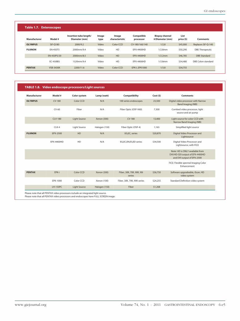
Table 1.7. Enteroscopes
Manufacturer Model #Insertion tube length/
Diameter (mm)Imagetype
Imagecharacteristic
Compatibleprocessor
Biopsy channel#/Diameter (mm)
Listprice ($) Comments
OLYMPUS SIF-Q180 2000/9.2 Video Color CCD CV-180/160/140 1/2.8 $45,000 Replaces SIF-Q-140
FUJINON EN-450T5 2000mm/9.4 Video HD EPX-4400HD 1/2.8mm $50,290 DBE Therapeutic
EN-450P5/20 2000mm/8.5 Video HD EPX-4400HD 1/2.2mm $46,785 DBE Standard
EC-450BI5 1520mm/9.4 Video HD EPX-4400HD 1/2.8mm $34,480 DBE Colon standard
PENTAX VSB-3430K 2200/11.6 Video Color CCD EPK-I, EPK1000 1/3.8 $34,755
TABLE 1.8. Video endoscope processors/Light sources
Manufacturer Model # Color system Lamp (watt) Compatibility Cost ($) Comments
OLYMPUS CV-180 Color CCD N/A 100 series endoscopes 23,500 Digital video processor with NarrowBand Imaging (NBI)
CV-60 Fiber N/A Fiber Optic (OSF-V60) 7,300 Combed video processor, lightsource and air pump
CLV-180 Light Source Xenon (300) CV-180 13,400 Light source for color CCD withNarrow Band Imaging (NBI)
CLK-4 Light Source Halogen (150) Fiber Optic (OSF-4) 1,165 Simplified light source
FUJINON EPX-2500 HD N/A EG,EC, series $20,870 Digital Video Processor andLightsource
EPX-4400HD HD N/A EG,EC,EN,ES,ED series $34,500 Digital Video Processor andLightsource, with FICE
Note: HD is ONLY available fromDVI/HD-SDI output of EPX-4400HD
and DVI output of EPX-2500
FICE: Flexible spectral Imaging ColorEnhancement
PENTAX EPK-i Color CCD Xenon (300) Fiber, 30K, 70K, 90K, 90iseries
$36,750 Software upgradeable, iScan, HDvideo system
EPK-1000 Color CCD Xenon (100) Fiber, 30K, 70K, 90K series $24,255 Standard Definition video system
LH-150PC Light Source Halogen (150) Fiber $1,268
Please note that all PENTAX video processors include an integrated light source.
Please note that all PENTAX video processors and endoscopes have FULL SCREEN image.www.giejournal.org Volume 74, No. 1 : 2011 GASTROINTESTINAL ENDOSCOPY 6.e5
GI endoscopes
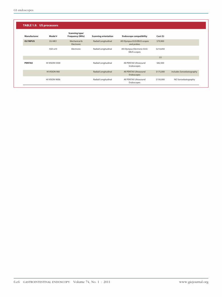
TABLE 1.9. US processors
Manufacturer Model #Scanning type/
Frequency (MHz) Scanning orientation Endoscope compatibility Cost ($)
OLYMPUS EU-ME1 Mechanical &Electronic
Radial/Longitudinal All Olympus EUS/EBUS scopesand probes
$79,900
SSD-a10 Electronic Radial/Longitudinal All Olympus Electronic EUS/EBUS scopes
$214,450
???
PENTAX HI VISION 5500 Radial/Longitudinal All PENTAX UltrasoundEndoscopes
$82,500
HI VISION 900 Radial/Longitudinal All PENTAX UltrasoundEndoscopes
$175,000 includes Sonoelastography
HI VISION 900b Radial/Longitudinal All PENTAX UltrasoundEndoscopes
$150,000 NO Sonoelastography
6.e6 GASTROINTESTINAL ENDOSCOPY Volume 74, No. 1 : 2011 www.giejournal.org