Embed Size (px)
Citation preview

8/9/2019 Gigi tiruan lepasan pada kasus Sindrom Kombinasi
http://slidepdf.com/reader/full/gigi-tiruan-lepasan-pada-kasus-sindrom-kombinasi 1/4
22 Volume 1, Number 1, 2012
Sheng-Wei Feng
School of Dentistry, Taipei Medical
University, Taipei, Taiwan
Pei-Bang Liao
Taipei Medical University Hospital,
Taipei, Taiwan
May-Show Chen
School of Dentistry, Taipei MedicalUniversity, Taipei, Taiwan
Taipei Medical University Hospital,
Taipei, Taiwan
Corresponding author:
Sheng-Wei Feng
School of Dentistry, Taipei Medical
University, Taipei, Taiwan
250 Wu-Hsing Street, Taipei, Taiwan
Tel: 886-2-2736-1661 ext. 5148; Fax:
886-2-27362295
AbstractCombination syndrome commonly occurs in patients witha completely edentulous maxilla opposed by a bilateraldistal-extension removable partial denture. is syndrome poses a considerable challenge to dentists. e symptomsof the syndrome consist of anterior maxillary boneloss, mandibular bone loss, tuberosity overgrowth, andalveolar ridge canting. All of these effects render prosthetictreatment more difficult, and although it is preferable touse dental implants for functional support, complex casesstill require conventional prosthetic treatments for medical
or financial reasons.This c l inical report presents the prosthodonticmanagement of a patient exhibiting combination syndromealong with a discussion of relevant literature.
Keywords: Combination syndrome, distal-extension RPD
Introduction
The oral rehabilitation of patients with an edentulousmaxilla opposed by natural mandibular anterior teeth
is a considerable challenge for many clinicians. These casespose many potential problems, including loss of bone
from the anterior edentulous maxilla and super-eruptionof unopposed mandibular anterior teeth. Kelly (1972)proposed the term combination syndrome for this oralcondition and its resultant clinical features. e Glossary ofProsthodontic Terms1 has defined combination syndromeas: the characteristic features that occur when an edentulousmaxilla is opposed by natural mandibular anterior teeth,including loss of bone from the anterior portion of themaxillary ridge, overgrowth of the tuberosities, papillaryhyperplasia of the hard palatal mucosa, extrusion ofmandibular anterior teeth, and loss of alveolar bone andridge height beneath the posterior mandibular removabledental prosthesis bases – also called anterior hyperfunctionsyndrome.
Kelly (1972)2 observed 20 patients equipped withcomplete max illary dentures opposing distal-extensionremovable partial dentures (RPD). After three years offollow-up , six of these patients showed a reduction of theanterior bony ridge height (1.350.83 mm) on lateralcephalometric radiography. Meanwhile, an increasing bonelevel of the tuberosities (1.380.36 mm) was noted in fivepatients. Kelly (1972) proposed the preservation of posteriorteeth to support lower partial dentures and a more stableocclusion to avoid combination syndrome. Preservation
Case Report
Prosthodontic Treatment of a Patient with
Combination Syndrome: A Clinical Case Report
8/9/2019 Gigi tiruan lepasan pada kasus Sindrom Kombinasi
http://slidepdf.com/reader/full/gigi-tiruan-lepasan-pada-kasus-sindrom-kombinasi 2/4
Journal of Prosthodontics and Implantology 23
Case Report
Case Report
A 73-year-old male patient was referred tothe Dentistry Department of Taipei MedicalUniversity Hospital in Taipei, Taiwan, forrestorative treatment. The patient's chiefcomplaints were inadequate retention ofmaxillary complete denture and inability
to chew comfortably. No major systemicdiseases or drug allergies were reported. Onexamination, the patient had an edentulousmaxilla and nine natural mandibular anteriorteeth (Figure 1). Clinically, the patientdisplayed anterior bone loss and flabbytissue of the maxillary ridge, overgrowth ofthe maxillary tuberosities, and over-eruptedmandibular anterior teeth (Figure 2). Thepatient rejected any surgery and implanttherapy due to financial considerations. Thepatient agreed to have a new complete dentureand a mandibular removable partial denture
of posterior occlusion and avoidance ofanterior hyperfunction are considered theprimary treatment suggestions for thiscomplex condition. Saunder et al (1979)3 and Jameson (2001)4 suggested the use of analternative tooth form and occlusal concept(linear occlusion) and minimum anterior
contact for reducing further bone loss caused by hyperfunction of anterior teeth. Previousstudies advocated osseointegrated implant-retained or implant-supported prostheses tochange the occlusal force distribution anddecrease the traumatic stress to the alveolar
bone resulting from combination syndrome.5
T h e p r e s e n t r e p o r t d e t a i l s t h eprosthodontic management of a specificpatient exhibiting symptoms of combinationsyndrome.
Figure 1. Panoramic radiograph showing
a typical case of combination syndrome
with severe resorption of the anterior
maxillary and super-eruption of unopposed
mandibular anterior teeth.
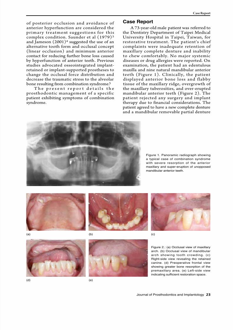
Figure 2.: (a) Occlusal view of maxillary
arch. (b) Occlusal view of mandibular
arch showing tooth crowding. (c)
Right-side view revealing the retained
canine. (d) Preoperative frontal view
showing greater bone resorption of the
premaxillary area. (e) Left-side view
indicating sufficient restoration space.
(a)
(d)
(b)
(e)
(c)
8/9/2019 Gigi tiruan lepasan pada kasus Sindrom Kombinasi
http://slidepdf.com/reader/full/gigi-tiruan-lepasan-pada-kasus-sindrom-kombinasi 3/4
24 Volume 1, Number 1, 2012
Case Report
semi-adjustable articulator (Whip Mix 3040,Louisville, Kentucky, USA). In addition,an intraoral Gothic Arch Tracer (Simplex®,Dentsply, New York, USA) was applied to
verif y accurate and reproduc ible occlusal vertical dimension (OVD) and centric relation(CR)(Figure 3c).
e selection of maxillary anterior artificialteeth was determined by patient gender andpersonality. Balanced occlusion was indicatedfor this case to assure an even distributionof occlusal force and prevent occlusalinterferences on the residual ridge. The tootharrangement was checked for esthetics and CRposition and then submitted for processing.
After prescrip, both casts were remounted,adjusted, and polished. At a subsequentappointment, the finished prostheses weredelivered and minimal occlusal adjust ment was needed. e patient was pleased with their
appearance and chewing ability. A maintenanceprogram including oral hygiene instruction andprosthesis home care was established. At the18-month maintenance visit, no complications
were observed.
Discussion
Treatment of patients with an edentulousmaxilla opposed to natural mandibularanterior teeth and a distal-extension RPD isconsidered a challenge for dental practitioners.Combination syndrome has a prevalence rate
aer some discussion.Initial therapy included oral hygiene
instructions, caries control, and nonsurgicalperiodontal therapy. At the first clinicalappointment for prosthodontic treatment,a preliminary impression of the maxillaryand mandibular arches was made with
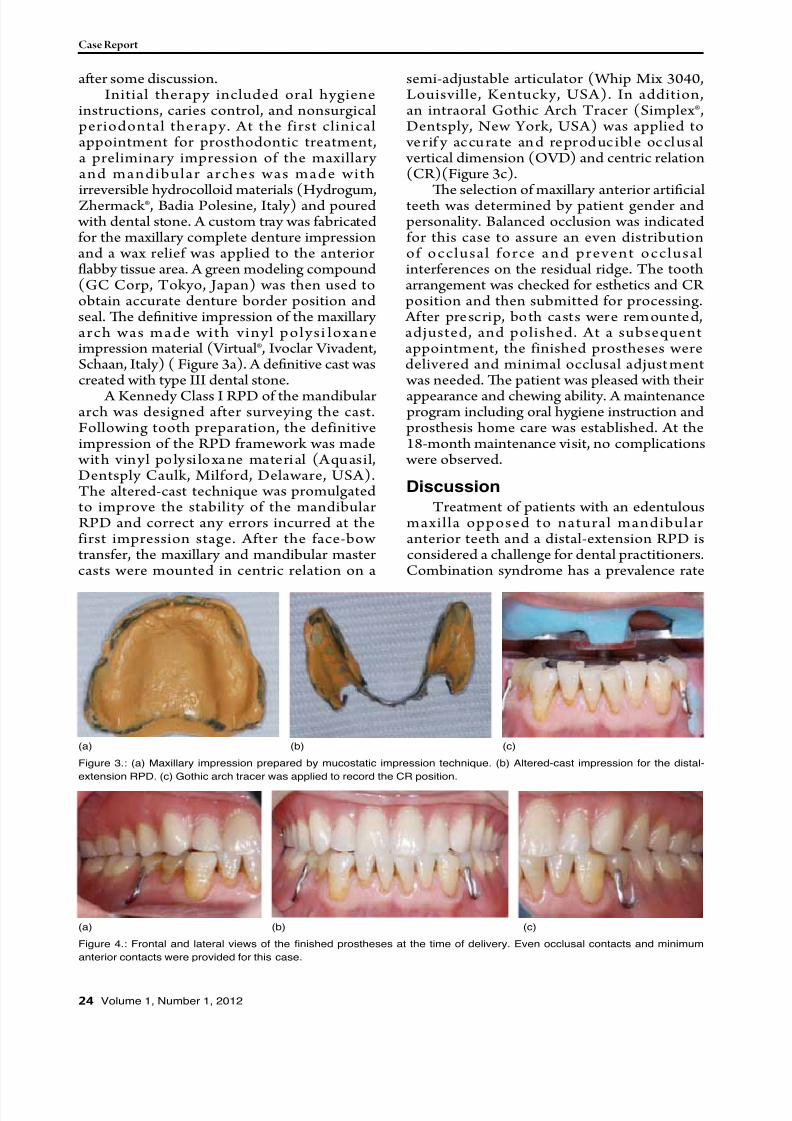
irreversible hydrocolloid materials (Hydrogum,Zhermack®, Badia Polesine, Italy) and poured with dental stone. A custom tray was fabricatedfor the maxillary complete denture impressionand a wax relief was applied to the anteriorflabby tissue area. A green modeling compound(GC Corp, Tokyo, Japan) was then used toobtain accurate denture border position andseal. e definitive impression of the maxillaryarch was made with vinyl polysi loxaneimpression material (Virtual®, Ivoclar Vivadent,Schaan, Italy) ( Figure 3a). A definitive cast wascreated with type III dental stone.
A Kennedy Class I RPD of the mandibulararch was designed after surveying the cast.Following tooth preparation, the definitiveimpression of the RPD framework was made
with vinyl polysi loxane material (Aquasil,Dentsply Caulk, Milford, Delaware, USA).The altered-cast technique was promulgatedto improve the stability of the mandibularRPD and correct any errors incurred at thefirst impression stage. After the face-bowtransfer, the maxillary and mandibular mastercasts were mounted in centric relation on a
(a)
(a)
(b)
(b)
(c)
(c)
Figure 3.: (a) Maxillary impression prepared by mucostatic impression technique. (b) Altered-cast impression for the distal-
extension RPD. (c) Gothic arch tracer was applied to record the CR position.
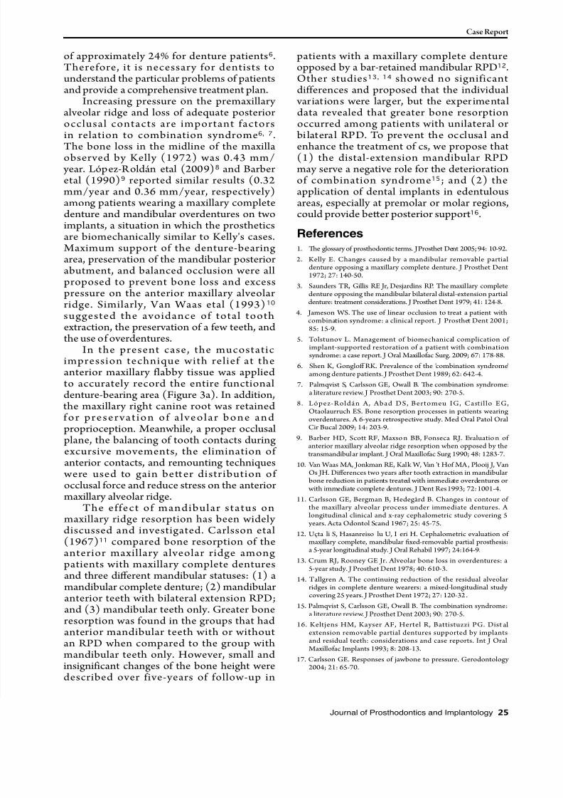
Figure 4.: Frontal and lateral views of the finished prostheses at the time of delivery. Even occlusal contacts and minimum
anterior contacts were provided for this case.
8/9/2019 Gigi tiruan lepasan pada kasus Sindrom Kombinasi
http://slidepdf.com/reader/full/gigi-tiruan-lepasan-pada-kasus-sindrom-kombinasi 4/4
Journal of Prosthodontics and Implantology 25
Case Report
patients with a maxillary complete dentureopposed by a bar-retained mandibular RPD12.Other studies13, 14 showed no significantdifferences and proposed that the individual
variations were larger, but the experimentaldata revealed that greater bone resorptionoccurred among patients with unilateral or
bi lateral RPD. To prevent the occlusal andenhance the treatment of cs, we propose that(1) the distal-extension mandibular RPDmay serve a negative role for the deteriorationof combination syndrome15; and (2) theapplication of dental implants in edentulousareas, especially at premolar or molar regions,could provide beer posterior support16.
References1. e glossary of prosthodontic terms. J Prosthet Dent 2005; 94: 10-92.
2. Kelly E. Changes caused by a mandibular removable partialdenture opposing a maxillary complete denture. J Prosthet Dent1972; 27: 140-50.
3. Saunders TR, Gillis RE Jr, Desjardins RP. The maxillary completedenture opposing the mandibular bilateral distal-extension partialdenture: treatment considerations. J Prosthet Dent 1979; 41: 124-8.
4. Jameson WS. The use of linear occlusion to treat a patient withcombination syndrome: a clinical report. J Prosthet Dent 2001;85: 15-9.
5. Tolstunov L. Management of biomechanical complication ofimplant-supported restoration of a patient with combinationsyndrome: a case report. J Oral Maxillofac Surg. 2009; 67: 178-88.
6. Shen K, Gongloff RK. Prevalence of the 'combination syndrome'among denture patients. J Prosthet Dent 1989; 62: 642-4.
7. Palmqvist S, Carlsson GE, Owall B. e combination syndrome:a literature review. J Prosthet Dent 2003; 90: 270-5.
8. López-Roldán A, Abad DS, Bertomeu IG, Casti llo EG,Otaolaurruch ES. Bone resorption processes in patients wearing
overdentures. A 6-years retrospective study. Med Oral Patol OralCir Bucal 2009; 14: 203-9.
9. Barber HD, Scott RF, Maxso n BB, Fonseca RJ. Evaluatio n ofanterior maxillary alveolar ridge resorption when opposed by thetransmandibular implant. J Oral Maxillofac Surg 1990; 48: 1283-7.
10. Van Waas MA, Jonkman RE, Kalk W, Van 't Hof MA , Plooij J, VanOs JH. Differences two years aer tooth extraction in mandibular bone reduction in patients treated with immediate overdentures or with immediate complete dentures. J Dent Res 1993; 72: 1001-4.
11. Carlsson GE, Bergman B, Hedegård B. Changes in contour ofthe maxillary alveolar process under immediate dentures. Alongitudinal clinical and x-ray cephalometric study covering 5
years. Acta Odontol Scand 1967; 25: 45-75.
12. Uçta li S, Hasanreiso lu U, I eri H. Cephalometric evaluation ofmaxillary complete, mandibular fixed-removable partial prosthesis:a 5-year longitudinal study. J Oral Rehabil 1997; 24:164-9.
13. Crum RJ, Rooney GE Jr. Alveolar bone loss in overdentures: a5-year study. J Prosthet Dent 1978; 40: 610-3.
14. Tallgren A. The continuing reduction of the residual alveolarridges in complete denture wearers: a mixed-longitudinal studycovering 25 years. J Prosthet Dent 1972; 27: 120-32.
15. Palmqvist S, Carlsson GE, Owall B. e combination syndrome:a literature review. J Prosthet Dent 2003; 90: 270-5.
16. Keltjens HM, Kayser AF, Hertel R, Battistuzzi PG. Dist alextension removable partial dentures supported by implantsand residual teeth: considerations and case reports. Int J OralMaxillofac Implants 1993; 8: 208-13.
17. Carlsson GE. Responses of jawbone to pressure. Gerodontology2004; 21: 65-70.
of approximately 24% for denture patients6.Therefore, it is necessary for dentists tounderstand the particular problems of patientsand provide a comprehensive treatment plan.
Increasing pressure on the premaxillaryalveolar ridge and loss of adequate posteriorocclusal contacts are important factors
in relation to combination syndrome6, 7
.The bone loss in the midline of the maxillaobserved by Kelly (1972) was 0.43 mm/
year. López-Roldán etal (2009)8 and Barberetal (1990)9 reported similar results (0.32mm/year and 0.36 mm/year, respectively)among patients wearing a maxillary completedenture and mandibular overdentures on twoimplants, a situation in which the prostheticsare biomechanically similar to Kelly's cases.Maximum support of the denture-bearingarea, preservation of the mandibular posteriorabutment, and balanced occlusion were all
proposed to prevent bone loss and excesspressure on the anterior maxillary alveolarridge. Similarly, Van Waas etal (1993)10 suggested the avoidance of total toothextraction, the preservation of a few teeth, andthe use of overdentures.
In the present case, the mucostaticimpression technique with relief at theanterior maxillary flabby tissue was appliedto accurately record the entire functionaldenture-bearing area (Figure 3a). In addition,the maxillary right canine root was retained
f or preservat ion of a lveolar bone andproprioception. Meanwhile, a proper occlusalplane, the balancing of tooth contacts duringexcursive movements, the elimination ofanterior contacts, and remounting techniques
were used to gain bett er distribution ofocclusal force and reduce stress on the anteriormaxillary alveolar ridge.
The effect of mandibular status onmaxillary ridge resorption has been widelydiscussed and investigated. Carlsson etal(1967)11 compared bone resorption of theanterior maxillary alveolar ridge amongpatients with maxillary complete denturesand three different mandibular statuses: (1) amandibular complete denture; (2) mandibularanterior teeth with bilateral extension RPD;and (3) mandibular teeth only. Greater boneresorption was found in the groups that hadanterior mandibular teeth with or withoutan RPD when compared to the group withmandibular teeth only. However, small andinsignificant changes of the bone height weredescribed over five-years of follow-up in


![Gigi Tiruan Sebagian Lepasan Kerangka Logam [Autosaved]](https://img.pdfslide.net/doc/110x75/55cf9a07550346d033a02c22/gigi-tiruan-sebagian-lepasan-kerangka-logam-autosaved.jpg)