Embed Size (px)
Citation preview

Gluconeogenesis;Regulation of Glycolysis & Gluconeogenesis
Copyright © 1999-2007 by Joyce J. Diwan. All rights reserved.
Molecular Biochemistry I
Gluconeogenesis occurs mainly in liver.
Gluconeogenesis occurs to a more limited extent in kidney & small intestine under some conditions.
Synthesis of glucose from pyruvate utilizes many of the same enzymes as Glycolysis.
Three Glycolysis reactions have such a large negative G that they are essentially irreversible. Hexokinase (or Glucokinase) Phosphofructokinase Pyruvate Kinase.
These steps must be bypassed in Gluconeogenesis.
Two of the bypass reactions involve simple hydrolysis reactions.
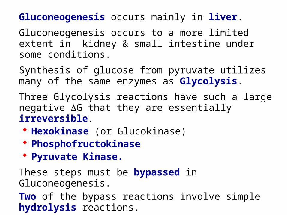
Hexokinase or Glucokinase (Glycolysis) catalyzes:glucose + ATP glucose-6-phosphate + ADP
Glucose-6-Phosphatase (Gluconeogenesis) catalyzes:
glucose-6-phosphate + H2O glucose + Pi
H O
OH
H
OHH
OH
CH2OH
H
OH
HH O
OH
H
OHH
OH
CH2OPO32
H
OH
HH2O
1
6
5
4
3 2
+ Pi
glucose-6-phosphate glucose
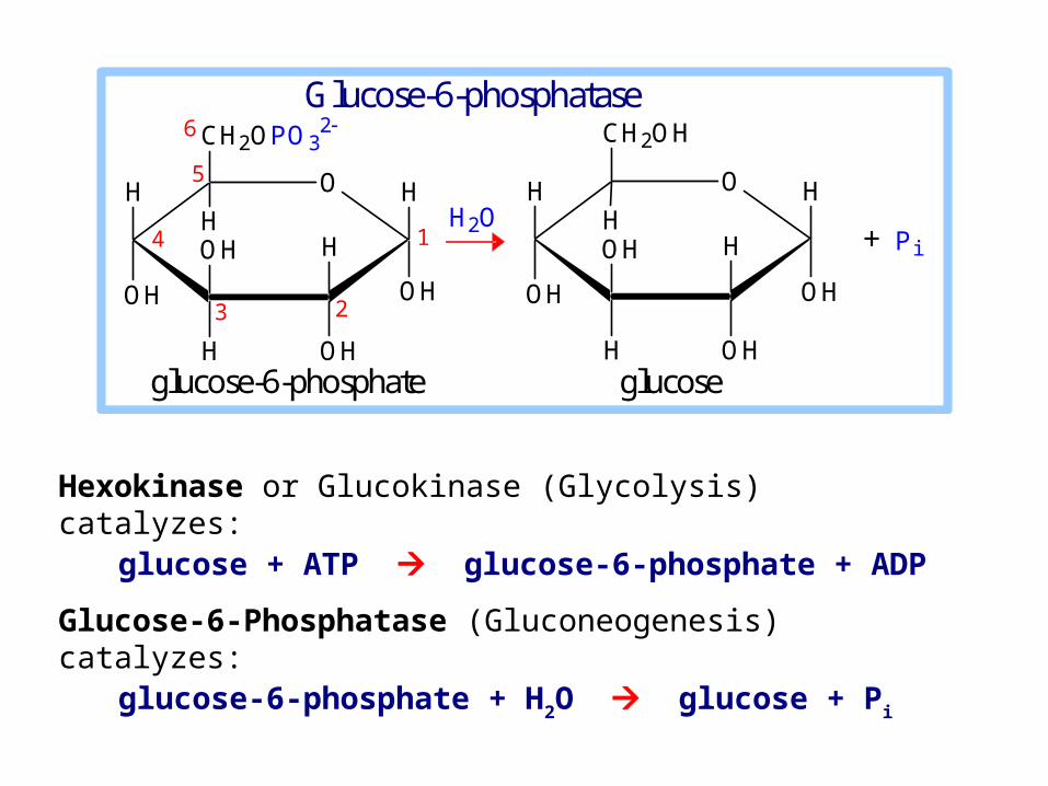
Glucose-6-phosphatase
Glucose-6-phosphatase enzyme is embedded in the endoplasmic reticulum (ER) membrane in liver cells.
The catalytic site is found to be exposed to the ER lumen. Another subunit may function as a translocase, providing access of substrate to the active site.
H O
OH
H
OHH
OH
CH2OH
H
OH
HH O
OH
H
OHH
OH
CH2OPO32
H
OH
HH2O
1
6
5
4
3 2
+ Pi
glucose-6-phosphate glucose
Glucose-6-phosphatase
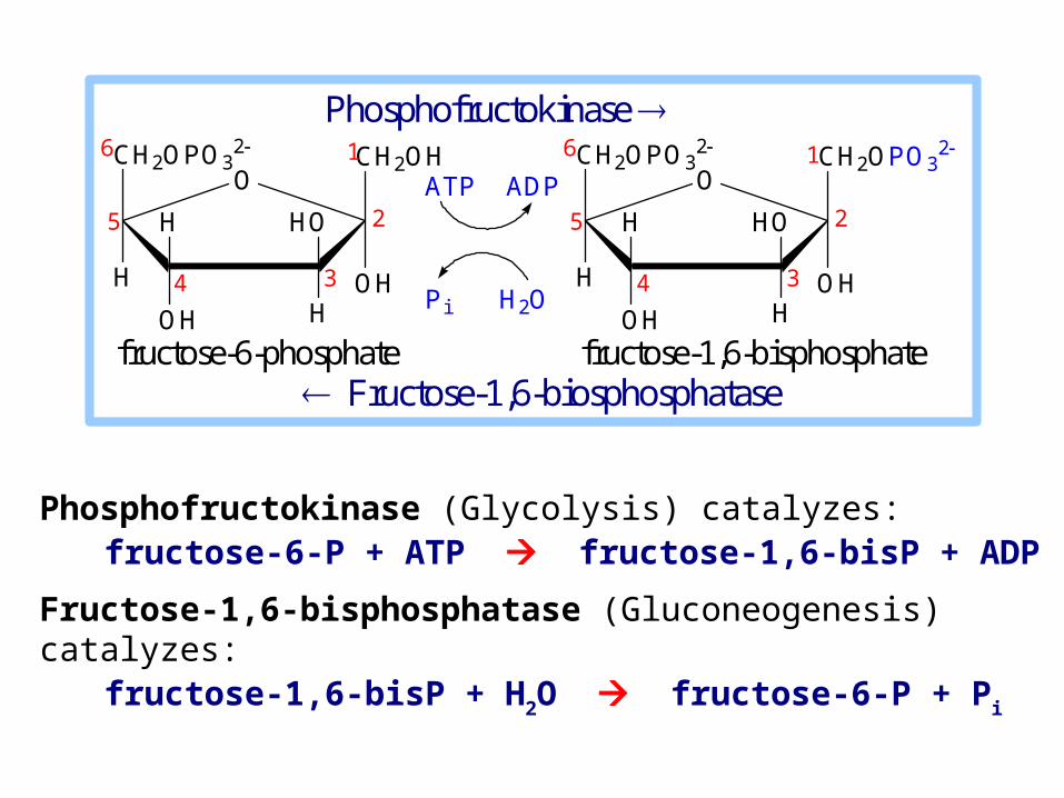
Phosphofructokinase (Glycolysis) catalyzes: fructose-6-P + ATP fructose-1,6-bisP + ADP
Fructose-1,6-bisphosphatase (Gluconeogenesis) catalyzes: fructose-1,6-bisP + H2O fructose-6-P + Pi
fructose-6-phosphate fructose-1,6-bisphosphate
Phosphofructokinase CH2OPO3
2
OH
CH2OH
H
OH H
H HO
O6
5
4 3
2
1 CH2OPO32
OH
CH2OPO32
H
OH H
H HO
O6
5
4 3
2
1ATP ADP
Pi H2O
Fructose-1,6-biosphosphatase
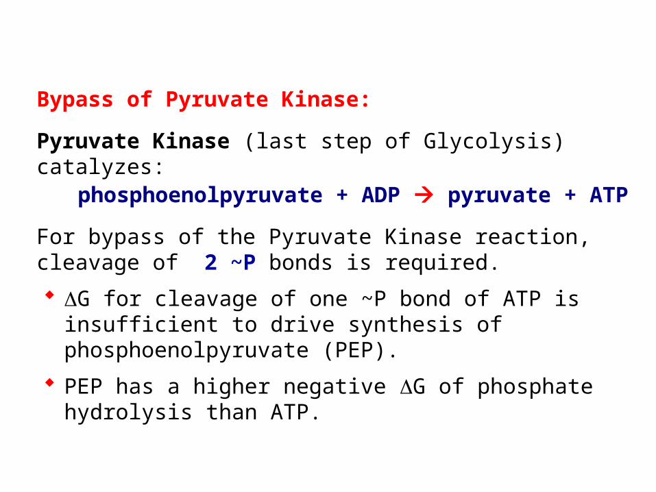
Bypass of Pyruvate Kinase:
Pyruvate Kinase (last step of Glycolysis) catalyzes: phosphoenolpyruvate + ADP pyruvate + ATP
For bypass of the Pyruvate Kinase reaction, cleavage of 2 ~P bonds is required.
G for cleavage of one ~P bond of ATP is insufficient to drive synthesis of phosphoenolpyruvate (PEP).
PEP has a higher negative G of phosphate hydrolysis than ATP.
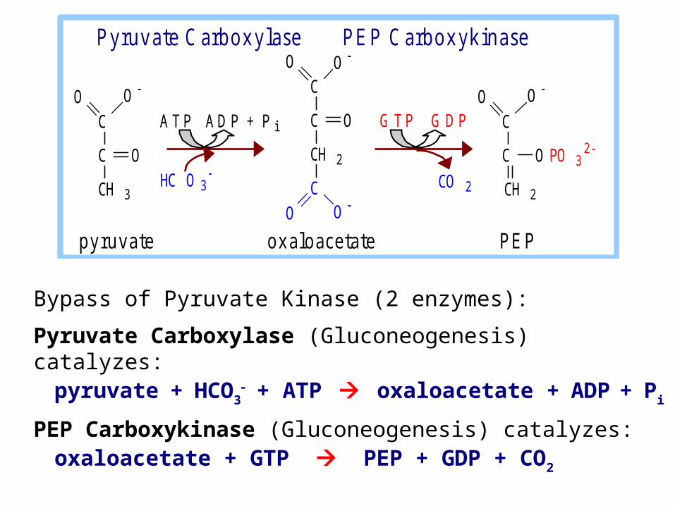
Bypass of Pyruvate Kinase (2 enzymes):
Pyruvate Carboxylase (Gluconeogenesis) catalyzes:pyruvate + HCO3
+ ATP oxaloacetate + ADP + Pi
PEP Carboxykinase (Gluconeogenesis) catalyzes:oxaloacetate + GTP PEP + GDP + CO2
C
C
CH 2
O O
O PO 32
C
C
CH 3
O O
O
A T P A D P + P i C
CH 2
C
C
O
O O
O O
HC O 3
G T P G D P
CO 2
p y r u v a te o x a lo a c e ta te P E P
P y ru v a te C a rb o x y la s e P E P C a rb o x y k in a s e
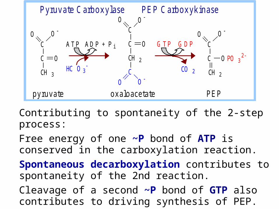
Contributing to spontaneity of the 2-step process:
Free energy of one ~P bond of ATP is conserved in the carboxylation reaction.
Spontaneous decarboxylation contributes to spontaneity of the 2nd reaction.
Cleavage of a second ~P bond of GTP also contributes to driving synthesis of PEP.
C
C
CH 2
O O
O PO 32
C
C
CH 3
O O
O
A T P A D P + P i C
CH 2
C
C
O
O O
O O
HC O 3
G T P G D P
CO 2
p y r u v a te o x a lo a c e ta te P E P
P y ru v a te C a rb o x y la s e P E P C a rb o x y k in a s e
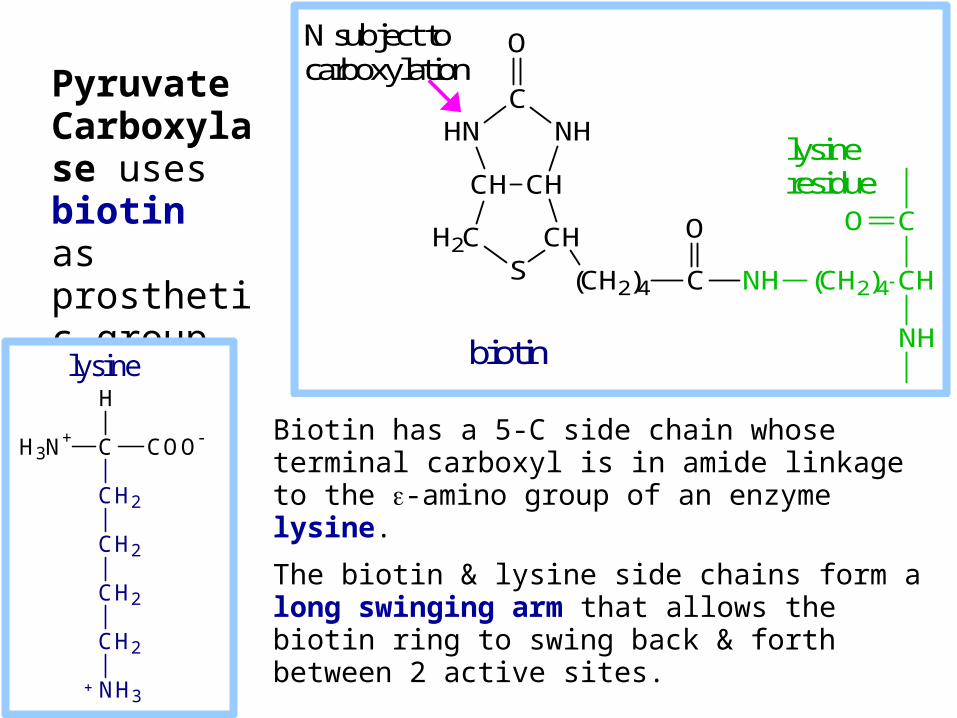
Biotin has a 5-C side chain whose terminal carboxyl is in amide linkage to the -amino group of an enzyme lysine.
The biotin & lysine side chains form a long swinging arm that allows the biotin ring to swing back & forth between 2 active sites.
Pyruvate Carboxylase uses biotin as prosthetic group.
CHCH
H2CS
CH
NHC
HN
O
(CH2)4 C NH (CH2)4 CH
CO
NH
O
biotin
N subject to carboxylation
lysine residue
H3N+ C COO
CH2
CH2
CH2
CH2
NH3
H
lysine
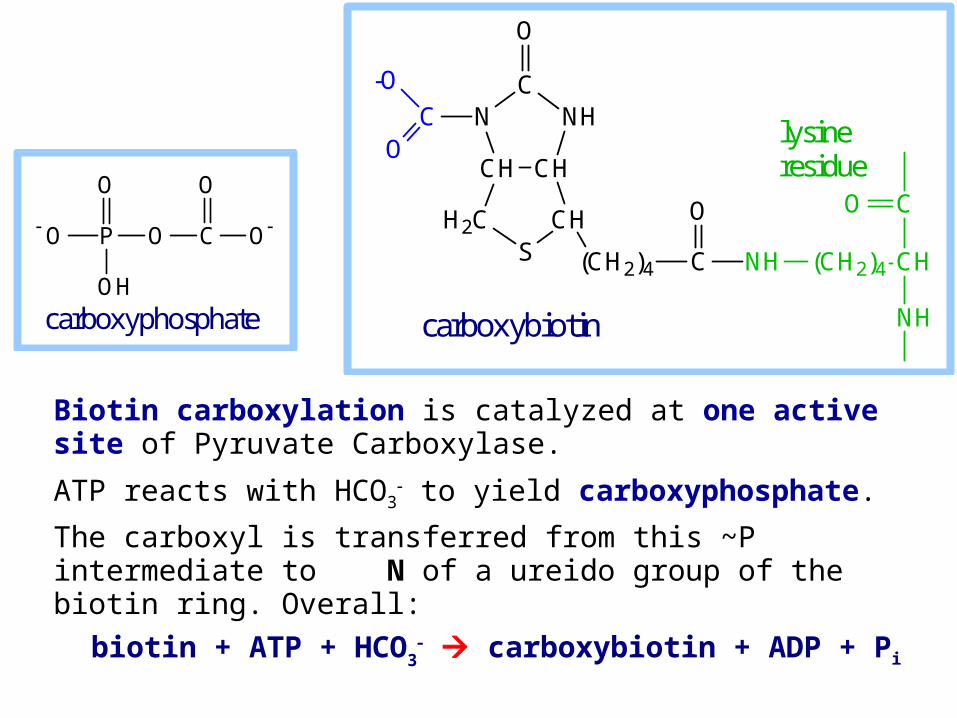
Biotin carboxylation is catalyzed at one active site of Pyruvate Carboxylase.
ATP reacts with HCO3 to yield carboxyphosphate.
The carboxyl is transferred from this ~P intermediate to N of a ureido group of the biotin ring. Overall:
biotin + ATP + HCO3 carboxybiotin + ADP + Pi
O P O
O
OH
C O
O
carboxyphosphate
CHCH
H2CS
CH
NHC
N
O
(CH2)4 C NH (CH2)4 CH
CO
NH
O
CO
-O
carboxybiotin
lysine residue
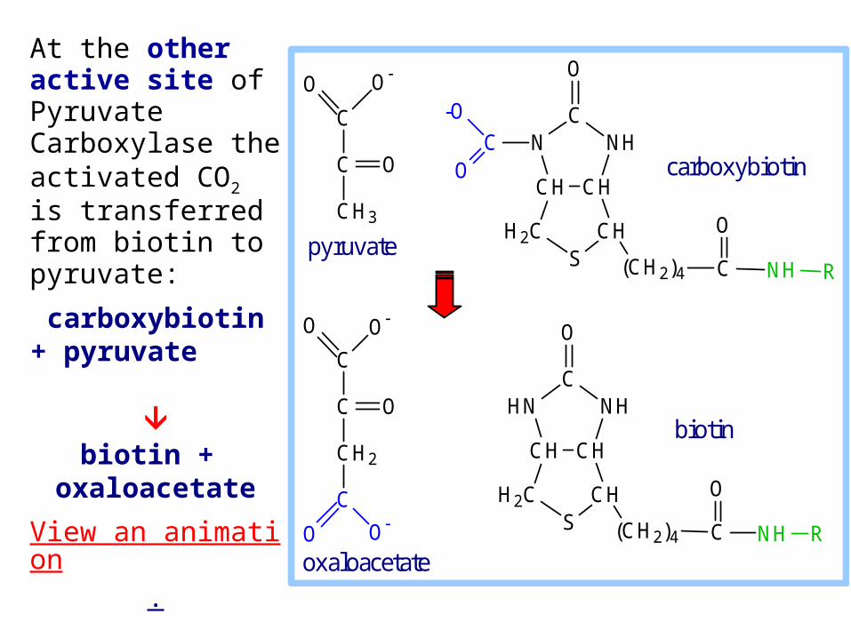
At the other active site of Pyruvate Carboxylase the activated CO2 is transferred from biotin to pyruvate:
carboxybiotin+ pyruvate
biotin +
oxaloacetate
View an animation.
CHCH
H2CS
CH
NHC
N
O
(CH2)4 C NH R
O
CO
-OC
C
CH3
O O
O
C
CH2
C
C
O
O O
OO
CHCH
H2CS
CH
NHC
HN
O
(CH2)4 C NH R
O
carboxybiotin
pyruvate
oxaloacetate
biotin
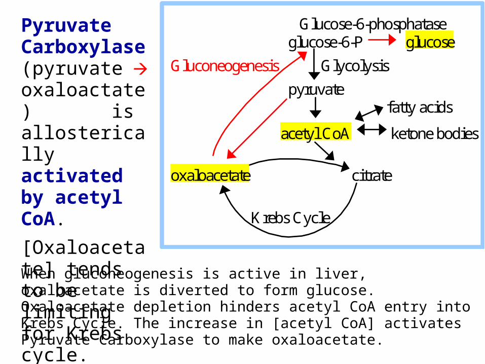
When gluconeogenesis is active in liver, oxaloacetate is diverted to form glucose. Oxaloacetate depletion hinders acetyl CoA entry into Krebs Cycle. The increase in [acetyl CoA] activates Pyruvate Carboxylase to make oxaloacetate.
Pyruvate Carboxylase (pyruvate oxaloactate) is allosterically activated by acetyl CoA.
[Oxaloacetate] tends to be limiting for Krebs cycle.
Glucose-6-phosphatase glucose-6-P glucose
Gluconeogenesis Glycolysis
pyruvate fatty acids
acetyl CoA ketone bodies oxaloacetate citrate
Krebs Cycle
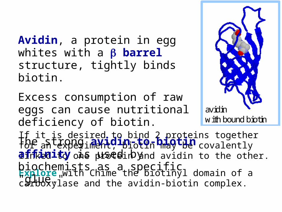
If it is desired to bind 2 proteins together for an experiment, biotin may be covalently linked to one protein and avidin to the other.
Explore with Chime the biotinyl domain of a carboxylase and the avidin-biotin complex.
avidin with bound biotin
Avidin, a protein in egg whites with a barrel structure, tightly binds biotin.
Excess consumption of raw eggs can cause nutritional deficiency of biotin.
The strong avidin-to-biotin affinity is used by biochemists as a specific "glue."
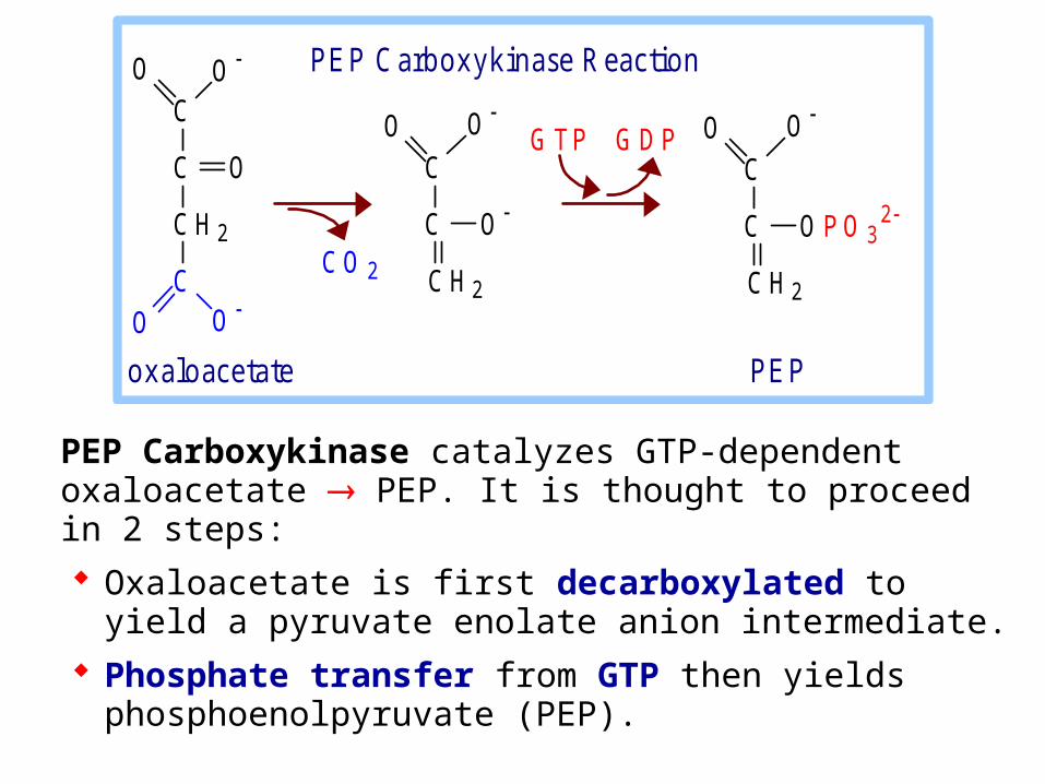
PEP Carboxykinase catalyzes GTP-dependent oxaloacetate PEP. It is thought to proceed in 2 steps:
Oxaloacetate is first decarboxylated to yield a pyruvate enolate anion intermediate.
Phosphate transfer from GTP then yields phosphoenolpyruvate (PEP).
C
C
C H 2
O O
O P O 32
C
C H 2
C
C
O
O O
O O
C O 2
C
C
C H 2
O O
O
G T P G D P
o x a lo a c e ta te P E P
P E P C a rb o x y k in a se R e a c tio n
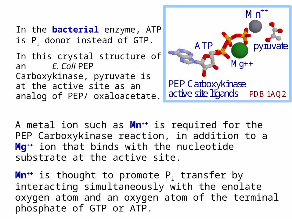
In the bacterial enzyme, ATP is Pi donor instead of GTP.
In this crystal structure of an E. Coli PEP Carboxykinase, pyruvate is at the active site as an analog of PEP/ oxaloacetate.
Mg++
pyruvate
Mn++
ATP
PEP Carboxykinase active site ligands PDB 1AQ2
A metal ion such as Mn++ is required for the PEP Carboxykinase reaction, in addition to a Mg++ ion that binds with the nucleotide substrate at the active site.
Mn++ is thought to promote Pi transfer by interacting simultaneously with the enolate oxygen atom and an oxygen atom of the terminal phosphate of GTP or ATP.
The source of pyruvate and oxaloacetate for gluconeogenesis during fasting or carbohydrate starvation is mainly amino acid catabolism.
Some amino acids are catabolized to pyruvate, oxaloacetate, or precursors of these.
Muscle proteins may break down to supply amino acids. These are transported to liver where they are deaminated and converted to gluconeogenesis inputs.
Glycerol, derived from hydrolysis of triacylglycerols in fat cells, is also a significant input to gluconeogenesis.
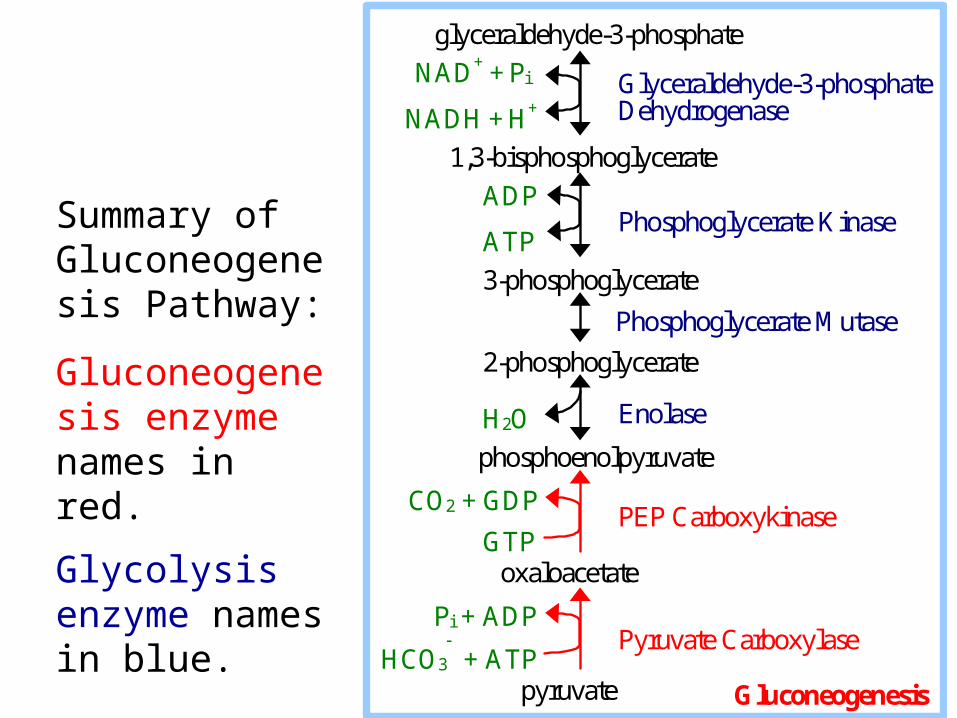
Glyceraldehyde-3-phosphate Dehydrogenase
Phosphoglycerate Kinase
Enolase
PEP Carboxykinase
glyceraldehyde-3-phosphate
NAD+ + Pi
NADH + H+
1,3-bisphosphoglycerate
ADP
ATP
3-phosphoglycerate
Phosphoglycerate Mutase
2-phosphoglycerate H2O
phosphoenolpyruvate
CO2 + GDP
GTP oxaloacetate
Pi + ADP
HCO3 + ATP
pyruvate
Pyruvate Carboxylase
Gluconeogenesis
Summary of Gluconeogenesis Pathway:
Gluconeogenesis enzyme names in red.
Glycolysis enzyme names in blue.
Glucose-6-phosphatase
Fructose-1,6-bisphosphatase
glucose Gluconeogenesis
Pi
H2O glucose-6-phosphate
Phosphoglucose Isomerase
fructose-6-phosphate
Pi
H2O fructose-1,6-bisphosphate
Aldolase
glyceraldehyde-3-phosphate + dihydroxyacetone-phosphate
Triosephosphate Isomerase (continued)

Glycolysis & Gluconeogenesis are both spontaneous. If both pathways were simultaneously active in a cell, it would constitute a "futile cycle" that would waste energy.
Glycolysis: glucose + 2 NAD+ + 2 ADP + 2 Pi
2 pyruvate + 2 NADH + 2 ATPGluconeogenesis: 2 pyruvate + 2 NADH + 4 ATP + 2 GTP glucose + 2 NAD+ + 4 ADP + 2 GDP + 6 Pi
Questions:
1. Glycolysis yields how many ~P ?
2. Gluconeogenesis expends how many ~P ? 3. A futile cycle of both pathways would waste how many ~P per cycle ?
26
4
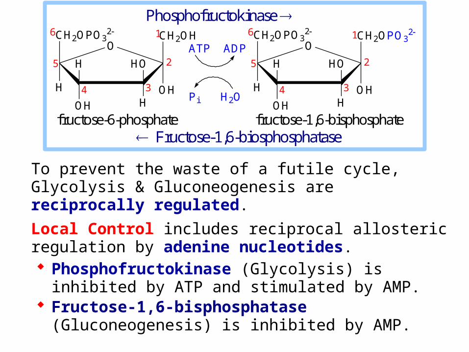
To prevent the waste of a futile cycle, Glycolysis & Gluconeogenesis are reciprocally regulated.
Local Control includes reciprocal allosteric regulation by adenine nucleotides. Phosphofructokinase (Glycolysis) is inhibited by
ATP and stimulated by AMP. Fructose-1,6-bisphosphatase (Gluconeogenesis) is
inhibited by AMP.
fructose-6-phosphate fructose-1,6-bisphosphate
Phosphofructokinase CH2OPO3
2
OH
CH2OH
H
OH H
H HO
O6
5
4 3
2
1 CH2OPO32
OH
CH2OPO32
H
OH H
H HO
O6
5
4 3
2
1
ATP ADP
Pi H2O
Fructose-1,6-biosphosphatase
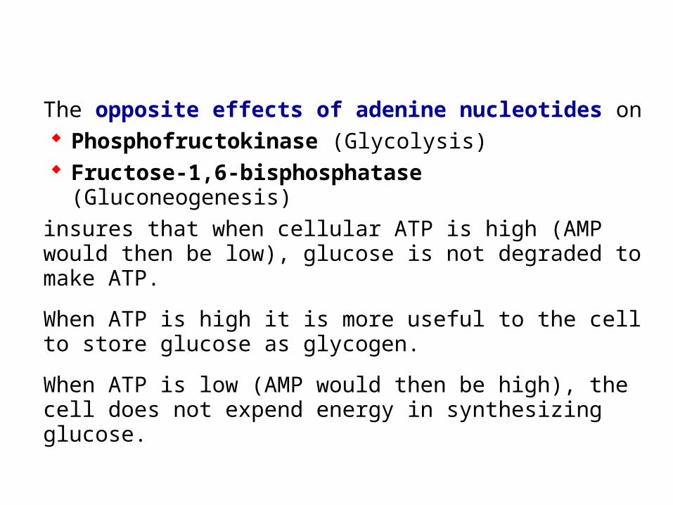
The opposite effects of adenine nucleotides on Phosphofructokinase (Glycolysis) Fructose-1,6-bisphosphatase (Gluconeogenesis)
insures that when cellular ATP is high (AMP would then be low), glucose is not degraded to make ATP.
When ATP is high it is more useful to the cell to store glucose as glycogen.
When ATP is low (AMP would then be high), the cell does not expend energy in synthesizing glucose.
Global Control in liver cells includes reciprocal effects of a cyclic AMP cascade, triggered by the hormone glucagon when blood glucose is low.
Phosphorylation of enzymes & regulatory proteins in liver by Protein Kinase A (cAMP Dependent Protein Kinase) results in inhibition of glycolysis stimulation of gluconeogenesis,
making glucose available for release to the blood.
Enzymes relevant to these pathways that are phosphorylated by Protein Kinase A include:
Pyruvate Kinase, a glycolysis enzyme that is inhibited when phosphorylated.
CREB (cAMP response element binding protein) which activates, through other factors, transcription of the gene for PEP Carboxykinase, leading to increased gluconeogenesis.
A bi-functional enzyme that makes and degrades an allosteric regulator, fructose-2,6-bisphosphate.
Reciprocal regulation by fructose-2,6-bisphosphate:
Fructose-2,6-bisphosphate stimulates Glycolysis.
Fructose-2,6-bisphosphate allosterically activates the Glycolysis enzyme Phosphofructokinase.
Fructose-2,6-bisphosphate also activates transcription of the gene for Glucokinase, the liver variant of Hexokinase that phosphorylates glucose to glucose-6-phosphate, the input to Glycolysis.
Fructose-2,6-bisphosphate allosterically inhibits the gluconeogenesis enzyme Fructose-1,6-bisphosphatase.
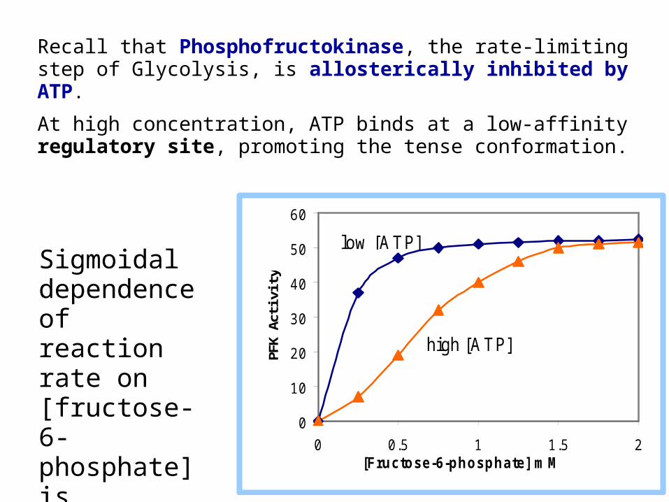
Recall that Phosphofructokinase, the rate-limiting step of Glycolysis, is allosterically inhibited by ATP.
At high concentration, ATP binds at a low-affinity regulatory site, promoting the tense conformation.
0
10
20
30
40
50
60
0 0.5 1 1.5 2[Fructose-6-phosphate] m M
PFK
Act
ivity
high [A T P]
low [A T P] Sigmoidal dependence of reaction rate on [fructose-6-phosphate] is observed at high [ATP].
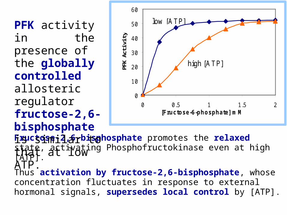
Fructose-2,6-bisphosphate promotes the relaxed state, activating Phosphofructokinase even at high [ATP].
Thus activation by fructose-2,6-bisphosphate, whose concentration fluctuates in response to external hormonal signals, supersedes local control by [ATP].
0
10
20
30
40
50
60
0 0.5 1 1.5 2[Fructose-6-phosphate] m M
PFK
Act
ivity
high [A T P]
low [A T P] PFK activity in the presence of the globally controlled allosteric regulator fructose-2,6-bisphosphate is similar to that at low ATP.
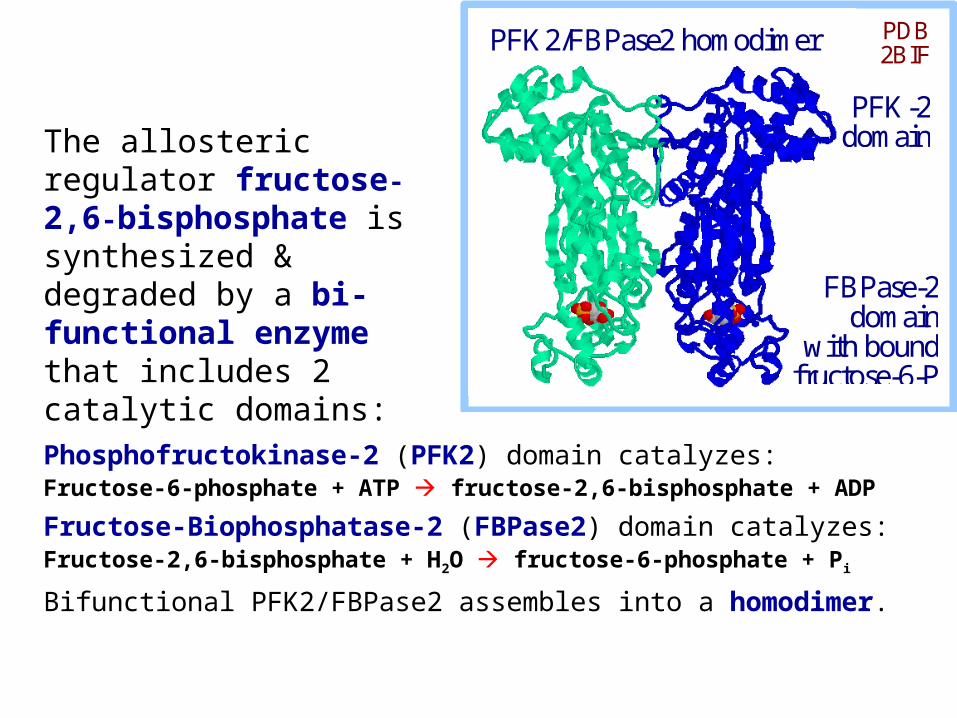
Phosphofructokinase-2 (PFK2) domain catalyzes:Fructose-6-phosphate + ATP fructose-2,6-bisphosphate + ADP
Fructose-Biophosphatase-2 (FBPase2) domain catalyzes:Fructose-2,6-bisphosphate + H2O fructose-6-phosphate + Pi
Bifunctional PFK2/FBPase2 assembles into a homodimer.
PFK2/FBPase2 homodimer PDB 2BIF
PFK-2 domain
FBPase-2 domain
with bound fructose-6-P in active site
The allosteric regulator fructose-2,6-bisphosphate is synthesized & degraded by a bi-functional enzyme that includes 2 catalytic domains:
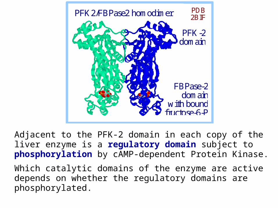
Adjacent to the PFK-2 domain in each copy of the liver enzyme is a regulatory domain subject to phosphorylation by cAMP-dependent Protein Kinase.
Which catalytic domains of the enzyme are active depends on whether the regulatory domains are phosphorylated.
PFK2/FBPase2 homodimer PDB 2BIF
PFK-2 domain
FBPase-2 domain
with bound fructose-6-P in active site
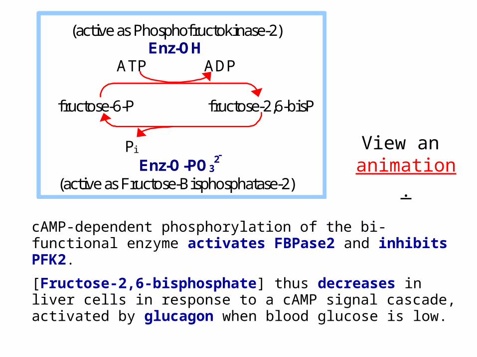
cAMP-dependent phosphorylation of the bi-functional enzyme activates FBPase2 and inhibits PFK2.
[Fructose-2,6-bisphosphate] thus decreases in liver cells in response to a cAMP signal cascade, activated by glucagon when blood glucose is low.
(active as Phosphofructokinase-2) Enz-OH
ATP ADP
fructose-6-P fructose-2,6-bisP
Pi
Enz-O-PO32
(active as Fructose-Bisphosphatase-2)
View an animation.
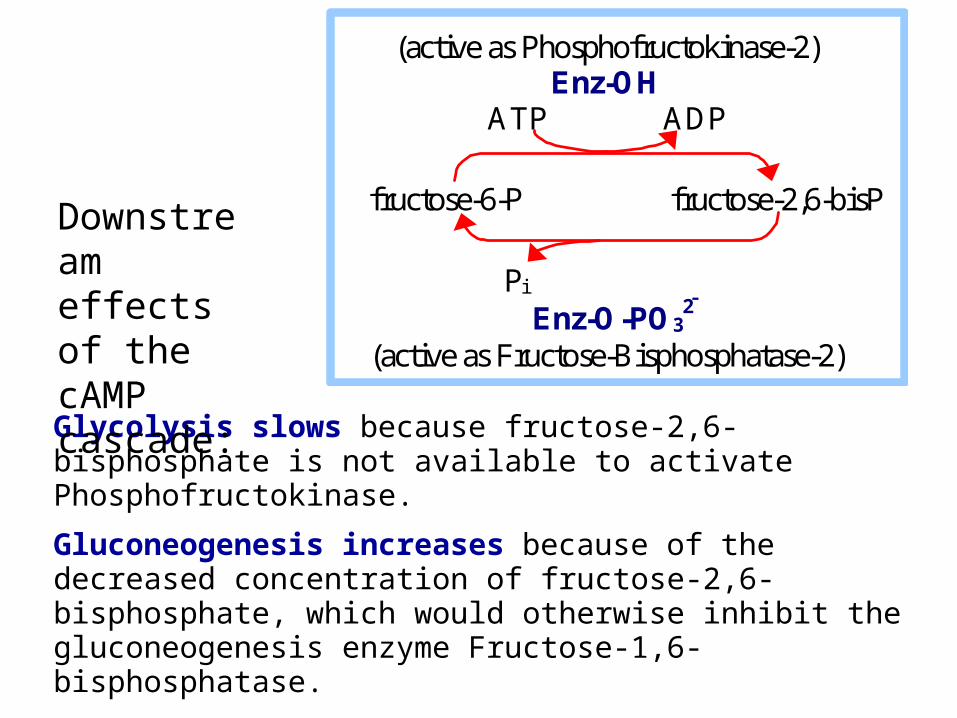
Glycolysis slows because fructose-2,6-bisphosphate is not available to activate Phosphofructokinase.
Gluconeogenesis increases because of the decreased concentration of fructose-2,6-bisphosphate, which would otherwise inhibit the gluconeogenesis enzyme Fructose-1,6-bisphosphatase.
(active as Phosphofructokinase-2) Enz-OH
ATP ADP
fructose-6-P fructose-2,6-bisP
Pi
Enz-O-PO32
(active as Fructose-Bisphosphatase-2)
Downstream effects of the cAMP cascade:
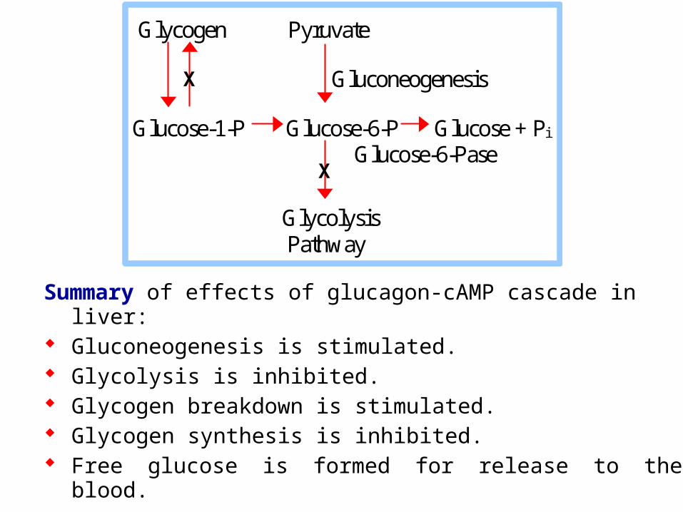
Summary of effects of glucagon-cAMP cascade in liver: Gluconeogenesis is stimulated. Glycolysis is inhibited. Glycogen breakdown is stimulated. Glycogen synthesis is inhibited. Free glucose is formed for release to the blood.
Glycogen Pyruvate Gluconeogenesis Glucose-1-P Glucose-6-P Glucose + Pi Glucose-6-Pase
Glycolysis Pathway
X
X
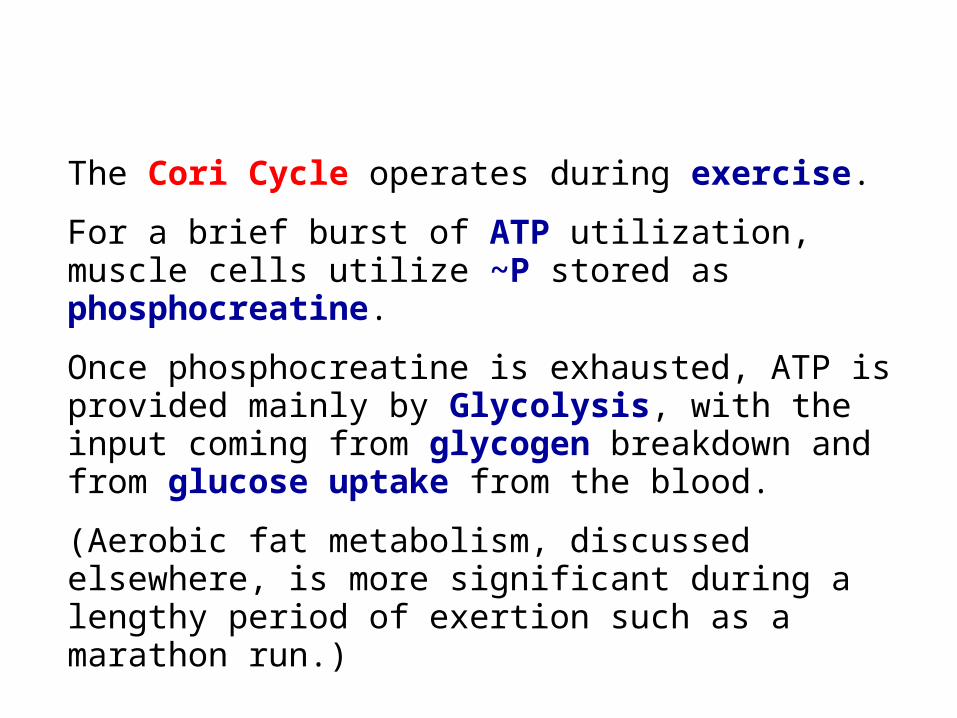
The Cori Cycle operates during exercise.
For a brief burst of ATP utilization, muscle cells utilize ~P stored as phosphocreatine.
Once phosphocreatine is exhausted, ATP is provided mainly by Glycolysis, with the input coming from glycogen breakdown and from glucose uptake from the blood.
(Aerobic fat metabolism, discussed elsewhere, is more significant during a lengthy period of exertion such as a marathon run.)
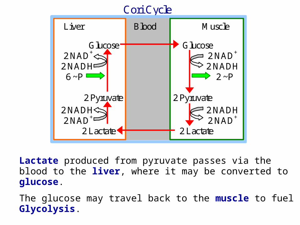
Lactate produced from pyruvate passes via the blood to the liver, where it may be converted to glucose.
The glucose may travel back to the muscle to fuel Glycolysis.
Cori Cycle
Liver Blood Muscle Glucose Glucose 2 NAD+ 2 NAD+
2 NADH 2 NADH 6 ~P 2 ~P 2 Pyruvate 2 Pyruvate 2 NADH 2 NADH 2 NAD+ 2 NAD+ 2 Lactate 2 Lactate
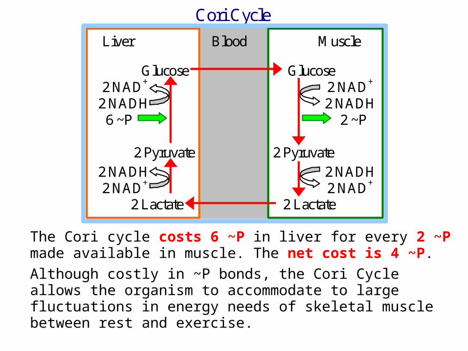
The Cori cycle costs 6 ~P in liver for every 2 ~P made available in muscle. The net cost is 4 ~P.
Although costly in ~P bonds, the Cori Cycle allows the organism to accommodate to large fluctuations in energy needs of skeletal muscle between rest and exercise.
Cori Cycle
Liver Blood Muscle Glucose Glucose 2 NAD+ 2 NAD+
2 NADH 2 NADH 6 ~P 2 ~P 2 Pyruvate 2 Pyruvate 2 NADH 2 NADH 2 NAD+ 2 NAD+ 2 Lactate 2 Lactate
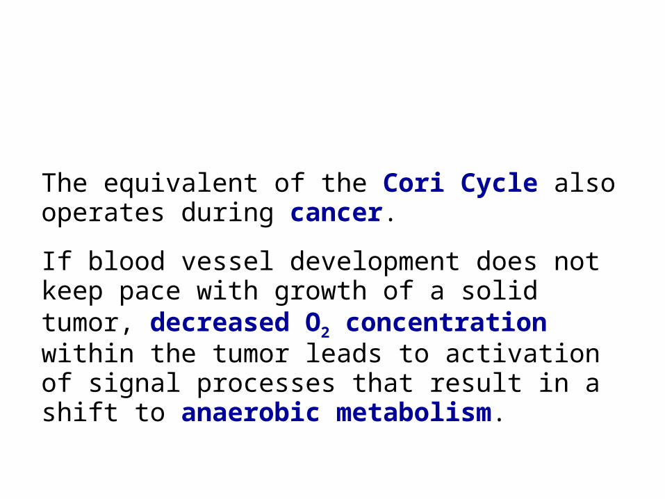
The equivalent of the Cori Cycle also operates during cancer.
If blood vessel development does not keep pace with growth of a solid tumor, decreased O2 concentration within the tumor leads to activation of signal processes that result in a shift to anaerobic metabolism.
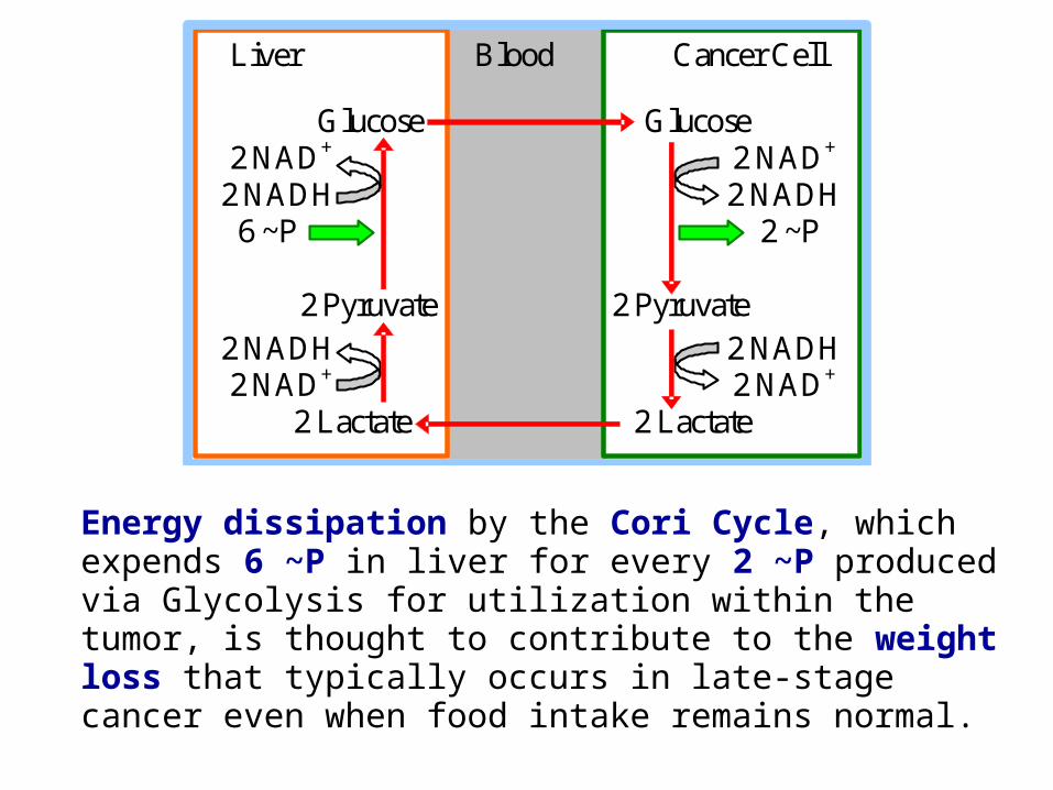
Energy dissipation by the Cori Cycle, which expends 6 ~P in liver for every 2 ~P produced via Glycolysis for utilization within the tumor, is thought to contribute to the weight loss that typically occurs in late-stage cancer even when food intake remains normal.
Liver Blood Cancer Cell Glucose Glucose 2 NAD+ 2 NAD+
2 NADH 2 NADH 6 ~P 2 ~P 2 Pyruvate 2 Pyruvate 2 NADH 2 NADH 2 NAD+ 2 NAD+ 2 Lactate 2 Lactate