Embed Size (px)
Citation preview
Vol. 112, No. 3, 1983 BIOCHEMICAL AND BIOPHYSICAL RESEARCH COMMUNICATIONS
May 16, 1983 Pages 1013-1020
GLUTATHIONE CONJUGATES OF HISC?iIDAZOLE
A.J. VARGHESE
Physics Division, Ontario Cancer Institute,
500 Sherbourne Street, Toronto, Ontario M4X lK9, Canada
Rf!ceived March 30, 1983
SUMMARY: The hydroxylamine derivative of misonidazole reacts with qlutathione -- under physiological conditions to form two isomeric conjugates. Eased on physical and chemical properties, the two conjugates have been identified as l-~[2-amino-(4-glutathion-S-yl)-l-imidazolyl]-3-methoxypropanol and l--[2-amino-(5-glutathion-~-vl)-l-imidazolyl]-3-methoxypropano1. The formation of the glutathione conjugates of reduced misonidazole offers a molecular mechanism for the depletion of GSH in mammalian cells after exposure to misonidazole under hypoxic conditions.
Misonidazole (Fig. 1) is currently undergoing clinical trials (l-5)
as a radiation sensitizer in the treatment of certain types of human tumors.
Neurotoxicity is a limiting factor in the clinical use of the drug. Another
inportant property that is of potential clinical significance in cancer
chemotherapy of the drug is that it is more toxic to hypoxic cells than to
aerobic cells (6-12). A number of studies (9, 11-16) indicate that reductive
metabolism of the drug has a role in the hypoxic cell toxicity of the drug.
E\en though there is evidence for the reductive metabolism of the drug in
man, its role in neurotoxicity is not known. Since reduction of nitro
ccmpounds proceeds through reactive intermediates such as the nitroso and
hldroxylamine derivatives, one possible mechanism for the toxic nroperty of
misonidazole is the modification of cell constituents by these reactive
reduction intermediates. Results of a number of recent studies (18-22)
suggest that intracellular thiols play a significant role in the hypoxic
cell toxicity of misonidazole. In this paper, iJe present evidence that
misonidazole, after reduction to the hydroxylamine state, reacts with
glutathione under physiological conditions to form stable qlutathione
ccnjuqates.
0006-291X/83 $1.50
Copyright 0 I983 by Academic Press, Inc. 1013 Ail rights of reproduction in an), form reserved.
Vol. 112, No. 3, 1983 BIOCHEMICAL AND BIOPHYSICAL RESEARCH COMMUNICATIONS
H-C =C-H
I I N\Q CH,-E- CH2- OCH3
I NO2
I
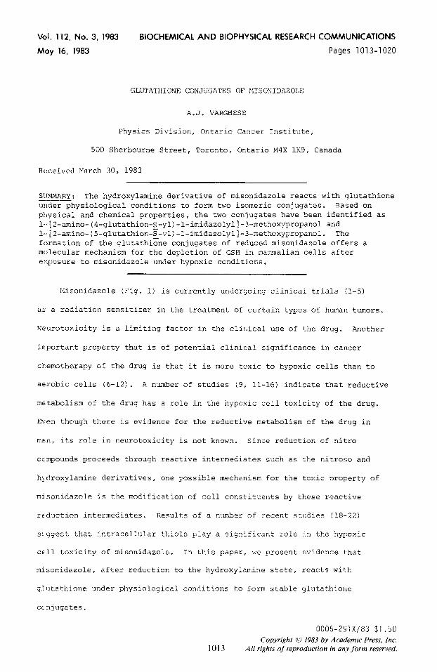
Fig. 1 Structure of misonidazole. The asterisk shows the [14Cl label.
MATERIALS
Misonidazole was obtained from Dr. Carey Snithen of Roche Products, Ltd., Welwyn City, Hertfordshire, England. GSH and ascorbic acid were purchased from Sigma Chemical Cc., St. Louis, MO. [2-14C]-Misonidazole was synthesized as described previously (20 uCi/n mole) (131. [Glycine-2-3H] GSH was purchased from New England Nuclear, Boston, MA and diluted to 0.05 mCi/m mole. The amino and hydrazo derivatives of misonidazole were prepared as described previously (15). Raney Nickel (activated) was purchased from Aldrich Chemical co., Milwaukee, WI, U.S.A.
METHODS
Reaction with GSH
Misonidazole (2 mM) was reduced with zinc dust in the presence of ammonium chloride as described previously (15). When the drug was fully reduced as indicated by the absence of absorbance above 240 nm, the suspension was filtered. Unless otherwise stated, one volume of the filtrate was added to an equal volume of a solution (10 n-&l) of GSH in 0.05M phosphate buffer (pH 7) and the reaction was allowed to proceed at 37O in the absence of air for one hour.
HPLC Analysis
A Waters liquid chromtograph equipped with a Waters Model 440 absorbance detector operated at 254 nrn and a Waters u Bondapak ClS column (0.9 x 30 cm) with water:methanol:acetic acid (94:5:1) as solvent were used, the flow rate was 2 ml/min, and 200 ~1 samples were injected. When radioactivity measurements were required, one ml fractions were collected and counted. For the large scale preparation of GS-M, the fractions containing the product were pooled and lyophilized. This was done successively until milligram quantities were obtained. The lyophilized sample was used for all subsequent studies.
Spectroscopic Studies
Proton NMR analysis was performed by Dr. Arthur Gray, University of Toronto on the 360-MHz FT systems. Fast Atom Bombardment (FAB) spectra were obtained by Dr. J.L. Holmes, Department of Chemistry, University of Ottawa, Ontario, Canada.
RESULTS
Results of preliminary studies indicated that misonidazole, after
reduction with zinc dust to the hydroxylamine derivative reacted with GSH in
phosphate buffer (pH 7) and the reaction product (GS-MIS) could be separated
1014
Vol. 112, No. 3, 1983 BIOCHEMICAL AND BIOPHYSICAL RESEARCH COMMUNICATIONS
r I I 1
9
b)
I
I-
,_
e r,
1
a)
IO 0 - 30 20 IO 0 Fraction number
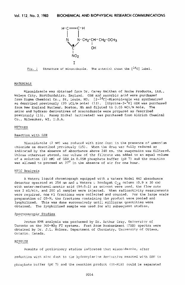
Fig. 2 HPLC separation of GS-MIS. Reduced misoniaazole was reacted with -- GSH and the reaction mixture was analyzed as described in Methods. Fig. 2a shows the absorbance monitored at 254 nm. The retention volume in ml is the x-axis. Fig. 2b shows the distribution of [14C] activity in one ml fractions when [2-l*C] misonidazole was reduced and reacted with GSH. The distribution of [3H] activity in one ml fractions when [3H] GSH was reacted with reduced misonidazble is shown in Fig. 2c.
b> the HPLC separation procedure described above. A typical separation of
tt.e reaction mixture is shown in Fig. 2a. GS-MIS has a retention volume of
2:. ml. Plisonidazole had a retention volume of 60 ml under the same
conditions. The optimum pH for the formation of GS-KIS was pH 8. In
solutions of pH > 4.5, detectable amounts of GS-MIS were not detected and
in solutions of pH < 9 the yield decreased markedly. The optimum pH for the
fclrmation of GS-MIS was pH 8 - 8.5. When GSH was reacted with either the
amine or hydrazo derivative of misonidazole, there was no evidence for the
formation of the glutathione product. Both the amine and the hydrazo
dt:rivatives have been reported to be formed when misonidazole is reduced with
zinc dust (15).
When the experiment was repeated using reduced [2- .4C] misonidazole, the
d>stribution of radioactivity in the HPLC fractions is shown in Figure 2b.
Pz-ior to the addition of GSH, radioactivity was not detected in fractions
beyond the 20th. The results of a similar study using [3H] GSH for the
1015
Vol. 112, No. 3, 1983 BIOCHEMICAL AND BIOPHYSICAL RESEARCH COMMUNICATIONS
Ml t -1
FAB spectrum I in glycerol 1 t
peoks from glycerol *
60 -
* Lu L. I
Doped wth NaCL
Doped with KCL
60
300 400 500 600 700 M/Z
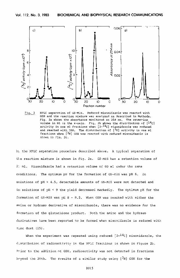
Fig. 3 FAB spectra of GS-MIS. M represents the Molecular Weight.
reaction are shown in Figure 2c. Again, when GSH without the addition of
reduced misonidazole was analyzed, radioactivity was not detected in fractions
after the 20th. The presence of both [l*C] and [3H] activities in the
fractions (nos. 22-24) containing GS-MIS suggests the presence of both the
GSH and misonidazole residues in the product.
Additional information on the structure of GS-EIIS was obtained by mass
spectrometry by Fast Atom Bombardment ionization technique. From the spectra
shown in Figure 3, it is apparently clear that GS-MIS has a molecular weight
Vol. 112, No. 3, 1983 BIOCHEMICAL AND BIOPHYSICAL RESEARCH COMMUNICATIONS
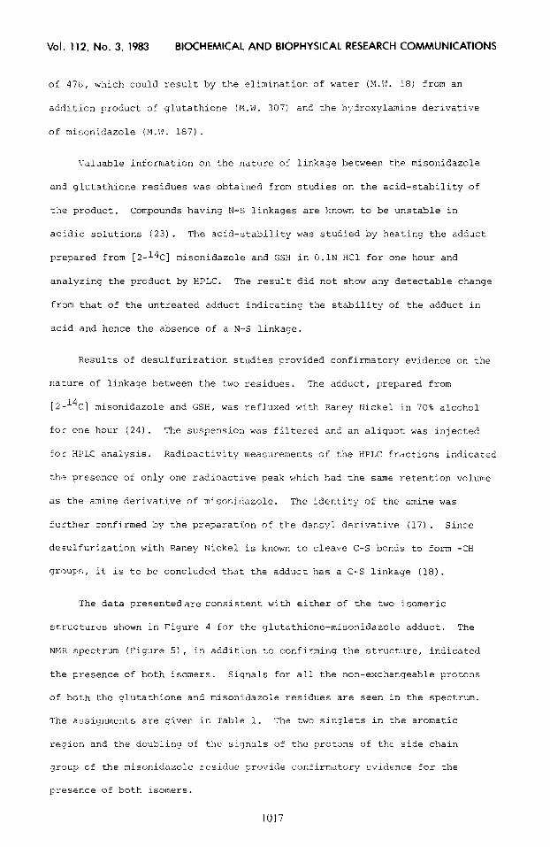
of 476, which could result by the elimination of water (M.W. 18) from an
addition product of qlutathione (M.W. 307) and the h;.droxylamine derivative
of misonidazole (?I.\<. 187).
Valuable information on the nature of lir:<age between the misonidazole
an'1 glutathione residues was obtained from studies on the acid-stability of
the product. Compounds having N-S linkages are known to be unstable in
acidic solutions (23). The acid-stability was studied by heating the adduct
prepared from [2-14C] misonidazole and GSH in O.lN HCl for one hour and
analyzing the product by HPLC. The result did not show any detectable change
from that of the untreated adduct indicating the stability of the adduct in
acid and hence the absence of a N-S linkage.
Results of desulfurization studies provided confirmatory evidence on the
nature of linkage between the two residues. The adduct, prepared from
[2-14c] misonidazole and GSH, was refluxed with Raney Nickel in 70% alcohol
for one hour (24). The suspension was filtered and an aliquot was injected
for HPLC analysis. Radioactivity measurements of the HPLC fractions indicated
th,a presence of only one radioactive peak which had the same retention volume
as the amine derivative of misonidazole. The identity of the amine was
further confirmed by the preparati'on of the dansyl derivative (17). Since
desulfurization with Raney Nickel is known to cleave C-S bonds to form -CH
grl2ups, it is to be concluded that the adduct has a C-S linkage (18).
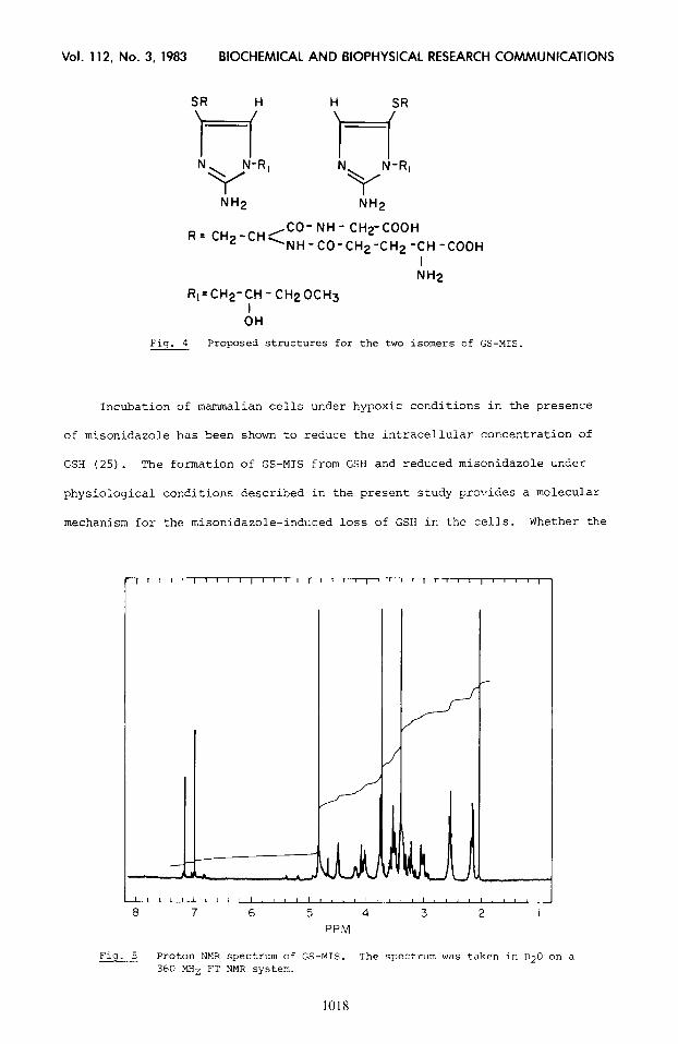
The data presentedare consistent with either of the two isomeric
structures shown in Figure 4 for the qlutathione-misonidazole adduct, The
NMR spectrum (Figure 51, in addition to confirming the structure, indicated
the presence of both isomers. Signals for all the non-exchangeable protons
of both the glutathione and misonidazole residues are seen in the spectrum.
The assignments are given in Table 1. The two singlets in the aromatic
region and the doubling of the signals oL + the protons of the side chain
group of the misonidazole residue provide confirmatory evidence for the
presence of both isomers.
1017
Vol. 112, No. 3, 1983 BIOCHEMICAL AND BIOPHYSICAL RESEARCH COMMUNICATIONS
,CO- NH - CHTCOOH R= CH2-CH, NH-CO-CH2-CH2-CH-COOH
I NH2
Rt=CH2-CH - CH20CH3
AH
4 Fig. Proposed structures for the two isomers of GS-MIS.
Incubation of mammalian cells under hypoxic conditions in the presence
of misonidazole has been shown to reduce the intracellular concentration of
GSH (25). The formation of GS-MIS from GSH and reduced misonidazole under
physiological conditions described in the present study provides a molecular
mechanism for the misonidazole-induced loss of GSH in the cells. Whether the
h A
Fig. 5 Proton NMR spectrum of GS-MIS. The spec:rum was taken in D20 on a 360 MHz FT NMR system.
Vol. 112, No. 3, 1983 8lOCHEMlCAL AND BIOPHYSICAL RESEARCH COMMUNICATIONS
TABLE 1
360 MHZ 'H-NMR Spectral Properties of MIS-GS
Assignment Chemical Shift (in ppm from TMS)
Glutathione residue
C*S-lY cys-6 9lY-a
qlu-cX 91-y qlu-B
Misonidazole residue
= N - CH = CH - N - -
= N - CH = CH - N - -
'- N - 5 - CHOH - CH2 - OCH3
'- N - CH2 - CHOH - CH2 - OCH3 -
N - CH2 - CHOH - CH2 - OCH3
CH2 - 0 - CH 3
4.52 (1) 3.02; 3.22
3.74 (2)
3.56 (1) 2.12 (2) 2.56 (2)
6.96 (f)
7.15 (%)
4.02; 3.76
4.2 (1)
3.38 (2)
3.36 (3)
(2)
(2)
removal of GSH as GS-MIS contributes to the hypoxic cell toxicity of
misonidazole is a topic for further investigation. Sowever, it is of
interest to note that the intracellular concentration of [2-14C] misonidazole
leading to 50% toxicity in EM-6 tumor cells is estimated to be about 3.7 mM,
wh:ch is equivalent to the concentration of GSH in the cell 126). The
formation of GS-MIS0 is also of importance as a possible pathway for the
removal of the toxic metabolites resulting from the reduction of misonidazole.
In either case, additional studies are required to determine the biological
imljortance of GS-MIS.
ACKNOWLEDGEMENTS --
This work was supported by the Ontario Cancer Treatment and Research Foundation and the National Cancer Institute of Canada.
REFERENCES -
1. Phillips, T.L., Wasserman, T.H., Stetz, J. and Brady, L.W. (1982) Int. J. Radiat. Oncol Biol. Phys. 8: 327-334.
2. Dische, S. (1980) Cancer Clin. Trials 3: 175-178. -
1019
Vol. 112, No. 3, 1983 BIOCHEMICAL AND BIOPHYSICAL RESEARCH COMMUNICATIONS
3.
4.
5.
6. 7. 8.
9. 10. 11.
12.
13.
14. 15.
16. 17. 18.
19.
20.
21. 22.
23.
24.
25.
26.
Saunders, M.I., Anderson, P., Dische, S. and Martin, W.M. (1982) Int. J. Radiat. Oncol. Biol. Phys. 8: 347-350. Ydrach, A.A., Martial, V.A., Parsons, J., Concannon, J., Asbell, S.O. and George, F. (1982) Int. J. Radiat. Oncol. Biol. Phys. 8: 357-359. Spooner, D., Bugden, R.D., Peckham, M.J. and Wist, E.A. (1582) Int. J. Radiat. Oncol. Biol. Phys. 8: 387-389. Brown, J.M. (1977) Radiat, Res. 72: 469-486. Brown, J.M. and Yu, N.Y. (1979) z. J. Radiol. 52: 893-896. Moore, B.A., Palcic, B. and Skarsgaard, L.D. (1976) Radiat. Res. 67: - 459-573.
Taylor, Y.C. and Rauth, A.M. (1978) Cancer Res. 38: 2745-2752. Stratford, 1-J. and Adams, G.E. (1978) Er. J. RaKol. 51: 745-746. - Whitmore, G.F., Gulyas, S. and Varghese, A.J. (1978) Br. J. Cancer 37 - (Suppl. 3): 115-119. Sridhar,R., Koch, C. and Sutherland, R. (19763 Int. J. Radiat. Oncol. Biol. Phys. 1: 1149-1157. Varghese, A.J., Gulyas, S. and Mohindra, J.K. (1976) Cancer Fes. 36: - 3761-3765. Varghese, A.J. and Whitmore, G.F. (1980! Cancer Res. 40: 2165-2169. Varghese, A.J. and Whitmore, G.F. (1981) Chem. Biol. Gteract. 36: - 141-151. Varghese, A.J. and Whitmore, G.F. (1983) Cancer Res. 43: 78-82. Varghese, A.J. (1981) Anal. Biochem. 110: 197-200. - - Astor, M., Hall, E.J., Martin, J., Flynn, M. and Biaglow, J.F. (1982) Int. J. Radiat. Oncol. Biol. Phys. 8: 409-413. Bump, E.A., Yu, N.Y. and Brown, J.MT (1982) Int. J. Radiat. Oncol. Biol. Phys. 8: 449- Varnes, M.E.: Biaglow, J.E., Hall, E.J. and Koch, C.J. (1980) Cancer Management, Vol. 5, Chapter 18. L. Brady (Ed.). N.Y., Masson, pp. 121- 127. Taylor, Y.C. and Rauth, A.M. (1980) Br. J. Cancer 41: 892-900. Biaglow, J.E., Varnes, M.P., Aston, M. and Hall, E.JT (1962) Int. J. Radiat. Oncol. Biol. Phys. 8: 719-723. Ketterer, B. and Kadlubar, F.F. (1982) Chem. Biol. Interact. 39: lll- - 127. Mozingo, R., Ilolf, D.E., Harris, S.A. and Folkers, K. (1943) J. Am. Chem. Sot. 65: 1013-1016. Brown, M. 982) Int. J. Radiat. Oncol. Biol. Phys. 8: 675-682. Miller, C.G., Nagan-Lee, J. and Chapman, J.D. (1982) -1nt. J. Radiat. Oncol. Biol. Phys. 8: 741-744. -


![Review Article Role of Glutathione in Cancer Progression ...downloads.hindawi.com/journals/omcl/2013/972913.pdf · GCL and glutathione S-transferases [ ]. 2. GSH Biosynthesis Glutathione](https://img.pdfslide.net/doc/110x75/5edbd12aad6a402d666637cd/review-article-role-of-glutathione-in-cancer-progression-gcl-and-glutathione.jpg)