Embed Size (px)
Citation preview
Carbohydrate Research 389 (2014) 115–122
Contents lists available at ScienceDirect
Carbohydrate Research
journal homepage: www.elsevier .com/locate /carres
Glycans in immune recognition and response
http://dx.doi.org/10.1016/j.carres.2014.02.0040008-6215/� 2014 Elsevier Ltd. All rights reserved.
⇑ Corresponding author. Tel.: +972 3 640 6792; fax: +972 3 642 2046.E-mail address: [email protected] (V. Padler-Karavani).
Ron Amon, Eliran Moshe Reuven, Shani Leviatan Ben-Arye, Vered Padler-Karavani ⇑Department of Cell Research and Immunology, The George S. Wise Faculty of Life Sciences, Tel Aviv University, Tel Aviv 69978, Israel
a r t i c l e i n f o a b s t r a c t
Article history:Received 27 November 2013Received in revised form 29 January 2014Accepted 2 February 2014Available online 12 February 2014
Keywords:Sialic acidNeu5GcAntibodiesCancerImmunotherapyBiomarker
Glycans at the forefront of cells facilitate immune recognition processes. Cancer cells commonly presentaltered cell surface glycosylation, especially manifested in the expression of sialic acid at the termini ofglycolipids and glycoproteins. Although tumor-associated carbohydrate antigens (TACAs) result inexpression of altered-self, most such carbohydrates do not elicit strong humoral responses. Various strat-egies had been devised to elicit increased immunogenicity of such TACA aiming for potent immunother-apeutic antibodies or cancer vaccines. However some carbohydrates are immunogenic in humans andhold potential for novel glycotherapies. N-Glycolylneuraminic acid (Neu5Gc) is a foreign immunogenicsugar in humans originating from the diet (e.g., red meat) and subsequently expressed on the cell surface,especially accumulating on carcinoma. Consequently, the human immune system detects this non-selfcarbohydrate generating a broad anti-Neu5Gc antibody response. The co-existence of Neu5Gc/anti-Neu5Gc within humans spurs chronic inflammation mediated disease, including cancer. Concurrently,anti-Neu5Gc antibodies hold potential for novel targeted therapy. aGal is another foreign immunogeniccarbohydrate antigen in humans and all humans have circulating anti-Gal antibodies. This review aims todescribe the immunogenicity of Neu5Gc and its implications for human diseases, highlighting differencesand similarities with aGal and its potential for novel targeted theranostics.
� 2014 Elsevier Ltd. All rights reserved.
1. Introduction
The immune system discriminates self from non-self and elimi-nates particles carrying such non-self determinants. Pathogens canevade immune recognition either by masking non-self antigensand/or by disguising with host self-antigens through molecularmimicry. However, some pathological conditions present altered-self determinants that cause breaching of tolerance and lead torejection through an autoimmune response.103 Cell surface glyco-sylation is universal to all living cells and strategically positionedto mediate such immune recognition processes.107,155
Carbohydrate chains (glycans) that decorate glycoproteins andglycolipids (glycoconjugates) on the cell surface hold tremendousstructural diversity.120,155 In vertebrates, glycans usually terminatewith sialic acids (Sia) that function as markers of normal self andcan be recognized by a variety of receptors (e.g., Siglecs) mediatinginter- and intra-cellular communication.129,152 Cancer cells com-monly present altered cell surface glycosylation as a result ofabnormal expression of glycosyltransferases giving rise tochanges in the typical glycan structures and/or to theirexpression levels.11,153 Such changes especially affect sialylation
patterns33,85,86,122 that correlate with an advanced cancer stage,progression and/or metastasis.11,57,78,122,139 Although such tumor-associated carbohydrate antigens (TACAs) result in expression ofaltered-self, most of these carbohydrates are largely poorly immu-nogenic and do not result in potent antibody responses,60 with afew exceptions.90,96 Nevertheless, carbohydrates have been shownto be involved in other aspects of tumor immunology and can con-tribute to cellular immune responses within immunoediting andimmunosurveillance processes.14,24,116,137 For example, it wasshown that tumor cells with a lower degree of sialylation can inter-act better with immunosurveilling cells.24
TACA holds tremendous potential for targeted cancer therapyand in recent years efforts have been put into eliciting increasedimmunogenicity of such TACA aiming to generate potent antibod-ies for immunotherapy or a vaccine that can provoke a specific im-mune response against cancer.52,71,164,166 Several approaches toimproving TACA immunogenicity have been investigated: cova-lently coupling of carbohydrates to immunologically active proteincarriers such as bovine serum albumin (BSA), keyhole limpethemocyanin (KLH), tetanus toxoid (TT), or Bacille Calmette–Guérin(BCG);52,166 treating with a mixture of several mono-epitopic vac-cines;95,133 or fully synthetic homogeneous vaccines designed tocontain an adjuvant or other immunological epitopes to furtherenhance the immunogenicity of resulting vaccines.12,13,73 Manyof these new strategies were very successful at improving
116 R. Amon et al. / Carbohydrate Research 389 (2014) 115–122
immunogenicity, at least in animals.36,52 However, although sev-eral promising TACA-based cancer vaccines have entered clinicaltrials (including in Phase III),34,77,100 none have been approved forclinical use yet. Most tested vaccines failed in clinical trials mainlydue to the lack of a robust T cell-mediated immunity and/or lack ofsurvival benefit for patients.34,36,52,77,94,100,166,172 Importantly,many of the new strategies that trigger much stronger immune re-sponses have not yet been tested in humans.26,71,164
aGal is a foreign immunogenic carbohydrate antigen in humansdue to a specific gene inactivation, and all humans have circulatinganti-Gal antibodies that could potentially be used for immunother-apy if the antigen had been present on the target cells.42 aGal wasused to generate autologous tumor-vaccines by incorporation ofthis xenogenic carbohydrate antigen through intra-tumoral injec-tion of aGal glycolipids.47 This proved to then tag the cells fordestruction by complement-mediated cytotoxicity (CDC) and byantibody-dependent cellular cytotoxicity (ADCC) following anti-Gal binding to the aGal epitopes de novo expressed on the tumorcells in mice.42,43,47 Another potent approach to increase tumorimmunogenicity was to metabolically engineer cells to expressunnatural TACA analogues18 followed by treatment with antibod-ies specifically generated against these unnatural carbohydratesthat could promote CDC in vitro, though evidence for therapeuticefficacy in vivo is still pending.52,159
However, recent research suggests that such metabolic engi-neering with a foreign carbohydrate actually occurs in humansthrough dietary consumption of N-Glycolylneuraminic acid(Neu5Gc) that accumulates on carcinoma and also provokes an im-mune response in humans.99,115,122,123,127,146,150,174 Neu5Gc is anon-human sialic acid since humans uniquely cannot synthesizeit due to a specific inactivation of the gene encoding the enzymeCMP-Neu5Ac hydroxylase (CMAH).22,151 Thus both aGal andNeu5Gc are immunogenic in humans because they are foreign tothe human immune system, and therefore serve as targets for cir-culating antibodies.98,126,141 This review aims to summarize recentresearch on the immunogenicity of Neu5Gc and its implications forcancer and other human diseases, emphasizing its potential for no-vel targeted theranostics (therapy and diagnostics). In addition, thedifferences and similarities between the immunogenic sugars aGaland Neu5Gc will be highlighted.
2. Sialic acid diversity
Sialic acids (Sias) are a diverse family of �50 alpha-keto aldonicacid carbohydrates with a nine-carbon carboxylated backbone,found predominantly as the terminal units on glycans and glyco-conjugates in vertebrates.4 Sia diversity arise from various modifi-cations at either the C5-amino group (with acetyl or glycolyl) orthe hydroxyl groups at C4, C7, C8, and C9 by acetate, lactate, sul-fate, or phosphate esters or by methyl ethers.4,140,154 The two mostcommon Sias in mammals are N-Acetylneuraminic acid (Neu5Ac)and its hydroxylated form N-Glycolylneuraminic acid (Neu5Gc),as described in Figure 1140,154. Sia is a-linked to underlying sugarsthrough their C2 to either C3/6 of galactose, C6 of N-acetylgalactos-amine, or to C8 of another Sia (a2-3Gal or a2-6Gal; a2-6GalNAc;and a2-8Sia, respectively).4 Overall sialoglyconjugate diversity re-sults from Sia-modification, linkage to underlying sugars and theircomposition, the conjugated scaffold (protein/lipid), the glycanmode of attachment (e.g., O/N-linked to proteins), and finally theirspatial organization (density).4,25,140
3. Recent advances in the synthesis of sialic acid derivatives
Chemical sialylation was one of the most challenging glycosyl-ation reactions in the past largely due to low yields, poor
stereo-selectivity, and difficulties in product purification.19 Recentadvancements in chemical and especially chemoenzymatic synthe-sis provided chemically well-defined and structurally homoge-neous sialic acid-containing glycans (sialosides).1,10,84,112 Thenewly developed chemical ‘one-pot glycosylation’ reaction alloweddirect sequential assembly of monosaccharides into glycans in thesame reaction flask without intermediates isolation,70,158 howeverthis approach remains impractical in generating sialosides, espe-cially for certain Sia-derivatives that are labile to the final depro-tection steps.112 Importantly, subsequent development of ahighly efficient one-pot multiple-enzyme (OPME) system allowedthe chemoenzymatic synthesis of naturally occurring and non-nat-ural sialosides.1,15,112,167,170,171 This approach exploits the high re-gio-selectivity, chemo-selectivity, and stereo-selectivity ofenzymes with the flexibility and diversity of chemical synthesisto achieve an efficient synthesis of such complex carbohy-drates.1,112 This breakthrough allowed to generate a wide collec-tion of sialosides15,19,20,32,93,142,167–171 finally paving the way forextensive investigation of sialic acid’s diversity and their biologicalroles.19,21,29,83,124,143
4. Hanganutziu–Deicher (HD) antigens and antibodies
Almost a century ago human ‘heterophile’ antibodies had beennoticed and later on suggested to recognize Neu5Gc-antigens. Inthe 1920s Hanganutziu54 and Deicher28 independently noticedthat injection of horse antisera (e.g., to tetanus toxin or diphtheria)into humans caused ‘Serum-sickness’ with allergy-like symptomsdue to hemagglutinins. These human ‘heterophile’ antibodies laternamed Hanganutziu–Deicher-(HD)-antibodies could agglutinateanimal erythrocytes from many species, except human and chick-en. Such antibodies were then detected in patients who had neverbeen exposed to animal sera, including patients with variousinflammatory or infectious conditions and cancer (reviewed in99).Subsequently HD-antigens were defined in late 1970s as aNeu5Gc-containing ganglioside (Neu5Gc-GM3, Neu5Gca2-3Galb1-4G1cb1-1’Ceramide)5,64,104 or Neu5Gc-containing glyco-protein.111,118 However, these early studies used crude methodsfor detection of the HD-antigens39,62,65,69,72,80,81,87,119,138 or HD-antibodies,108,109,114,117,145 apparently assuming that normal hu-mans are negative.53,75,114 With the advent of modern glycobiologytools detection of Neu5Gc-antigens and anti-Neu5Gc antibodieshad been extensively revisited providing compelling evidence fortheir presence not only in patients but also in healthy individuals,followed by investigation of their implications for various humandiseases, as described below.
5. Neu5Gc in human tissues
5.1. Neu5Gc in healthy humans
Healthy human tissues had been inspected in the past by vari-ous chromatographic and immunochemical techniques for detec-tion of Neu5Gc, however those failed to provide unequivocalchemical evidence for its presence99 or failed to detect it at all.82
The expression of this sialic acid had been recently re-examinedmainly by highly characterized antibodies to Neu5Gc-containingantigens, HPLC, and finally mass-spectrometry (Table 1). Varkiand colleagues had generated a highly sensitive polyclonal chickenanti-Neu5Gc antibody (chickens immunized with GM3(Neu5Gc))that was further affinity-purified and extensively characterizedby various protocols (e.g. ELISA, Western blot, flow cytometry,immunohistochemistry, and glycan microarray)31,124,146 demon-strating broad monospecificity to various Neu5Gc-containingglycoconjugates.124 Immunohistochemistry staining with this
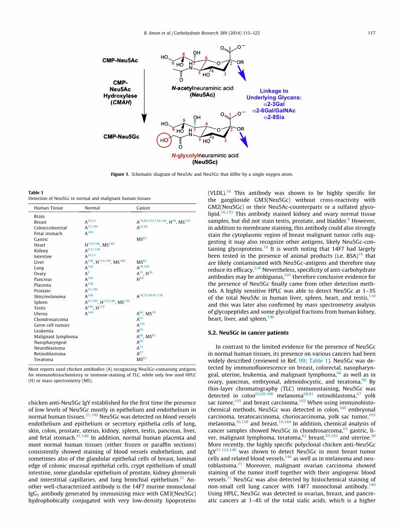
Figure 1. Schematic diagram of Neu5Ac and Neu5Gc that differ by a single oxygen atom.
Table 1Detection of Neu5Gc in normal and malignant human tissues
Human Tissue Normal Cancer
BrainBreast A16,31 A16,66,102,144,146, H58, MS102
Colon/colorectal A31,146 A62,66
Fetal stomach A146
Gastric MS82
Heart H110,146, MS146
Kidney A9,31,146
Intestine A16,31
Liver A146, H110,146, MS146 MS82
Lung A146 A56,149
Ovary A9 A31, H58
Pancreas A146 H58
Placenta A146
Prostate A31,146
Skin/melanoma A146 A16,31,68,81,138
Spleen A31,146, H110,146, MS146
Testis A146, H110
Uterus A146 A66, MS30
Chondrosarcoma A63
Germ cell tumors A105
Leukemia A66
Malignant lymphoma A66, MS82
Nasopharyngeal A66
Neuroblastoma A31
Retinoblastoma A67
Teratoma MS82
Most reports used chicken antibodies (A) recognizing Neu5Gc-containing antigensfor immunohistochemistry or immune-staining of TLC, while only few used HPLC(H) or mass spectrometry (MS).
R. Amon et al. / Carbohydrate Research 389 (2014) 115–122 117
chicken anti-Neu5Gc IgY established for the first time the presenceof low levels of Neu5Gc mostly in epithelium and endothelium innormal human tissues.31,146 Neu5Gc was detected on blood vesselsendothelium and epithelium or secretory epithelia cells of lung,skin, colon, prostate, uterus, kidney, spleen, testis, pancreas, liver,and fetal stomach.31,146 In addition, normal human placenta andmost normal human tissues (either frozen or paraffin sections)consistently showed staining of blood vessels endothelium, andsometimes also of the glandular epithelial cells of breast, luminaledge of colonic mucosal epithelial cells, crypt epithelium of smallintestine, some glandular epithelium of prostate, kidney glomeruliand interstitial capillaries, and lung bronchial epithelium.31 An-other well-characterized antibody is the 14F7 murine monoclonalIgG1 antibody generated by immunizing mice with GM3(Neu5Gc)hydrophobically conjugated with very low-density lipoproteins
(VLDL).16 This antibody was shown to be highly specific forthe ganglioside GM3(Neu5Gc) without cross-reactivity withGM2(Neu5Gc) or their Neu5Ac-counterparts or a sulfated glyco-lipid.16,135 This antibody stained kidney and ovary normal tissuesamples, but did not stain testis, prostate, and bladder.9 However,in addition to membrane staining, this antibody could also stronglystain the cytoplasmic region of breast malignant tumor cells sug-gesting it may also recognize other antigens, likely Neu5Gc-con-taining glycoproteins.16 It is worth noting that 14F7 had largelybeen tested in the presence of animal products (i.e. BSA)16 thatare likely contaminated with Neu5Gc-antigens and therefore mayreduce its efficacy.126 Nevertheless, specificity of anti-carbohydrateantibodies may be ambiguous,101 therefore conclusive evidence forthe presence of Neu5Gc finally came from other detection meth-ods. A highly sensitive HPLC was able to detect Neu5Gc at 1–3%of the total Neu5Ac in human liver, spleen, heart, and testis,110
and this was later also confirmed by mass spectrometry analysisof glycopeptides and some glycolipid fractions from human kidney,heart, liver, and spleen.146
5.2. Neu5Gc in cancer patients
In contrast to the limited evidence for the presence of Neu5Gcin normal human tissues, its presence on various cancers had beenwidely described (reviewed in Ref. 99; Table 1). Neu5Gc was de-tected by immunofluorescence on breast, colorectal, nasopharyn-geal, uterine, leukemia, and malignant lymphoma,66 as well as inovary, pancreas, embryonal, adenoidocystic, and teratoma.66 Bythin-layer chromatography (TLC) immunostaining, Neu5Gc wasdetected in colon62,69,106 melanoma68,81 retinoblastoma,67 yolksac tumor,105 and breast carcinoma.102 When using immunohisto-chemical methods, Neu5Gc was detected in colon,160 embryonalcarcinoma, teratocarcinoma, choriocarcinoma, yolk sac tumor,105
melanoma,16,138 and breast.16,144 In addition, chemical analysis ofcancer samples showed Neu5Gc in chondrosarcoma,63 gastric, li-ver, malignant lymphoma, teratoma,82 breast,55,102 and uterine.30
More recently, the highly specific polyclonal chicken anti-Neu5GcIgY31,124,146 was shown to detect Neu5Gc in most breast tumorcells and related blood vessels,146 as well as in melanoma and neu-roblastoma.31 Moreover, malignant ovarian carcinoma showedstaining of the tumor itself together with their angiogenic bloodvessels.31 Neu5Gc was also detected by histochemical staining ofnon-small cell lung cancer with 14F7 monoclonal antibody.149
Using HPLC, Neu5Gc was detected in ovarian, breast, and pancre-atic cancers at 1–4% of the total sialic acids, which is a higher
118 R. Amon et al. / Carbohydrate Research 389 (2014) 115–122
percentage than what was found for normal human tissues.110
Finally, Neu5Gc in malignant tissues was also confirmed bymass-spectrometry.30,82,102
5.3. Metabolism of Neu5Gc in humans
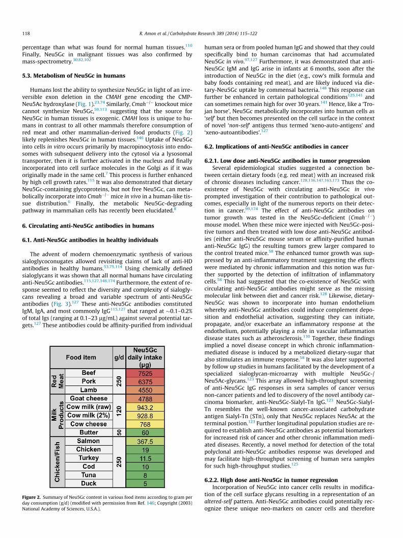
Humans lost the ability to synthesize Neu5Gc in light of an irre-versible exon deletion in the CMAH gene encoding the CMP-Neu5Ac hydroxylase (Fig. 1).23,74 Similarly, Cmah�/� knockout micecannot synthesize Neu5Gc,59,113 suggesting that the source forNeu5Gc in human tissues is exogenic. CMAH loss is unique to hu-mans in contrast to all other mammals therefore consumption ofred meat and other mammalian-derived food products (Fig. 2)likely replenishes Neu5Gc in human tissues.146 Uptake of Neu5Gcinto cells in vitro occurs primarily by macropinocytosis into endo-somes with subsequent delivery into the cytosol via a lysosomaltransporter, then it is further activated in the nucleus and finallyincorporated into cell surface molecules in the Golgi as if it wasoriginally made in the same cell.7 This process is further enhancedby high cell growth rates.115 It was also demonstrated that dietaryNeu5Gc-containing glycoproteins, but not free Neu5Gc, can meta-bolically incorporate into Cmah�/� mice in vivo in a human-like tis-sue distribution.6 Finally, the metabolic Neu5Gc-degradingpathway in mammalian cells has recently been elucidated.8
6. Circulating anti-Neu5Gc antibodies in humans
6.1. Anti-Neu5Gc antibodies in healthy individuals
The advent of modern chemoenzymatic synthesis of varioussialoglycoconugates allowed revisiting claims of lack of anti-HDantibodies in healthy humans.53,75,114 Using chemically definedsialoglycans it was shown that all normal humans have circulatinganti-Neu5Gc antibodies.115,127,146,174 Furthermore, the extent of re-sponse seemed to reflect the diversity and complexity of sialogly-cans revealing a broad and variable spectrum of anti-Neu5Gcantibodies (Fig. 3).127 These anti-Neu5Gc antibodies constitutedIgM, IgA, and most commonly IgG115,127 that ranged at �0.1–0.2%of total Igs (ranging at 0.1–23 lg/mL) against several potential tar-gets.127 These antibodies could be affinity-purified from individual
Figure 2. Summary of Neu5Gc content in various food items according to gram perday consumption (g/d) (modified with permission from Ref. 146; Copyright (2003)National Academy of Sciences, U.S.A.).
human sera or from pooled human IgG and showed that they couldspecifically bind to human carcinomas that had accumulatedNeu5Gc in vivo.97,127 Furthermore, it was demonstrated that anti-Neu5Gc IgM and IgG arise in infants at 6 months, soon after theintroduction of Neu5Gc in the diet (e.g., cow’s milk formula andbaby foods containing red meat), and are likely induced via die-tary-Neu5Gc uptake by commensal bacteria.148 This response canfurther be enhanced in certain pathological conditions125,141 andcan sometimes remain high for over 30 years.141 Hence, like a ‘Tro-jan horse’, Neu5Gc metabolically incorporates into human cells as‘self’ but then becomes presented on the cell surface in the contextof novel ‘non-self’ antigens thus termed ‘xeno-auto-antigens’ and‘xeno-autoantibodies’.127
6.2. Implications of anti-Neu5Gc antibodies in cancer
6.2.1. Low dose anti-Neu5Gc antibodies in tumor progressionSeveral epidemiological studies suggested a connection be-
tween certain dietary foods (e.g. red meat) with an increased riskof chronic diseases including cancer.128,136,147,163,173 Thus the co-existence of Neu5Gc with circulating anti-Neu5Gc in vivoprompted investigation of their contribution to pathological out-comes, especially in light of the numerous reports on their detec-tion in cancer.99,174 The effect of anti-Neu5Gc antibodies ontumor growth was tested in the Neu5Gc-deficient (Cmah�/�)mouse model. When these mice were injected with Neu5Gc-posi-tive tumors and then treated with low dose anti-Neu5Gc antibod-ies (either anti-Neu5Gc mouse serum or affinity-purified humananti-Neu5Gc IgG) the resulting tumors grew larger compared tothe control treated mice.58 The enhanced tumor growth was sup-pressed by an anti-inflammatory treatment suggesting the effectswere mediated by chronic inflammation and this notion was fur-ther supported by the detection of infiltration of inflammatorycells.58 This had suggested that the co-existence of Neu5Gc withcirculating anti-Neu5Gc antibodies might serve as the missingmolecular link between diet and cancer risk.128 Likewise, dietary-Neu5Gc was shown to incorporate into human endotheliumwhereby anti-Neu5Gc antibodies could induce complement depo-sition and endothelial activation, suggesting they can initiate,propagate, and/or exacerbate an inflammatory response at theendothelium, potentially playing a role in vascular inflammationdisease states such as atherosclerosis.130 Together, these findingsimplied a novel disease concept in which chronic inflammation-mediated disease is induced by a metabolized dietary-sugar thatalso stimulates an immune response.58 It was also later supportedby follow up studies in humans facilitated by the development of aspecialized sialoglycan-microarray with multiple Neu5Gc-/Neu5Ac-glycans.123 This array allowed high-throughput screeningof anti-Neu5Gc IgG responses in sera samples of cancer versusnon-cancer patients and led to discovery of the novel antibody car-cinoma biomarker, anti-Neu5Gc-Sialyl-Tn IgG.123 Neu5Gc-Sialyl-Tn resembles the well-known cancer-associated carbohydrateantigen Sialyl-Tn (STn), only that Neu5Gc replaces Neu5Ac at theterminal postion.123 Further longitudinal population studies are re-quired to establish anti-Neu5Gc antibodies as potential biomarkersfor increased risk of cancer and other chronic inflammation medi-ated diseases. Recently, a novel method for detection of the totalpolyclonal anti-Neu5Gc antibodies response was developed andmay facilitate high-throughput screening of human sera samplesfor such high-throughput studies.125
6.2.2. High dose anti-Neu5Gc in tumor regressionIncorporation of Neu5Gc into cancer cells results in modifica-
tion of the cell surface glycans resulting in a representation of analtered-self pattern. Anti-Neu5Gc antibodies could potentially rec-ognize these unique neo-markers on cancer cells and therefore
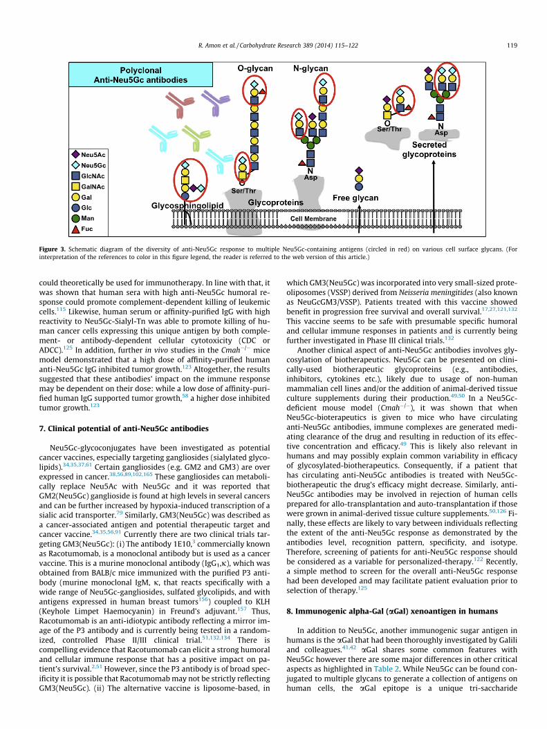
Figure 3. Schematic diagram of the diversity of anti-Neu5Gc response to multiple Neu5Gc-containing antigens (circled in red) on various cell surface glycans. (Forinterpretation of the references to color in this figure legend, the reader is referred to the web version of this article.)
R. Amon et al. / Carbohydrate Research 389 (2014) 115–122 119
could theoretically be used for immunotherapy. In line with that, itwas shown that human sera with high anti-Neu5Gc humoral re-sponse could promote complement-dependent killing of leukemiccells.115 Likewise, human serum or affinity-purified IgG with highreactivity to Neu5Gc-Sialyl-Tn was able to promote killing of hu-man cancer cells expressing this unique antigen by both comple-ment- or antibody-dependent cellular cytotoxicity (CDC orADCC).125 In addition, further in vivo studies in the Cmah�/� micemodel demonstrated that a high dose of affinity-purified humananti-Neu5Gc IgG inhibited tumor growth.123 Altogether, the resultssuggested that these antibodies’ impact on the immune responsemay be dependent on their dose: while a low dose of affinity-puri-fied human IgG supported tumor growth,58 a higher dose inhibitedtumor growth.123
7. Clinical potential of anti-Neu5Gc antibodies
Neu5Gc-glycoconjugates have been investigated as potentialcancer vaccines, especially targeting gangliosides (sialylated glyco-lipids).34,35,37,61 Certain gangliosides (e.g. GM2 and GM3) are overexpressed in cancer.38,56,89,102,165 These gangliosides can metaboli-cally replace Neu5Ac with Neu5Gc and it was reported thatGM2(Neu5Gc) ganglioside is found at high levels in several cancersand can be further increased by hypoxia-induced transcription of asialic acid transporter.79 Similarly, GM3(Neu5Gc) was described asa cancer-associated antigen and potential therapeutic target andcancer vaccine.34,35,56,91 Currently there are two clinical trials tar-geting GM3(Neu5Gc): (i) The antibody 1E10,3 commercially knownas Racotumomab, is a monoclonal antibody but is used as a cancervaccine. This is a murine monoclonal antibody (IgG1,j), which wasobtained from BALB/c mice immunized with the purified P3 anti-body (murine monoclonal IgM, j, that reacts specifically with awide range of Neu5Gc-gangliosides, sulfated glycolipids, and withantigens expressed in human breast tumors156) coupled to KLH(Keyhole Limpet Haemocyanin) in Freund’s adjuvant.157 Thus,Racotumomab is an anti-idiotypic antibody reflecting a mirror im-age of the P3 antibody and is currently being tested in a random-ized, controlled Phase II/III clinical trial.51,132,134 There iscompelling evidence that Racotumomab can elicit a strong humoraland cellular immune response that has a positive impact on pa-tient’s survival.2,51 However, since the P3 antibody is of broad spec-ificity it is possible that Racotumomab may not be strictly reflectingGM3(Neu5Gc). (ii) The alternative vaccine is liposome-based, in
which GM3(Neu5Gc) was incorporated into very small-sized prote-oliposomes (VSSP) derived from Neisseria meningitides (also knownas NeuGcGM3/VSSP). Patients treated with this vaccine showedbenefit in progression free survival and overall survival.17,27,121,132
This vaccine seems to be safe with presumable specific humoraland cellular immune responses in patients and is currently beingfurther investigated in Phase III clinical trials.132
Another clinical aspect of anti-Neu5Gc antibodies involves gly-cosylation of biotherapeutics. Neu5Gc can be presented on clini-cally-used biotherapeutic glycoproteins (e.g., antibodies,inhibitors, cytokines etc.), likely due to usage of non-humanmammalian cell lines and/or the addition of animal-derived tissueculture supplements during their production.49,50 In a Neu5Gc-deficient mouse model (Cmah�/�), it was shown that whenNeu5Gc-bioterapeutics is given to mice who have circulatinganti-Neu5Gc antibodies, immune complexes are generated medi-ating clearance of the drug and resulting in reduction of its effec-tive concentration and efficacy.49 This is likely also relevant inhumans and may possibly explain common variability in efficacyof glycosylated-biotherapeutics. Consequently, if a patient thathas circulating anti-Neu5Gc antibodies is treated with Neu5Gc-biotherapeutic the drug’s efficacy might decrease. Similarly, anti-Neu5Gc antibodies may be involved in rejection of human cellsprepared for allo-transplantation and auto-transplantation if thosewere grown in animal-derived tissue culture supplements.50,126 Fi-nally, these effects are likely to vary between individuals reflectingthe extent of the anti-Neu5Gc response as demonstrated by theantibodies level, recognition pattern, specificity, and isotype.Therefore, screening of patients for anti-Neu5Gc response shouldbe considered as a variable for personalized-therapy.122 Recently,a simple method to screen for the overall anti-Neu5Gc responsehad been developed and may facilitate patient evaluation prior toselection of therapy.125
8. Immunogenic alpha-Gal (aGal) xenoantigen in humans
In addition to Neu5Gc, another immunogenic sugar antigen inhumans is the aGal that had been thoroughly investigated by Galiliand colleagues.41,42 aGal shares some common features withNeu5Gc however there are some major differences in other criticalaspects as highlighted in Table 2. While Neu5Gc can be found con-jugated to multiple glycans to generate a collection of antigens onhuman cells, the aGal epitope is a unique tri-saccharide
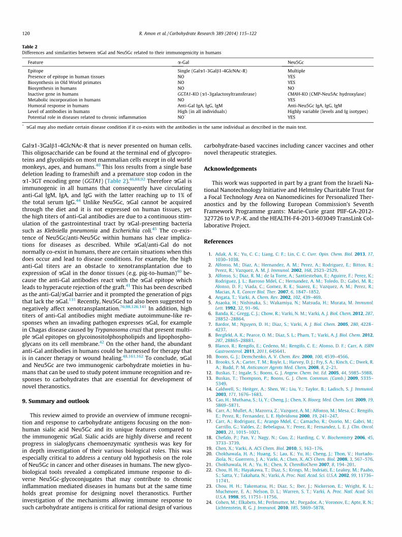
Table 2Differences and similarities between aGal and Neu5Gc related to their immunogenicity in humans
Feature a-Gal Neu5Gc
Epitope Single (Gala1-3Galb1-4GlcNAc-R) MultiplePresence of epitope in human tissues NO YESBiosynthesis in Old World primates NO YESBiosynthesis in humans NO NOInactive gene in humans GGTA1-KO (a1-3galactosyltransferase) CMAH-KO (CMP-Neu5Ac hydroxylase)Metabolic incorporation in humans NO YESHumoral response in humans Anti-Gal IgA, IgG, IgM Anti-Neu5Gc IgA, IgG, IgMLevel of antibodies in humans High (in all individuals) Highly variable (levels and Ig isotypes)Potential role in diseases related to chronic inflammation NO* YES
* aGal may also mediate certain disease condition if it co-exists with the antibodies in the same individual as described in the main text.
120 R. Amon et al. / Carbohydrate Research 389 (2014) 115–122
Gala1-3Galb1-4GlcNAc-R that is never presented on human cells.This oligosaccharide can be found at the terminal end of glycopro-teins and glycolipids on most mammalian cells except in old worldmonkeys, apes, and humans.40 This loss results from a single basedeletion leading to frameshift and a premature stop codon in thea1-3GT encoding gene (GGTA1) (Table 2).46,88,92 Therefore aGal isimmunogenic in all humans that consequently have circulatinganti-Gal IgM, IgA, and IgG with the latter reaching up to 1% ofthe total serum IgG.44 Unlike Neu5Gc, aGal cannot be acquiredthrough the diet and it is not expressed on human tissues, yetthe high titers of anti-Gal antibodies are due to a continuous stim-ulation of the gastrointestinal tract by aGal-presenting bacteriasuch as Klebsiella pneumonia and Escherichia coli.45 The co-exis-tence of Neu5Gc/anti-Neu5Gc within humans has clear implica-tions for diseases as described. While aGal/anti-Gal do notnormally co-exist in humans, there are certain situations when thisdoes occur and lead to disease conditions. For example, the highanti-Gal titers are an obstacle to xenotransplantation due toexpression of aGal in the donor tissues (e.g. pig-to-human)40 be-cause the anti-Gal antibodies react with the aGal epitope whichleads to hyperacute rejection of the graft.41 This has been describedas the anti-Gal/aGal barrier and it prompted the generation of pigsthat lack the aGal.131 Recently, Neu5Gc had also been suggested tonegatively affect xenotransplantation.76,98,126,141 In addition, hightiters of anti-Gal antibodies might mediate autoimmune-like re-sponses when an invading pathogen expresses aGal, for examplein Chagas disease caused by Trypanosoma cruzi that present multi-ple aGal epitopes on glycoinositolphospholipids and lipophospho-glycans on its cell membrane.42 On the other hand, the abundantanti-Gal antibodies in humans could be harnessed for therapy thatis in cancer therapy or wound healing.48,161,162 To conclude, aGaland Neu5Gc are two immunogenic carbohydrate moieties in hu-mans that can be used to study potent immune recognition and re-sponses to carbohydrates that are essential for development ofnovel theranostics.
9. Summary and outlook
This review aimed to provide an overview of immune recogni-tion and response to carbohydrate antigens focusing on the non-human sialic acid Neu5Gc and its unique features compared tothe immunogenic aGal. Sialic acids are highly diverse and recentprogress in sialoglycans chemoenzymatic synthesis was key forin depth investigation of their various biological roles. This wasespecially critical to address a century old hypothesis on the roleof Neu5Gc in cancer and other diseases in humans. The new glyco-biological tools revealed a complicated immune response to di-verse Neu5Gc-glycoconjugates that may contribute to chronicinflammation mediated diseases in humans but at the same timeholds great promise for designing novel theranostics. Furtherinvestigation of the mechanisms allowing immune response tosuch carbohydrate antigens is critical for rational design of various
carbohydrate-based vaccines including cancer vaccines and othernovel therapeutic strategies.
Acknowledgements
This work was supported in part by a grant from the Israeli Na-tional Nanotechnology Initiative and Helmsley Charitable Trust fora Focal Technology Area on Nanomedicines for Personalized Ther-anostics and by the following European Commission’s SeventhFramework Programme grants: Marie-Curie grant PIIF-GA-2012-327726 to V.P.-K. and the HEALTH-F4-2013-603049 TransLink Col-laborative Project.
References
1. Adak, A. K.; Yu, C. C.; Liang, C. F.; Lin, C. C. Curr. Opin. Chem. Biol. 2013, 17,1030–1038.
2. Alfonso, M.; Diaz, A.; Hernandez, A. M.; Perez, A.; Rodriguez, E.; Bitton, R.;Perez, R.; Vazquez, A. M. J. Immunol. 2002, 168, 2523–2529.
3. Alfonso, S.; Diaz, R. M.; de la Torre, A.; Santiesteban, E.; Aguirre, F.; Perez, K.;Rodriguez, J. L.; Barroso Mdel, C.; Hernandez, A. M.; Toledo, D.; Gabri, M. R.;Alonso, D. F.; Viada, C.; Gomez, R. E.; Suarez, E.; Vazquez, A. M.; Perez, R.;Macias, A. E. Cancer Biol. Ther. 2007, 6, 1847–1852.
4. Angata, T.; Varki, A. Chem. Rev. 2002, 102, 439–469.5. Asaoka, H.; Nishinaka, S.; Wakamiya, N.; Matsuda, H.; Murata, M. Immunol.
Lett. 1992, 32, 91–96.6. Banda, K.; Gregg, C. J.; Chow, R.; Varki, N. M.; Varki, A. J. Biol. Chem. 2012, 287,
28852–28864.7. Bardor, M.; Nguyen, D. H.; Diaz, S.; Varki, A. J. Biol. Chem. 2005, 280, 4228–
4237.8. Bergfeld, A. K.; Pearce, O. M.; Diaz, S. L.; Pham, T.; Varki, A. J. Biol. Chem. 2012,
287, 28865–28881.9. Blanco, R.; Rengifo, E.; Cedeno, M.; Rengifo, C. E.; Alonso, D. F.; Carr, A. ISRN
Gastroenterol. 2011, 2011, 645641.10. Boons, G. J.; Demchenko, A. V. Chem. Rev. 2000, 100, 4539–4566.11. Brooks, S. A.; Carter, T. M.; Royle, L.; Harvey, D. J.; Fry, S. A.; Kinch, C.; Dwek, R.
A.; Rudd, P. M. Anticancer Agents Med. Chem. 2008, 8, 2–21.12. Buskas, T.; Ingale, S.; Boons, G. J. Angew. Chem. Int. Ed. 2005, 44, 5985–5988.13. Buskas, T.; Thompson, P.; Boons, G. J. Chem. Commun. (Camb.) 2009, 5335–
5349.14. Caldwell, S.; Heitger, A.; Shen, W.; Liu, Y.; Taylor, B.; Ladisch, S. J. Immunol.
2003, 171, 1676–1683.15. Cao, H.; Muthana, S.; Li, Y.; Cheng, J.; Chen, X. Bioorg. Med. Chem. Lett. 2009, 19,
5869–5871.16. Carr, A.; Mullet, A.; Mazorra, Z.; Vazquez, A. M.; Alfonso, M.; Mesa, C.; Rengifo,
E.; Perez, R.; Fernandez, L. E. Hybridoma 2000, 19, 241–247.17. Carr, A.; Rodriguez, E.; Arango Mdel, C.; Camacho, R.; Osorio, M.; Gabri, M.;
Carrillo, G.; Valdes, Z.; Bebelagua, Y.; Perez, R.; Fernandez, L. E. J. Clin. Oncol.2003, 21, 1015–1021.
18. Chefalo, P.; Pan, Y.; Nagy, N.; Guo, Z.; Harding, C. V. Biochemistry 2006, 45,3733–3739.
19. Chen, X.; Varki, A. ACS Chem. Biol. 2010, 5, 163–176.20. Chokhawala, H. A.; Huang, S.; Lau, K.; Yu, H.; Cheng, J.; Thon, V.; Hurtado-
Ziola, N.; Guerrero, J. A.; Varki, A.; Chen, X. ACS Chem. Biol. 2008, 3, 567–576.21. Chokhawala, H. A.; Yu, H.; Chen, X. ChemBioChem 2007, 8, 194–201.22. Chou, H. H.; Hayakawa, T.; Diaz, S.; Krings, M.; Indriati, E.; Leakey, M.; Paabo,
S.; Satta, Y.; Takahata, N.; Varki, A. Proc. Natl. Acad. Sci. U.S.A. 2002, 99, 11736–11741.
23. Chou, H. H.; Takematsu, H.; Diaz, S.; Iber, J.; Nickerson, E.; Wright, K. L.;Muchmore, E. A.; Nelson, D. L.; Warren, S. T.; Varki, A. Proc. Natl. Acad. Sci.U.S.A. 1998, 95, 11751–11756.
24. Cohen, M.; Elkabets, M.; Perlmutter, M.; Porgador, A.; Voronov, E.; Apte, R. N.;Lichtenstein, R. G. J. Immunol. 2010, 185, 5869–5878.
R. Amon et al. / Carbohydrate Research 389 (2014) 115–122 121
25. Cohen, M.; Varki, A. OMICS 2010, 14, 455–464.26. Dalziel, M.; Crispin, M.; Scanlan, C. N.; Zitzmann, N.; Dwek, R. A. Science 2014,
343, 1235681.27. de la Torre, A.; Hernandez, J.; Ortiz, R.; Cepeda, M.; Perez, K.; Car, A.; Viada, C.;
Toledo, D.; Guerra, P. P.; Garcia, E.; Arbolaez, M.; Fernandez, L. E. Breast Cancer(Auckl.) 2012, 6, 151–157.
28. Deicher, H. Serums Z. Hyg. 1926, 106, 561–579.29. Deng, L.; Chen, X.; Varki, A. Biopolymers 2013, 99, 650–665.30. Devine, P. L.; Clark, B. A.; Birrell, G. W.; Layton, G. T.; Ward, B. G.; Alewood, P.
F.; McKenzie, I. F. Cancer Res. 1991, 51, 5826–5836.31. Diaz, S. L.; Padler-Karavani, V.; Ghaderi, D.; Hurtado-Ziola, N.; Yu, H.; Chen, X.;
Brinkman-Van der Linden, E. C.; Varki, A.; Varki, N. M. PLoS One 2009, 4,e4241.
32. Ding, L.; Yu, H.; Lau, K.; Li, Y.; Muthana, S.; Wang, J.; Chen, X. Chem. Commun.(Camb.) 2011, 8691–8693.
33. Dube, D. H.; Bertozzi, C. R. Nat. Rev. Drug Disc. 2005, 4, 477–488.34. Durrant, L. G.; Noble, P.; Spendlove, I. Clin. Exp. Immunol. 2012, 167, 206–215.35. Fernandez, L. E.; Gabri, M. R.; Guthmann, M. D.; Gomez, R. E.; Gold, S.;
Fainboim, L.; Gomez, D. E.; Alonso, D. F. Clin. Dev. Immunol. 2010, 2010,814397.
36. Franco, A. Anticancer Agents Med. Chem. 2008, 8, 86–91.37. Fuentes, D.; Avellanet, J.; Garcia, A.; Iglesias, N.; Gabri, M. R.; Alonso, D. F.;
Vazquez, A. M.; Perez, R.; Montero, E. Breast Cancer Res. Treat. 2010, 120, 379–389.
38. Fuentes, R.; Allman, R.; Mason, M. D. Lung Cancer 1997, 18, 21–33.39. Fukui, Y.; Maru, M.; Ohkawara, K.; Miyake, T.; Osada, Y.; Wang, D. Q.; Ito, T.;
Higashi, H.; Naiki, M.; Wakamiya, N. Biochem. Biophys. Res. Commun. 1989,160, 1149–1154.
40. Galili, U. Biochimie 2001, 83, 557–563.41. Galili, U. Xenotransplantation 2013, 20, 138–147.42. Galili, U. Immunology 2013, 140, 1–11.43. Galili, U.; Albertini, M. R.; Sondel, P. M.; Wigglesworth, K.; Sullivan, M.;
Whalen, G. F. Cancers (Basel) 2010, 2, 773–793.44. Galili, U.; Macher, B. A.; Buehler, J.; Shohet, S. B. J. Exp. Med. 1985, 162, 573–
582.45. Galili, U.; Mandrell, R. E.; Hamadeh, R. M.; Shohet, S. B.; Griffiss, J. M. Infect.
Immun. 1988, 56, 1730–1737.46. Galili, U.; Swanson, K. Proc. Natl. Acad. Sci. U.S.A. 1991, 88, 7401–7404.47. Galili, U.; Wigglesworth, K.; Abdel-Motal, U. M. J. Immunol. 2007, 178, 4676–
4687.48. Galili, U.; Wigglesworth, K.; Abdel-Motal, U. M. Burns 2010, 36, 239–251.49. Ghaderi, D.; Taylor, R. E.; Padler-Karavani, V.; Diaz, S.; Varki, A. Nat. Biotechnol.
2010, 28, 863–867.50. Ghaderi, D.; Zhang, M.; Hurtado-Ziola, N.; Varki, A. Biotechnol. Genet. Eng. Rev.
2012, 28, 147–175.51. Gomez, R. E.; Ardigo, M. L. Front. Oncol. 2012, 2, 147.52. Guo, Z.; Wang, Q. Curr. Opin. Chem. Biol. 2009, 13, 608–617.53. Halbert, S. P.; Anken, M.; Henle, W.; Golubjatnikov, R. J. Clin. Microbiol. 1982,
15, 610–616.54. Hanganutziu, M. C. R. Séances Soc. Biol. 1924, 91, 1457–1459.55. Hanisch, F. G.; Stadie, T. R.; Deutzmann, F.; Peter-Katalinic, J. Eur. J. Biochem.
1996, 236, 318–327.56. Hayashi, N.; Chiba, H.; Kuronuma, K.; Go, S.; Hasegawa, Y.; Takahashi, M.;
Gasa, S.; Watanabe, A.; Hasegawa, T.; Kuroki, Y.; Inokuchi, J.; Takahashi, H.Cancer Sci. 2013, 104, 43–47.
57. Hedlund, M.; Ng, E.; Varki, A.; Varki, N. M. Cancer Res. 2008, 68, 388–394.58. Hedlund, M.; Padler-Karavani, V.; Varki, N. M.; Varki, A. Proc. Natl. Acad. Sci.
U.S.A. 2008, 105, 18936–18941.59. Hedlund, M.; Tangvoranuntakul, P.; Takematsu, H.; Long, J. M.; Housley, G. D.;
Kozutsumi, Y.; Suzuki, A.; Wynshaw-Boris, A.; Ryan, A. F.; Gallo, R. L.; Varki,N.; Varki, A. Mol. Cell. Biol. 2007, 27, 4340–4346.
60. Heimburg-Molinaro, J.; Lum, M.; Vijay, G.; Jain, M.; Almogren, A.;Rittenhouse-Olson, K. Vaccine 2011, 29, 8802–8826.
61. Hernandez, A. M.; Rodriguez, N.; Gonzalez, J. E.; Reyes, E.; Rondon, T.; Grinan,T.; Macias, A.; Alfonso, S.; Vazquez, A. M.; Perez, R. J. Immunol. 2011, 186,3735–3744.
62. Higashi, H.; Hirabayashi, Y.; Fukui, Y.; Naiki, M.; Matsumoto, M.; Ueda, S.;Kato, S. Cancer Res. 1985, 45, 3796–3802.
63. Higashi, H.; Ito, M.; Fukaya, N.; Yamagata, S.; Yamagata, T. Anal. Biochem.1990, 186, 355–362.
64. Higashi, H.; Naiki, M.; Matuo, S.; Okouchi, K. Biochem. Biophys. Res. Commun.1977, 79, 388–395.
65. Higashi, H.; Nishi, Y.; Fukui, Y.; Ikuta, K.; Ueda, S.; Kato, S.; Fujita, M.; Nakano,Y.; Taguchi, T.; Sakai, S. Gann 1984, 75, 1025–1029.
66. Higashi, H.; Nishi, Y.; Fukui, Y.; Ikuta, K.; Ueda, S.; Kato, S.; Fujita, M.; Nakano,Y.; Taguchi, T.; Sakai, S., et al Gann 1984, 75, 1025–1029.
67. Higashi, H.; Sasabe, T.; Fukui, Y.; Maru, M.; Kato, S. Jpn. J. Cancer Res. 1988, 79,952–956.
68. Hirabayashi, Y.; Higashi, H.; Kato, S.; Taniguchi, M.; Matsumoto, M. Jpn. J.Cancer Res. 1987, 78, 614–620.
69. Hirabayashi, Y.; Kasakura, H.; Matsumoto, M.; Higashi, H.; Kato, S.; Kasai, N.;Naiki, M. Jpn. J. Cancer Res. 1987, 78, 251–260.
70. Hsu, C. H.; Hung, S. C.; Wu, C. Y.; Wong, C. H. Angew. Chem., Int. Ed. 2011, 50,11872–11923.
71. Hudak, J. E.; Bertozzi, C. R. Chem. Biol. 2014, 21, 16–37.
72. Ikuta, K.; Nishi, Y.; Shimizu, Y.; Higashi, H.; Kitamoto, N.; Kato, S.; Fujita, M.;Nakano, Y.; Taguchi, T.; Naiki, M. Biken J. 1982, 25, 47–50.
73. Ingale, S.; Wolfert, M. A.; Gaekwad, J.; Buskas, T.; Boons, G. J. Nat. Chem. Biol.2007, 3, 663–667.
74. Irie, A.; Koyama, S.; Kozutsumi, Y.; Kawasaki, T.; Suzuki, A. J. Biol. Chem. 1998,273, 15866–15871.
75. Iznaga, N.; Carr, A.; Fernández, L. E.; Solozabal, J.; Núñez, G.; Perdomo, Y.;Morales, A. J. Clin. Lab. Immunol. 1996, 48, 75–85.
76. Jang, K. S.; Kim, Y. G.; Adhya, M.; Park, H. M.; Kim, B. G. Xenotransplantation2013, 20, 199–208.
77. Julien, S.; Videira, P. A.; Delannoy, P. Biomolecules 2012, 2, 435–466.78. Kannagi, R.; Izawa, M.; Koike, T.; Miyazaki, K.; Kimura, N. Cancer Sci. 2004, 95,
377–384.79. Kannagi, R.; Yin, J.; Miyazaki, K.; Izawa, M. Biochim. Biophys. Acta 2008, 1780,
525–531.80. Kawachi, S.; Saida, T. J. Dermatol. 1992, 19, 827–830.81. Kawachi, S.; Saida, T.; Uhara, H.; Uemura, K.; Taketomi, T.; Kano, K. Int. Arch.
Allergy Appl. Immunol. 1988, 85, 381–383.82. Kawai, T.; Kato, A.; Higashi, H.; Kato, S.; Naiki, M. Cancer Res. 1991, 51, 1242–
1246.83. Khedri, Z.; Muthana, M. M.; Li, Y.; Muthana, S. M.; Yu, H.; Cao, H.; Chen, X.
Chem. Commun. (Camb.) 2012, 3357–3359.84. Kiefel, M. J.; von Itzstein, M. Chem. Rev. 2002, 102, 471–490.85. Kim, Y. J.; Varki, A. Glycoconj. J. 1997, 14, 569–576.86. Kobata, A.; Amano, J. Immunol. Cell Biol. 2005, 83, 429–439.87. Koda, T.; Shimosakoda, T.; Nishinaka, S.; Asaoka, H.; Nakaba, H.; Tamura, I.;
Matsuda, H. Gan To Kagaku Ryoho 1994, 21, 2771–2777.88. Koike, C.; Fung, J. J.; Geller, D. A.; Kannagi, R.; Libert, T.; Luppi, P.; Nakashima,
I.; Profozich, J.; Rudert, W.; Sharma, S. B.; Starzl, T. E.; Trucco, M. J. Biol. Chem.2002, 277, 10114–10120.
89. Kootstra, N. A.; Schuitemaker, H. AIDS Res. Hum. Retroviruses 1998, 14, 339–345.
90. Kurtenkov, O.; Klaamas, K.; Rittenhouse-Olson, K.; Vahter, L.; Sergejev, B.;Miljukhina, L.; Shljapnikova, L. Exp. Oncol. 2005, 27, 136–140.
91. Labrada, M.; Clavell, M.; Bebelagua, Y.; Leon, J.; Alonso, D. F.; Gabri, M. R.;Veloso, R. C.; Verez, V.; Fernandez, L. E. Expert Opin. Biol. Ther. 2010, 10, 153–162.
92. Larsen, R. D.; Rivera-Marrero, C. A.; Ernst, L. K.; Cummings, R. D.; Lowe, J. B. J.Biol. Chem. 1990, 265, 7055–7061.
93. Lau, K.; Yu, H.; Thon, V.; Khedri, Z.; Leon, M. E.; Tran, B. K.; Chen, X. Org.Biomol. Chem. 2011, 9, 2784–2789.
94. Liu, C. C.; Ye, X. S. Glycoconj. J. 2012, 29, 259–271.95. Livingston, P. O. Semin. Cancer Biol. 1995, 6, 357–366.96. Lloyd, K. O. Semin. Cancer Biol. 1991, 2, 421–431.97. Lu, Q.; Padler-Karavani, V.; Yu, H.; Chen, X.; Wu, S. L.; Varki, A.; Hancock, W. S.
Anal. Chem. 2012, 84, 2761–2768.98. Lutz, A. J.; Li, P.; Estrada, J. L.; Sidner, R. A.; Chihara, R. K.; Downey, S. M.;
Burlak, C.; Wang, Z. Y.; Reyes, L. M.; Ivary, B.; Yin, F.; Blankenship, R. L.; Paris,L. L.; Tector, A. J. Xenotransplantation 2013, 20, 27–35.
99. Malykh, Y. N.; Schauer, R.; Shaw, L. Biochimie 2001, 83, 623–634.100. Mander, L.; Liu, H.-W. Comprehensive Natural Products II: Chemistry and
Biology. In 1; Access Online via Elsevier: 2010.101. Manimala, J. C.; Roach, T. A.; Li, Z.; Gildersleeve, J. C. Glycobiology 2007, 17,
17C–23C.102. Marquina, G.; Waki, H.; Fernandez, L. E.; Kon, K.; Carr, A.; Valiente, O.; Perez,
R.; Ando, S. Cancer Res. 1996, 56, 5165–5171.103. Medzhitov, R.; Janeway, C. A. J. Science 2002, 296, 298–300.104. Merrick, J. M.; Zadarlik, K.; Milgrom, F. Int. Arch. Allergy Appl. Immunol. 1978,
57, 477–480.105. Miyake, M.; Hashimoto, K.; Ito, M.; Ogawa, O.; Arai, E.; Hitomi, S.; Kannagi, R.
Cancer 1990, 65, 499–505.106. Miyoshi, I.; Higashi, H.; Hirabayashi, Y.; Kato, S.; Naiki, M. Mol. Immunol. 1986,
23, 631–638.107. Moremen, K. W.; Tiemeyer, M.; Nairn, A. V. Nat. Rev. Mol. Cell Biol. 2012, 13,
448–462.108. Morito, T.; Kano, K.; Milgrom, F. J. Immunol. 1982, 129, 2524–2528.109. Morito, T.; Nishimaki, T.; Masaki, M.; Yoshida, H.; Kasukawa, R.; Nakarai, H.;
Kano, K. Int. Arch. Allergy Appl. Immunol. 1986, 81, 204–208.110. Muchmore, E. A.; Diaz, S.; Varki, A. Am. J. Phys. Anthropol. 1998, 107, 187–198.111. Mukuria, C. J.; Fujii, Y.; Kato, S.; Naiki, M. J. Biochem. (Tokyo) 1986, 100, 469–
475.112. Muthana, S.; Cao, H.; Chen, X. Curr. Opin. Chem. Biol. 2009, 13, 573–581.113. Naito, Y.; Takematsu, H.; Koyama, S.; Miyake, S.; Yamamoto, H.; Fujinawa, R.;
Sugai, M.; Okuno, Y.; Tsujimoto, G.; Yamaji, T.; Hashimoto, Y.; Itohara, S.;Kawasaki, T.; Suzuki, A.; Kozutsumi, Y. Mol. Cell. Biol. 2007, 27, 3008–3022.
114. Nakarai, H.; Chandler, P. J.; Kano, K.; Morton, D. L.; Irie, R. F. Int. Arch. AllergyAppl. Immunol. 1990, 91, 323–328.
115. Nguyen, D. H.; Tangvoranuntakul, P.; Varki, A. J. Immunol. 2005, 175, 228–236.116. Nicoll, G.; Avril, T.; Lock, K.; Furukawa, K.; Bovin, N.; Crocker, P. R. Eur. J.
Immunol. 2003, 33, 1642–1648.117. Nishimaki, T.; Kano, K.; Milgrom, F. J. Immunol. 1979, 122, 2314–2318.118. Nowak, J. A.; Jain, N. K.; Stinson, M. W.; Merrick, J. M. Mol. Immunol. 1986, 23,
693–700.119. Ohashi, Y.; Sasabe, T.; Nishida, T.; Nishi, Y.; Higashi, H. Am. J. Ophthalmol.
1983, 96, 321–325.
122 R. Amon et al. / Carbohydrate Research 389 (2014) 115–122
120. Ohtsubo, K.; Marth, J. D. Cell 2006, 126, 855–867.121. Osorio, M.; Gracia, E.; Reigosa, E.; Hernandez, J.; de la Torre, A.; Saurez, G.;
Perez, K.; Viada, C.; Cepeda, M.; Carr, A.; Avila, Y.; Rodriguez, M.; Fernandez, L.E. Cancer Manage. Res. 2012, 4, 341–345.
122. Padler-Karavani, V. Cancer Lett. 2013. pii: S0304-3835, doi: http://dx.doi.org/10.1016/j.canlet.2013.10.005. [Epub ahead of print].
123. Padler-Karavani, V.; Hurtado-Ziola, N.; Pu, M.; Yu, H.; Huang, S.; Muthana, S.;Chokhawala, H. A.; Cao, H.; Secrest, P.; Friedmann-Morvinski, D.; Singer, O.;Ghaderi, D.; Verma, I. M.; Liu, Y. T.; Messer, K.; Chen, X.; Varki, A.; Schwab, R.Cancer Res. 2011, 71, 3352–3363.
124. Padler-Karavani, V.; Song, X.; Yu, H.; Hurtado-Ziola, N.; Huang, S.; Muthana,S.; Chokhawala, H. A.; Cheng, J.; Verhagen, A.; Langereis, M. A.; Kleene, R.;Schachner, M.; de Groot, R. J.; Lasanajak, Y.; Matsuda, H.; Schwab, R.; Chen, X.;Smith, D. F.; Cummings, R. D.; Varki, A. J. Biol. Chem. 2012, 287, 22593–22608.
125. Padler-Karavani, V.; Tremoulet, A. H.; Yu, H.; Chen, X.; Burns, J. C.; Varki, A.PLoS One 2013, 8, e58443.
126. Padler-Karavani, V.; Varki, A. Xenotransplantation 2011, 18, 1–5.127. Padler-Karavani, V.; Yu, H.; Cao, H.; Chokhawala, H.; Karp, F.; Varki, N.; Chen,
X.; Varki, A. Glycobiology 2008, 18, 818–830.128. Pan, A.; Sun, Q.; Bernstein, A. M.; Schulze, M. B.; Manson, J. E.; Stampfer, M. J.;
Willett, W. C.; Hu, F. B. Arch. Intern. Med. 2012, 172, 555–563.129. Paulson, J. C.; Macauley, M. S.; Kawasaki, N. Ann. N. Y. Acad. Sci. 2012, 1253,
37–48.130. Pham, T.; Gregg, C. J.; Karp, F.; Chow, R.; Padler-Karavani, V.; Cao, H.; Chen, X.;
Witztum, J. L.; Varki, N. M.; Varki, A. Blood 2009, 114, 5225–5235.131. Phelps, C. J.; Koike, C.; Vaught, T. D.; Boone, J.; Wells, K. D.; Chen, S. H.; Ball, S.;
Specht, S. M.; Polejaeva, I. A.; Monahan, J. A.; Jobst, P. M.; Sharma, S. B.;Lamborn, A. E.; Garst, A. S.; Moore, M.; Demetris, A. J.; Rudert, W. A.; Bottino,R.; Bertera, S.; Trucco, M.; Starzl, T. E.; Dai, Y.; Ayares, D. L. Science 2003, 299,411–414.
132. Rabu, C.; McIntosh, R.; Jurasova, Z.; Durrant, L. Future Oncol. 2012, 8, 943–960.133. Ragupathi, G.; Coltart, D. M.; Williams, L. J.; Koide, F.; Kagan, E.; Allen, J.;
Harris, C.; Glunz, P. W.; Livingston, P. O.; Danishefsky, S. J. Proc. Natl. Acad. Sci.U.S.A. 2002, 99, 13699–13704.
134. Reichert, J. M. mAbs 2013, 5, 1–4.135. Rojas, G.; Pupo, A.; Gomez, S.; Krengel, U.; Moreno, E. ACS Chem. Biol. 2013, 8,
376–386.136. Rose, D. P.; Boyar, A. P.; Wynder, E. L. Cancer 1986, 58, 2363–2371.137. Rughetti, A.; Pellicciotta, I.; Biffoni, M.; Backstrom, M.; Link, T.; Bennet, E. P.;
Clausen, H.; Noll, T.; Hansson, G. C.; Burchell, J. M.; Frati, L.; Taylor-Papadimitriou, J.; Nuti, M. J. Immunol. 2005, 174, 7764–7772.
138. Saida, T.; Ikegawa, S.; Takizawa, Y.; Kawachi, S. Arch. Dermatol. Res. 1990, 282,179–182.
139. Saldova, R.; Wormald, M. R.; Dwek, R. A.; Rudd, P. M. Dis. Markers 2008, 25,219–232.
140. Schauer, R.; Srinivasan, G. V.; Wipfler, D.; Kniep, B.; Schwartz-Albiez, R. Adv.Exp. Med. Biol. 2011, 705, 525–548.
141. Scobie, L.; Padler-Karavani, V.; Le Bas-Bernardet, S.; Crossan, C.; Blaha, J.;Matouskova, M.; Hector, R. D.; Cozzi, E.; Vanhove, B.; Charreau, B.; Blancho,G.; Bourdais, L.; Tallacchini, M.; Ribes, J. M.; Yu, H.; Chen, X.; Kracikova, J.;Broz, L.; Hejnar, J.; Vesely, P.; Takeuchi, Y.; Varki, A.; Soulillou, J. P. J. Immunol.2013, 191, 2907–2915.
142. Shengshu, H.; Hai, Y.; Xi, C. Sci. China Chem. 2011, 54, 117–128.143. Song, X.; Yu, H.; Chen, X.; Lasanajak, Y.; Tappert, M. M.; Air, G. M.; Tiwari, V.
K.; Cao, H.; Chokhawala, H. A.; Zheng, H.; Cummings, R. D.; Smith, D. F. J. Biol.Chem. 2011, 286, 31610–31622.
144. Stacker, S. A.; Thompson, C.; Riglar, C.; McKenzie, I. F. J. Natl Cancer Inst. 1985,75, 801–811.
145. Takiguchi, M.; Tamura, T.; Goto, M.; Kusakawa, S.; Milgrom, F.; Kano, K. Clin.Exp. Immunol. 1984, 56, 345–352.
146. Tangvoranuntakul, P.; Gagneux, P.; Diaz, S.; Bardor, M.; Varki, N.; Varki, A.;Muchmore, E. Proc. Natl. Acad. Sci. U.S.A. 2003, 100, 12045–12050.
147. Tavani, A.; La Vecchia, C.; Gallus, S.; Lagiou, P.; Trichopoulos, D.; Levi, F.;Negri, E. Int. J. Cancer 2000, 86, 425–428.
148. Taylor, R. E.; Gregg, C. J.; Padler-Karavani, V.; Ghaderi, D.; Yu, H.; Huang, S.;Sorensen, R. U.; Chen, X.; Inostroza, J.; Nizet, V.; Varki, A. J. Exp. Med. 2010,207, 1637–1646.
149. van Cruijsen, H.; Ruiz, M. G.; van der Valk, P.; de Gruijl, T. D.; Giaccone, G. BMCCancer 2009, 9, 180.
150. Varki, A. Biochimie 2001, 83, 615–622.151. Varki, A. Glycoconj. J. 2009, 26, 231–245.152. Varki, A. Glycobiology 2011, 21, 1121–1124.153. Varki, A.; Kannagi, R.; Toole, B. P. Essentials of Glycobiology; Cold Spring Harbor
Laboratory Press: Cold Spring Harbor, NY, 2009.154. Varki, A.; Schauer, R. Essentials of Glycobiology; Cold Spring Harbor Laboratory
Press: Cold Spring Harbor, NY, 2009.155. Varki, A.; Sharon, N. Essentials of Glycobiology; Cold Spring Harbor Laboratory
Press: Cold Spring Harbor, NY, 2009.156. Vazquez, A. M.; Alfonso, M.; Lanne, B.; Karlsson, K. A.; Carr, A.; Barroso, O.;
Fernandez, L. E.; Rengifo, E.; Lanio, M. E.; Alvarez, C., et al Hybridoma 1995, 14,551–556.
157. Vazquez, A. M.; Perez, A.; Hernandez, A. M.; Macias, A.; Alfonso, M.; Bombino,G.; Perez, R. Hybridoma 1998, 17, 527–534.
158. Wang, C. C.; Lee, J. C.; Luo, S. Y.; Kulkarni, S. S.; Huang, Y. W.; Lee, C. C.; Chang,K. L.; Hung, S. C. Nature 2007, 446, 896–899.
159. Wang, Q.; Zhang, J.; Guo, Z. Bioorg. Med. Chem. 2007, 15, 7561–7567.160. Watarai, S.; Kushi, Y.; Shigeto, R.; Misawa, N.; Eishi, Y.; Handa, S.; Yasuda, T. J.
Biochem. 1995, 117, 1062–1069.161. Whalen, G. F.; Sullivan, M.; Piperdi, B.; Wasseff, W.; Galili, U. Anticancer Res.
2012, 32, 3861–3868.162. Wigglesworth, K. M.; Racki, W. J.; Mishra, R.; Szomolanyi-Tsuda, E.; Greiner,
D. L.; Galili, U. J. Immunol. 2011, 186, 4422–4432.163. Willett, W. C. Oncologist 2000, 5, 393–404.164. Wolfert, M. A.; Boons, G. J. Nat. Chem. Biol. 2013, 9, 776–784.165. Yamashiro, S.; Okada, M.; Haraguchi, M.; Furukawa, K.; Lloyd, K. O.; Shiku, H.;
Furukawa, K. Glycoconj. J. 1995, 12, 894–900.166. Yin, Z.; Huang, X. J. Carbohydr. Chem. 2012, 31, 143–186.167. Yu, H.; Cao, H.; Tiwari, V. K.; Li, Y.; Chen, X. Bioorg. Med. Chem. Lett. 2011, 21,
5037–5040.168. Yu, H.; Cheng, J.; Ding, L.; Khedri, Z.; Chen, Y.; Chin, S.; Lau, K.; Tiwari, V. K.;
Chen, X. J. Am. Chem. Soc. 2009, 131, 18467–18477.169. Yu, H.; Chokhawala, H.; Karpel, R.; Yu, H.; Wu, B.; Zhang, J.; Zhang, Y.; Jia, Q.;
Chen, X. J. Am. Chem. Soc. 2005, 127, 17618–17619.170. Yu, H.; Chokhawala, H. A.; Huang, S.; Chen, X. Nat. Protoc. 2006, 1, 2485–
2492.171. Yu, H.; Huang, S.; Chokhawala, H.; Sun, M.; Zheng, H.; Chen, X. Angew. Chem.,
Int. Ed. 2006, 45, 3938–3944.172. Zeichner, S. B. J. Am. Osteopath. Assoc. 2012, 112, 482–483.173. Zhang, J.; Kesteloot, H. Nutr. Cancer 2005, 53, 65–72.174. Zhu, A.; Hurst, R. Xenotransplantation 2002, 9, 376–381.