Embed Size (px)
Citation preview
AGRICULTURAL RESEARCH COMMUNICATION CENTRE
www.arccjournals.comIndian J. Anim. Res., 48 (3) : 298-300, 2014
doi:10.5958/j.0976-0555.48.3.063
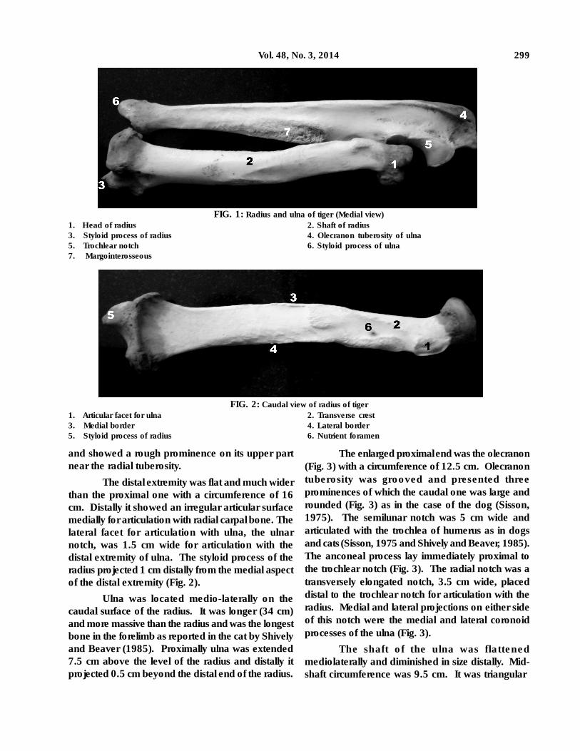
of 11.5 cm. It showed a concave fovea capitis radii(2.5 cm in diameter) for humerus. There was aconvex marginal area on the caudomedial aspect ofthe head for articulation with the ulna which hasbeen termed as the articular circumference in cat(Mc Clure et al., 1973) and dog (Sisson, 1975 andSmith, 1999). On the ventro-lateral aspect of thehead of the radius, there was a roughened, 2.5 cmlong prominence, the radial tuberosity for theinsertion of biceps brachii muscle, as reported incat by Mc Clure et al. (1973) and Senning (1977).The area between the radial tuberosity and the headwas the neck of the radius.
Flattened shaft of the radius had cranial andcaudal surfaces and medial and lateral borders. Thecaudal surface and the lateral border were concaveand the cranial surface and medial border wereconvex. A nutrient foramen was located on thecaudal surface, approximately 3 cm distal to theradial tuberosity (Fig. 2) as reported in dog by Sisson(1975). A transverse crest (6 cm long) extendedobliquely from the radial tuberosity downwards andmedially along this surface (Fig. 2). Lateral borderwas roughened in its middle for the attachment ofinterosseous ligament. Cranial surface was smooth
In felines, the strongly built forelimbs areheavily muscled and the manus can be supinated.These features allow them to clutch and grapple withthe prey with the forelimbs. Literature on theanatomical aspects of tiger is limited. Current studyhas been aimed to elucidate the macroscopicfeatures of the skeleton antebrachii of an adult tiger.
Carcass of an adult male tiger was receivedafter postmortem from the Zoo, Thrissur. The boneswere macerated and processed (Young, 1980) andprepared for the study.
Skeleton antebrachii consisted of two longbones, the radius and ulna. They were relativelylonger and unlike in the case of herbivores, not fused,allowing pronation and supination (Pasquini andSpurgeon, 1989). Radius was 27 cm long andflattened cranio-caudally. Proximally, radius formedarticulation with the capitulum of the humerus,crossed medially cranial to the ulna and articulateddistally with the radial carpal bone. Mid-shaftcircumference was 8 cm.
Proximal extremity of radius was small withan expanded caput radii and a distinct neck(Fig. 1). Caput radii was ovoid with a circumference
GROSS ANATOMY OF SKELETON ANTEBRACHII OF A TIGER(PANTHERA TIGRIS)
K.M. Lucky* and K.R. Harshan
College of Veterinary and Animal Sciences,Mannuthy-680 651, India.
Received: 31-12-2012 Accepted: 25-08-2013
ABSTRACTGross anatomical features of the skeleton of forearm of an adult tiger revealed that the ulna was
longer and more massive than the radius and was the longest bone of the forelimb. The oval caputradii showed concave fovea capitis radii for humerus and a convex articular circumference for ulna.Radial tuberosity was situated ventrolateral to the caput. A transverse crest extended obliquely fromthe radial tuberosity ventromedially along the caudal surface of the radius. The olecranon tuberosityof ulna was grooved and presented three prominences of which the caudal one was large androunded. The shaft of ulna presented a rough, elevated margointer osseus along the cranialsurface. The spatium interosseum was wide. The radius and ulna were not fused allowing pronationand supination movements to clutch the prey with the powerful forelimbs.
Key words: Anatomy, Radius, Tiger, Ulna.
*Corresponding author’s e-mail: [email protected].
299Vol. 48, No. 3, 2014
FIG. 2: Caudal view of radius of tiger1. Articular facet for ulna 2. Transverse crest3. Medial border 4. Lateral border5. Styloid process of radius 6. Nutrient foramen
FIG. 1: Radius and ulna of tiger (Medial view)1. Head of radius 2. Shaft of radius3. Styloid process of radius 4. Olecranon tuberosity of ulna5. Trochlear notch 6. Styloid process of ulna7. Margointerosseous
and showed a rough prominence on its upper partnear the radial tuberosity.
The distal extremity was flat and much widerthan the proximal one with a circumference of 16cm. Distally it showed an irregular articular surfacemedially for articulation with radial carpal bone. Thelateral facet for articulation with ulna, the ulnarnotch, was 1.5 cm wide for articulation with thedistal extremity of ulna. The styloid process of theradius projected 1 cm distally from the medial aspectof the distal extremity (Fig. 2).
Ulna was located medio-laterally on thecaudal surface of the radius. It was longer (34 cm)and more massive than the radius and was the longestbone in the forelimb as reported in the cat by Shivelyand Beaver (1985). Proximally ulna was extended7.5 cm above the level of the radius and distally itprojected 0.5 cm beyond the distal end of the radius.
The enlarged proximal end was the olecranon(Fig. 3) with a circumference of 12.5 cm. Olecranontuberosity was grooved and presented threeprominences of which the caudal one was large androunded (Fig. 3) as in the case of the dog (Sisson,1975). The semilunar notch was 5 cm wide andarticulated with the trochlea of humerus as in dogsand cats (Sisson, 1975 and Shively and Beaver, 1985).The anconeal process lay immediately proximal tothe trochlear notch (Fig. 3). The radial notch was atransversely elongated notch, 3.5 cm wide, placeddistal to the trochlear notch for articulation with theradius. Medial and lateral projections on either sideof this notch were the medial and lateral coronoidprocesses of the ulna (Fig. 3).
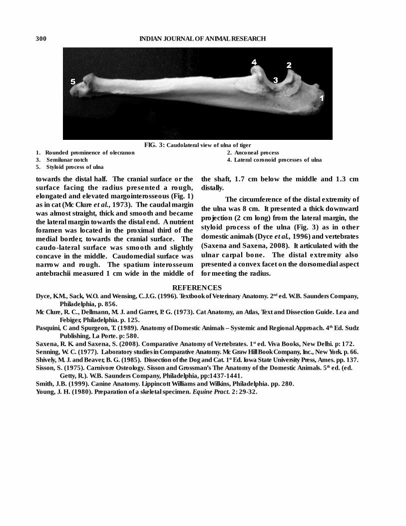
The shaft of the ulna was flattenedmediolaterally and diminished in size distally. Mid-shaft circumference was 9.5 cm. It was triangular
300 INDIAN JOURNAL OF ANIMAL RESEARCH
FIG. 3: Caudolateral view of ulna of tiger1. Rounded prominence of olecranon 2. Anconeal process3. Semilunar notch 4. Lateral coronoid processes of ulna5. Styloid process of ulna
towards the distal half. The cranial surface or thesurface facing the radius presented a rough,elongated and elevated margointerosseous (Fig. 1)as in cat (Mc Clure et al., 1973). The caudal marginwas almost straight, thick and smooth and becamethe lateral margin towards the distal end. A nutrientforamen was located in the proximal third of themedial border, towards the cranial surface. Thecaudo-lateral surface was smooth and slightlyconcave in the middle. Caudomedial surface wasnarrow and rough. The spatium interosseumantebrachii measured 1 cm wide in the middle of
the shaft, 1.7 cm below the middle and 1.3 cmdistally.
The circumference of the distal extremity ofthe ulna was 8 cm. It presented a thick downwardprojection (2 cm long) from the lateral margin, thestyloid process of the ulna (Fig. 3) as in otherdomestic animals (Dyce et al., 1996) and vertebrates(Saxena and Saxena, 2008). It articulated with theulnar carpal bone. The distal extremity alsopresented a convex facet on the dorsomedial aspectfor meeting the radius.
REFERENCESDyce, K.M., Sack, W.O. and Wensing, C.J.G. (1996). Textbook of Veterinary Anatomy. 2nd ed. W.B. Saunders Company,
Philadelphia, p. 856.Mc Clure, R. C., Dellmann, M. J. and Garret, P. G. (1973). Cat Anatomy, an Atlas, Text and Dissection Guide. Lea and
Febiger, Philadelphia. p. 125.Pasquini, C and Spurgeon, T. (1989). Anatomy of Domestic Animals – Systemic and Regional Approach. 4th Ed. Sudz
Publishing, La Porte. p: 580.Saxena, R. K. and Saxena, S. (2008). Comparative Anatomy of Vertebrates. 1st ed. Viva Books, New Delhi. p: 172.Senning, W. C. (1977). Laboratory studies in Comparative Anatomy. Mc Graw Hill Book Company, Inc., New York. p. 66.Shively, M. J. and Beaver, B. G. (1985). Dissection of the Dog and Cat. 1st Ed. Iowa State University Press, Ames. pp. 137.Sisson, S. (1975). Carnivore Osteology. Sisson and Grossman’s The Anatomy of the Domestic Animals. 5th ed. (ed.
Getty, R.). W.B. Saunders Company, Philadelphia, pp:1437-1441.Smith, J.B. (1999). Canine Anatomy. Lippincott Williams and Wilkins, Philadelphia. pp. 280.Young, J. H. (1980). Preparation of a skeletal specimen. Equine Pract. 2: 29-32.