Embed Size (px)
Citation preview
1 | P a g e -
40th EWGCCE meeting
The official meeting of the ESC Working
Group on Cardiac Cellular Electrophysiology
www.escardio.org/ewgcce-meeting
2 | P a g e -
40th EWGCCE meeting
Table of Contents
Welcome .................................................................................... 3
General Information ................................................................... 4
Scientific Programme ................................................................. 5
Carmeliet-Coraboeuf-Weidmann lecture ................................... 8
Posters ..................................................................................... 11
Best of Science selection @ 40th EWGCCE Meeting ................ 23
41th EWGCCE: Mark your calendar right now! ........................ 31
3 | P a g e -
40th EWGCCE meeting
Welcome
Dear Participant,
Welcome to Glasgow, a vibrant and cosmopolitan place with a wealth of
cultural heritage to explore. You will spend three days discussing the latest on
Cardiac Cellular Electrophysiology. We are grateful for your interest and
attendance to this meeting. Much has been done to make this a unique
occasion: an excellent scientific program, excellent speakers and the warm
ambiance of Glasgow. Especially, we thank you for submitting your scientific
contribution.
We have had great fun in organizing this year’s Scientific Meeting of the
European Working Group on Cardiac Cellular Electrophysiology.
It would not have been possible without the involvement of the Scientific
Committee, the Nucleus members of the Working Group and the Working
Groups Office team at the European Heart House.
Enjoy and learn!
The local organizers,
Godfrey Smith, Francis Burton, Peter Macfarlane, Niall Macquaide, Rachel
Myles, Antony Workman, Christopher Loughrey
4 | P a g e -
40th EWGCCE meeting
General Information
Venue
Kelvin Gallery - University of Glasgow , Gilbert Scott Building, G12 8QQGlasgow, United Kingdom
Registration Desk
Your badge and the Meeting documentation can be collected on the first meeting day, Saturday 2
September at the venue, from 8.00 am.
Our staff at the registration desk will assist you in case of any inquiry.
Language
The offi cial language of the Meeti ng is English.
Badge
Participants of the Meeting are requested to wear their badge at all times during the Meeting.
Oral Presentations
The oral sessions will be held in the Kelvin Gallery, in the Gilbert Scott Building at the Meeting venue.
Presenters are kindly asked to provide their presentati ons on USB stick well before the start of the
session in which they are scheduled.
Poster Presentations
Posters will be displayed at the ground floor of the Meeti ng venue.
Poster dimensions should not exceed 130 cm width x 90 cm height.
Posters should be mounted on the poster boards on arrival to the Meeting venue and will be
displayed on Saturday and Sunday. Posters should be removed Sunday before the evening program
(18.00 h).
Coff ee/Tea
Coff ee, tea and water will be available during the breaks.
Lunch
Lunch is included in the registration fee and will be offered Saturday and Sunday at 12.30 and on
General Assembly
We kindly invite all EWGCCE members to attend the General Assembly scheduled on Sunday 4
September from 17.00 h – 18.00h. Important information will come by then, and this is also the
moment when the poster prizes will be notified and awarded.
Welcome Reception
Friday 2nd September, from 18.00, the welcome reception will be held in the Hunterian Museum
(Gilbert Scott Building - University of Glasgow).
Meeting Dinner
The Meeting dinner will be served in the Hilton Glasgow Grosvenor , 1-9 Grosvenor Terrace,
Glasgow. Walking distance from the University. The dinner is included in the registration fee.
WiFi
At the Meeting venue, free, open, WiFi will be available.
5 | P a g e -
40th EWGCCE meeting
Scientific Programme
Saturday 3rd September
08:30-10:00- Session 1: Ventricular arrhythmia mechanisms:
Chairs: Peter MacFarlane, Paul Volders
Title: Hereditary repolarization disorders: ECG imaging of the clinical substrate and
mathematical modeling of the molecular mechanism
Speaker: Yoram Rudy (St Louis, MO) 40+5mins
Oral presentations: 3 x (10+5mins)
The different possible mechanisms of the perpertuation of Torsade de Pointes in the drug-
induced Chronic AV Block Dog - N. Vandersickel, A.Dunnink, A. Bossu, V. Meyboom, M. van
der Heyden J. Beekman, J.M.T. de Bakker, M.A.Vos, A.V. Panfilov (Ghent BE)
The differential effects of hypothermia on cardiac conduction and excitability -Karen
McGlynn, Erik Sveberg Dietrichs, Andrew Allan, Adam Connolly, Martin Bishop, Francis
Burton, Torkjel Tveita, Godfrey L Smith (Glasgow UK)
Variability in the kinetics of cardiac INa - Michael Clerx, Roel L.H.M.G. Spätjens, Sandrine R.M.
Seyen, Pieter Collins, Ralf L.M. Peeters, Paul G.A. Volders (Maastricht NL)
10:00-10.30- Coffee Break
10:30-12:00- Session 2: Atrial arrhythmia mechanisms:
Chairs: Tony Workman, Dobromir Dobrev
Title: Translational assessment of cellular mechanisms of atrial fibrillation
Speaker: Uli Schotten (Maastricht, Netherlands) 40+5mins
Oral presentations: 3 x (10+5mins)
A dynamic-clamp study of L-type Ca2+ current in rabbit and human atrial myocytes: the
contribution of window ICaL to early afterdepolarisations -Kettlewell S, Dempster J, Colman
MA, Rankin AC, Myles RC, Smith GL, Workman AJ (Glasgow UK)
Loss of myocardial nNOS begets atrial fibrillation by abolish the physiological right-left action
potential duration gradient in human and mouse atrial myocytes -Xing Liu, Ricardo Carnicer,
Alice Recalde, Alfonso Bueno-Orovio, Blanca Rodriguez, Barbara Casadei (Oxford UK)
PDE8 is a novel regulator of cAMP signaling in human atrial fibrillation - C E Molina, S
Ghezelbash, E Jacquet, A Garnier, R Fischmeister, D Dobrev (Essen DE)
12:30 – 13:30: Lunch
14:00-15:30- posters (number 1 to number 43)
15:30-16:00- Coffee Break
6 | P a g e -
40th EWGCCE meeting
16:00-17:30- Keynote CCW lecture: Denis Noble (Oxford, UK)
The golden trio of cardiac electrophysiology, Coraboeuf, Carmeliet & Weidmann, chasing
thresholds and conductance changes in the early days of micro electrode recording.
19:00 - Conference dinner
At the Hilton Glasgow Grosvenor
1-9 Grosvenor Terrace, Glasgow
Sunday 4th September
08:30-10:00- Session 3: Emerging therapies to address myocardial disease
Chairs: Godfrey Smith, Matteo Mangoni
Title: iPS-cell derived engineered heart tissue improves left-ventricular function after myocardial
injury.
Speaker: Thomas Eschenhagen (Hamburg Germany) 40+5min
Oral presentations: 3 x (10+5mins)
Elevated Ventricular Wall Stress Disrupts Cardiomyocyte T-tubular Structure and Ca2+
Homeostasis - Ruud M, Frisk M, Espe EK, Aronsen M, Zhang L, Norseng PA, Sjaastad I,
Sejersted O, Christensen G, Louch WE (Oslo NO)
Measuring electrical conductivity of cardiac T-tubular systems - M. Scardigli, C. Crocini, C.
Ferrantini, T. Gabbrielli, L. Silvestri, C. Tesi, E. Cerbai, C. Poggesi, F. S. Pavone, L. Sacconi
(Sesto Fiorentino IT)
Intramural structural discontinuities underlie right ventricular conduction abnormalities in
the Scn5a haplo-insufficient mouse model - Allen Kelly, Simona Salerno, Adam Connelly,
Martin Bishop, Ulrik Wisloff, Flavien Charpentier, Tomas Stolen, Godfrey Smith (Glasgow UK)
10:00-10.30- Coffee Break
10:30-12:00- Session 4: Excitation-contraction coupling in health and disease
Chairs: Chris Loughrey, Frank Heinzel
Title: Control of diastolic calcium.
Speaker: David Eisner (Manchester UK)
Oral presentations: 3 x (10+5mins)
The efficacy of late sodium current blockers in hypertrophic cardiomyopathy is dependent
on genotype: a study on transgenic mouse models with different mutations.
L. Santini, R. Coppini, L. Mazzoni, C. Ferrantini, F. Gentile, JM. Pioner, L. Sartiani, C. Poggesi,
A. Mugelli, E. Cerbai (Florence IT)
Physical coupling between SERCA2 and PDE3A regulates SERCA2 activity in cardiomyocytes
Jan Magnus Aronsen MD, Jonas Skogestad MD, Karina Hougen MD PhD, Marianne Lunde,
Gustav Lothe, Per Kristian Lunde PhD, Jens Preben Morth PhD, Kjetil Taskén MD PhD,
Cathrine Rein Carlson PhD, Ivar Sjaastad MD PhD (Oslo NO)
7 | P a g e -
40th EWGCCE meeting
Phosphodiesterase-5 inhibitor sildenafil suppresses calcium waves by reducing sarcoplasmic
reticulum content - D. Hutchings, K. Dibb, D. Eisner, A. Trafford (Manchester UK)
12:15 – 13:15: Lunch
13:30-15:00- posters (number 44 to number 86)
15:30-17:00- Session 5: Novel pathways regulating cardiac electrophysiology
Chairs: Rachel Myles, Carol Ann Remme
Title: The role of sarcoplasmic reticulum calcium handling during alternans and fibrillation
Speaker: Crystal Ripplinger (Davis CA) 40+5min
Oral presentations: 3 x (10+5mins)
Optogenetic termination of atrial fibrillation in the mouse heart
Tobias Bruegmann, Thomas Beiert, Jan Wilko Schrickel, Philipp Sasse (Bonn DE)
Optical treatment of cardiac arrhythmias
Claudia Crocini, Cecilia Ferrantini, Raffaele Coppini, Marina Scardigli, Leslie M. Loew,
Godfrey Smith, Corrado Poggesi, Elisabetta Cerbai, Francesco S. Pavone, Leonardo Sacconi
(Sesto Fiorentino IT)
Optogenetic monitoring of endocardial calcium transients in vivo using a minimally invasive
fiber optic approach. L. Menke, I. van Asten, L. van Stuijvenberg, T.A.B. van Veen, M.A. Vos, T
P. de Boer (Utrecht NL)
17:00-18:00 - General Assembly
8 | P a g e -
40th EWGCCE meeting
Carmeliet-Coraboeuf-Weidmann lecture
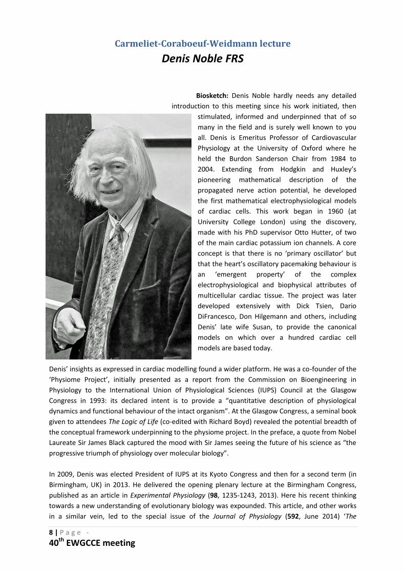
Denis Noble FRS
Biosketch: Denis Noble hardly needs any detailed
introduction to this meeting since his work initiated, then
stimulated, informed and underpinned that of so
many in the field and is surely well known to you
all. Denis is Emeritus Professor of Cardiovascular
Physiology at the University of Oxford where he
held the Burdon Sanderson Chair from 1984 to
2004. Extending from Hodgkin and Huxley’s
pioneering mathematical description of the
propagated nerve action potential, he developed
the first mathematical electrophysiological models
of cardiac cells. This work began in 1960 (at
University College London) using the discovery,
made with his PhD supervisor Otto Hutter, of two
of the main cardiac potassium ion channels. A core
concept is that there is no ‘primary oscillator’ but
that the heart’s oscillatory pacemaking behaviour is
an ‘emergent property’ of the complex
electrophysiological and biophysical attributes of
multicellular cardiac tissue. The project was later
developed extensively with Dick Tsien, Dario
DiFrancesco, Don Hilgemann and others, including
Denis’ late wife Susan, to provide the canonical
models on which over a hundred cardiac cell
models are based today.
Denis’ insights as expressed in cardiac modelling found a wider platform. He was a co-founder of the
‘Physiome Project’, initially presented as a report from the Commission on Bioengineering in
Physiology to the International Union of Physiological Sciences (IUPS) Council at the Glasgow
Congress in 1993: its declared intent is to provide a “quantitative description of physiological
dynamics and functional behaviour of the intact organism”. At the Glasgow Congress, a seminal book
given to attendees The Logic of Life (co-edited with Richard Boyd) revealed the potential breadth of
the conceptual framework underpinning to the physiome project. In the preface, a quote from Nobel
Laureate Sir James Black captured the mood with Sir James seeing the future of his science as “the
progressive triumph of physiology over molecular biology”.
In 2009, Denis was elected President of IUPS at its Kyoto Congress and then for a second term (in
Birmingham, UK) in 2013. He delivered the opening plenary lecture at the Birmingham Congress,
published as an article in Experimental Physiology (98, 1235-1243, 2013). Here his recent thinking
towards a new understanding of evolutionary biology was expounded. This article, and other works
in a similar vein, led to the special issue of the Journal of Physiology (592, June 2014) ‘The
9 | P a g e -
40th EWGCCE meeting
integration of evolutionary biology with physiological science’. This issue was provocatively sub-titled
in the editorial ‘Evolution evolves: physiology returns to centre stage’. Some 35 eminent authors
contributed to the 19 papers published there.
Denis wrote what is perhaps the first popular book on Systems Biology, The Music of Life. It has
rightly enjoyed widespread critical acclaim. Over the last decade and more, his lectures and
presentations have principally concerned the implications of physiological knowledge for
evolutionary biology and vice versa. Denis has established himself as one of the leading thinkers in
modern bioscience. (That rather fuzzy notion of the ‘public philosopher’ perhaps fits him well.) His
deep scholarship ranges over the central ideas of biology and of the philosophy of science and is
truly that of a polymath. As a physiologist and a leading proponent of the notion of ‘emergence’, he
will relish that he continues a strand of philosophical thought whose name was coined by a founder
member of The Physiological Society, George Henry Lewes (1817-1878).
Denis has published more than 500 papers as well as 11 books. His latest opus Dance to the Tune of
Life; Biological Relativity will soon be published (by Cambridge University Press).
David Miller July 2016
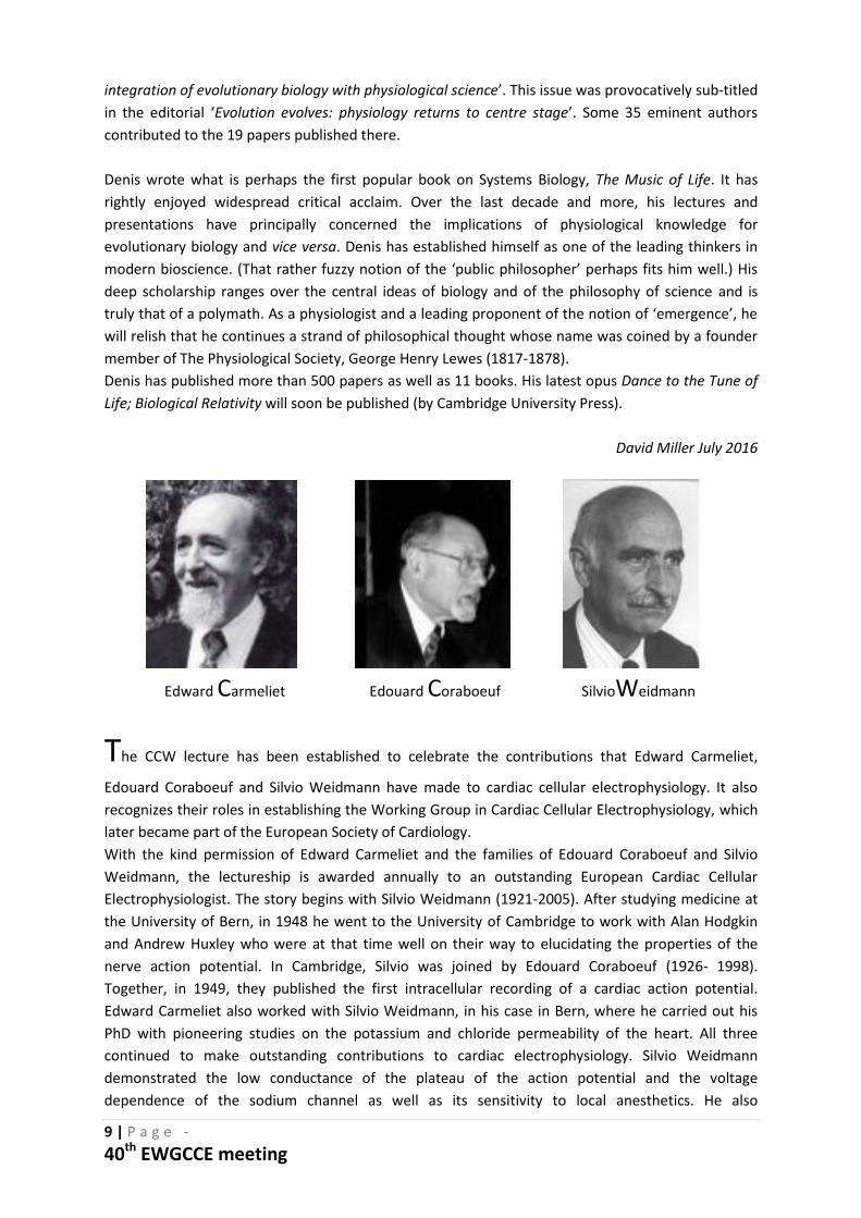
The CCW lecture has been established to celebrate the contributions that Edward Carmeliet,
Edouard Coraboeuf and Silvio Weidmann have made to cardiac cellular electrophysiology. It also
recognizes their roles in establishing the Working Group in Cardiac Cellular Electrophysiology, which
later became part of the European Society of Cardiology.
With the kind permission of Edward Carmeliet and the families of Edouard Coraboeuf and Silvio
Weidmann, the lectureship is awarded annually to an outstanding European Cardiac Cellular
Electrophysiologist. The story begins with Silvio Weidmann (1921-2005). After studying medicine at
the University of Bern, in 1948 he went to the University of Cambridge to work with Alan Hodgkin
and Andrew Huxley who were at that time well on their way to elucidating the properties of the
nerve action potential. In Cambridge, Silvio was joined by Edouard Coraboeuf (1926- 1998).
Together, in 1949, they published the first intracellular recording of a cardiac action potential.
Edward Carmeliet also worked with Silvio Weidmann, in his case in Bern, where he carried out his
PhD with pioneering studies on the potassium and chloride permeability of the heart. All three
continued to make outstanding contributions to cardiac electrophysiology. Silvio Weidmann
demonstrated the low conductance of the plateau of the action potential and the voltage
dependence of the sodium channel as well as its sensitivity to local anesthetics. He also
Edward Carmeliet Edouard Coraboeuf SilvioWeidmann
10 | P a g e -
40th EWGCCE meeting
demonstrated the diffusion of potassium between cells. Edouard Coraboeuf went on to identify
early afterdepolarizations, which lead to torsades-de-pointes arrhythmias. He subsequently
pioneered cellular studies on the human heart as well as characterizing the maintained component
of the sodium current and its contribution to the plateau of the action potential. As mentioned
above, Edouard and Edward had both worked with Silvio. Subsequently the two collaborated on
work characterizing the chloride current. Edward Carmeliet (1930-) also pioneered studies of the
control of the action potential duration; in particular the effects of heart rate and metabolism. He
published seminal papers on virtually every cardiac potassium channel and on the mechanisms of
action of antiarrhythmic agents. He also demonstrated the interaction of ionic gradients with
channels and transporters.
As well as their own scientific contributions, all three have established their own schools of
research as represented by countless successful careers of younger scientists worldwide.
Our Working Group owes much to this trio. Edward Carmeliet organized the first meeting in
Leuven in 1977. The next year Edouard Coraboeuf organized a meeting in Orsay and, in 1980, the
Working Group met in Bern at the invitation of Silvio Weidmann.
Past CCW Lecturers:
2012: Ursula Ravens (36th EGWCCE Meeting, Nantes, FR)
2013 David Eisner (37th EGWCCE Meeting, Athens, GR)
2014: András Varró (38th EWGCCE Meeting, Maastricht, NL)
2015: Barbara Casadei (39th EWGCCE Meeting, Milan, IT)
The CCW lecture is supported by CAIRN Research
11 | P a g e -
40th EWGCCE meeting
Posters
Saturday 3rd September
Poster presenters should be available at their poster from 14:00 – 15:30
Poster number 1 to poster number 43
1. Short QT in a murine model of metabolic dysfunction
Shiraz Ahmad, Haseeb Valli, Samantha Salvage, Andrew Grace, Kamalan Jeevaratnam, Christopher Huang. Centre of affiliation: University of Cambridge. London, UK. 2. Biphasic time course of response to activation of sympathetic nerves in isolated rabbit ventricular myocardium
Stephanie Anderson, Francis Burton, Godfrey Smith, Rachel Myles.
Centre of affiliation: British Heart Foundation funded PhD student. Glasgow, UK.
3. Risk assessment models for myocardial infarction
Galya Atanasova, PhD.
Centre of affiliation: University Hospital, Department of Internal Medicine. Pleven, Bulgaria.
4. Rad GTPase as a new potential actor in Brugada syndrome
Nadjet Belbachir, Vincent Portero, Eva LePogam, Nathalie Gaborit, Solena Le Scouarnec, Isabelle Baró, Christophe Guilluy, Celine Marionneau, Vincent Probst, Jean-Jacques Schott, Richard Redon, Flavien Charpentier.
Centre of affiliation: l'institut du thorax. Nantes, France.
5. Modifier genes in the LQT1 syndrome: mechanistic analysis of NOS1AP polymorphism Joyce Bernardi, Carlotta Ronchi, Eleonora Torre, Marcella Rocchetti, Antonio Zaza.
Centre of affiliation: University of Milano-Bicocca. Milan, Italy.
6. The arrhythmogenic electrophysiological and structural substrates of the RVOT: insights from large animal models and human hearts
D. Benoist, V. Dubes, M. Constantin, S. Charron, S. H. Gilbert, C. N. W. Belterman, M. Haissaguerre, E. White, R. Coronel, O. Bernus.
Centre of affiliation: Electrophysiology and Heart Modeling Institute LIRYC - University of Bordeaux. Bordeaux, France.
7. Effects of exercise training on myocardial and skeletal muscle function in a post-myocardial infarction heart failure rat model
Aline Bezerra Gurgel, Michael Dunne, Godfrey Smith, Ole Kemi.
Centre of affiliation: University of Glasgow. UK.
12 | P a g e -
40th EWGCCE meeting
8. Rotors in AF and impact of ablation
Caroline Cros, Remi Chayvel, Richard Walton, Caroline Auclerc-Pascarel, Valentin Meillet, Remi Dubois, Olivier Bernus, Pierre Jais, Fabien Brette.
Centre of affiliation: IHU LIRYC. Bordeaux, France.
9. A simple method to extract movement information from video images of contracting heart cells
Francis Burton.
Centre of affiliation: Institute of Cardiovascular and Medical Sciences, University of Glasgow. UK.
10. Are transverse tubules restored in the atria following recovery from heart failure?
J.L. Caldwell, J D Clarke, D.A. Eisner, A.W. Trafford, K.M. Dibb.
Centre of affiliation: University of Manchester.UK.
11. Absence of Nav1.8-based (late) sodium current in rabbit cardiomyocytes and human iPSC derived cardiomyocytes
Simona Casini, Isabella Mengarelli, Cees A. Schumacher, Marieke W. Veldkamp, Arie O. Verkerk, Carol Ann Remme.
Centre of affiliation: Experimental Cardiology. Amsterdam, The Netherlands.
12. Alpha1-adrenergic regulation of hERG Current is mediated by PKC dependent channel phosphorylation
Janire Urrutia, Aintzane Alday, Layse Malagueta-Vieira, Monica Gallego, Oscar Casis.
Centre of affiliation: Universidad del País Vasco UPV/EHU. Vitoria-Gasteiz, Spain.
13. The threshold behavior of the cardiac sodium current is potentiated by ephaptic effects: insights from a high resolution mathematical model of a narrow intercellular cleft
Echrak Hichri, Hugues Abriel, Jan P. Kucera.
Centre of affiliation: University of Bern. Switzerland.
14. Massively parallel all-optical cardiac electrophysiology
Emilia Entcheva.
Centre of affiliation: George Washington University. Washington, USA.
15. Arrhythmias and electrophysiological abnormalities in a mouse model of hypertrophic cardiomyopathy
Frederik Flenner, Felix W. Friedrich, Katrin Gurr, Klaus-Dieter Soehren, Thomas Eschenhagen, Torsten Christ, Lucie Carrier.
Centre of affiliation: Department of Experimental Pharmacology, UKE Hamburg. Germany.
13 | P a g e -
40th EWGCCE meeting
16. Sodium channel expression, distribution and (dys)function in atrial myocytes: relevance for arrhythmogenesis
G.A. Marchal, S.Z. Ibrahim, C. Schumacher, M.W. Veldkamp and C.A. Remme.
Centre of affiliation: Academic Medical Centre. Amsterdam, The Netherlands.
17. Enhanced expression and PKCdelta-mediated hyperphosphorylation underlie the proarrythmic increase in sodium-calcium-exchanger activity in patients with chronic atrial fibrillation
Ghezelbash S, Molina CE, Badimon L, Kamler M, Heijman J, Dobrev D.
Centre of affiliation: Institute of Pharmacology, University Duisburg Duisburg-Essen. Germany.
18. Circadian rhythm in QT interval is preserved in Cardiomyocyte specific Bmal1 knockout mice despite changed J wave morphology
Lisa A Gottlieb, Morten B Thomsen.
Centre of affiliation: Department of Biomedical Sciences, Faculty of Health and Medical Sciences, University of Copenhagen. Denmark.
19. The effects of TBQ on cardiac intracellular ATP
Natasha Hadgraft, Mustafa Naeem, Gina Galli, Louise Miller, David Greensmith.
Centre of affiliation: University of Salford. UK.
20. Long-term stimulation of iPS-derived cardiomyocytes using optogenetic techniques to promote phenotypic changes in E-C coupling
Craig Hamilton, Niall MacQuaide, Victor Zamora, Godfrey Smith.
Centre of affiliation: University of Glasgow. UK.
22. Inward rectifier ion currents in human induced pluripotent stem cell-derived cardiomyocytes
András Horváth, Ahmet Uzun, Ingra Mannhardt, Kaja Breckwoldt, Christiane Neuber, Alexandra Löser, Arne Hansen, András Varró, Thomas Eschenhagen, Torsten Christ.
Centre of affiliation: UKE Hamburg, Department of Experimental Pharmacology and Toxicology. Hamburg, Germany.
23. Effects of Omecamtiv mecarbil on cellular electrophysiology and contractile properties of canine left ventricular myocytes
Balázs Horváth, Norbert Szentandrássy, Krisztina Váczi, Kornél Kistamás, Tamás Bányász, János Magyar, Péter P. Nánási.
Centre of affiliation: Department of Physiology, Faculty of Medicine, University of Debrecen.
Hungary.
14 | P a g e -
40th EWGCCE meeting
24. Antiarrhythmic treatment of ventricular fibrillation due to acute myocardial infarction in rats
Laura Amalie Hundahl, Lasse Skibsbye, Thomas Jespersen.
Centre of affiliation: Cardiac Physiology Group, Department of Biomedical Sciences. Copenhagen, Denmark.
25. Regulation of basal and norepinephrine-increased Ca2+-currents by PDEs in human heart: differences between stem cell-derived and mature cardiomyocytes
Ismaili D, Mannhardt I, Hansen A, Eschenhagen T, Christ T.
Centre of affiliation: University Medical Center Hamburg-Eppendorf. Hamburg, Germany.
26. Allosteric modulator LUF7244 uncouples dofetilide-mediated rescue of aberrant hERG trafficking from dofetilide-induced hERG blockade
Y. Ji, M.J.C. Houtman, F. Romunde, D. Fransen, A.P. IJzerman, L.H. Heitman, M.A. Vos, M.A.G. van der Heyden.
Centre of affiliation: University Medical Center Utrecht. UTRECHT. The Netherlands.
27. Receptor-species dependent desensitization controls IKs as a downstream effector of Gq protein-coupled receptors
Marie-Cécile Kienitz, Dilyana Vladimirova, Christian Müller, Lutz Pott and Andreas Rinne. Centre of affiliation: Department of Cellular Physiology, Ruhr-University. Bochum, Germany.
28. Dependence of diastolic calcium levels on frequency and extracellular calcium concentration
Kornel Kistamas, Luigi Venetucci, Andrew Trafford, David Eisner.
Centre of affiliation: Institute of Cardiovascular Sciences, Faculty of Biology, Medicine and Health, University of Manchester. Manchester, UK.
29. Human-induced pluripotent stem cell-derived cardiomyocytes: phenotypic and functional variability
Jussi Koivumaeki, Nikolay Naumenko, Tomi Tuomainen, Jouni Takalo, Pasi Tavi.
Centre of affiliation: University of Eastern Finland. Kuopio, Finland.
30. Dispersion of ryanodine receptor clusters disrupts calcium sparks in failing cardiomyocytes
Kolstad.T, MacQuaide.N, Edwards.A.G, Aronsen.J.M, Frisk.M, Sjaastad.I, Sejersted.O.M, Louch.W.E.
Centre of affiliation: Oslo University Hospital Ullevål and Institute for Experimental Medical Research. Oslo, Norway.
15 | P a g e -
40th EWGCCE meeting
31. Optogenetic modulation of cardiomyocyte excitability
Ramona Kopton, Eva Rog-Zielinska, Urszula Siedlecka, Jonas Wietek, Peter Hegemann, Peter Kohl, Franziska Schneider.
Centre of affiliation: Institute for Experimental Cardiovascular Medicine, University Heart Centre Freiburg - Bad Krozingen, Medical Center - University of Freiburg, Germany. Freiburg, Germany.
32. Two pore mutations convert the depolarized-activated Shaker Kv channel into a hyperpolarized-conducting cation selective channel
Alain J. Labro, Evelyn Martinez-Morales, Dirk J. Snyders.
Centre of affiliation: University of Antwerp. Antwerp, Belgium.
33. Screening of adult rabbit cardiomyocytes action potential characteristics using FluoVolt and di-4-ANEPPS indicators
Quentin Lachaud, Niall MacQuaide, Francis Burton, Godfrey Smith.
Centre of affiliation: University of Glasgow. UK.
34. The Rho kinase inhibitor, fasudil, ameliorates diabetes-induced cardiac dysfunction via improving calcium removal modulation and actin remodeling
Lai Dongwu, Gao jing, Bi xukun, He Hong, Shi xiao lu, Yang ying, Ye yang, Fu guosheng. Centre of affiliation: division of cardiology, sir run run shaw hospital, Zhejiang University. Hangzhou, China.
35. The transcription factor NFIX: a novel modulator of cardiac rhythm in the adult heart
S. Landi, G. Campostrini, V. Fontana, L. Carnevali, G. Rossi, G. Messina, A. Bucchi, M.
Baruscotti, D. DiFrancesc, A. Barbuti.
Centre of affiliation: Department of Biosciences, University of Milan. Italy.
36. Ultrasound-guided venous access for pacemakers and defibrillators. Randomized trial.
Mattia Liccardo Pasquale Nocerino Antonella Borrino Cristina Carbone Gaia Salzano.
Centre of affiliation: Ospedale Santa Maria delle Grazie, Pozzuoli, Napoli. Italy.
37. Sarcolemmal structure and function reverts to an immature phenotype in failing
cardiomyocytes Lipsett DB, Frisk M, Aronsen JM, Sjaastad I, Sejersted OM, Christensen G,
Louch, WE.
Centre of affiliation: Institute for Experimental Medical Research, University of Oslo & Oslo
University Hospital. Oslo, Norway.
16 | P a g e -
40th EWGCCE meeting
38. Mapping the in vitro interactome of cardiac NCX1
T. Lubelwana Hafver, G.A. de Souza, P. Wanichawan, M. Lunde, M. Martinsen, O.M.
Sejersted, C.R. Carlson.
Centre of affiliation: Institute for Experimental Medical Research, Oslo University Hospital
and University of Oslo. Norway.
39. Altered RyR structure and function in static culture
Connor Blair, Bracy Fertig, Godfrey Smith, Niall MacQuaide.
Centre of affiliation: University of Glasgow. UK.
40. Voltage dependence mechanism of the cardiac potassium channel hERG: a ligand/
receptor model
Olfat A. Malak, Zeineb Es-Salah-Lamoureux, Gildas Loussouarn.
NB: These authors contributed equally to this work.
Centre of affiliation: Inserm, UMR 1087, Institut du thorax, Université de Nantes. France.
41. Epac2 inhibition reduces arrhythmogenic Ca2+ wave frequency in cardiomyocytes from
mice with catecholaminergic polymorphic ventricular tachycardia type 1
Sadredini M, Manotheepan R, Danielsen TK, Lehnart SL, Sjaastad I, Stokke MK.
Centre of affiliation: Institute for Experimental medical research. Oslo, Norway.
42. The coxsackievirus and adenovirus receptor regulates calcium homeostasis in the
developing heart
Claudia Matthäus, René Jüttner, Fritz G. Rathjen.
Centre of affiliation: Max-Delbrück-Center for Molecular Medicine. Berlin, Germany.
43. L-type Cav1.3 and T-type Cav3.1 calcium channels in cardiac pacemaker activity
Matthias Baudot, Matteo Mangoni, Pietro Mesirca, Isabelle Bidaud, Antony Chung You
Chong, Leila Talssi.
Centre of affiliation: Institut de génomique fonctionnelle. Montpellier, France.
17 | P a g e -
40th EWGCCE meeting
Sunday 4th September:
Poster presenters should be available at their poster from 13:30-15:00
Poster number 44 to poster number 86
44. Importance of Cav1.3-mediated Ca2+ current in relation to If and Na+/Ca2+ exchanger
during the diastolic depolarization of mouse pacemaker cells
Mesirca P., Bidaud I., Striessnig J., Mangoni M.E.
Centre of affiliation: Institute for Functional Genomics. Montpellier, France.
45. Ageing alters calcium spark characteristics in the mouse sinus node
Louise Miller, Derek S. Steele, George Hart, Mark R. Boyett, Halina Dobrzynski.
Centre of affiliation: University of Manchester. UK.
46. Priming for action: interventions that promote IKs-channel opening restore defective
cAMP-dependent upregulation in Long-QT1 syndrome
Cristina Moreno Vadillo, Roel L.H.M.G. Spätjens, Sandrine R.M. Seyen, Gabriele Menini, Paul
G.A. Volders.
Centre of affiliation: CARIM institute, Maastricht University. The Netherlands.
47. Computer models of human atrial myocytes and whole atria facilitate experimental
investigations into nNOS-mediated mechanisms for atrial fibrillation
Anna Muszkiewicz, Xing Liu, Alfonso Bueno-Orovio, Jose F. Rodriguez, Barbara Casadei,
Blanca Rodriguez.
Centre of affiliation: University of Oxford. UK.
48. Calcium handling in pigs with progressive septic shock
Nalos L, Jarkovská D, Horák J, Beneš J, Martínková V, Švíglerová J, Matějovič M, Štengl M.
Centre of affiliation: Charles University in Prague, Medical Faculty in Pilsen. Plzeň.
49. Role of Iroquois transcription factors in cardiac arrhythmic diseases using induced
pluripotent stem cells
Gaborit Nathalie, N. Gaborit, Z. Al Sayed, N. Jacob, D. Harkous, V. Forest, A. Girardeau, G.
Lamirault, P. Lemarchand.
Centre of affiliation: L'institut du thorax, INSERM U1087. Nantes, France.
50. Einstein's equation and the role of electrons in cardiac electrophysiology
Mark I.M. Noble.
Centre of affiliation: University of Aberdeen. Scotland.
18 | P a g e -
40th EWGCCE meeting
51. The rate-dependent effects of If blockade are influenced by exercise training in man
Charles M Pearman, Jessica Coulson, Laura C Smith, Moinuddin Choudhury, Emeka Oguguo,
Mark Boyett, Gwilym M Morris.
Centre of affiliation: The University of Manchester. UK.
52. Electrophysiological characterization of human ipscs-cms obtained from hypertrophic
cardiomyopathy patients
Chandra Prajapati, Kim Larsson, Katriina Aalto-Setälä.
Centre of affiliation: University of Tampere. Finland.
53. Heart failure; a nervous approach
E. Radcliffe, D. Eisner, E. Murphy, A.Trafford.
Centre of affiliation: University of Manchester. UK.
54. Cardiac drug and safety screening platforms for the future: automated patch clamp,
extracellular field potential and impedance approach
Gesa Rascher-Eggstein, Elena Dragicevic, Krisztina Juhasz, Sonja Stölzle-Feix, Ulrich Thomas,
Nadine Becker, Leo Doerr, Markus Rapedius, Matthias Beckler, Michael George, Andrea
Brüggemann, Niels Fertig.
Centre of affiliation: München, Germany.
55. Arrhythmogenic mechanisms of LQTS-CALM1-F142L mutation in patient-specific
induced pluripotent stem cell-derived cardiomyocytes
M. Rocchetti, L. Sala, L. Dreizehnter, L. Crotti, D. Sinnecker, M. Mura, L.S. Pane, C. Altomare,
G. Mostacciuolo, S. Severi, A. Porta, A.L. George, P.J. Schwartz, M. Gnecchi, A. Moretti, A.
Zaza.
Centre of affiliation: Università Milano Bicocca. Italy.
56. Antibodies levels anti sCha as diagnostic and prognosis marker of malignant
arrhythmias in chronic Chagasic patients
H O. Rodriguez Angulo(1), CP. Cristina Poveda(2), JDR. Juan D Ramirez(3), JSR. Julien Santi
Rocca(2), JI. Javier Isoler(2), FG. Felipe Guhl(3), IM. Ivan Mendoza(4), JM. Juan Marques(4),
NG. Nuria Girones(2), MF. Manuel Fresno(2)
Centre of affiliation: (1) Venezualan Institute of Scientific Research (IVIC), Caracas,
Venezuela; (2) Autonomous University of Madrid, Madrid, Spain; (3) University of Los Andes,
Bogota, Colombia; (4) Institute of Tropical Medicines (IMT UCV), Caracas, Venezuela.
57. Modulation ectopic atrial activity and ST alterations by Ivrabadine in a Chagas disease
model
Hector Rodriguez Juan Marques Ivan Mendoza Rafael Bonfante Diana Colombet Manuel
Fresno Nuria Girones.
Centre of affiliation: IVIC-CBB. Caracas, Venezuela.
19 | P a g e -
40th EWGCCE meeting
58. Striatin-gene loss of function induces functional alterations in a mouse model for
cardiomyogenic differentiation
Gennaccaro Laura, Marcelo Rosato-Siri, Broso Francesca, Luisa Foco, Leibbrandt Andreas,
Elling Ulrich, Penninger Josef M., Pramstaller Peter, Rossini Alessandra and Piubelli Chiara.
Centre of affiliation: Center for Biomedicine, EURAC Research. Bolzano/Bozen, Italy.
59. Comparative study between active and passive fixation mechanisms of right
ventricular apical pacing leads
Mostafa Nawar, MD Sameh Arab, MD, Mohamed Sadaka, MD, Mohamed Sanhoury, MSc.
Centre of affiliation: Faculty of medicine-Alexandria University. Egypt.
60. Optogenetic strategies to unravel heterocellular coupling in the heart
Franziska Schneider, Ramona Kopton, Callum Johnston, Eva Rog-Zielinska, Urszula Siedlecka
and Peter Kohl.
Centre of affiliation: Heart centre, University of Freiburg. Germany.
61. Stretch dependent regulation of calcium handling in intact atrial myocytes: a source
for arrhythmias?
Schönleitner P., Schotten U., Antoons G.
Centre of affiliation: University Maastricht, Department of Physiology Maastricht. The
Netherlands.
62. Electrical properties of gap junction channels: dependence on the ratio of co-
expressed Cx43:Cx40 and Cx43:Cx45
Sebastien Chaigne, Sebastien Dupuis , Marion Constantin, Thomas Desplantez.
Centre of affiliation: 1 IHU Institut de Rythmologie et Modélisation Cardiaque, Fondation
Bordeaux Université. France.
63. Quantitative 3D localization of cardiac RyRs by dSTORM super-resolution imaging
Xin Shen, Terje Kolstad, William E. Louch.
Centre of affiliation: Institute of Experimental and Medical Research. Oslo, Norway.
64. Is there a role for ICDs in LVAD patients? A meta-analysis.
Mohammed Shurrab, MD, MSc, Stephen Pettit, MD, Soon J. Park, MD, Safaa Atturman, MD,
Aesha Sbaih, MD, Ghaith Khaleel, MD, David Newman, MD, Eugene Crystal, MD, Mark
Petrie, MD and Saleem Haj-Yahia, MD.
Centre of affiliation: An-Najah National University Hospital. Nablus, Palestine.
65. Cardiac ryanodine receptor gating and ion conduction at physiological temperature
Sam El-Ajouz, Katja Witschas, Elisa Venturi, Rebecca Sitsapesan.
Centre of affiliation: University of Oxford. UK.
20 | P a g e -
40th EWGCCE meeting
66. Calcium handling in rats with volume overload
Milan Stengl, Dagmar Jarkovska, Lukas Nalos, Vojtech Melenovsky, Jitka Sviglerova.
Centre of affiliation: Faculty of Medicine in Pilsen, Charles University. Czech Republic.
67. Cardiac effect of sildenafil in rat with volume overload
Sviglerova J, Jarkovska D, Melenovsky V, Skaroupkova P, Cervenka L, Stengl M.
Centre of affiliation: Charles University in Prague, Faculty of Medicine in Pilsen, Dept. of
Physiology. Czech Republic.
68. Multicellular localized oxidative stress causes arrhythmias as uncovered by
optogenetics and patterned illumination
Alexander Teplenin, Wanchana Jangsangthong, Iolanda Feola, Antoine A.F. De Vries, Daniël
A. Pijnappels.
Centre of affiliation: Leiden University Medical Center. The Netherlands.
69. Spatiotemporal control of ectopic pacemaker activity in optogenetically engineered
cardiac monolayers
Alexander Teplenin, Antoine A.F. De Vries, Daniël A. Pijnappels.
Centre of affiliation: Leiden University Medical Center. The Netherlands.
70. L-type Cav1.3 Channels facilitate Ryanodine Receptor-mediated sarcoplasmic Ca2+
release during sino-atrial node pacemaker activity
Angelo G. Torrente, Pietro Mesirca, Patricia Neco, Riccardo Rizzetto, Christian Barrere, Joerg
Striessnig, Sylvain Richard, Joël Nargeot, Ana Maria Gomez, Matteo E. Mangoni.
Centre of affiliation: IGF - CNRS. Montpellier, France.
71. Effects of retigabine on Kv7.1, Kv7.5 and Kv7.1/Kv7.5 channels
de la Cruz A, Prieto A, Peraza DA, Gonzalez T, Valenzuela C.
Centre of affiliation: Instituto de Investigaciones Biomedicas Alberto Sols CSIC-UAM. Madrid.
72. Slowed cardiac conduction associated with ageing in PGC-1 beta knockout hearts
Haseeb Valli, Shiraz Ahmad, Samantha Salvage, Ali Al-Hadithi, Andrew Grace, Kamalan
Jeevaratnam, Christopher Huang.
Centre of affiliation: University of Cambridge. London, UK.
73. Statin modulation of cardiac and skeletal ryanodine receptor channel gating
Elisa Venturi, Katja Witschas, Christopher Lindsay, Angela Russell, Rebecca Sitsapesan.
Centre of affiliation: University of Oxford. UK.
21 | P a g e -
40th EWGCCE meeting
74. Role of microtubule and end tracking proteins in cardiac conduction
Portero Vincent, Veerman Christiaan, Podliesna Svitlana, Verkerk Arie, Marchal Gerard,
Klerk Mischa, Lodder Elisabeth, Mengarelli Isabella, Bezzina Connie, Remme Carol Ann.
Centre of affiliation: Department of Experimental Cardiology Academic Medical Centre.
Amsterdam, The Netherlands.
75. Abnormalities in cellular electrophysiology and calcium homeostasis may predispose
patients to development of postoperative atrial fibrillation
Niels Voigt, Azinwi P Khan, Jordi Heijman, Ursula Ravens, Stanley Nattel, Dobromir Dobrev.
Centre of affiliation: University Duisburg-Essen. Germany.
76. The N-terminal Membrane Occupation and Recognition Nexus (MORN) Domains of
junctophilin-2 binds to Na+/Ca2+ exchanger
Pimthanya Wanichawan (1,2), Tandekile Lubelwana Hafver (1,2), Marianne Lunde (1,2),
Marita Martinsen (1,2), William Edward Louch (1,2), Ole Mathias Sejersted (1,2) and
Cathrine Rein Carlson (1,2)
Centre of affiliation: (1) Institute for Experimental Medical Research, Oslo University Hospital
and University of Oslo, Norway; (2) KG Jebsen Cardiac Research Center and Center for Heart
Failure Research, University of Oslo, Norway.
77. Computer simulations of HCN4 channelopathies - insights into today's rabbit sinoatrial
cell models rather than channelopathies
Ronald Wilders, Arie O. Verkerk.
Centre of affiliation: Academic Medical Center, University of Amsterdam. The Netherlands.
78. Human atrial t-tubule density increases with increasing cardiomyocyte cross-section
area
Macquaide N, Partha Sarathy P, Kettlewell S, Smith GL, Myles RC, Rankin AC, Workman AJ.
Centre of affiliation: University of Glasgow. UK.
79. An miRNA genomics study of atrial fibrillation radiofrequency ablation on atrial ion
channel protein
Licheng Lei, Nannan Zhao, Guiyu Xu, Shuixiang Yang.
Centre of affiliation: Beijing Capital Medical University. China.
80. The radiofrequency ablation can reverse the abnormal coronary circulating miRNAs in
patients of atrial fibrillation
Nannan Zhao, Guiyu Xu, Shuixiang Yang.
Centre of affiliation: Beijing Capital Medical University. China.
22 | P a g e -
40th EWGCCE meeting
81. The miRNAs regulating transcription factor of TBX5 and NKx2.5 in patients of atrial
fibrillation are altered by radiofrequency ablation
Nan Jing, Wang rupeng, Yang Shuixiang.
Centre of affiliation: Beijing Capital Medical University. China.
82. Transcription factor Tbx3 controls the pacemaker function of the adult sinoatrial node
via Ca2+ clock
J. Yanni, X. Cai, E. Cartwright, H. Dobrzynski, G. Hart, M.R. Boyett.
Centre of affiliation: Institute of Cardiovascular Sciences. Manchester, UK.
83. Development of excitation-contraction coupling in young rat hearts
Zahradníková jr., A., Macková, K., Zahradník I., Zahradníková A..
Centre of affiliation: Institute of Molecular Physiology and Genetics. Bratislava.
84. Fatty acid regulation of Ca2+ homoestasis and contraction in cardiac myocytes
Yin Hua Zhang.
Centre of affiliation: Seoul National University, College of Medicine. South Korea.
85. Multiplexed measurement of physiological responses of human pluripotent stem cell
derived cardiomyocytes to drugs and disease
Berend J. van Meer, M.C. Ribeiro, L.G.J. Tertoolen, R. Passier, C.L. Mummery.
Centre of affiliation: Leiden University Medical Center, Dept. of Anatomy & Embryology. The
Netherlands.
86. A possible mechanism underlying the weak phenotype in Long QT syndrome type 5 Balazs Ordog, Teodora Hartai, Szilvia Deri, Laszlo Virag, Norbert Jost, Isvtan Baczko, Andras Varro Centre of affiliation: Department of Pharmacology and Pharmacotherapy, University of
Szeged. Hungary.
23 | P a g e -
40th EWGCCE meeting
Best of Science selection @ 40th EWGCCE Meeting
Optogenetic termination of atrial fibrillation in the mouse heart
Tobias Bruegmann1, Thomas Beiert2, Jan Wilko Schrickel2, Philipp Sasse1
Centre of affiliation : 1 Institute of Physiology I, Life&Brain Center, University of Bonn 2 Department
of Internal Medicine II, Cardiology, University of Bonn, German.
Atrial fibrillation (AF) is the most common arrhythmia of the atrium with an increased risk of
thrombus formation and stroke. Restoration of sinus rhythm is still the main goal in the initial
treatment of patients with AF, especially if symptomatic. Because of the low efficacy of
pharmaceutic termination of AF, electrical cardioversion using high amplitude electrical current is
often the only option. Because these electrical shocks are painful for the patients, this has to be
performed during general anesthesia and implantable AF cardioverters are not accepted by patients.
In clear contrast to electrical current, optogenetic methods enable cell-type selective stimulation of
cells expressing light-sensitive proteins with light. Optogenetic stimulation of the heart has been
demonstrated before in transgenic embryonic zebrafish and adult mice expressing the light-gated
non-selective cation channel Channelrhodopsin2 (ChR2). Importantly, constant illumination leads to
constant depolarization and refractoriness of ChR2-expressing cardiomyocytes in vitro suggesting
that optogenetics could be used for the termination of AF.
To proof this principle we have performed experiments with Langendorff perfused hearts from
double transgenic mice expressing ChR2 and the AF-promoting loss-of-function A96S mutation in
Connexin40. ECG and atrial electrogramms were recorded by bipolar surface electrodes and a 2-
French octapolar mouse electrophysiological catheter (CibaMouse, NuMED Inc).
Vulnerability for AF induction was enhanced by perfusion with the atrial KATP-channel activator
Diazoxide (300 µM) in low K+ (2mM) Tyrode solution. Atrial arrhythmia were induced by atrial
epicardial electrical burst (5 s long, 30-100 Hz, 2 ms pulses, 2-10 mA) stimulation protocols.
This procedure induced long lasting episodes of AF or atrial flutter that could be optogenetically
terminated by epicardial illumination of the atria. We found that 1 s long light pulses focused on
both atria (100 mm2) with 0.4 mW/mm2 terminated the arrhythmia in all tested mice (n=7) with an
average efficacy of each illumination protocol of 91.4 ± 8.6% (s.e.m.).
In preliminary experiments, we investigated different illumination parameters and found decreased
efficacies with reduced light intensity, smaller size of the illuminated area and shorter illuminations.
Importantly, optogenetic termination of AF was also effective by 1 sec long illuminations through a
small light guide (Ø 400 µm core, 0.48 N.A., 70 mW/mm2 at the tip) placed epicardially on the right
atrium demonstrating how illumination could be performed in an implantable device.
In summary we provide the first evidence for optogenetic termination of atrial tachyarrhythmia. This
report could lay out the foundation for the development of implantable devices for pain-free
termination of atrial flutter and AF.
PDE8 is a novel regulator of cAMP signaling in human atrial fibrillation
C E Molina, S Ghezelbash, E Jacquet, A Garnier, R Fischmeister, D Dobrev Centre of affiliation : 1Institute of Pharmacology, West German Heart and Vascular Center, University
Duisburg-Essen. Germany. 2INSERM, UMR-S 1180, Univ. Paris-Sud, Université Paris-Saclay, F-92296,
Châtenay-Malabry, France. 3UPR 2301 CNRS, IMAGIF CTPF and qPCR Platform, Centre de Recherche
de Gif, Gif-sur-Yvette, France.
24 | P a g e -
40th EWGCCE meeting
Purpose: Atrial fibrillation (AF) is associated with reduced L-type Ca2+ current (ICa,L) and altered
cAMP-dependent signaling. Cyclic nucleotide phosphodiesterases (PDEs) degrade cAMP and regulate
cAMP-mediated PKA-dependent phosphorylation of various proteins, including ICa,L channel subunits.
PDE1-4 are the main PDE isozymes hydrolyzing cAMP in heart, but recent studies demonstrate the
existence of a novel isoform PDE8 in ventricle. Here we assess the expression, localization and
function of PDE8 in human atria of patients with sinus rhythm (SR), paroxysmal AF (pAF) and
longstanding persistent (chronic) AF (cAF).
Methods: mRNA (RT-qPCR) and protein (Western blot) levels of PDE8A and PDE8B isoforms were
assessed in right atria of SR, pAF and cAF patients. Localization of PDE8A and PDE8B in human atrial
cardiomyocytes was determined by immunofluorescence. Protein-protein interaction between ICa,L
1C channel subunit and PDE8B was studied by co-immunoprecipitation in the three rhythm groups.
Multicellular action potentials (APs) were recorded at 1 Hz in right atrial trabeculae from patients in
SR, pAF and cAF.
Results: PDE8A mRNA is present in human atrium and increases significantly in pAF (ratio
SR=0.93±0.04 n=16 vs pAF=1.13±0.04 n=8 and cAF =1.08±0.05 n=8, p<0.01, ANOVA). By contrast, in
samples from cAF patients only PDE8B mRNA was increased (ratio SR=0.94±0.07 n=16 vs
pAF=1.04±0.1 n=8 and cAF =1.28±0.13 n=8, p<0.05, ANOVA). Accordingly, GAPDH-normalized
protein expression levels of PDE8A were 2-fold higher in pAF, but unaltered in cAF patients while
PDE8B protein abundance was increased by 77% in cAF only (p<0.05). Immunostaining confirmed
the presence of PDE8A and PDE8B in human atrial cardiomyocytes, with PDE8A being localized in the
cytosol, and PDE8B preferentially localized at the plasma membrane. Immunoprecipitation of ICa,L
1C subunit resulted in strongly enhanced co-immunoprecipitation of PDE8B in cAF but not pAF
(PDE8B/1C ratio, SR=0.31±0.28 n=5, pAF =0.14±0.08 n=4, cAF mean=4.97±2.21 n=5, p<0.05,
ANOVA), identifying PDE8B as part of the ICa,L channel complex and pointing to potential contribution
of PDE8B to reduced ICa,L in cAF. Finally, preliminary results indicate that the selective PDE8 inhibitor
PF-04957325 (1 µM) increased the plateau potential to more positive values and slightly prolonged
the AP duration at 50% of repolarization, which is consistent with a stimulation of ICa,L.
Conclusions: Our results show for the first time that PDE8A and B are expressed in human atrium.
PDE8B localizes at the plasma membrane of human atrial cardiomyocytes, and upregulates and
accumulates in the ICa,L channel complex of cAF patients, likely contributing to the reduction of ICa,L
and the related AP shortening in cAF patients. PDE8B may constitute a novel regulator of atrial ICa,L,
with potential implications for AF pathophysiology.
Physical coupling between SERCA2 and PDE3A regulates SERCA2 activity in cardiomyocytes
Jan Magnus Aronsen* MD, Jonas Skogestad* MD, Karina Hougen MD PhD, Marianne Lunde, Gustav Lothe, Per Kristian Lunde PhD, Jens Preben Morth PhD, Kjetil Taskén MD PhD, Cathrine Rein Carlson PhD, Ivar Sjaastad MD PhD * = equal contribution Centre of affiliation: 1Institute for Experimental Medical Research, Oslo University Hospital and University of Oslo, Oslo, Norway 2KG Jebsen Cardiac Research Center and Center for Heart Failure Research, University of Oslo, Oslo, Norway
3Bjørknes College, Oslo, Norway 4Centre for Molecular Medicine Norway, Nordic EMBL Partnership, University of Oslo and Oslo University Hospital, Oslo, Norway 5Biotechnology Centre, University of Oslo, Oslo, Norway
25 | P a g e -
40th EWGCCE meeting
Rationale: SERCA2 controls cardiac contractility, and its activity is negatively regulated by the cAMP
phosphodiesterase PDE3A through an unknown mechanism. Recent clinical trials have shown
upregulation of SERCA2 gene therapy as beneficial in heart failure.
Objective: To examine whether PDE3A is physically associated with SERCA, and to further evaluate
whether this protein interaction represent a novel drug target to increase SERCA2 activity.
Methods and results: PDE3 inhibition increased Ca2+ transients, SR Ca2+ load and SERCA2 activity
without altering global cytosolic cAMP levels in field stimulated cardiomyocytes. SERCA2 activity was
increased by PDE3 inhibition in cardiomyocytes dialyzed with 5 µmol/l cAMP by patch pipettes.
Active PDE3A co-purified and precipitated with SERCA2 from left ventricular myocardium, and
proximity ligation assay demonstrated co-localization of PDE3A and SERCA2 in intact
cardiomyocytes. A combination of immunoprecipitation and peptide interaction experiments
revealed interaction between specific cytosolic regions between amino acids 277 and 493 in PDE3A
and amino acid 169 to 216 in SERCA2. By whole cell voltage clamp of intact cardiomyocytes,
increased SERCA2 activity was induced by dialysis with disruptor peptides of the SERCA2-PDE3A
interaction. TAT-labeled PDE3A-SERCA2 disruptor peptide fragments were further able to increase
SERCA2 activity in field stimulated cardiomyocytes. PDE3A-SERCA2 disruptor peptides were able to
increase SERCA2 activity in cardiomyocytes in presence of either PKI or Rp-cAMP and without
concomitant phospholamban phosphorylation. Finally, PDE3A-SERCA2 disruptor peptides increased
SERCA2 activity in ventricular myocytes from phospholamban-deficient mice (PLB-KO), further
suggesting independence of phospholamban.
Conclusion: PDE3A is physically associated to SERCA2, and this direct interaction regulates SERCA2
activity in cardiomyocytes possibly by direct regulation of SERCA2. Cell permeable disruptor peptides
of the PDE3A-SERCA2 protein-protein interaction is able to increase SERCA2 activity and may
potentially offer a new therapeutic approach in chronic heart disease.
Modifier genes in the LQT1 syndrome: mechanistic analysis of NOS1AP polymorphism.
Joyce Bernardi, Carlotta Ronchi, Eleonora Torre, Marcella Rocchetti, Antonio Zaza. Centre of affiliation : Laboratory of Cardiac Cellular Physiology, dept. of Biotechnologies and
Biosciences, University of Milano-Bicocca.
Background: Recently, minor SNP variants of the NOS1AP gene have been reported to be associated
with QT prolongation and increased incidence of sudden death in LQT1 patients. The NOS1AP gene
encodes for CAPON protein that localizes NOS1 close to the sarcoplasmic reticulum (SR). NOS1
activity accounts for NO-mediated modulation of ICaL, RyR2 channels and SERCA, thus interfering
with regulation of Ca2+ handling and SR stability. Therefore we hypothesize that NOS1AP SNPs might
affect NOS1 localization/function to decrease SR stability. In this setting, mutation-induced QT
prolongation would induce Ca2+ overload, whose proarrhythmic effect would be unveiled by
abnormal NOS1 localization/function.
Aim: To evaluate the effect of changes in NOS1 activity on SR functional stability, repolarization and
arrhythmogenesis in the context of IKs deficiency (LQT1).
Methods: In guinea-pig ventricular myocytes subjected to IKs blockade (to reproduce the LQT1
phenotype) and adrenergic stimulation (isoproterenol, ISO), we measured electrical activity and
evaluated SR functional stability, in basal condition and under selective inhibition of NOS1 (SMTC
3µM).
26 | P a g e -
40th EWGCCE meeting
Results: Under basal conditions, NOS1 inhibition prolonged AP duration (APD) (128.37.6 ms vs
100.78.8 ms; 27.5%. p<0.03) enhanced ICaL density (peak current density at +10 mV SMTC vs ctr: -
16.61.2 pA/pF vs -13.51.0 pA/pF; p<0.05) and did not affect IKs (tail current density SMTC vs ctr:
2.30.5 pA/pF vs 2.70.7 pA/pF) and IKr (tail current density SMTC vs ctr: 0.830.09 pA/pF vs
0.840.07 pA/pF). ISO (1nM) induced delayed afterdepolarizations (DADs), an index of SR instability,
in SMTC treated cells, but not in control ones (6 out of 9 treated cells; p<0.05).
Conclusions: These results indicate that NOS1 deficiency may contribute to APD prolongation and
enhance Ca2+ influx; moreover, these effects may compromise SR stability in the presence of
adrenergic stimulation. Therefore, the effects of NOS1 inhibition are such as to account for the
arrhythmogenic effect of NOS1AP polymorphism.
Loss of myocardial nNOS begets atrial fibrillation by abolish the physiological right-left
action potential duration gradient in human and mouse atrial myocytes
Xing Liu1,*, Ricardo Carnicer1, Alice Recalde1, Alfonso Bueno-Orovio2, Blanca Rodriguez2, Barbara
Casadei1
Centre of affiliation : 1Division of Cardiovascular Medicine, Radcliffe Department of Medicine,
University of Oxford, John Radcliffe Hospital, UK
2Department of Computer Science, University of Oxford, UK
Rationale: In both humans and several mammalian models, right to left action potential duration
(APD) gradient (APD in right atria (RA) is longer than that in left atria (LA)) has been reported to
ensure that impulses from both LA and RA reach the atrioventricular (AV) node at the same time
given that the earlier reception of electrical impulses from the sinoatrial node in RA then LA. This
action potential synchronization therefore plays a key role in maintaining the physiological heart
rhythm. Recent evidences indicate that loss of this synchrony is another hallmark of onset of atrial
fibrillation (AF) by associating with both ectopic beats origination and re-entry. However, the
mechanisms underlying this phenomenon and its importance in the begetting of AF are still unclear.
Methodology: Whole-cell current and voltage patch clamps were used to record APs and ion
currents in both human and mouse LA and RA myocytes. AF was induced in isoflurane anaesthetised
mice by using trans-oesophageal electrical stimulation.
Results: Both human and mouse atrial myocytes exhibit normal RA-LA APD gradient under
physiological conditions. Investigations of the ionic mechanism show that a smaller L-type calcium
current (ICa,L, N=10,n=22 in LA vs N=5,n=8 in RA, P<0.05) and a larger inwardly rectifying potassium
current (IK1, N=2,n=7 in LA vs N=6, n=14 in RA) in LA than RA are key contributors to this APD
gradient in mice atrial myocytes. Whereas disruption of nitric oxide synthase (nNOS) either by acute
inhibition (100nM SMTC, N=3, n=9 in both groups; P=0.1762) or gene deletion (nNOS-/-, LA: N=3,
n=15 vs RA: N=9, n=28; P=0.1788) erase this phenomenon. In addition, computer modelling
illustrates that abolishing this gradient (caused by loss of nNOS) promotes rotor stability between
RA-LA junctions and is associated with its arrhythmogenic consequences. Indeed, nNOS-/-
mice displays a more then 2-fold increase in AF inducibility (N=18 per genotype, P<0.05) measured
by in vivo atrial burst pacing. Further investigation indicates that SMTC, abolishes differences in ICa,L
(LA: N=10, n=26 vs RA: N=6, n=9; P=0.45) and IK1 (LA: N=2, n=6 vs RA: N=5, n=11; P=0.64) between
two atriums, is responsible for the disruption of the physiological right-left APD gradient in mouse
atrial myocytes.
27 | P a g e -
40th EWGCCE meeting
Conclusions: Taken together, our findings identify a novel role of nNOS in atrial
physiological APD gradient. Loss of nNOS is an important factor in begetting AF by
abolishing this gradient through mediating ICa,L and IK1 differently within two atriums.
Measuring electrical conductivity of cardiac T-tubular systems
M. Scardigli, C. Crocini, C. Ferrantini, T. Gabbrielli, L. Silvestri, C. Tesi, E. Cerbai, C. Poggesi, F. S.
Pavone, L. Sacconi
Centre of affiliation: LENS
Introduction: The transverse axial tubular system (TATS) propagates the action potential to the core
of cardiomyocytes, triggering Ca2+ release also in the deepest parts of the cell. TATS structural
alterations are generally associated to cardiac pathological settings and, more recently, additional
electrical defects of TATS have been observed. Moreover, electrical defects are linked to non-
homogeneous Ca2+ release and delayed myofilaments activation, significantly contributing to
mechanical dysfunction.
Purpose: Structural and ultrastuctural alterations of TATS can modify the conductivity of the system.
Here, we aim at studying if structural remodelling can represent the source of electrical resistance
changes in TATS.
Methods: We employ fluorescence recovery after photo-bleaching (FRAP) microscopy to probe the
diffusional properties in TATS lumen of isolated cardiomyocytes. Fluorescent dextran that freely
diffuses from extracellular space to TATS, is used to stain T-tubules lumen of cardiomyocytes from
different rodent models. The fluorescent molecular probe inside TATS lumen is then photo-bleached
and the diffusion of unbleached fluorescent dextran from the extracellular space into TATS is
monitored using confocal imaging.
Results: We designed a mathematical model, in which the apparent diffusion of dextran inside TATS
lumen is directly correlated to the geometrical properties of the TATS. Then, inspired by a geological
article (Klinkenberg, GSA Bulletin, 1951), we exploited the analogy between electrical conductivity
and diffusion, eventually attempting to measure the electrical resistance of TATS. First, we validated
the method using acute detubuled cardiomyocytes and then we applied FRAP microscopy to probe
the electrical conductivity in Spontaneously Hypertensive Rats with overt heart failure (SHR/HF).
Conclusion: We demonstrated that TATS geometry is crucial for the electrical conductivity of T-
tubules and structural and ultra-structural alterations can affect the electrical resistance of the T-
tubular system.
The transcription factor NFIX: a novel modulator of cardiac rhythm in the adult heart
S. Landi1, G. Campostrini1, V. Fontana1, L. Carnevali1, G. Rossi1, G. Messina1, A. Bucchi1, M. Baruscotti1, D. DiFrancesco1, A. Barbuti1 1Department of Biosciences, University of Milano, Milano 20133, Italy Centre of affiliation : University of Milan. Italy. Nfix is a transcription factor involved in the development of various organs. In particular, in skeletal
muscle Nfix induces the switch from embryonic to fetal myogenesis. Here we investigated for the
first time the expression profile and role of Nfix in the heart.
We found that NFIX is expressed in the adult sinoatrial node (SAN), ventricles and atria
(SAN<ventricles<atria) at higher levels than in skeletal muscle. Nfix expression shows a double peak
of expression, the first around E12.5 when cardiac chambers form and the second at birth.
In order to study the role of Nfix in the heart we compared wild-type and NFIX-knockout (KO) mice.
Since no apparent morphological alterations are visible we recorded electrocardiograms in free-
28 | P a g e -
40th EWGCCE meeting
moving mice. Preliminary data show that NFIX-KO mice have a higher heart rate (day 577 bpm; night
593 bpm) than age-matched WT mice (day 514 bpm; night 554 bpm). Because of the limited number
of adult NFIX-KO mice, we carried out in vitro experiments on neonatal rat ventricular
cardiomyocytes infected with a virus carrying a shRNA able to silence specifically NFIX. NFIX-silenced
cardiomyocytes show faster action potential rate (2.03±1.45 Hz, n=4) than non-silenced control
cardiomyocytes (0.76±0.56 Hz, n=7).
HCN channels are key regulators of heart rate; we thus evaluated their expression levels in the heart
of WT and KO mice and, in agreement with the faster beating rate observed both in vitro and in vivo,
we found an upregulation of HCN4 in the SAN and of HCN2 in ventricles, of NFIX-KO mice.
We also evaluated the role of Nfix in maintaining structural properties of the heart. We compared
the expression levels of several genes by quantitative PCR but found no differences in the cardiac
myosins (MYH6, MYH7, MLC2V), troponin (cTNI) and NKX2.5 between wild-type (n=3) and NFIX
knockout (n=3) mice, indicating that the sarcomeric structure is unaltered.
In conclusion our data suggest that in the heart NFIX is a novel factor that contributes to set the
heart rate by modulating HCN channels even tough further studies are necessary to properly clarify
the detailed molecular mechanisms.
The coxsackievirus and adenovirus receptor regulates calcium homeostasis in the
developing heart
Claudia Matthäus, René Jüttner, Fritz G. Rathjen Centre of affiliation : Max-Delbrück-Center for Molecular Medicine, Berlin, Germany The coxsackievirus and adenovirus receptor (CAR) is a cell adhesion protein of the Ig superfamily
which serves as receptor for different coxsackie- and adenoviruses. CAR is strongly expressed
throughout the developing heart and in contrast, during adulthood CAR is only concentrated found
at the intercalated discs. Surprisingly, a strong re-expression of CAR was found in patients suffering
from dilated cardiomyopathy. CAR knockout (KO) mouse models revealed extensive malformations
of the heart which lead to death between embryonic days E12 and E13.5 indicating an important
function of CAR during heart development. Conditional CAR KO mouse models showed impairment
during excitation conduction in the mature heart. The aim of this study was to investigate the
physiological function of CAR during early heart beats with regard to intercellular communication
and Ca2+ cycling in embryonic cardiomyocytes. By using a global CAR KO mouse model, the
investigation of cultivated E10.5 CAR KO cardiomyocytes and E10.5 CAR KO hearts revealed a
significant higher spontaneous beating frequency. Calcium imaging recordings of Ca2+ transients in
CAR KO cardiomyocytes showed a significant faster systolic Ca2+ decline compared to wildtype. The
analysis of the cardiac Ca2+ extrusion mechanism revealed a higher activity for NCX and SERCA2 in
CAR KO cardiomyocytes. Gene expression and protein level of both NCX and SERCA2 was not
changed in CAR KO hearts. Surprisingly, the protein level of connexin 40, 43 and 45 was decreased in
CAR KO hearts. Investigation of intercellular cell coupling between neighbouring cardiomyocytes
revealed for CAR KO cardiomyocytes increased dye spreading. Colocalisation of CAR with Cx45 and
ZO-1 suggests that CAR is involved in a larger protein complex at the junctional sites. There, CAR may
promote correct localisation of Cx45 and ZO-1 and cell-to-cell coupling. Taken together, CAR
regulates intercellular communication between embryonic cardiomyocytes, is able to influence
spontaneous Ca2+ cycling and is therefore an important regulator for early, embryonic heart beating.
29 | P a g e -
40th EWGCCE meeting
Optogenetic modulation of cardiomyocyte excitability
Ramona Kopton1,2, Eva Rog-Zielinska3, Urszula Siedlecka3, Jonas Wietek4, Peter Hegemann4, Peter Kohl1,2,3 and Franziska Schneider1,2 1 Institute for Experimental Cardiovascular Medicine, University Heart Centre Freiburg - Bad Krozingen, Medical Center - University of Freiburg, Germany 2 Faculty of Medicine, University of Freiburg, Germany 3 National Heart and Lung Institute, Imperial College London, UK 4 Experimental Biophysics, Institute for Biology, Humboldt-University Berlin, Germany Centre of affiliation : Institute for Experimental Cardiovascular Medicine, University Heart Centre Freiburg - Bad Krozingen, Medical Center - University of Freiburg, Germany Introduction:
Optogenetics - the use of light-activated proteins to modulate cellular behaviour - is a fast-
developing technology, first applied to neuroscience research about ten years ago. A number of
studies have begun to transfer the optogenetic approach to cardiovascular research, with a focus on
electrical regulation of specific cell types in the heart. There is demand to develop suitable tools to
selectively trigger or silence electrical activity in cardiomyocytes (CM) and to interfere with
membrane currents in non-myocytes (NM) to advance our understanding of heterocellular
electrotonic coupling in vitro and in vivo. We here report on cell-type specific activation of co-
cultured CM and NM using the light-gated cation channel ChR2. Furthermore, we tested three
anion-selective channelrhodopsins (ACR) for their potential to inhibit action potential initiation and
propagation [1, 2].
Experimental approach and preliminary results:
Neonatal hearts of the lines WT1-VSFP+;+, αMHC-VSFP+;+ and WT1-ChR2-H134R+;+ were isolated and
digested. Cultured cells were transfected with cDNA of ACRs coupled to fluorescent marker proteins.
In order to analyse heterocellular coupling we performed whole-cell patch-clamp recordings on CM
cocultured with NM from the WT1-ChR2-H134R+;+ line (external buffer: pH 7.4; [Cl-] 153 mM,
internal solution: pH 7.2; [Cl-] 52mM ). In a first set of experiments, pulsed ChR2 activation in NM
evoked action potentials in patched CM, indicating direct electrical coupling between both cell types.
Next, we tested three different ACR for their potential to hyperpolarise cardiac cells (GtACR1-eGFP,
iC++ mCherry and Phobos-mCherry) [3, 4]. Transfected cells were patch-clamped in the current-clamp
mode to directly follow light-induced changes in membrane voltage (external buffer: pH 7.4; [Cl-] 153
mM, internal solution: pH 7.2; [Cl-] 12 mM). Notably, ACR activation lead to hyperpolarisation in
isolated cardiac NM and to depolarisation in CM, in line with the prediction of chloride reversal
potentials from the Nernst equation.
Outlook:
We would like to inhibit cardiac electrical activity by optogenetic hyperpolarisation of NM. As we
have shown ACR photocurrents depolarise CM and even evoke action potentials, different to the
inhibitory effect reported in neurons [1, 2, 4]. We expect that NM, which are connected to CM, have
more negative potentials, resulting in depolarising ACR currents also in NM. Therefore, we are
looking for a stronger tool to inhibit cardiac activity and plan to generate a light-activated K+-
channel.
References: [1] Wietek J et al. Science 2014/344:409-12. [2] Berndt A et al. Science 2014/344:420-4. [3] Govorunova EG et al. Science 2015/349:647-50. [4] Berndt A et al. Proc Natl Acad Sci U S A. 2016/113:822-9.
30 | P a g e -
40th EWGCCE meeting
Variability in the kinetics of cardiac Ina
Michael Clerx, Roel L.H.M.G. Spätjens, Sandrine R.M. Seyen, Pieter Collins, Ralf L.M. Peeters, Paul G.A. Volders Centre of affiliation: Maastricht University, The Netherlands. We aimed to establish the existence of variability in the kinetical parameters of cardiac INa,
distinguish it from experimental noise, and shed light on the possible range and distribution such
variability might take.
Our study had two parts: First, we conducted a meta-analysis of the midpoints of activation and
inactivation reported in over 100 patch-clamp experiments on wild-type SCN5A in expression
systems. Here we investigated the correlation between reported midpoints of activation and of
inactivation, and made a worst-case estimate of the variability that could be introduced by
differences in experimental protocol and conditions. Next, we performed patch-clamp experiments
under controlled conditions wherein we measured the current elicited by a single step from -120mV
to -20mV. Using model cell experiments and arguments from identifiability theory we hypothesized
that the time constants of inactivation could be obtained consistently and accurately, making them
good candidates to measure any cell-to-cell variability.
In our meta-analysis, we found a very wide range of reported values, with midpoints of activation
between -60mV and -30mV all falling within two standard deviations of the mean. For inactivation,
this range stretched from -110mV to -60mV. A strong correlation between midpoint of activation and
inactivation was seen, with an average distance of 40mV between the midpoints. Removing this
linear component showed that midpoints of inactivation still varied over a range of 20mV. This was
equivalent to a worst-case estimate of the variance introduced by different experimental conditions,
making it unlikely that this is purely a measurement artifact.
In the single voltage-step experiments we observed variance in the time constants of inactivation,
obtained either via direct fits of bi-exponentially decaying curves or by fitting a full model. For both
slow and fast inactivation, time constants varied between half and double the mean value for all
cells. By performing repeated fits (with randomly chosen starting positions) to the same data,
we showed that the difference in time constants found between cells was considerably larger
than the variance introduced by uncertainty in the fitting method.
We conclude there is a strong suggestion of variability between the midpoints of activation and
inactivation of INa in expression systems, and clear evidence of variance between individual cells'
time constants of inactivation.
Our work provides an experimental foundation for the cell-to-cell variability already
hypothesized in simulation studies. This is a vital step in characterizing and understanding the
role of inter-subject variability in the genesis and treatment of cardiac arrhythmia.
31 | P a g e -
40th EWGCCE meeting
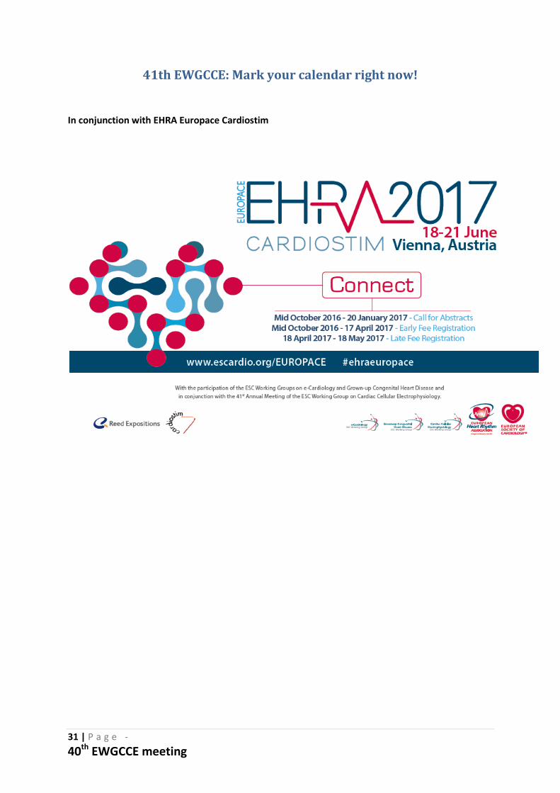
41th EWGCCE: Mark your calendar right now!
In conjunction with EHRA Europace Cardiostim