Embed Size (px)
Citation preview
H2O2-responsive liposomal nanoprobe for photoacousticinflammation imaging and tumor theranosticsvia in vivo chromogenic assayQian Chena, Chao Lianga, Xiaoqi Suna, Jiawen Chena, Zhijuan Yanga, He Zhaob, Liangzhu Fenga, and Zhuang Liua,1
aInstitute of Functional Nano & Soft Materials, Jiangsu Key Laboratory for Carbon-Based Functional Materials and Devices, Soochow University, Suzhou,Jiangsu 215123, China; and bDepartment of Pediatric Research, Children’s Hospital of Soochow University, Suzhou, Jiangsu 215123, China
Edited by Hongjie Dai, Stanford University, Stanford, CA, and approved April 12, 2017 (received for review February 6, 2017)
Abnormal H2O2 levels are closely related to many diseases, includinginflammation and cancers. Herein, we simultaneously load HRP andits substrate, 2,2′-azino-bis(3-ethylbenzothiazoline-6-sulfonic acid)(ABTS), into liposomal nanoparticles, obtaining a Lipo@HRP&ABTSoptical nanoprobe for in vivo H2O2-responsive chromogenic assaywith great specificity and sensitivity. In the presence of H2O2, colorlessABTS would be converted by HRP into the oxidized form with strongnear-infrared (NIR) absorbance, enabling photoacoustic detection ofH2O2 down to submicromolar concentrations. Using Lipo@HRP&ABTSas an H2O2-responsive nanoprobe, we could accurately detect theinflammation processes induced by LPS or bacterial infection inwhich H2O2 is generated. Meanwhile, upon systemic administra-tion of this nanoprobe we realize in vivo photoacoustic imagingof small s.c. tumors (∼2 mm in size) as well as orthotopic braingliomas, by detecting H2O2 produced by tumor cells. Interestingly,local injection of Lipo@HRP&ABTS further enables differentiationof metastatic lymph nodes from those nonmetastatic ones, basedon their difference in H2O2 contents. Moreover, using the H2O2-dependent strong NIR absorbance of Lipo@HRP&ABTS, tumor-specific photothermal therapy is also achieved. This work thus de-velops a sensitive H2O2-responsive optical nanoprobe useful not onlyfor in vivo detection of inflammation but also for tumor-specifictheranostic applications.
hydrogen peroxide | inflammation | tumor | photoacoustic imaging |photothermal therapy
Hydrogen peroxide (H2O2) plays an active role in variousphysiological processes including cell growth, immune re-
sponse, and senescence (1, 2). An imbalance in H2O2 productionis closely associated with diseases such as cancers, diabetes, in-flammation, and cardiovascular and neurodegenerative diseases (3,4). Therefore, the accurate and sensitive detection of H2O2 wouldhave significant clinical value not only for disease diagnosis but alsofor better understanding of disease mechanisms. However, in vivoimaging of H2O2 for clinical applications remains a challenging taskowing to the relatively low concentrations of H2O2 in physiologicalenvironments (below ∼50 μM) (5, 6). Previously, boronate-basedfluorescent probes have been explored for detection of physiologicH2O2 (with the detection limit at a few micromoles), relying on theH2O2-mediated transformation of arylboronates to phenols, gen-erating fluorescence under visible light excitation (5–8). Moreover,a number of peroxalate-based nanoparticles have also been de-veloped for in vivo H2O2-responsive bioluminescence imaging (9).However, the performance of in vivo H2O2 detection by eitherfluorescence or bioluminescence imaging is limited by the tissuepenetration depth of those conventional optical imaging techniques,in particular with visible light used in those systems, hamperingaccurate detection of H2O2 in deep tissues (3, 9). Thus, it is nec-essary to design nanoprobes that allow accurate in vivo imaging ofH2O2 in deep tissues with high specificity and sensitivity. In addi-tion, it may be of great interest to realize H2O2-specific treatment tofight certain types of diseases such as cancer with enhanced selec-tivity for precision medicine.
Photoacoustic (PA) imaging, which relies on ultrasound signalsgenerated by photothermal expansion of light-absorbing tissues orcontrast probes under pulsed laser irradiation, has emerged as apromising type of biomedical imaging technique combining ad-vantages of both optical and ultrasound imaging (10–12). Differentfrom conventional fluorescence or bioluminescence imaging tech-niques that are suitable mostly for small animals, PA imaging hasalready moved into clinical trials owing to its significantly improvedin vivo imaging depth (as deep as ∼12 cm) and spatial resolutioncompared with traditional optical imaging modalities (10, 11, 13).A large variety of nanoscale agents, including both organic andinorganic ones, with absorption in the near-infrared (NIR) “tissuetransparent” optical window have been extensively explored as PAimaging probes (14–20). For example, many types of nanoparticleswith strong and constant NIR absorbance are often used for en-hanced blood pool imaging and PA imaging of tumors (15, 16, 19).In addition, smart PA imaging nanoprobes with their absorbancespectra responsive to certain physiological signals such as reactiveoxygen species (ROS), pH, and enzymes have also received sub-stantial interest in recent years (12, 21, 22), because such a type ofimaging would provide critical information directly related to dis-ease progression and mechanisms. However, a sensitive and bio-compatible nanoprobe that can precisely detect H2O2 in vivo by PAimaging has not yet been reported, to our best knowledge.In this work, a liposomal nanocarrier simultaneously loaded
with HRP and its substrate, 2,2′-azino-bis(3-ethylbenzothiazoline-6-sulfonic acid) (ABTS), is fabricated for in vivo H2O2 detectionby PA imaging. The obtained Lipo@HRP&ABTS nanoprobe inthe presence of H2O2 would show strong NIR absorbance owing
Significance
Hydrogen peroxide is closely associated with many importantphysiological and pathological events. Herein, we develop a li-posomal nanoprobe for in vivo H2O2-responsive chromogenic as-say, which is highly specific and sensitive to H2O2. Using such ananoprobe, photoacoustic imaging of H2O2-related inflammationprocesses in mice induced by either LPS or bacteria is realized.Meanwhile, such a nanoprobe, by reactingwith endogenous H2O2
produced by tumor cells, allows sensitive photoacoustic imagingof early-stage small tumors and orthotopic brain glioma and evenenables accurate differentiation of metastatic sentinel lymphnodes from nonmetastatic ones. Furthermore, owing to its H2O2-responsive near-infrared absorbance, selective photothermal ab-lation of tumors is also achieved, illustrating the promise of usingthis nanoproble for tumor theranostics with great specificity.
Author contributions: Q.C., X.S., and Z.L. designed research; Q.C., C.L., X.S., J.C., Z.Y., H.Z.,and L.F. performed research; Q.C. and Z.L. analyzed data; and Q.C. and Z.L. wrote the paper.
The authors declare no conflict of interest.
This article is a PNAS Direct Submission.1To whom correspondence should be addressed. Email: [email protected].
This article contains supporting information online at www.pnas.org/lookup/suppl/doi:10.1073/pnas.1701976114/-/DCSupplemental.
www.pnas.org/cgi/doi/10.1073/pnas.1701976114 PNAS | May 23, 2017 | vol. 114 | no. 21 | 5343–5348
CHEM
ISTR
YBIOCH
EMISTR
Y
to the HRP-catalyzed oxidization of colorless ABTS substrate intoits green oxidized form, relying on a well-established chromogenicreaction extensively applied in bioanalysis techniques such asELISA. Notably, such a nanoprobe is highly specific and sensitiveto H2O2, with detection sensitivity down to the submicromolarlevel by PA imaging. Thus, using Lipo@HRP&ABTS nanoprobe,PA imaging of H2O2-related inflammation process is realized,revealing the development of inflammation induced by LPS orbacterial infection as well as the slowing down of inflammationafter treatment by an antiinflammatory drug. In additional, such aLipo@HRP&ABTS nanoprobe can also be used for in vivo PAimaging of s.c. tumors and orthotopic brain glioma upon systemici.v. injection, or metastatic tumor cells within sentinel lymph nodes(SLNs), owing to the endogenous H2O2 production by tumor cells.Furthermore, using Lipo@HRP&ABTS accumulated in the tu-mor and its H2O2-induced NIR absorbance, tumor-specific pho-tothermal ablation is successfully realized under NIR laserirradiation, which meanwhile induces minimal nonspecific heatingto normal tissues without abnormal accumulation of H2O2. Ourwork achieves in vivo PA imaging of H2O2 using a liposomaloptical nanoprobe, which is useful not only for in vivo detection ofinflammations or tumors with great sensitivity but also for tumor-specific photothermal ablation with high efficacy and minimalnonspecific heating damage to normal tissues.
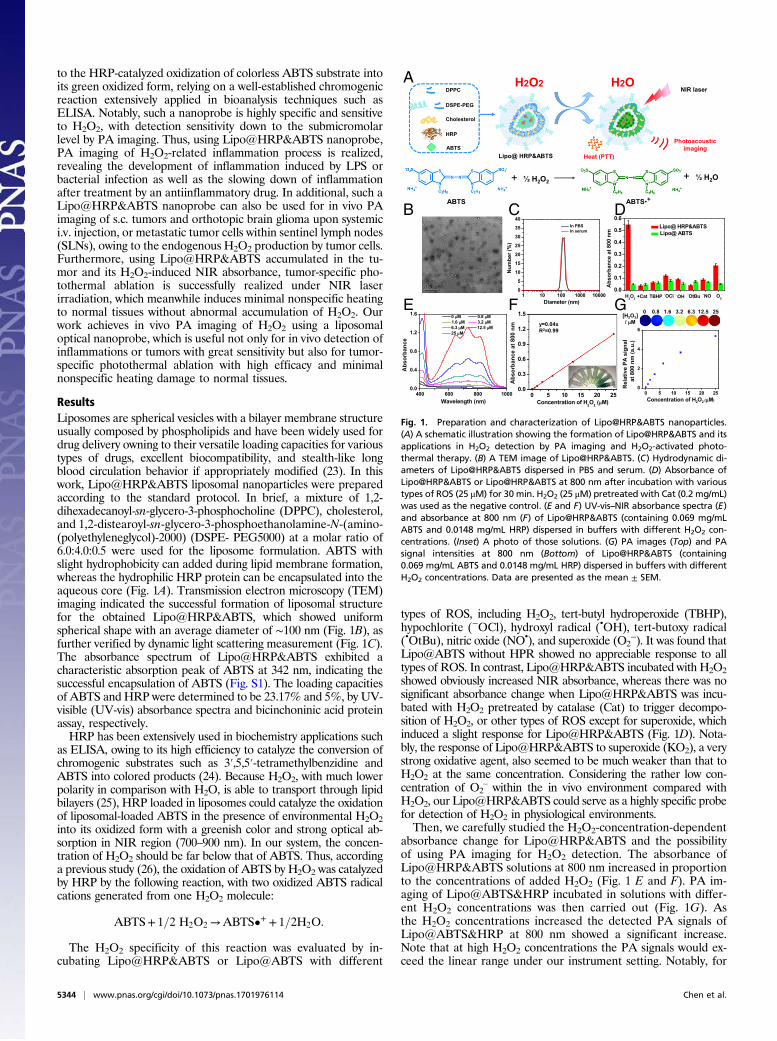
ResultsLiposomes are spherical vesicles with a bilayer membrane structureusually composed by phospholipids and have been widely used fordrug delivery owning to their versatile loading capacities for varioustypes of drugs, excellent biocompatibility, and stealth-like longblood circulation behavior if appropriately modified (23). In thiswork, Lipo@HRP&ABTS liposomal nanoparticles were preparedaccording to the standard protocol. In brief, a mixture of 1,2-dihexadecanoyl-sn-glycero-3-phosphocholine (DPPC), cholesterol,and 1,2-distearoyl-sn-glycero-3-phosphoethanolamine-N-(amino-(polyethyleneglycol)-2000) (DSPE- PEG5000) at a molar ratio of6.0:4.0:0.5 were used for the liposome formulation. ABTS withslight hydrophobicity can added during lipid membrane formation,whereas the hydrophilic HRP protein can be encapsulated into theaqueous core (Fig. 1A). Transmission electron microscopy (TEM)imaging indicated the successful formation of liposomal structurefor the obtained Lipo@HRP&ABTS, which showed uniformspherical shape with an average diameter of ∼100 nm (Fig. 1B), asfurther verified by dynamic light scattering measurement (Fig. 1C).The absorbance spectrum of Lipo@HRP&ABTS exhibited acharacteristic absorption peak of ABTS at 342 nm, indicating thesuccessful encapsulation of ABTS (Fig. S1). The loading capacitiesof ABTS and HRP were determined to be 23.17% and 5%, by UV-visible (UV-vis) absorbance spectra and bicinchoninic acid proteinassay, respectively.HRP has been extensively used in biochemistry applications such
as ELISA, owing to its high efficiency to catalyze the conversion ofchromogenic substrates such as 3′,5,5′-tetramethylbenzidine andABTS into colored products (24). Because H2O2, with much lowerpolarity in comparison with H2O, is able to transport through lipidbilayers (25), HRP loaded in liposomes could catalyze the oxidationof liposomal-loaded ABTS in the presence of environmental H2O2into its oxidized form with a greenish color and strong optical ab-sorption in NIR region (700–900 nm). In our system, the concen-tration of H2O2 should be far below that of ABTS. Thus, accordinga previous study (26), the oxidation of ABTS by H2O2 was catalyzedby HRP by the following reaction, with two oxidized ABTS radicalcations generated from one H2O2 molecule:
ABTS+ 1=2 H2O2 →ABTS•+ + 1=2H2O.
The H2O2 specificity of this reaction was evaluated by in-cubating Lipo@HRP&ABTS or Lipo@ABTS with different
types of ROS, including H2O2, tert-butyl hydroperoxide (TBHP),hypochlorite (−OCl), hydroxyl radical (•OH), tert-butoxy radical(•OtBu), nitric oxide (NO•), and superoxide (O2
−). It was found thatLipo@ABTS without HPR showed no appreciable response to alltypes of ROS. In contrast, Lipo@HRP&ABTS incubated with H2O2showed obviously increased NIR absorbance, whereas there was nosignificant absorbance change when Lipo@HRP&ABTS was incu-bated with H2O2 pretreated by catalase (Cat) to trigger decompo-sition of H2O2, or other types of ROS except for superoxide, whichinduced a slight response for Lipo@HRP&ABTS (Fig. 1D). Nota-bly, the response of Lipo@HRP&ABTS to superoxide (KO2), a verystrong oxidative agent, also seemed to be much weaker than that toH2O2 at the same concentration. Considering the rather low con-centration of O2
– within the in vivo environment compared withH2O2, our Lipo@HRP&ABTS could serve as a highly specific probefor detection of H2O2 in physiological environments.Then, we carefully studied the H2O2-concentration-dependent
absorbance change for Lipo@HRP&ABTS and the possibilityof using PA imaging for H2O2 detection. The absorbance ofLipo@HRP&ABTS solutions at 800 nm increased in proportionto the concentrations of added H2O2 (Fig. 1 E and F). PA im-aging of Lipo@ABTS&HRP incubated in solutions with differ-ent H2O2 concentrations was then carried out (Fig. 1G). Asthe H2O2 concentrations increased the detected PA signals ofLipo@ABTS&HRP at 800 nm showed a significant increase.Note that at high H2O2 concentrations the PA signals would ex-ceed the linear range under our instrument setting. Notably, for
0 5 10 15 20 250.0
0.3
0.6
0.9
1.2
1.5
Abs
orba
nce
at 8
00 n
m
Concentration of H2O2 (µµM)
H2O2 H2ODPPC
DSPE-PEG
Cholesterol
HRP
ABTSLipo@ HRP&ABTS Heat (PTT)
NIR laser
Photoacousticimaging
y=0.04x R2=0.99
A
B DC
E GF0.0
0.1
0.2
0.3
0.4
0.5
0.6
Abs
orba
nce
at 8
00 n
m Lipo@ HRP&ABTS
Lipo@ ABTS
H2O2 +Cat TBHP -OCl .OH .OtBu .NO O2-
[H2O2]/ µM
0 0.8 1.6 3.2 6.3 12.5 25
1 10 100 1000 100000
5
10
15
20
25
30
35
40
Num
ber (
%)
Diameter (nm)
In PBS In serum
+ ½ H2O2 + ½ H2O
ABTS ABTS•+
400 600 800 10000.0
0.4
0.8
1.2
1.6
Abs
orba
nce
Wavelength (nm)
0 µM 0.8 µM1.6 µM 3.2 µM6.3 µM 12.5 µM25 µM
0 5 10 15 20 250
2
4
6
Rel
ativ
e PA
sig
nal
at
800
nm
(a.u
.)
Concentration of H2O2 (µM)
Fig. 1. Preparation and characterization of Lipo@HRP&ABTS nanoparticles.(A) A schematic illustration showing the formation of Lipo@HRP&ABTS and itsapplications in H2O2 detection by PA imaging and H2O2-activated photo-thermal therapy. (B) A TEM image of Lipo@HRP&ABTS. (C) Hydrodynamic di-ameters of Lipo@HRP&ABTS dispersed in PBS and serum. (D) Absorbance ofLipo@HRP&ABTS or Lipo@HRP&ABTS at 800 nm after incubation with varioustypes of ROS (25 μM) for 30 min. H2O2 (25 μM) pretreated with Cat (0.2 mg/mL)was used as the negative control. (E and F) UV-vis–NIR absorbance spectra (E)and absorbance at 800 nm (F) of Lipo@HRP&ABTS (containing 0.069 mg/mLABTS and 0.0148 mg/mL HRP) dispersed in buffers with different H2O2 con-centrations. (Inset) A photo of those solutions. (G) PA images (Top) and PAsignal intensities at 800 nm (Bottom) of Lipo@HRP&ABTS (containing0.069 mg/mL ABTS and 0.0148 mg/mL HRP) dispersed in buffers with differentH2O2 concentrations. Data are presented as the mean ± SEM.
5344 | www.pnas.org/cgi/doi/10.1073/pnas.1701976114 Chen et al.
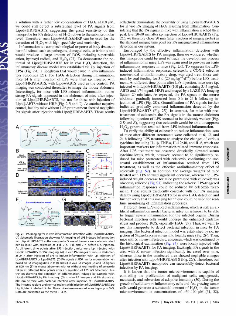
a solution with a rather low concentration of H2O2 at 0.8 μM,we could still detect a substantial level of PA signals fromLipo@HRP&ABTS, suggesting the great sensitivity of thisnanoprobe for PA detection of H2O2 down to the submicromolarlevel. Therefore, such Lipo@ABTS&HRP can be used for thedetection of H2O2 with high specificity and sensitivity.Inflammation is a complex biological response of body tissues to
harmful stimuli such as pathogens, damaged cells, or irritants andwould produce a large amount of ROS, including superoxideanion, hydroxyl radical, and H2O2 (27). To demonstrate the po-tential of Lipo@HRP&ABTS for in vivo H2O2 detection, theinflammatory disease model was established via i.p. injection ofLPSs (Fig. 2A), a lipoglycan that would cause in vivo inflamma-tory responses (28). For H2O2 detection during inflammation,mice 24 h after injection of LPS were then i.p. injected withLipo@HRP&ABTS, with Lipo@ABTS used as the control. PAimaging was conducted thereafter to image the mouse abdomen.Interestingly, for mice with LPS-induced inflammation, ratherstrong PA signals appeared in the abdomen of mice after injec-tion of Lipo@HRP&ABTS, but not for those with injection ofLipo@ABTS without HRP (Fig. 2 B and C). As another negativecontrol, healthy mice without LPS pretreatment showed negligiblePA signals after injection with Lipo@HRP&ABTS. Those results
collectively demonstrate the possibility of using Lipo@HRP&ABTSfor in vivo PA imaging of H2O2 resulting from inflammation. Con-sidering that the PA signals in mice with inflammation reached theirpeak level 20–30 min after i.p. injection of Lipo@HRP&ABTS (Fig.2C), we therefore chose 20 min (after injection of imaging probe) asthe standard imaging time point for PA imaging-based inflammationdetection in our system.Encouraged by the effective inflammation detection with
Lipo@HRP&ABTS by PA imaging, then we wondered whetherthis nanoprobe could be used to track the development processof inflammation in mice. LPS was again used to provoke an acuteinflammatory response in mice (Fig. 2A). To suppress the LPS-induced inflammation responses, celecoxib, a COX-2 selectivenonsteroidal antiinflammatory drug, was used treat those ani-mals by oral feeding for 3 d (20 mg·kg−1·d−1) before LPS treat-ment. At different time points after LPS injection, mice were i.p.injected with Lipo@HRP&ABTS (100 μL, containing 3.45 mg/mLABTS and 0.74 mg/mL HRP) and imaged by a LAZR PA ImagingSystem 20 min later. As expected, the PA signals in the mouseabdomen gradually increased from 0 h to 24 h following in-jection of LPS (Fig. 2D). Quantification of PA signals furtherindicated gradually enhanced inflammation detected by theLipo@HRP&ABTS (Fig. 2E). In contrast, for mice with pre-treatment of celecoxib, the PA signals in the mouse abdomenfollowing injection of LPS seemed to be obviously weaker (Fig.2 D and E), suggesting that celecoxib would be able to suppressH2O2 generation resulted from LPS-induced inflammation.To verify the ability of celecoxib to reduce inflammation, sera
of mice after different treatments were collected at 6, 12, and24 h following LPS treatment to analyze the changes of variouscytokines including IL-1β, TNF-α, IL-12p40, and IL-6, which areimportant markers for inflammation-related immune responses.After LPS treatment we observed drastically elevated serumcytokine levels, which, however, seemed to be significantly re-duced for mice pretreated with celecoxib, confirming the suc-cessful establishment of inflammation resulted from LPStreatment, as well as the effective antiinflammatory effect ofcelecoxib (Fig. S2). In addition, the average weights of micetreated with LPS showed significant decrease, whereas the LPS-induced weight decrease for mice pretreated with celecoxib wasobviously relieved (Fig. S3), indicating the adverse LPS-triggeredinflammation responses could be reduced by celecoxib treat-ment. Those results excellently correlate with our PA imagingdata by using Lipo@HRP&ABTS for in vivo H2O2 detection andfurther verify that this imaging technique could be used for real-time monitoring of inflammation processes.Different from LPS-induced inflammation, which is still an ar-
tificial inflammation model, bacterial infection is known to be ableto trigger severe inflammation for the infected organs. Duringbacterial infection cells would undergo the enhanced oxidativestress and produce ROS, especially H2O2 (29). Thus, we want touse this nanoprobe to detect bacterial infection in mice by PAimaging. The bacterial infection model was established by s.c. in-jection of Staphylococcus aureus into healthy mice (Fig. 2F). Then,mice with S. aureus-infected s.c. abscesses, which was confirmed bythe histological examination (Fig. S4), were locally injected withLipo@HRP&ABTS for PA imaging. Excitingly, PA signals in thearea with S. aureus infection significantly increased over time,whereas those in the uninfected area showed negligible changesafter injection with Lipo@HRP&ABTS (Fig. 2G). Therefore, ourLipo@HRP&ABTS nanoprobe can successfully detect bacterialinfection by PA imaging.It is known that the tumor microenvironment is capable of
controlling the proliferation of malignant cells, angiogenesis,metastasis, and subversion of adaptive immunity (30). During thegrowth of solid tumors inflammatory cells and fast-growing tumorcells would generate a substantial amount of H2O2 in the tumormicroenvironment at concentrations of ∼50–100 μM (31, 32).
Lipo@ HRP&ABTS
LPS &Lipo@ABTS
0 h 4 h 8 h 24 h
Celecoxib &LPS treated
LPS treated
0 5 10 15 20 250.2
0.4
0.6
0.8
1.0
Rel
ativ
e PA
sig
nals
(a.u
.)
Time (h)
LPS treated Celecoxib & LPS treated
A
B
D E
F 0 h 2 h
Normal Infected
4 h
Normal Infected Normal Infected
24 h
Normal Infected0 5 10 15 20 25
0
2
4
6
Rel
ativ
e PA
sig
nals
(a.u
.)
Time (h)
Infected Normal
12 h
Normal Infected
Bacterial infection model
S. aureuss.c. injection
At different
time points
Lipo@HRP&ABTSs.c. injection
20 min later
PA imaging
G
i.p. LPS
At different
time points
i.p.Lipo@ HRP&ABTS
20 min laterNIR laser
PA imaging
Celecoxib
W/t or w/o p.o.
celecoxib 3 days
C10 min 20 min 30 min 40 min0 min
LPS & Lipo@ HRP&ABTS
0 10 20 30 400.2
0.4
0.6
0.8
1.0
1.2
1.4
Rel
ativ
e PA
sig
nals
(a.u
.)
Time (min)
Lipo@ HRP&ABTS LPS & Lipo@ABTS LPS & Lipo@HRP&ABTS
2 m
m2
mm
2 m
m2
mm
2 m
m
2 m
m
2 m
m
Fig. 2. PA imaging for in vivo inflammation detection with Lipo@HRP&ABTS.(A) Schematic illustration showing PA imaging of LPS-induced inflammationwith Lipo@HRP&ABTS as the nanoprobe. Some of themice were administratedper os (p.o.) with celecoxib at 3 d, 2 d, 1 d, and 2 h before LPS injection.At different time points after LPS injection, mice were i.p. injected withLipo@HRP&ABTS for PA imaging. (B) In vivo PA images of mouse abdomenat 24 h after injection of LPS to induce inflammation with i.p. injection ofLipo@HRP&ABTS or Lipo@ABTS. (C) PA signals at 800 nm for mouse abdomenbased on PA imaging data in B. (D and E) In vivo PA images (D) and PA signalsat 800 nm (E) in mouse abdomen with or without oral feeding of celecoxibtaken at different time points after i.p. injection of LPS. (F) Schematic illus-tration showing the detection of inflammation induced by bacteria withLipo@HRP&ABTS by PA imaging. (G) In vivo PA images and PA signals at800 nm of mice with bacterial infection after injection of Lipo@HRP&ABTS.The infected regions and normal regions with injection of Lipo@HRP&ABTS arehighlighted in dashed circles. Three mice were measured in each group in B–G.Data are presented as the mean ± SEM.
Chen et al. PNAS | May 23, 2017 | vol. 114 | no. 21 | 5345
CHEM
ISTR
YBIOCH
EMISTR
Y
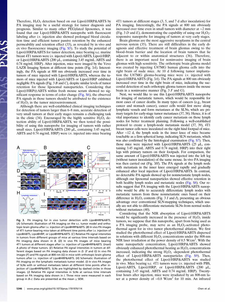
Therefore, H2O2 detection based on our Lipo@HRP&ABTS byPA imaging may be a useful strategy for tumor diagnosis andprognosis. Similar to many other PEGylated liposomes, it wasfound that our Lipo@HRP&ABTS nanoprobe with fluorescentlabeling after i.v. injection also showed prolonged blood circula-tion time and efficient tumor passive retention by the enhancedpermeability and retention effect (33), as revealed by in vivo andex vivo fluorescence imaging (Fig. S5). To study the potential ofLipo@HRP&ABTS for tumor detection, mice bearing s.c. murinebreast 4T1 tumors were i.v. injected with Lipo@ABTS, Lipo@HRP,or Lipo@HRP&ABTS (200 μL, containing 3.45 mg/mL ABTS and0.74 mg/mL HRP). After injection, mice were imaged by the VevoLAZR Imaging System at different time points (Fig. 3A). Interest-ingly, the PA signals at 800 nm obviously increased over time intumors of mice injected with Lipo@HRP&ABTS, whereas the tu-mors of mice injected with Lipo@ABTS or Lipo@HRP exhibitednegligible PA signals (Fig. 3 B and C), despite similar levels of tumorretention for those liposomal nanoparticles. Considering thatLipo@HRP@ABTS within fresh mouse serum showed no sig-nificant response in terms of color change (Fig. S6), the observedPA signals in those tumors should be attributed to the existenceof H2O2 in the tumor microenvironment.Although there are well-established clinical imaging techniques
for detection of tumors larger than 6–8 mm, accurate detection ofvery small tumors at their early stages remains a challenging taskin the clinic (34). Encouraged by the highly sensitive H2O2 de-tection ability of Lipo@HRP&ABTS, we then tested the possi-bility of using this nanoprobe for imaging of tumors with rathersmall sizes. Lipo@HRP&ABTS (200 μL, containing 3.45 mg/mLABTS and 0.74 mg/mL HRP) were i.v. injected into mice bearing
4T1 tumors at different stages (3, 5, and 7 d after inoculation) forPA imaging. Interestingly, the PA signals at 800 nm obviouslyincreased over time even in small tumors with diameter at ∼2 mm(Fig. 3 D and E), demonstrating the capability of using our H2O2-responsive nanoprobe for imaging of tumors at very early stages.Brain gliomas are the most aggressive neoplasms in the central
nervous system (35). There are still difficulties in the early di-agnosis and effective treatment of brain gliomas owing to theblood–brain barrier and the location of brain tumors that lieadjacent to or within anatomical structures (36). Therefore,there is an important need for noninvasive imaging of braingliomas with high sensitivity. The orthotopic brain glioma modelwas created by injecting U87MG human glioma cells into theright brain of nude mice. At 10 d following tumor implanta-tion the U87MG glioma-bearing mice were i.v. injected withLipo@HRP&ABTS (Fig. 3A). The PA signals at 800 nm obviouslyincreased over time in the right brain of mice, indicating the suc-cessful detection of such orthotopic glioma tumors inside the mousebrain in a noninvasive manner (Fig. 3 F and G).Next, we would like to use this Lipo@HRP&ABTS nanoprobe
for imaging of metastatic tumors, which would be responsible formost cases of cancer deaths. In many types of cancers (e.g., breastcancer and stomach cancer), cancer cells would first move alonglymphatic vessels and form metastases at the SLNs, which are pri-mary targets for early-stage tumor metastases (37, 38). Hence, it is ofvital importance to identify early cancer metastasis on those lymphnodes for better treatment planning. Following a well-establishedprotocol to create a lymph-node metastasis model (37, 39), 4T1breast tumor cells were inoculated on the right hind footpad of mice.After ∼12 d, the lymph node in the inner knee of mice becametouchable as a firm spherical lump, indicating SLN metastasis, whichwas also confirmed by the histological examination (Fig. S7). Then,those mice were injected with Lipo@HRP&ABTS (25 μL, con-taining 3.45 mg/mL ABTS and 0.74 mg/mL HRP) into their rightlegs with primary tumors on their footpads. For comparison, thesame amount of Lipo@HRP&ABTS was injected into the left leg(without tumor inoculation) of the same mouse. In vivo PA imagingwas then carried out (Fig. 3H). The PA signals at the lymph nodewith metastasis in the inner knee emerged rapidly and graduallyenhanced after local injection of Lipo@HRP&ABTS. In contrast,no detectable PA signals showed up for nonmetastatic lymph nodes,although our liposomal nanoparticles showed effective retention inboth healthy lymph nodes and metastatic nodes (Fig. S8). Our re-sults suggest that PA imaging with the Lipo@HRP&ABTS nanop-robe would be able to accurately differentiate lymph nodes withmetastatic tumors from those nonmetastatic ones based on theirdifference in H2O2 contents (Fig. 3 I and J), presenting an obviousadvantage over conventional SLN-mapping techniques, which usu-ally are not able to differentiate metastatic SLNs from normal nodeswithout metastases (40).Considering that the NIR absorption of Lipo@HRP&ABTS
would be significantly increased in the presence of H2O2 insidetumors, we suppose that this nanoprobe, apart from being used asa PA imaging probe, may serve as an H2O2-activated photo-thermal agent for in vivo tumor photothermal ablation. We firststudied the photothermal effect of Lipo@HRP&ABTS dispersedin solutions with different H2O2 concentrations under the 808-nmNIR laser irradiation at the power density of 0.5 W/cm2. With thesame nanoparticle concentration, Lipo@HRP&ABTS showedobviously enhanced photothermal heating as H2O2 concentrationsincreased, indicating the strong H2O2 -dependent photothermaleffect of Lipo@HRP&ABTS nanoparticles (Fig. S9). Then,the photothermal effect of Lipo@HRP&ABTS was studiedin vivo. Mice bearing s.c. 4T1 tumors were i.v. injected with PBS,Lipo@ABTS, Lipo@HRP, or Lipo@HRP&ABTS (200 μL,containing 3.45 mg/mL ABTS and 0.74 mg/mL HRP). Twenty-four hours after injection, mice were irradiated by an 808-nm la-ser at a power density of ∼0.8 W/cm2 for 10 min. An infrared
high
low
Lipo@HRP
Lipo@ABTS
Lipo@ HRP&ABTS
5 min 2 h 4 h 8 h 24 h
B
H
CSubcutaneous /
orthotopic tumors
Lipo@ HRP&ABTSi.v. injection
Healthy SLN
SLN with metastasis
Lipo@ HRP&ABTSi.t. injection
Lymphatic metastasis I J
A
0 5 10 15 20 250.0
0.2
0.4
0.6
Rel
ativ
e PA
sig
nals
(a.u
.)
Time (h)
3 day 5 day 7 day
0 5 10 15 20 250.0
0.2
0.4
0.6
Rel
ativ
e PA
sig
nals
(a.u
.)
Time (h)
FE GD 0 h 8 h 24 h
0 h 0.5 h 1 h 2 h 3 h 4 h
3 day
5 day
7 day
2 m
m2
mm
2 m
m
1 m
m1
mm
1 m
m
0 h 4 h
8 h 24 h
2 m
m
SLN with metastasis
Healthy SLN
2 m
m2
mm
Orthotopic brain glioma
0 5 10 15 20 25
0.1
0.2
0.3
0.4
0.5
Rel
ativ
e PA
sig
nals
(a.u
.)
Time (h)
Lipo@ABTSLipo@HRPLipo@HRP&ABTS
0 1 2 3 40.0
0.1
0.2
0.3
0.4
0.5
Rel
ativ
e PA
sig
nals
(a.u
.)
Time (h)
Healthy SLN SLN with metastasis
Fig. 3. PA imaging for in vivo tumor detection with Lipo@HRP&ABTS.(A) Schematic illustration of PA imaging on the s.c. tumor model and ortho-topic brain glioma after i.v. injection of Lipo@HRP&ABTS. (B) In vivo PA imagesof 4T1 tumor-bearing mice taken at different time points after i.v. injection ofLipo@ABTS, Lipo@HRP, or Lipo@HRP&ABTS. (C) Relative PA signal intensitiesin tumors from different groups of mice at various time intervals based onPA imaging data shown in B. (D) In vivo PA images of mice bearing4T1 tumors at different stages after i.v. injection of Lipo@HRP&ABTS. (Inset)A photo of these tumors. (E) Relative PA signal intensities in tumors at dif-ferent stages based on PA imaging data shown in D. (F and G) In vivo PAimages (F) and PA signals at 800 nm (G) in mice with orthotopic brain gliomatumors after i.v. injection of Lipo@HRP&ABTS. (H) Schematic illustration ofPA imaging on the lymphatic metastasis tumor model. (I) In vivo PA imagesof SLNs with or without metastasis taken at different time points after in-jection of Lipo@HRP&ABTS. SLNs are highlighted by dashed circles in thoseimages. (J) Relative PA signal intensities in SLNs at various time intervalsbased on PA imaging data shown in I. Three mice were measured in eachgroup in B–J. Data are presented as the mean ± SEM.
5346 | www.pnas.org/cgi/doi/10.1073/pnas.1701976114 Chen et al.
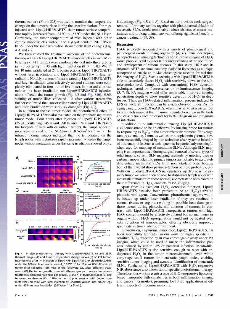
thermal camera (Fotric 225) was used to monitor the temperaturechange on the tumor surface during the laser irradiation. For miceinjected with Lipo@HRP&ABTS, their tumor surface tempera-ture rapidly increased from ∼34 °C to ∼55 °C under the NIR laser.Conversely, the tumor temperature of mice injected with othercontrol nanoparticles without the H2O2-dependent NIR absor-bance under the same irradiation showed only slight changes (Fig.4 A and B).We then studied the treatment outcome of the photothermal
therapy with such Lipo@HRP&ABTS nanoparticles in vivo. Micebearing s.c. 4T1 tumors were randomly divided into three groups(n = 5 per group): PBS with light irradiation (810 nm, 0.8 W/cm2
for 10 min, irradiated at 24 h postinjection), Lipo@HRP&ABTSwithout laser irradiation, and Lipo@HRP&ABTS with laser ir-radiation. Notably, tumors of mice treated by Lipo@HRP&ABTSand laser irradiation were effectively ablated (tumors were com-pletely eliminated in four out of five mice). In marked contrast,neither the laser irradiation nor Lipo@HRP&ABTS injectionalone affected the tumor growth (Fig. 4D and Fig. S10). H&Estaining of tumor slices collected 1 d after various treatmentsfurther confirmed that cancer cells treated by Lipo@HRP&ABTSand laser irradiation were seriously damaged (Fig. 4C).In addition to the s.c. tumor model, the photothermal effect of
Lipo@HRP&ABTS was also evaluated on the lymphatic metastasistumor model. Four hours after injection of Lipo@HRP&ABTS(25 μL, containing 3.45 mg/mL ABTS and 0.74 mg/mL HRP) intothe footpads of mice with or without tumors, the lymph nodes ofmice were exposed to the NIR laser (0.8 W/cm2 for 5 min). Theinfrared thermal images indicated that the temperature on thelymph nodes with metastases rapidly increased, whereas the lymphnodes without metastasis under the same irradiation showed only a
little change (Fig. 4 E and F). Based on our previous work, surgicalremoval of primary tumors together with photothermal ablation ofmetastatic SLNs would remarkably reduce chances of cancer me-tastases and prolong animal survival, offering significant benefit incancer treatment (37, 39).
DiscussionH2O2 is closely associated with a variety of physiological andpathological events in living organisms (4, 32). Thus, developingnanoprobes and imaging techniques for selective imaging of H2O2would provide useful tools for better understanding of the occurrenceand development of various diseases. In this study, HRP and itssubstrate ABTS are simultaneously loaded in liposomes as a uniquenanoprobe to enable an in vivo chromogenic reaction for real-timePA imaging of H2O2. Such a technique with Lipo@HRP&ABTS isable to selectively detect H2O2 with sensitivity down to the sub-micromolar level. Compared with conventional H2O2 detectiontechniques based on fluorescence or bioluminescence imaging(3, 7, 8), PA imaging would offer remarkably improved imagingpenetration depth to allow sensitive detection of H2O2 in deeptissues. Thus, an H2O2-related inflammation process induced byLPS or bacterial infection can be vividly observed under PA im-aging using Lipo@HRP&ABTS, which may serve as a useful toolto accurately map out the inflammation/infection areas in the bodyand closely track such processes for better diagnosis and prognosisof infections.In addition to the inflammation imaging, Lipo@HRP&ABTS is
also demonstrated to be an effective PA imaging probe for tumorsby responding to H2O2 in the tumor microenvironment. Early-stagetumors as small as 2 mm, as well as orthotopic brain gliomas, havebeen successfully imaged by our technique after systemic injectionof this nanoproble. Such a technique may be particularly meaningfulwhen used for mapping of metastatic SLNs. Although SLN map-ping is an important step during surgical removal of several types ofsolid tumors, current SLN mapping methods by injecting dyes orcarbon nanoparticles into primary tumors are not able to accuratelydifferentiate metastatic SLNs from nonmetastatic ones, becauseboth of them would show passive retention of those probes (37, 39).With our Lipo@HRP&ABTS nanoparticles injected near the pri-mary tumor we would then be able to distinguish lymph nodes withmetastatic tumors from those normal, nonmetastatic ones based ontheir differences in H2O2 contents by PA imaging.Apart from its excellent H2O2 detection function, Lipo@
HRP&ABTS has also been proven to be an H2O2-activatedphotothermal agent. Conventional photothermal agents wouldbe heated up under laser irradiation if they are retained innormal tissues or organs, resulting in possible heat damage tothose tissues during photothermal ablation of tumors. In con-trast, with Lipo@HRP&ABTS nanoparticles tumors with highH2O2 contents would be effectively ablated but normal issues ororgans without H2O2 up-regulation would not be heated evenwith retention of nanoparticles, offering obviously improvedspecificity in tumor ablation treatment.In conclusion, a liposomal nanoprobe, Lipo@HRP&ABTS, has
been successfully fabricated in our work for highly specific andsensitive H2O2 detection by in vivo chromogenic assay under PAimaging, which could be used to image the inflammation pro-cess induced by either LPS or bacterial infection. Meanwhile,Lipo@HRP&ABTS is also sensitive enough to react with en-dogenous H2O2 in the tumor microenvironment, even withinearly-stage small tumors or metastatic lymph nodes, enablingsensitive tumor imaging and accurate identification of metastaticSLNs. Furthermore, Lipo@HRP&ABTS with H2O2-responsiveNIR absorbance also allows tumor-specific photothermal therapy.Therefore, this work presents a type of H2O2-responsive liposome-based nanoprobe with capabilities in both inflammation imagingand cancer theranostics, promising for future applications in dif-ferent aspects of precision medicine.
0 2 4 6 8 10 12 140
200
400
600
800
1000
Tum
or v
olum
e (m
m3 )
Day
PBS + L Lipo@HRP&ABTS Lipo@HRP&ABTS + L
100 µm
PBS+LLipo@
HRP&ABTSLipo@
HRP&ABTS +L
PBS + L
Lipo@ HRP + L
Lipo@ ABTS + L
Lipo@ HRP&ABTS + L
550C
250C
0 min 2 min 4 min 6 min 8 min 10 min
Non-metastatic SLN
Metastatic SLN
0 min 1 min 2 min 3 min 4 min 5 min 50oC
25oC
F
A B
DC
E
0 100 200 300 400 500 600
35
40
45
50
55
Tem
pert
ure(
o C)
Time (s)
PBS + LLipo@HRP + LLipo@ABTS + LLipo@HRP&ABTS + L
0 50 100 150 200 250 30025
30
35
40
45
50
Tem
pert
ure (
o C)
Time (s)
Non-metastatic SLNMetastatic SLN
µ
Fig. 4. In vivo photothermal therapy with Lipo@HRP&ABTS. (A and B) IRthermal images (A) and tumor temperature change curves (B) of 4T1 tumor-bearing mice after i.v. injection of Lipo@HRP, Lipo@ABTS, or Lipo@HRP&ABTSunder the 808-nm laser irradiation (+L, 0.8 W/cm2 for 10 min). (C) H&E-stainedtumor slices collected from mice at the following day after different treat-ments. (D) The tumor growth curves of different groups of mice after varioustreatments indicated (five mice per group). (E and F) IR thermal images (E) andtemperature changes (F) of SLNs without (upper row) or with (lower row)metastases on mice with local injection of Lipo@HRP&ABTS into mouse legsunder 808-nm laser irradiation (0.8 W/cm2 for 5 min).
Chen et al. PNAS | May 23, 2017 | vol. 114 | no. 21 | 5347
CHEM
ISTR
YBIOCH
EMISTR
Y
Materials and MethodsFemale BALB/c mice (6–8 wk of age) were purchased from Nanjing PengSheng Biological Technology Co., Ltd. and used under protocols approved bythe Soochow University Laboratory Animal Center. To establish the perito-neal inflammation in mice, 100 μL (1 mg/mL) LPS was i.p. injected in eachmouse. To form s.c. abscesses, S. aureus (5 × 10⁶ cfu) was s.c. injected into theback of each mouse. To develop the s.c. tumor model, 1 × 10⁶ 4T1 murinebreast cancer cells in 50 μL PBS were s.c. injected into the back of eachmouse. The brain glioma model was created following the well-establishedmethod (41) by injecting 2.5 × 10⁵ U87MG human glioma cells in 10 μL PBSinto the right brain of each female nude mouse and used ∼10 d later. Tocreate the lymph node metastasis tumor model, 1 × 10⁶ 4T1 cells suspended
in 20 μL PBS were injected into the right hind footpad of each mouse. After∼12 d, we chose mice with spherical firm lumps touchable in their inner kneefor our experiments. Further experimental details can be found in SI Mate-rials and Methods.
ACKNOWLEDGMENTS. This work was partially supported by the NationalScience and Technology Major Project of China Grant 2016YFA0201200,National Natural Science Foundation of China Grant 51525203, the Collab-orative Innovation Center of Suzhou Nano Science and Technology, the“111 Project” from the Ministry of Education of China, and a projectfunded by the Priority Academic Program Development of Jiangsu HigherEducation Institutions.
1. Bai J, Jiang X (2013) A facile one-pot synthesis of copper sulfide-decorated reducedgraphene oxide composites for enhanced detecting of H2O2 in biological environ-ments. Anal Chem 85:8095–8101.
2. Winterbourn CC (2008) Reconciling the chemistry and biology of reactive oxygenspecies. Nat Chem Biol 4:278–286.
3. Van de Bittner GC, Dubikovskaya EA, Bertozzi CR, Chang CJ (2010) In vivo imaging ofhydrogen peroxide production in a murine tumor model with a chemoselective bio-luminescent reporter. Proc Natl Acad Sci USA 107:21316–21321.
4. Finkel T, Serrano M, Blasco MA (2007) The common biology of cancer and ageing.Nature 448:767–774.
5. Deng Z, et al. (2016) Engineering intracellular delivery nanocarriers and nanoreactorsfrom oxidation-responsive polymersomes via synchronized bilayer cross-linking andpermeabilizing inside live cells. J Am Chem Soc 138:10452–10466.
6. Bandyopadhyay A, Gao J (2016) Iminoboronate-based peptide cyclization that re-sponds to pH, oxidation, and small molecule modulators. J Am Chem Soc 138:2098–2101.
7. Miller EW, Albers AE, Pralle A, Isacoff EY, Chang CJ (2005) Boronate-based fluorescentprobes for imaging cellular hydrogen peroxide. J Am Chem Soc 127:16652–16659.
8. Chang MCY, Pralle A, Isacoff EY, Chang CJ (2004) A selective, cell-permeable opticalprobe for hydrogen peroxide in living cells. J Am Chem Soc 126:15392–15393.
9. Lee D, et al. (2007) In vivo imaging of hydrogen peroxide with chemiluminescentnanoparticles. Nat Mater 6:765–769.
10. Kim C, Favazza C, Wang LV (2010) In vivo photoacoustic tomography of chemicals:
High-resolution functional and molecular optical imaging at new depths. Chem Rev110:2756–2782.
11. Xu MH, Wang LHV (2006) Photoacoustic imaging in biomedicine. Rev Sci Instrum 77:041101.
12. Chen Q, et al. (2015) A self-assembled albumin-based nanoprobe for in vivo ratio-
metric photoacoustic pH imaging. Adv Mater 27:6820–6827.13. Zhou Y, et al. (2016) A phosphorus phthalocyanine formulation with intense absor-
bance at 1000 nm for deep optical imaging. Theranostics 6:688–697.14. Cheng K, et al. (2014) Construction and validation of nano gold tripods for molecular
imaging of living subjects. J Am Chem Soc 136:3560–3571.15. Song KH, Kim C, Cobley CM, Xia Y, Wang LV (2009) Near-infrared gold nanocages as a
new class of tracers for photoacoustic sentinel lymph node mapping on a rat model.
Nano Lett 9:183–188.16. Cheng L, et al. (2014) PEGylated WS(2) nanosheets as a multifunctional theranostic
agent for in vivo dual-modal CT/photoacoustic imaging guided photothermal ther-
apy. Adv Mater 26:1886–1893.17. De la Zerda A, et al. (2008) Carbon nanotubes as photoacoustic molecular imaging
agents in living mice. Nat Nanotechnol 3:557–562.18. Kim JW, Galanzha EI, Shashkov EV, Moon HM, Zharov VP (2009) Golden carbon
nanotubes as multimodal photoacoustic and photothermal high-contrast molecular
agents. Nat Nanotechnol 4:688–694.19. Liang XL, et al. (2015) PEGylated polypyrrole nanoparticles conjugating gadolinium
chelates for dual-modal MRI/photoacoustic imaging guided photothermal therapy ofcancer. Adv Funct Mater 25:1451–1462.
20. Li K, Liu B (2014) Polymer-encapsulated organic nanoparticles for fluorescence and
photoacoustic imaging. Chem Soc Rev 43:6570–6597.
21. Dragulescu-Andrasi A, Kothapalli SR, Tikhomirov GA, Rao J, Gambhir SS (2013) Acti-vatable oligomerizable imaging agents for photoacoustic imaging of furin-like ac-tivity in living subjects. J Am Chem Soc 135:11015–11022.
22. Pu K, et al. (2014) Semiconducting polymer nanoparticles as photoacoustic molecularimaging probes in living mice. Nat Nanotechnol 9:233–239.
23. Feng LZ, et al. (2016) Cisplatin-prodrug-constructed liposomes as a versatile thera-nostic nanoplatform for bimodal imaging guided combination cancer therapy. AdvFunct Mater 26:2207–2217.
24. Wei H, Wang E (2013) Nanomaterials with enzyme-like characteristics (nanozymes):Next-generation artificial enzymes. Chem Soc Rev 42:6060–6093.
25. Bienert GP, Schjoerring JK, Jahn TP (2006) Membrane transport of hydrogen perox-ide. Biochim Biophys Acta 1758:994–1003.
26. Kadnikova EN, Kosti�c NM (2002) Oxidation of ABTS by hydrogen peroxide catalyzedby horseradish peroxidase encapsulated into sol–gel glass: Effects of glass matrix onreactivity. J Mol Catal, B Enzym 18:39–48.
27. Hansson GK (2005) Inflammation, atherosclerosis, and coronary artery disease. N EnglJ Med 352:1685–1695.
28. Trent MS, Stead CM, Tran AX, Hankins JV (2006) Diversity of endotoxin and its impacton pathogenesis. J Endotoxin Res 12:205–223.
29. Korupalli C, et al. (2017) Acidity-triggered charge-convertible nanoparticles that cancause bacterium-specific aggregation in situ to enhance photothermal ablation offocal infection. Biomaterials 116:1–9.
30. Finger EC, Giaccia AJ (2010) Hypoxia, inflammation, and the tumor microenvironmentin metastatic disease. Cancer Metastasis Rev 29:285–293.
31. Chen Q, et al. (2016) Intelligent albumin-MnO2 nanoparticles as pH-/H2 O2 -responsivedissociable nanocarriers to modulate tumor hypoxia for effective combination therapy.Adv Mater 28:7129–7136.
32. Szatrowski TP, Nathan CF (1991) Production of large amounts of hydrogen peroxideby human tumor cells. Cancer Res 51:794–798.
33. Torchilin VP (2005) Recent advances with liposomes as pharmaceutical carriers. NatRev Drug Discov 4:145–160.
34. Lyman GH, et al.; American Society of Clinical Oncology (2005) American Society ofClinical Oncology guideline recommendations for sentinel lymph node biopsy inearly-stage breast cancer. J Clin Oncol 23:7703–7720.
35. Veiseh O, et al. (2009) Specific targeting of brain tumors with an optical/magneticresonance imaging nanoprobe across the blood-brain barrier. Cancer Res 69:6200–6207.
36. Galve-Roperh I, et al. (2000) Anti-tumoral action of cannabinoids: Involvement ofsustained ceramide accumulation and extracellular signal-regulated kinase activation.Nat Med 6:313–319.
37. Chen Q, et al. (2014) An albumin-based theranostic nano-agent for dual-modal im-aging guided photothermal therapy to inhibit lymphatic metastasis of cancer postsurgery. Biomaterials 35:9355–9362.
38. Skobe M, et al. (2001) Induction of tumor lymphangiogenesis by VEGF-C promotesbreast cancer metastasis. Nat Med 7:192–198.
39. Liang C, et al. (2014) Tumor metastasis inhibition by imaging-guided photothermaltherapy with single-walled carbon nanotubes. Adv Mater 26:5646–5652.
40. Shangguan D, et al. (2006) Aptamers evolved from live cells as effective molecularprobes for cancer study. Proc Natl Acad Sci USA 103:11838–11843.
41. Ruan S, et al. (2016) Increased gold nanoparticle retention in brain tumors by in situenzyme-induced aggregation. ACS Nano 10:10086–10098.
5348 | www.pnas.org/cgi/doi/10.1073/pnas.1701976114 Chen et al.