Embed Size (px)
Citation preview
HEAD INJURYBy
Dr C.K .Musau.
DEFINITION
• Trauma to the head.
• Neurological disruption.
• Variable presentation.
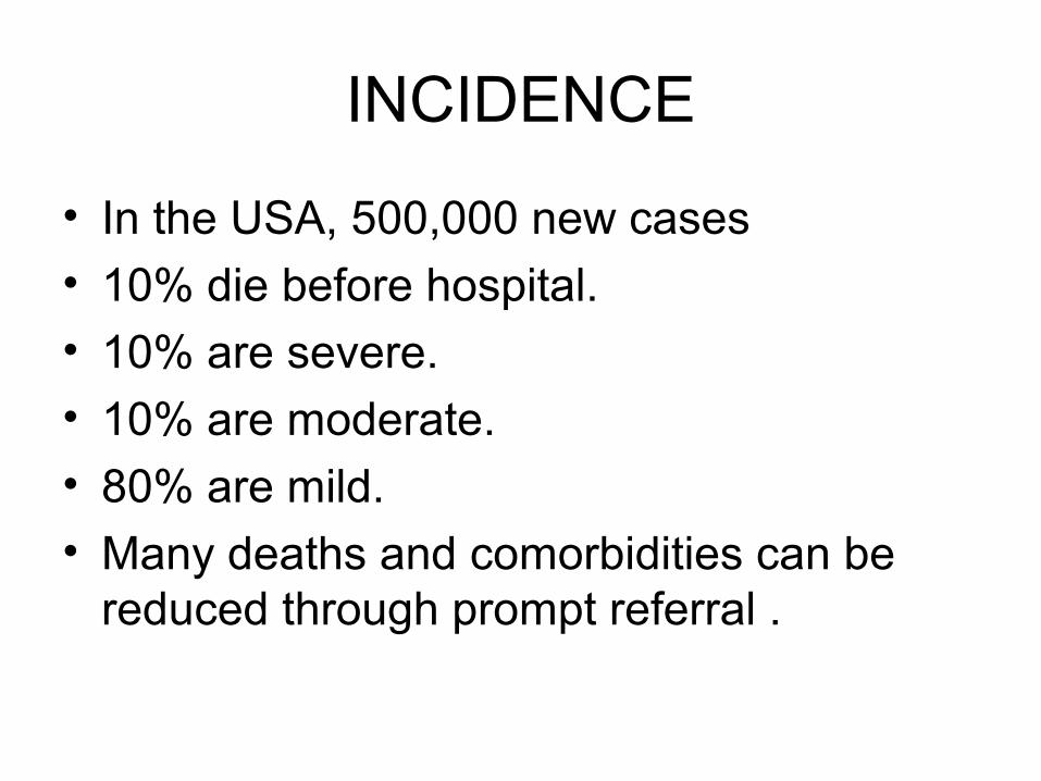
INCIDENCE
• In the USA, 500,000 new cases
• 10% die before hospital.
• 10% are severe.
• 10% are moderate.
• 80% are mild.
• Many deaths and comorbidities can be reduced through prompt referral .
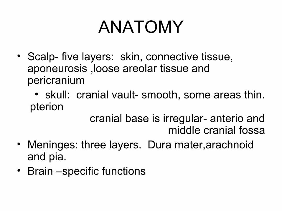
ANATOMY
• Scalp- five layers: skin, connective tissue, aponeurosis ,loose areolar tissue and pericranium
• skull: cranial vault- smooth, some areas thin. pterion
cranial base is irregular- anterio and middle cranial fossa
• Meninges: three layers. Dura mater,arachnoid and pia.
• Brain –specific functions
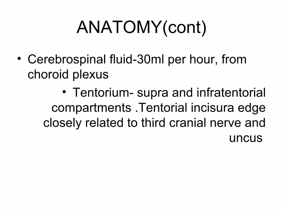
ANATOMY(cont)
• Cerebrospinal fluid-30ml per hour, from choroid plexus
• Tentorium- supra and infratentorial compartments .Tentorial incisura edge
closely related to third cranial nerve and uncus
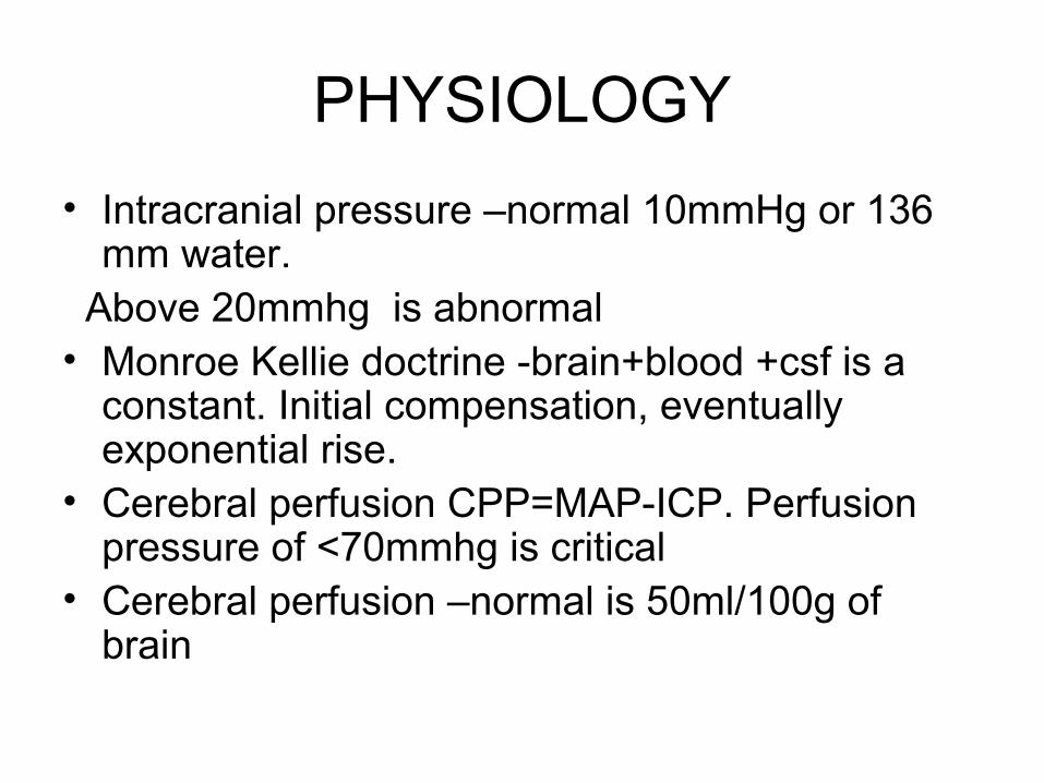
PHYSIOLOGY
• Intracranial pressure –normal 10mmHg or 136 mm water.
Above 20mmhg is abnormal• Monroe Kellie doctrine -brain+blood +csf is a
constant. Initial compensation, eventually exponential rise.
• Cerebral perfusion CPP=MAP-ICP. Perfusion pressure of <70mmhg is critical
• Cerebral perfusion –normal is 50ml/100g of brain
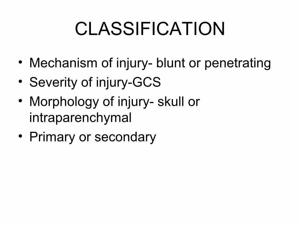
CLASSIFICATION
• Mechanism of injury- blunt or penetrating
• Severity of injury-GCS
• Morphology of injury- skull or intraparenchymal
• Primary or secondary
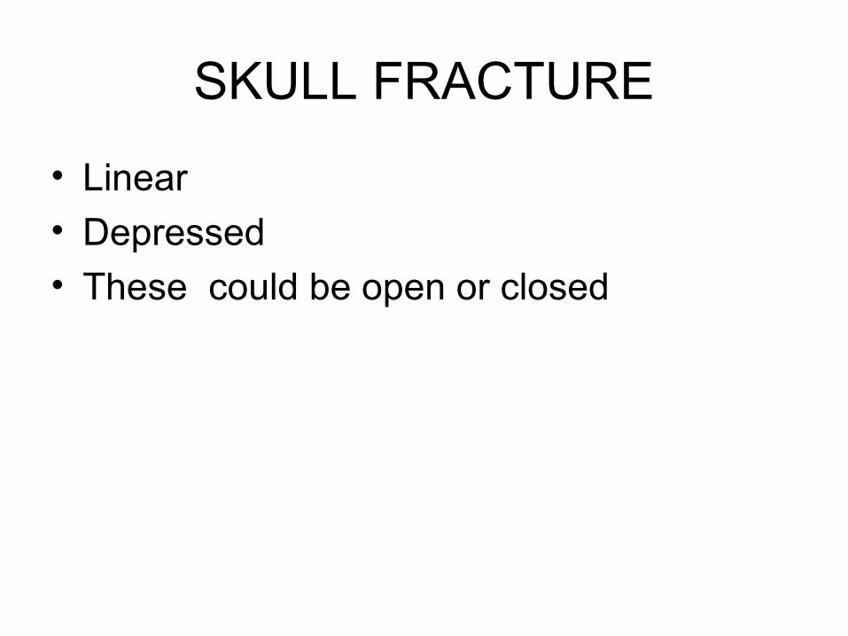
SKULL FRACTURE
• Linear
• Depressed
• These could be open or closed
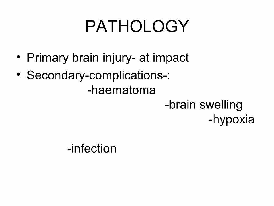
PATHOLOGY
• Primary brain injury- at impact
• Secondary-complications-: -haematoma -brain swelling -hypoxia -infection
INTRACRANIAL BLEED
• Epidural
• Subdural
• Subarachnoid
• intracerebral
MANAGEMENT
• History
• Physical examination
• Radiological investigations skull radiograph, cat scan, MRI
PRIMARY SURVEY
• A.-ABCDE
• B-Immobilize and stabilize the cervical spine
• C-Perform a brief neurological exam 1.pupillary response. 2.GCScore determination.
SECONDARY SURVEY
• A-.Inspect the entire head. Remove dressings ,look for lacerations or csf
• B-Palpate for fractures including the wounds• C-Inspect all scalp lacerations-look out for
brain,depressed fractures,debris or csf• D-Minineurological examination--GCS -BEST
- -Eye
-Motor - - Verbal Pupillary response
E-Examine cervical spineF-Determine the extend of the injuryG-Regular reassessment
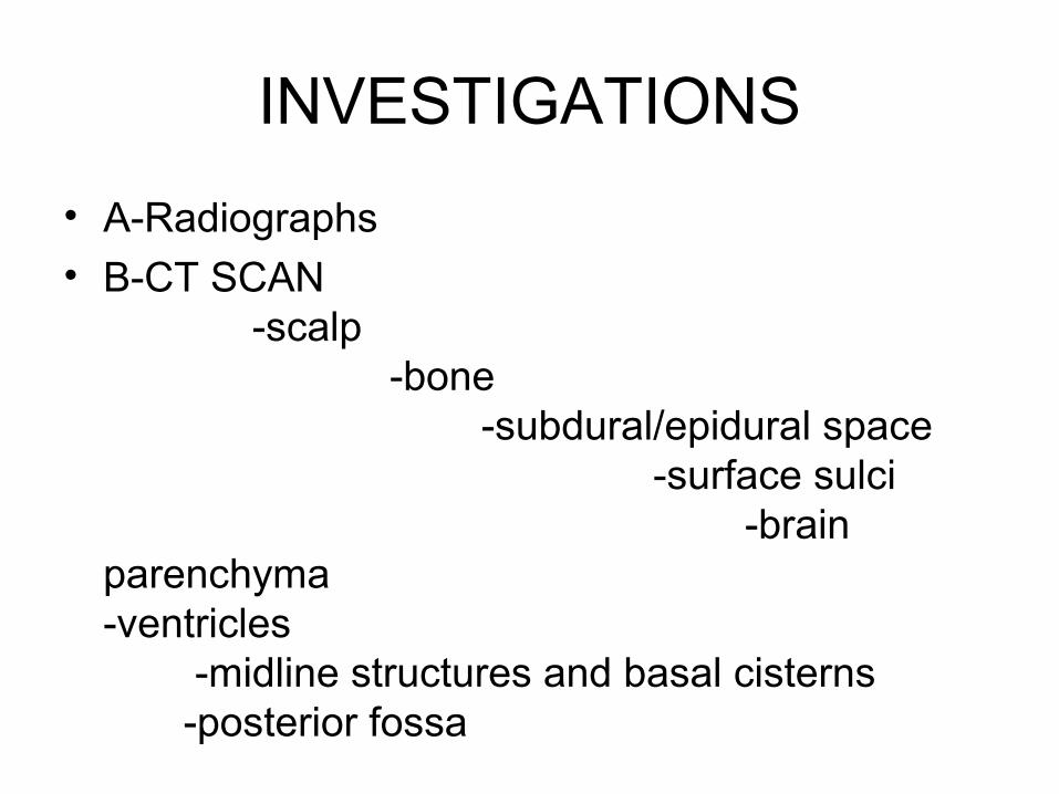
INVESTIGATIONS
• A-Radiographs• B-CT SCAN
-scalp -bone -subdural/epidural space -surface sulci -brain parenchyma -ventricles -midline structures and basal cisterns -posterior fossa
SPECIFIC MANAGEMENT
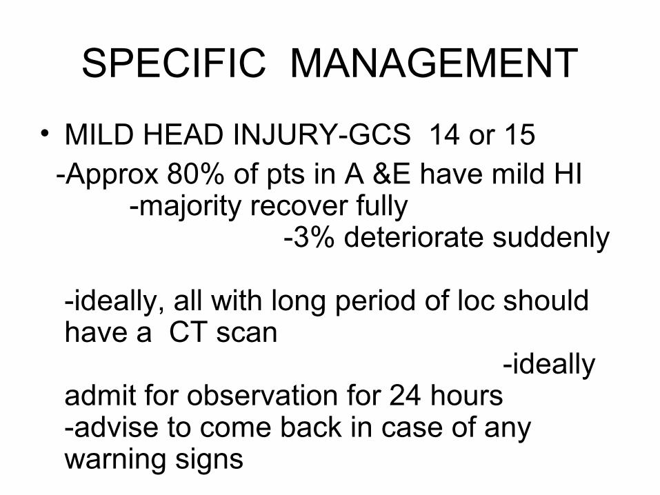
• MILD HEAD INJURY-GCS 14 or 15 -Approx 80% of pts in A &E have mild HI
-majority recover fully -3% deteriorate suddenly -ideally, all with long period of loc should have a CT scan -ideally admit for observation for 24 hours -advise to come back in case of any warning signs
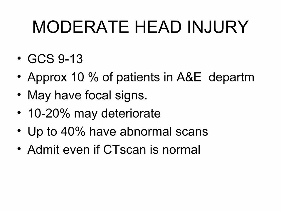
MODERATE HEAD INJURY
• GCS 9-13
• Approx 10 % of patients in A&E departm
• May have focal signs.
• 10-20% may deteriorate
• Up to 40% have abnormal scans
• Admit even if CTscan is normal
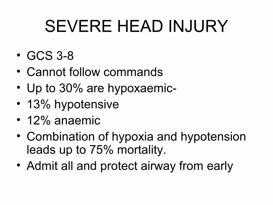
SEVERE HEAD INJURY
• GCS 3-8• Cannot follow commands• Up to 30% are hypoxaemic-• 13% hypotensive• 12% anaemic• Combination of hypoxia and hypotension
leads up to 75% mortality.• Admit all and protect airway from early
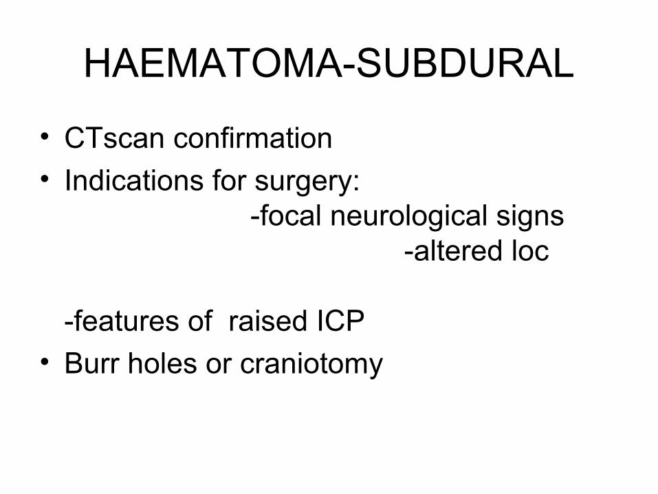
HAEMATOMA-SUBDURAL
• CTscan confirmation
• Indications for surgery: -focal neurological signs -altered loc -features of raised ICP
• Burr holes or craniotomy
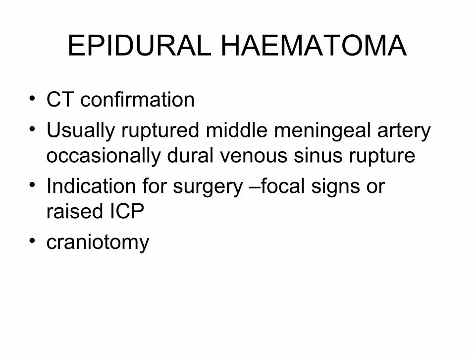
EPIDURAL HAEMATOMA
• CT confirmation
• Usually ruptured middle meningeal artery occasionally dural venous sinus rupture
• Indication for surgery –focal signs or raised ICP
• craniotomy
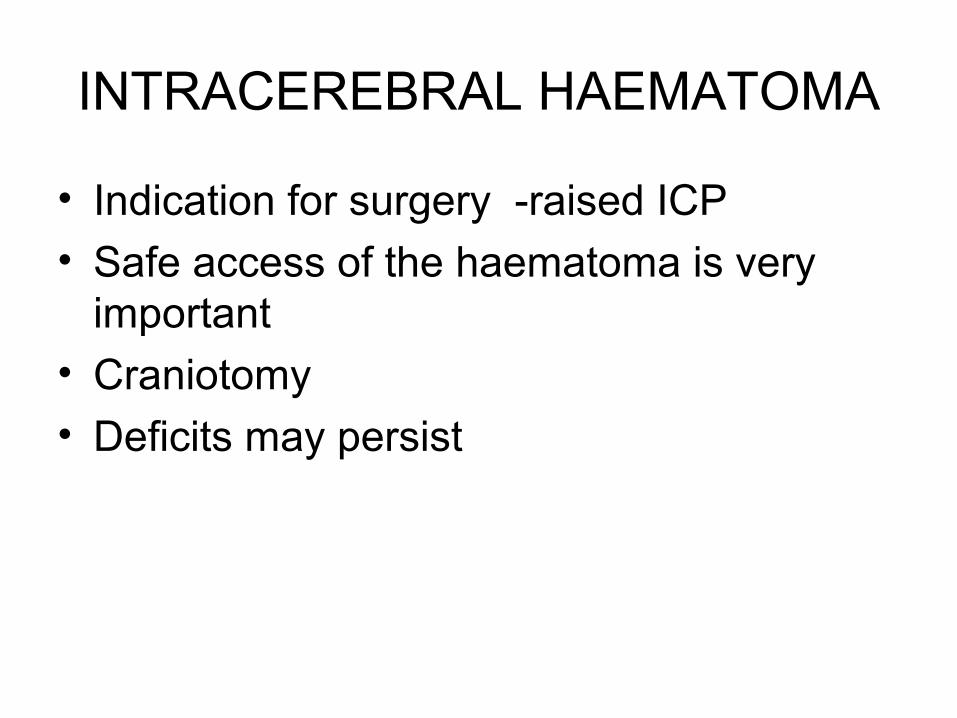
INTRACEREBRAL HAEMATOMA
• Indication for surgery -raised ICP
• Safe access of the haematoma is very important
• Craniotomy
• Deficits may persist
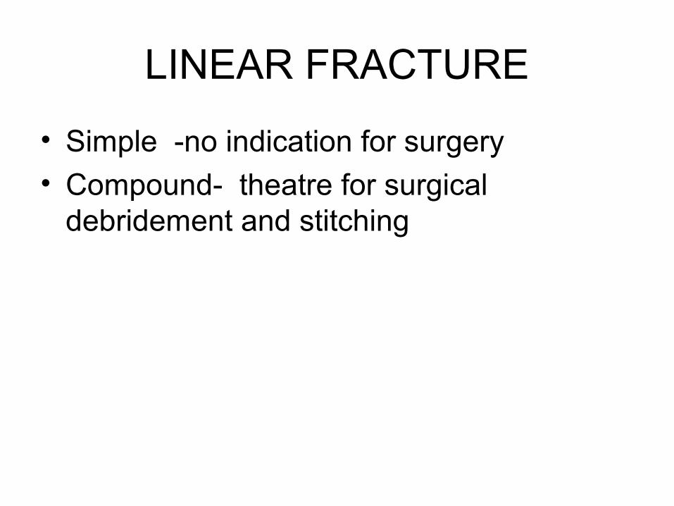
LINEAR FRACTURE
• Simple -no indication for surgery
• Compound- theatre for surgical debridement and stitching
DEPRESSED SKULL FRACTURE
• Closed elevation in case it is significant
• Compond- Theatre for surgical debridement and elevetion
• Antibiotic cover
RAISED ICP
• Ventillatory support
• Mannitol
• lasix
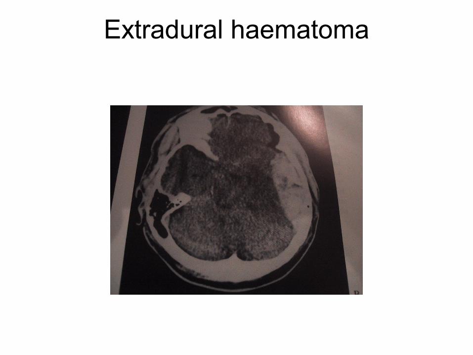
Extradural haematoma
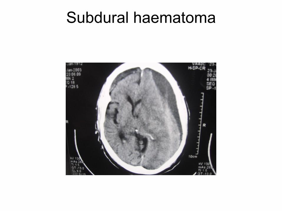
Subdural haematoma
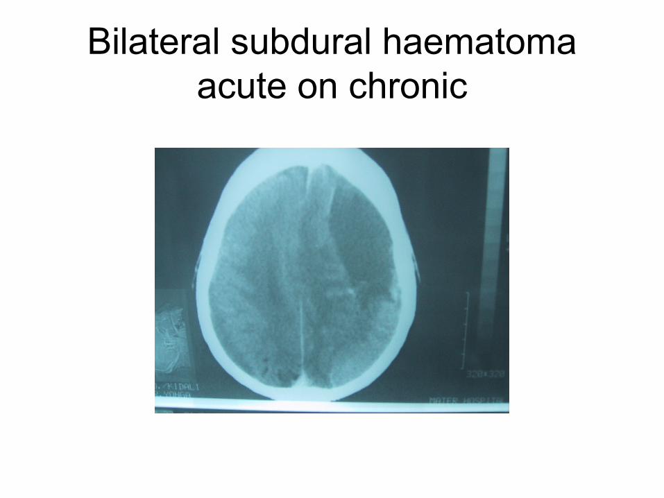
Bilateral subdural haematomaacute on chronic
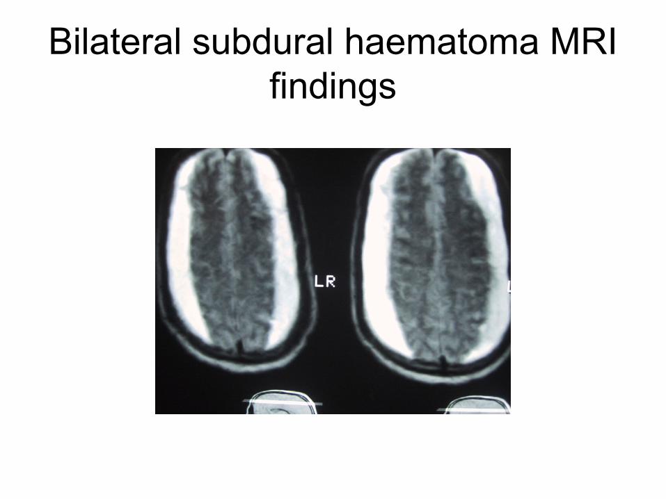
Bilateral subdural haematoma MRI findings
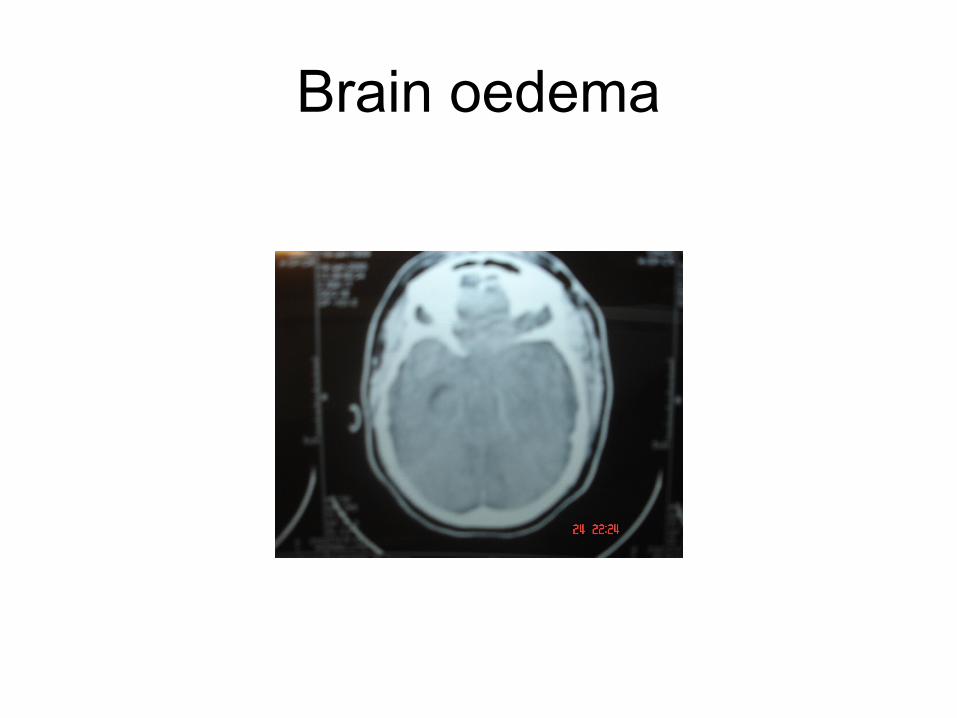
Brain oedema
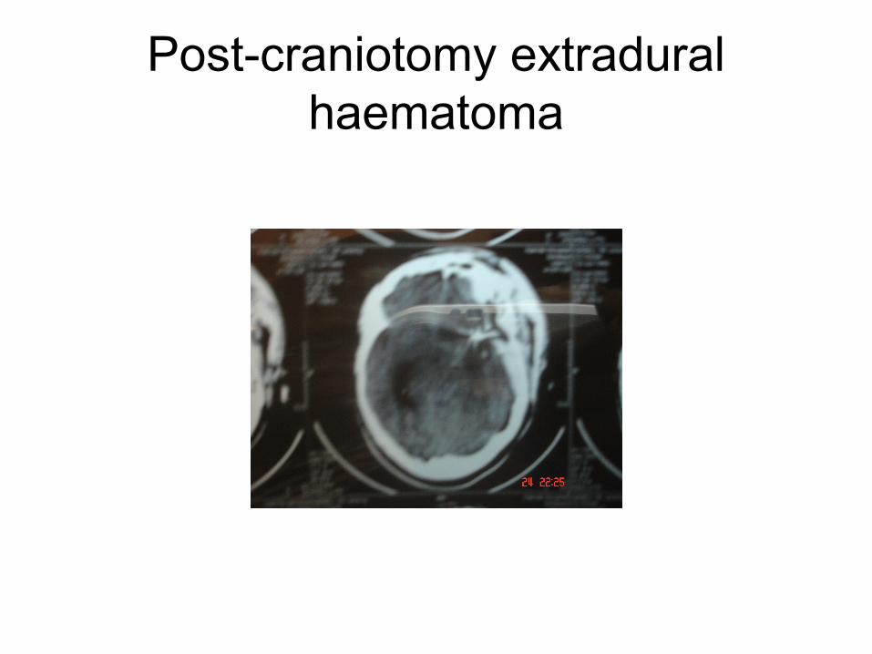
Post-craniotomy extradural haematoma
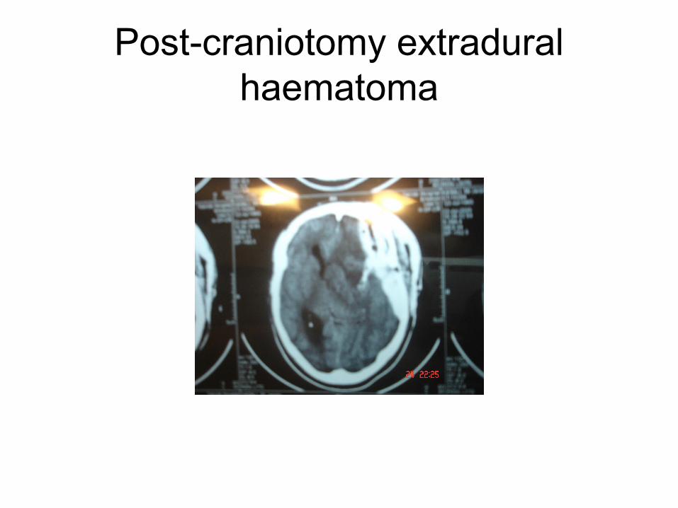
Post-craniotomy extradural haematoma
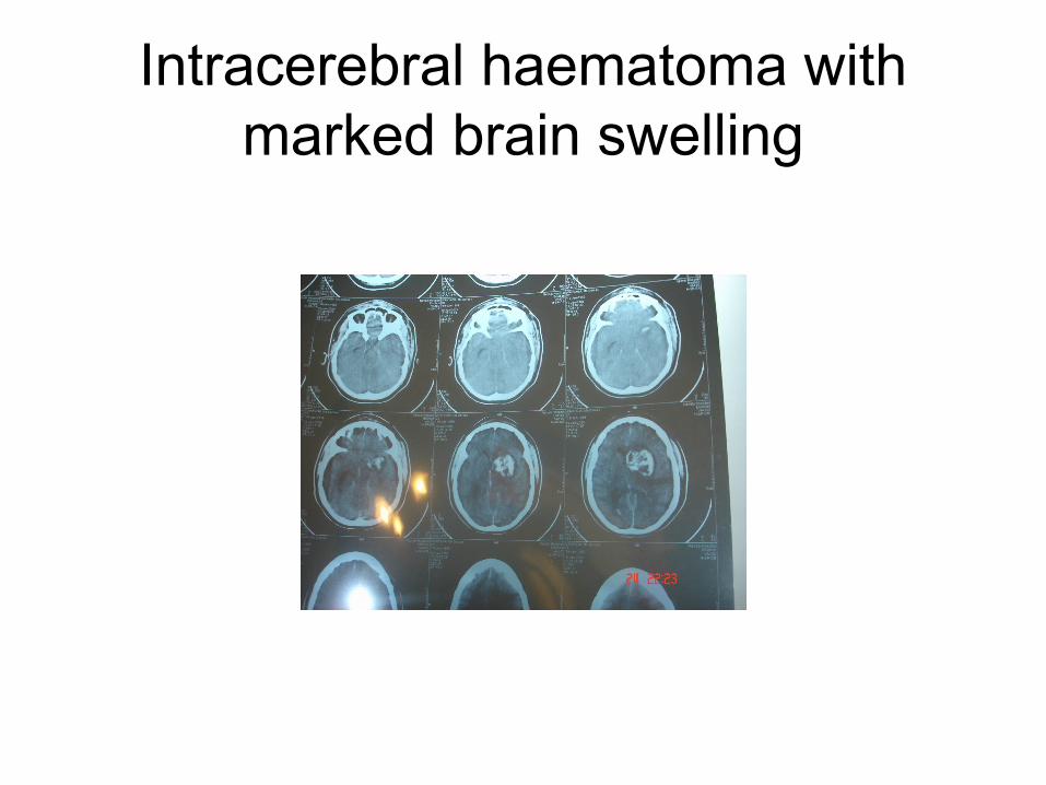
Intracerebral haematoma with marked brain swelling
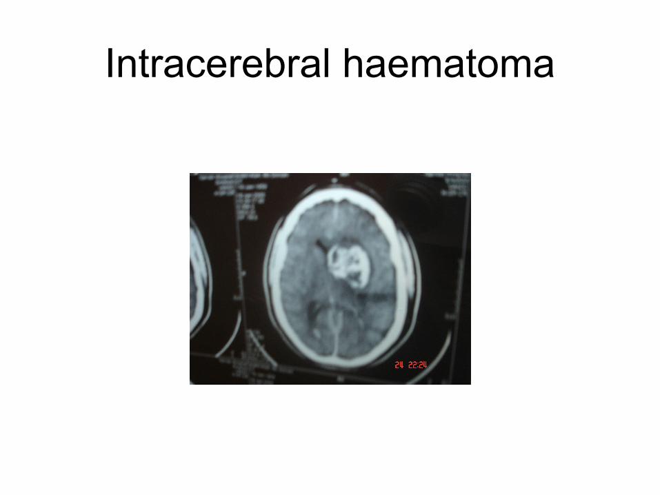
Intracerebral haematoma