Embed Size (px)
DESCRIPTION
HELICOBACTER PYLORI. Prepared by : Shefa EL –astal Islamic University-Gaza Medical Technology Department Prepared To :Dr.Abdelraouf A.Elmana. Helicobacter fact. Perviou name:campylobacter pylori Gram negative bacilli. Curved cells discovered in 1983 in stomach biopsied specimens. - PowerPoint PPT Presentation
Citation preview
HELICOBACTER PYLORI
Prepared by :Shefa EL –astalIslamic University-GazaMedical Technology DepartmentPrepared To:Dr.Abdelraouf
A.Elmana

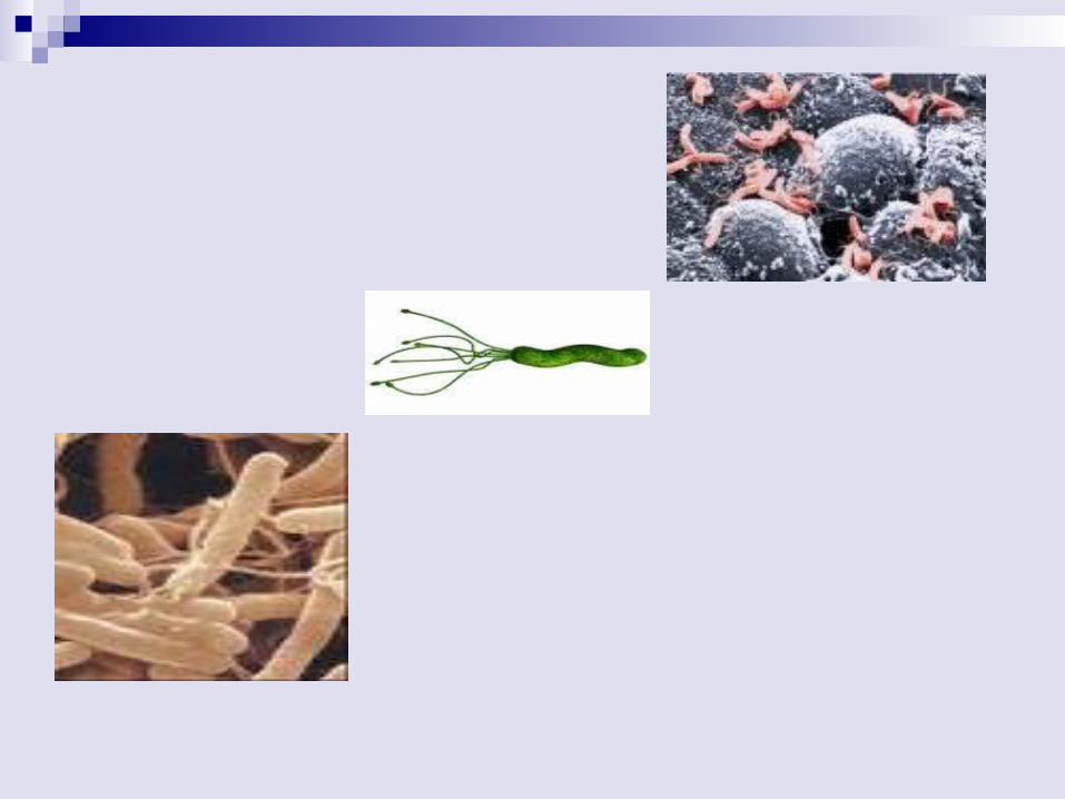
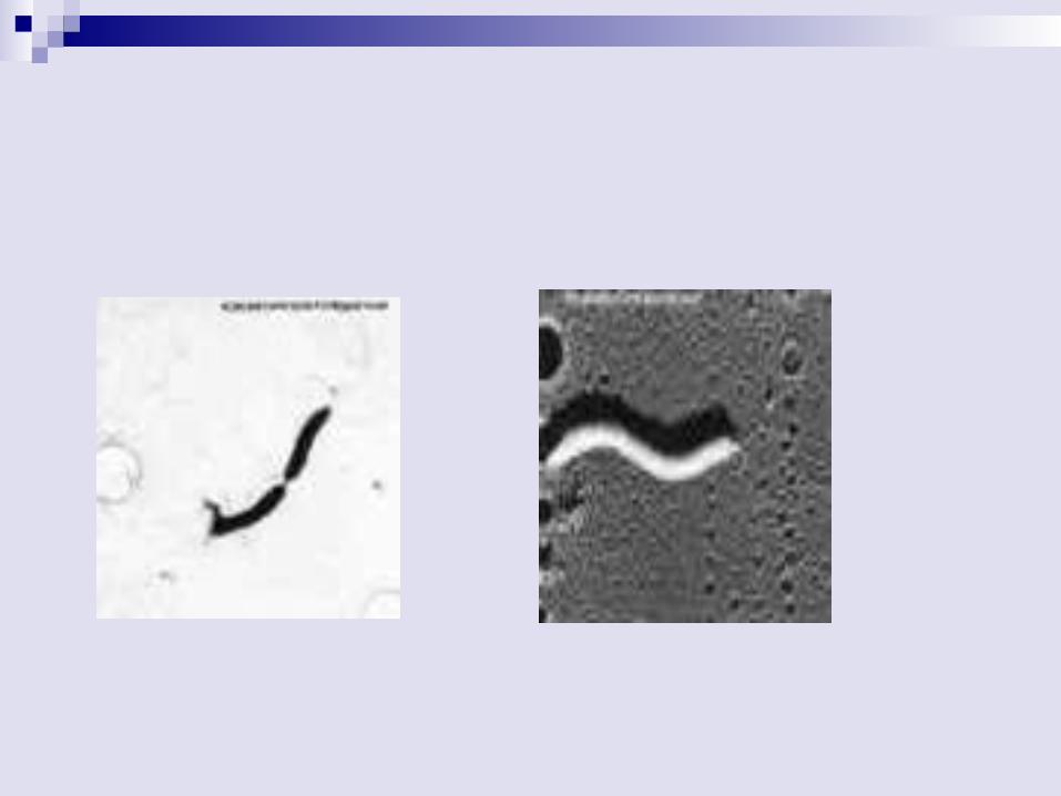
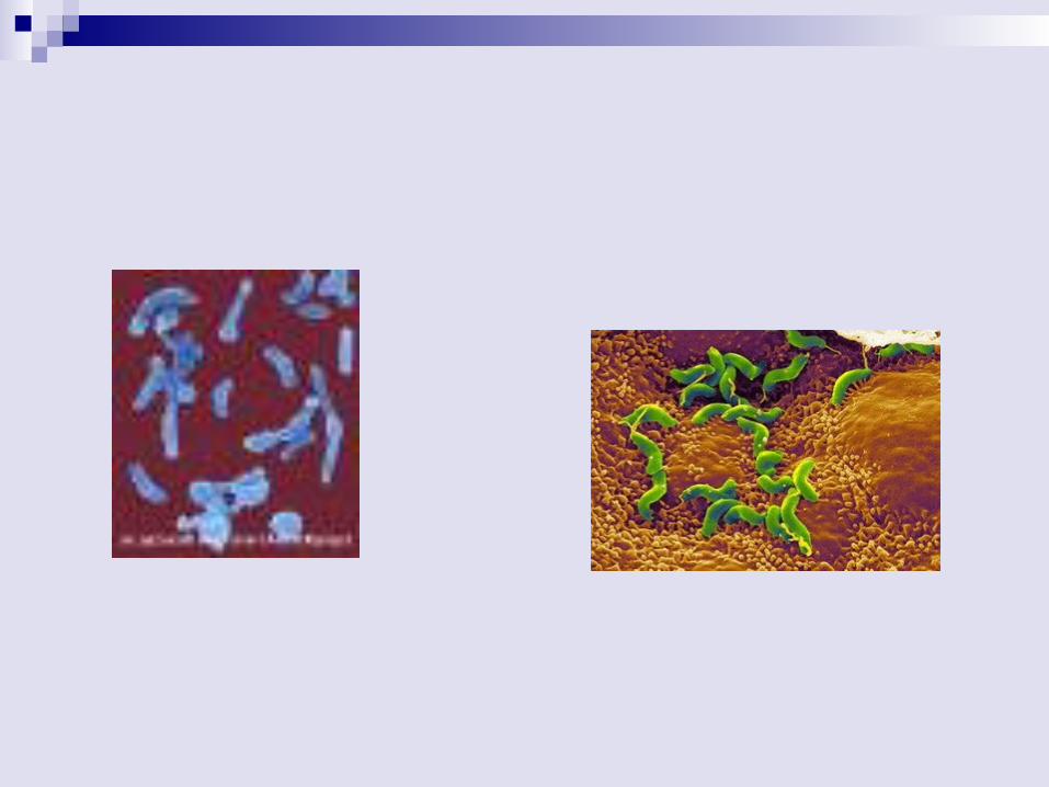
Helicobacter fact Perviou name:campylobacter pylori Gram negative bacilli. Curved cells discovered in 1983 in stomach
biopsied specimens. Causes 90% of stomach & duodenal
ulcers. Produces large amounts of urease. Infection common especially in lower
socioeconomic class/developing nations. Motile with multiple polarflagella.
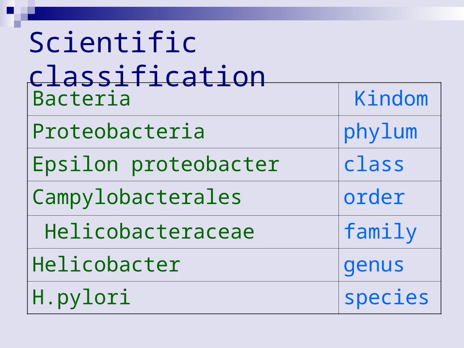
Scientific classificationKindom Bacteria
phylumProteobacteria
classEpsilon proteobacter
orderCampylobacterales
familyHelicobacteraceae
genusHelicobacter
speciesH.pylori
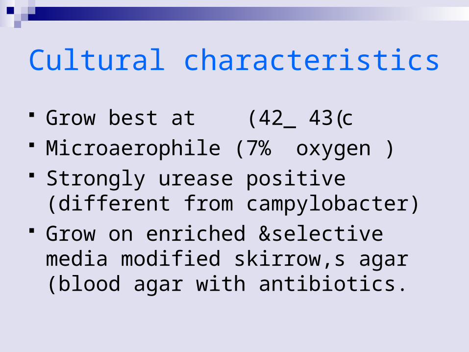
Cultural characteristics
Grow best at (42_ 43(c Microaerophile (7% oxygen ) Strongly urease positive (different from
campylobacter) Grow on enriched &selective media
modified skirrow,s agar (blood agar with antibiotics.

Habitat The bacteria are found everywhere in the
world It lives in the interface between the surface
of gastric epithelial cells The infection is rare in children, only about 50% of adults more than 60
years are infected.
Transmission (person to person)
H. pylori infection is most likely acquired by ingesting contaminated food and water
through person to person contact (oral _oral).
Person-to-person spread via fecal-oral rout.
Diseases by h.pylori
Gastritis (the irritation and inflammation of the lining of the stomach)
Gastric cancer (characterized by sores that form in the stomach or the upper part of the small intestine,
One out of every six patients with H. pylori infection will develop ulcers of the duodenum or stomach.
Clinical features In children, symptoms of gastritis may
include nausea, vomiting and frequent complaints about pain in the abdomen.
In older children and adults, the most
common symptom of peptic ulcer disease is a gnawing or burning pain in the abdomen.
Children who have peptic ulcer disease can have ulcers that bleed, causing
hematemesis (bloody vomit or vomit that looks like coffee grounds) or
melena (stool that's black, bloody, or looks like tar).
Diagnosis
H. pylori infection can be confirmed by invasive or noninvasive methods. Invasive tests require upper esophagogastroduodenal (EGD) endoscopy, which is considered the reference method of diagnosis. During endoscopy, biopsy specimens of the stomach and duodenum are obtained, and. If possible, noninvasive testing is done before tissue testing.
Serological testing has been the mainstay of H. pylori diagnosis, particularly in primary care, due to the accessibility, rapid results and low cost of this testing method.
Blood antibody tests may be good for diagnosing infection, but they are not good for determining if antibiotics have successfully eradicated the bacterium
Lab Identification Accurate and simple tests for the
detection of H. pylori infection are available. They include blood antibody tests, urea breath tests, stool antigen tests, and endoscopic biopsies.
specimenGastric biopsy
The diagnosis of H. pylori can be made by, 1. Culture2. Urease test 3. Methylene blue stain4. Giemsa stain
5. Fish technique
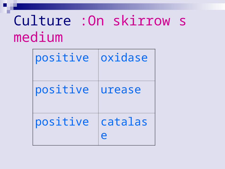
Culture :On skirrow s medium
oxidasepositive
ureasepositive
catalasepositive
Blood antibody tests Serological assays (blood serum) measure
specific H. pylori immunoglobulin G (IgG) antibodies that can determine if an individual has been infected.
The sensitivity and specificity of these assays range from 80–95%, depending on the assay used.
Urea breath test
The urea breath test (UBT) is a safe, easy, and accurate test for the presence of H. pylori in the stomach
The breath test relies on the ability of H. pylori to break down the naturally occurring chemical, urea, into carbon dioxide which is absorbed from the stomach and eliminated from the body in the breath.
Ten to 20 minutes after swallowing a capsule containing a minute amount of radioactive urea, a breath sample is collected and analyzed .
The presence of radioactive carbon dioxide in the breath (a positive test) means that there is active infection.
Has good sensitiviity &specificity.
stool antigen tests H. pylori stool antigen (HpSA) testing is based
on monoclonal antibody immunochromatography of stool samples.
This testing method identifies active infection and can be used to detect eradication after treatment.
A sensitivity and specificity range of 92–98% is reported in the literature for stool antigen testing.
endoscopic biopsies. Endoscopy is an accurate test for
diagnosing H. pylori as well as the inflammation and ulcers that it causes.
For endoscopy, the doctor inserts a flexible viewing tube (endoscope) through the mouth, down the esophagus, and into the stomach and duodenum.
A biopsy specimen is placed on a special slide containing urea (e.g., CLO test slides).
If the urea is broken down by H. pylori in the biopsy, there is a change in color around the biopsy on the slide.
This means that there is an infection with H. pylori in the stomach .

preventation
Right now, there's no vaccine against H. pylori. And because transmission isn't clearly understood, prevention guidelines aren't yet available. However, it's always important to make sure you and your family:
Wash your hand thoroughly. Eat food that's been properly prepared. Drink water from a safe source.


Treatment
Treatment of the infection involves theadministration of anti-microbial substance course of 14 days combined with bismuth salt.
Therapy with tetracycline, metronidazole, azithromycine, bismuth (Peptobismol).
Triple therapy (metronidazole + clarythromycin or amoxycillin+omeprazole)