Embed Size (px)
Citation preview
Hematuria
Urothelial Tumours
Renal Masses
Urolithiasis
UBC Department of Urologic Sciences Lecture Series
Disclaimer:
• This is a lot of information to cover and we are unlikely to cover it all today
• These slides are to be utilized for your reference to guide your self study
MCC Objectives
http://mcc.ca/examinations/objectives-overview/
For LMCC Part 1
Objectives applicable to this lecture: – Blood in Urine/Hematuria
Objectives
Hematuria: 1. To provide a framework for you to understand
the initial workup and management of patients presenting with hematuria
2. To discuss some common pathologies associated with hematuria and their risk factors, and management
1. Urothelial tumours
2. Renal Tumours
3. Urolithiasis
Hematuria
• 65 year old male presents with a 2 day history of gross painless hematuria.
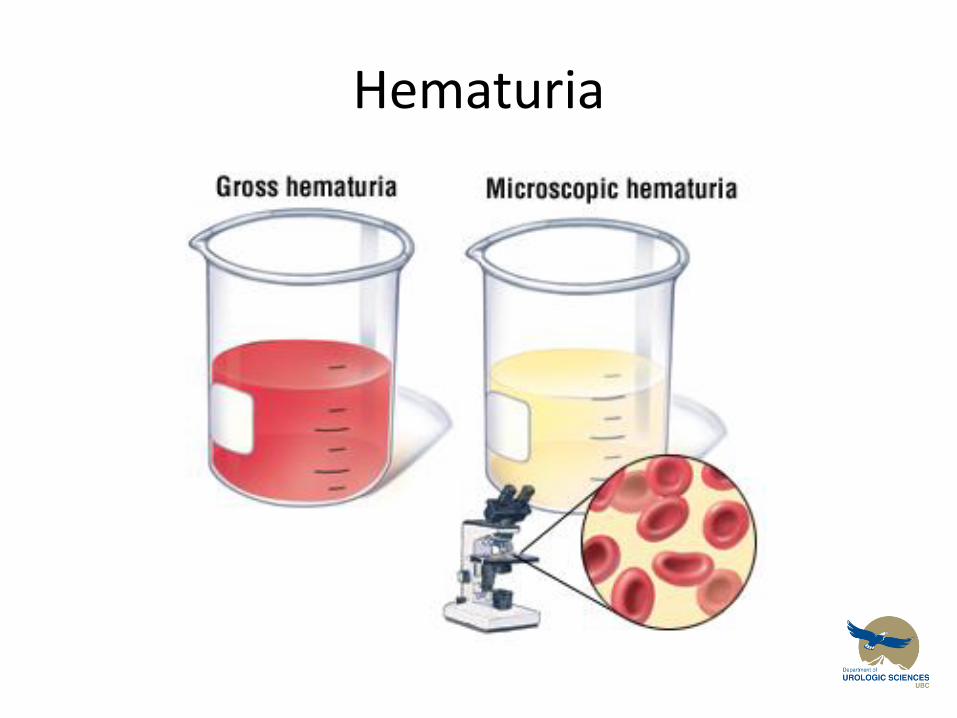
Hematuria
• 65 year old male presents with a 2 day history of gross painless hematuria.
• Important Points to Know – How to take a history in patient with hematuria
• What are the most common causes?
• Risk factors that this could be something worrisome?
– How do I work this patient up?
– When should I refer to a urologist?
Hematuria
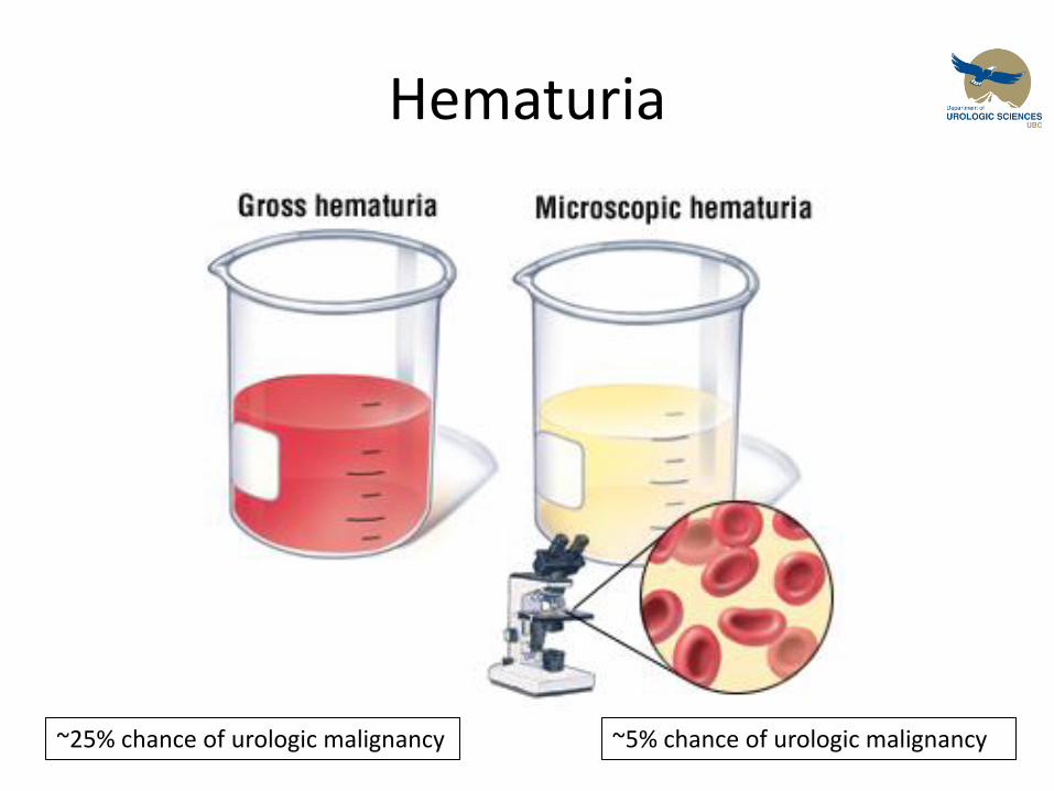
Hematuria
~25% chance of urologic malignancy ~5% chance of urologic malignancy
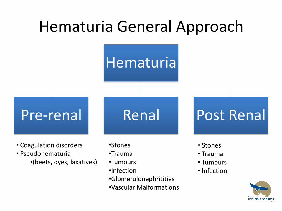
Hematuria General Approach
Hematuria
Pre-renal Renal Post Renal
• Coagulation disorders • Pseudohematuria
•(beets, dyes, laxatives)
•Stones •Trauma •Tumours •Infection •Glomerulonephritities •Vascular Malformations
• Stones • Trauma • Tumours • Infection
Hematuria
• Take home message #1
– The most common urologic causes of hematuria
• Stones
• Trauma
• Tumour
• Infections
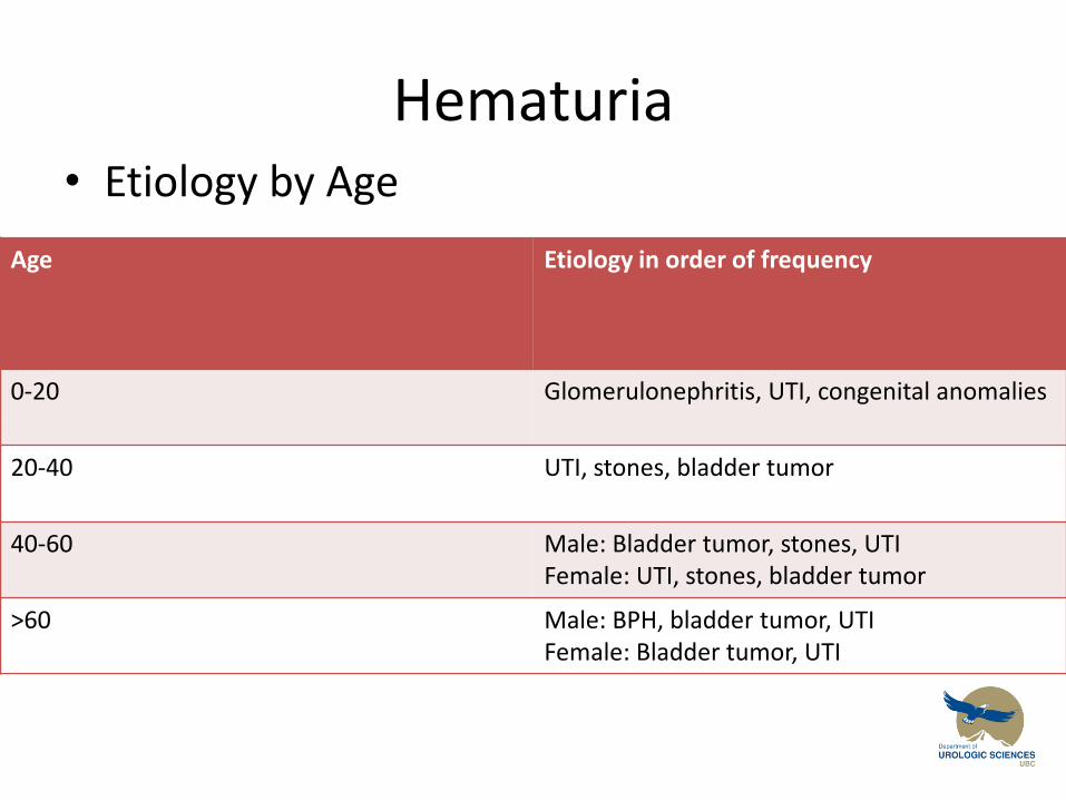
Hematuria • Etiology by Age
Age Etiology in order of frequency
0-20 Glomerulonephritis, UTI, congenital anomalies
20-40 UTI, stones, bladder tumor
40-60 Male: Bladder tumor, stones, UTI Female: UTI, stones, bladder tumor
>60 Male: BPH, bladder tumor, UTI Female: Bladder tumor, UTI
Hematuria History
• What questions should you ask this patient? – Stone
• Flank/Abdominal pain, dysuria, previous stones
– Trauma • Recent encounters with Chuck Norris
– Tumour • Weight loss, night sweats, flank pain, voiding changes
• Risk factors ????
– Infection • Suprapubic pain, dysuria, frequency, fever/chills +/- flank
pain
Risk Factors for Urothelial Tumours
• Smoking
• Smoking
• Smoking • Occupational exposures: Aniline dyes
– Hairdressers, leather tanners, textile workers, painters, dry cleaners
• Medications – Phenacetin – older analgesic, common in australiasia
– Cyclophoshamide
• Previous radiation exposure
• Chronic cystitis: catheters, infections
Hematuria
• Take home message #2
– Gross, painless hematuria is a malignancy until proven otherwise
Hematuria
• Take home message #2
– Gross, painless hematuria is a malignancy until proven otherwise!
• Stones, trauma, infections usually are symptomatic
• Anticoagulation / coagulopathy are not sufficient reasons for gross hematuria
Hematuria
• 65 year old male presents with a 2 day history of gross painless hematuria. He is a long term 2 pack per day smoker. He works as a hairdresser part-time, and part-time at a dry cleaners.
Hematuria
• 65 year old male presents with a 2 day history of gross painless hematuria. He is a long term 2 pack per day smoker. He works as a hairdresser part-time, and part-time at a dry cleaners
– We have determined this man significant enough risk that he requires a work up….
• But how
Hematuria Investigations
Laboratory Investigations:
1. U/A and culture – Leukocytes, Nitrites – Infection – R&M – if dysmorphic RBC’s +/- Protein = Glomerular cause, crystals stones – C&S – Infection
2. Urinary Cytology – Sensitivity and Specificity depend on grade of malignancy and number of specimens sampled
3. CBC
– Hgb - severity of blood loss – WBC – infection – Platelet loss/coagulopathy
4. Creatinine
– Renal impairment
5. INR/PTT
– Coagulopathy
Hematuria Investigations
Radiology Investigations
• Options for Imaging the Urinary Tract
– Ultrasound
– CT IVP
– MRI
– Intravenous pyelogram
Hematuria Investigations
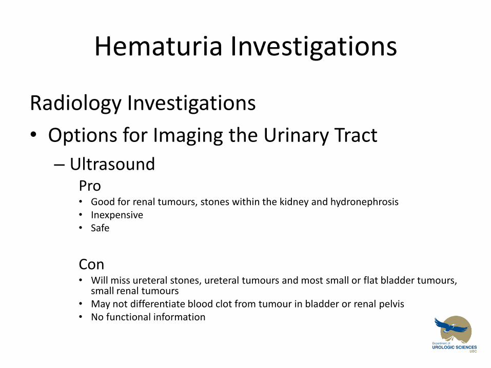
Radiology Investigations
• Options for Imaging the Urinary Tract
– Ultrasound Pro
• Good for renal tumours, stones within the kidney and hydronephrosis • Inexpensive • Safe
Con
• Will miss ureteral stones, ureteral tumours and most small or flat bladder tumours, small renal tumours
• May not differentiate blood clot from tumour in bladder or renal pelvis • No functional information
Hematuria Investigations
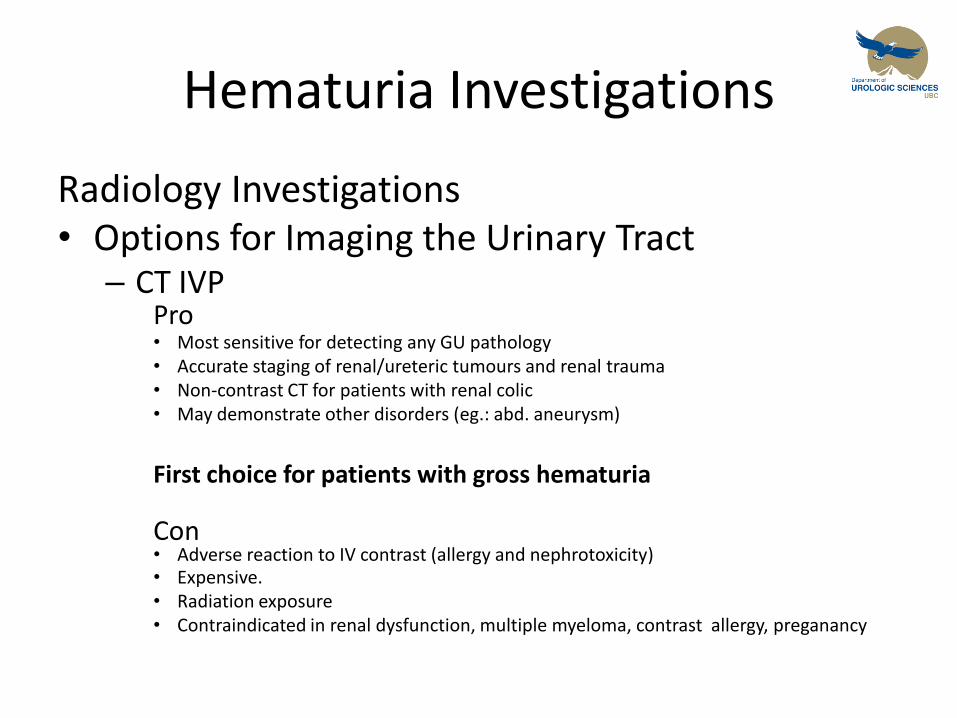
Radiology Investigations • Options for Imaging the Urinary Tract
– CT IVP Pro
• Most sensitive for detecting any GU pathology • Accurate staging of renal/ureteric tumours and renal trauma • Non-contrast CT for patients with renal colic • May demonstrate other disorders (eg.: abd. aneurysm)
First choice for patients with gross hematuria
Con
• Adverse reaction to IV contrast (allergy and nephrotoxicity) • Expensive. • Radiation exposure • Contraindicated in renal dysfunction, multiple myeloma, contrast allergy, preganancy
Hematuria Investigations
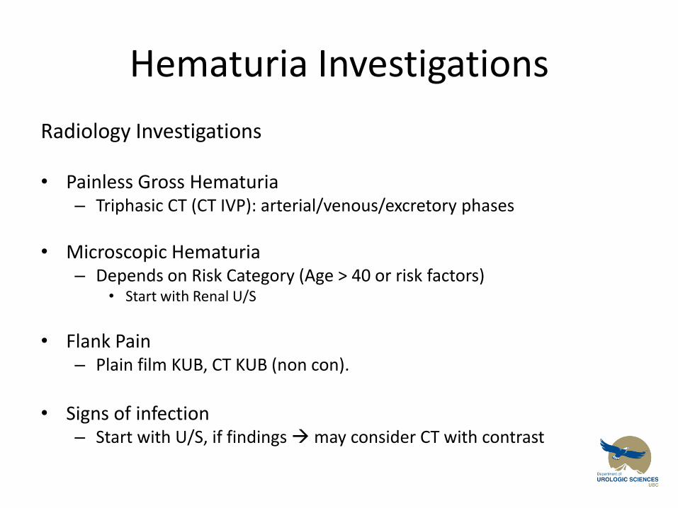
Radiology Investigations
• Painless Gross Hematuria – Triphasic CT (CT IVP): arterial/venous/excretory phases
• Microscopic Hematuria – Depends on Risk Category (Age > 40 or risk factors)
• Start with Renal U/S
• Flank Pain – Plain film KUB, CT KUB (non con).
• Signs of infection
– Start with U/S, if findings may consider CT with contrast
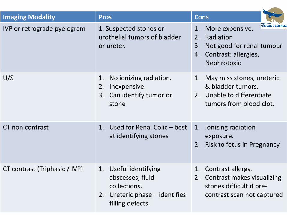
Hematuria Imaging Modality Pros Cons
IVP or retrograde pyelogram 1. Suspected stones or urothelial tumors of bladder or ureter.
1. More expensive. 2. Radiation 3. Not good for renal tumour 4. Contrast: allergies,
Nephrotoxic
U/S 1. No ionizing radiation. 2. Inexpensive. 3. Can identify tumor or
stone
1. May miss stones, ureteric & bladder tumors.
2. Unable to differentiate tumors from blood clot.
CT non contrast 1. Used for Renal Colic – best at identifying stones
1. Ionizing radiation exposure.
2. Risk to fetus in Pregnancy
CT contrast (Triphasic / IVP) 1. Useful identifying abscesses, fluid collections.
2. Ureteric phase – identifies filling defects.
1. Contrast allergy. 2. Contrast makes visualizing
stones difficult if pre-contrast scan not captured
Hematuria Referral
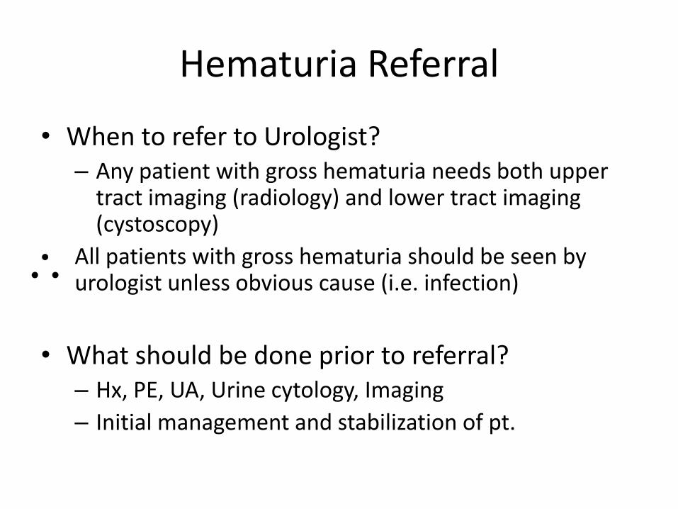
• When to refer to Urologist? – Any patient with gross hematuria needs both upper
tract imaging (radiology) and lower tract imaging (cystoscopy)
All patients with gross hematuria should be seen by urologist unless obvious cause (i.e. infection)
• What should be done prior to referral? – Hx, PE, UA, Urine cytology, Imaging
– Initial management and stabilization of pt.
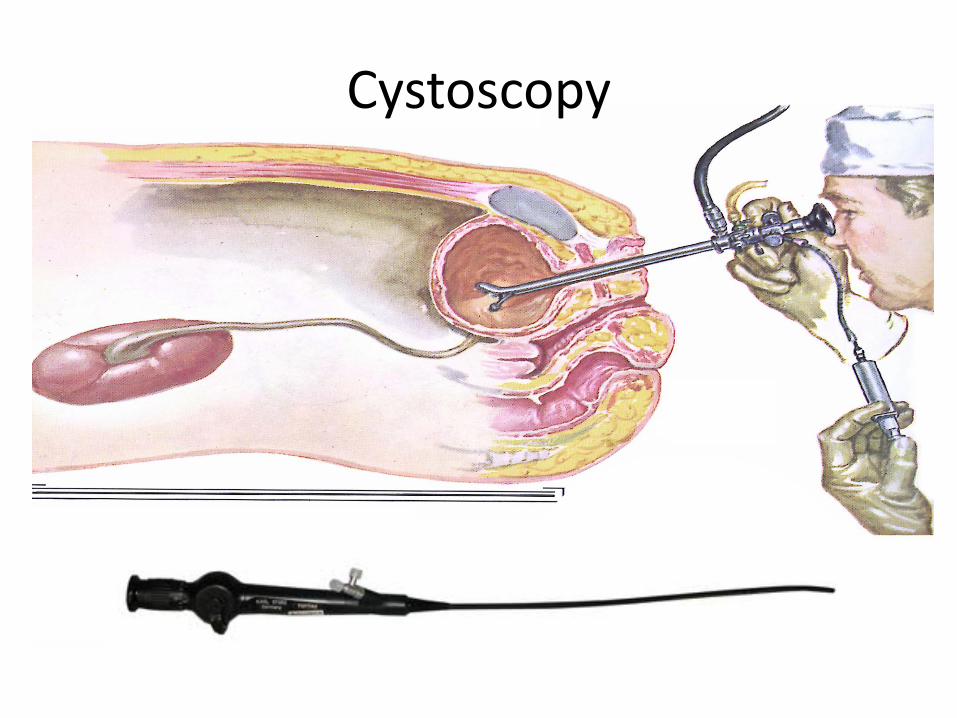
Cystoscopy
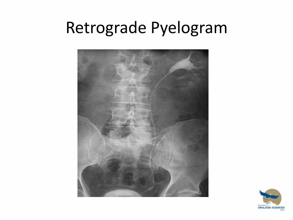
Retrograde Pyelogram
Hematuria
• Suggested learning resource
Canadian guidelines for the management of asymptomatic hematuria in adults
http://www.cua.org/userfiles/files/guidelines/amh_2008_en.pdf
Hematuria: Acutely Bleeding Patient
• ABC’s. – Stabilize Pt, Blood products if needed
• Investigations to determine site of bleeding (upper tract vs. lower tract)
– Treatment based on underlying cause
• Continuous Bladder Irrigation – Manually irrigate all clots out of bladder first! – Call Urology
• Surgical management – Cystoscopy + Fulgaration – Intravesical therapies: Alum, formalin, silver nitrate – Hyperbaric Oxygen – Vascular embolization. – Cystectomy and Urinary diversion.
Hematuria Summary
1. Painless Gross Hematuria – Malignancy until proven otherwise
2. Stones, infections & trauma – Rarely asymptomatic History!!!!
3. Workup – Hx, PE – Lab: U/A, Urine C&S, urine cytology, CBC, Cr, INR/PTT – Imaging: CT or U/S – Referral to Urologist: gross hematuria, microhematuria
with risk factors (See CUA guidelines)or abnormal cytology
4. Management – Stabilize Pt, +/- CBI, +/- Surgical intervention
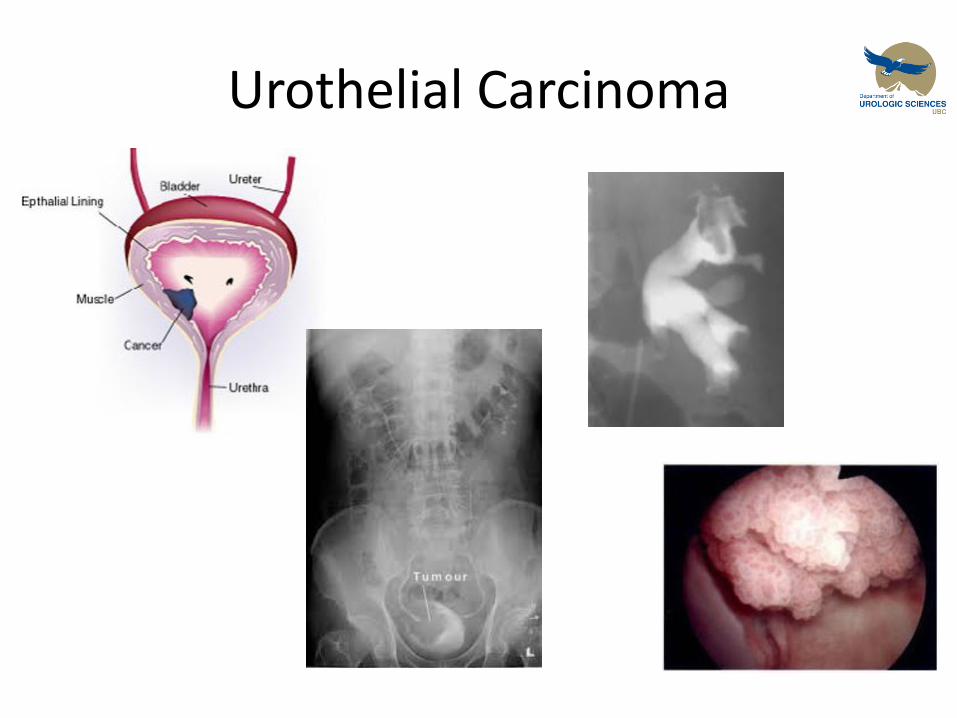
Urothelial Carcinoma
Objectives
Urothelial Carcinoma:
1. To provide a framework for you to understand the initial workup and management of patients diagnosed with urothelial malignancies
2. To discuss the classification of urothelial tumours by histological grade and stage, and the implications this has for treatment interventions
Urothelial Carcinoma
• 65 year old male presents with a 2 day history of gross painless hematuria. He is a long term 2 pack per day smoker. He works as a hairdresser part-time, and part-time at a dry cleaners UA: (+) RBC, (-) nitrites, (-) leuks
Urine culture negative
Urine cytology shows abnormal cells
Renal U/S normal
Next Step….
Urothelial Carcinoma
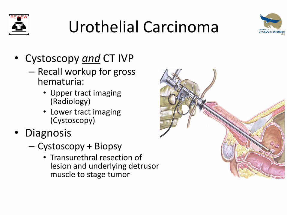
• Cystoscopy and CT IVP – Recall workup for gross
hematuria: • Upper tract imaging
(Radiology) • Lower tract imaging
(Cystoscopy)
• Diagnosis – Cystoscopy + Biopsy
• Transurethral resection of lesion and underlying detrusor muscle to stage tumor
Risk Factors for Urothelial Tumours
• Smoking
• Smoking
• Smoking • Occupational exposures: Aniline dyes
– Hairdressers, leather tanners, textile workers, painters, dry cleaners
• Medications – Phenacetin – older analgesic, common in australiasia
– Cyclophoshamide
• Previous radiation exposure
• Chronic cystitis: catheters, infections
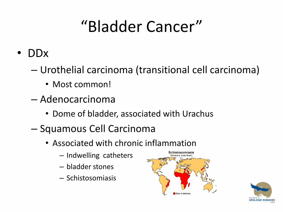
“Bladder Cancer”
• DDx
– Urothelial carcinoma (transitional cell carcinoma)
• Most common!
– Adenocarcinoma
• Dome of bladder, associated with Urachus
– Squamous Cell Carcinoma
• Associated with chronic inflammation – Indwelling catheters
– bladder stones
– Schistosomiasis
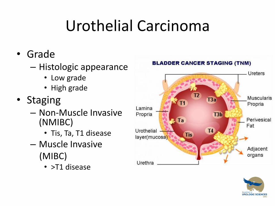
Urothelial Carcinoma
• Grade – Histologic appearance
• Low grade • High grade
• Staging – Non-Muscle Invasive
(NMIBC) • Tis, Ta, T1 disease
– Muscle Invasive (MIBC)
• >T1 disease
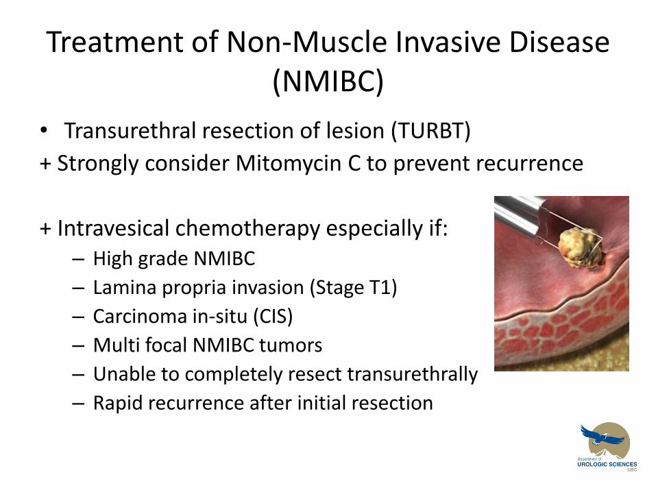
Treatment of Non-Muscle Invasive Disease (NMIBC)
• Transurethral resection of lesion (TURBT)
+ Strongly consider Mitomycin C to prevent recurrence
+ Intravesical chemotherapy especially if: – High grade NMIBC
– Lamina propria invasion (Stage T1)
– Carcinoma in-situ (CIS)
– Multi focal NMIBC tumors
– Unable to completely resect transurethrally
– Rapid recurrence after initial resection
Treatment of NMIBC
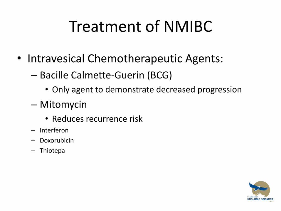
• Intravesical Chemotherapeutic Agents:
– Bacille Calmette-Guerin (BCG)
• Only agent to demonstrate decreased progression
– Mitomycin
• Reduces recurrence risk – Interferon
– Doxorubicin
– Thiotepa
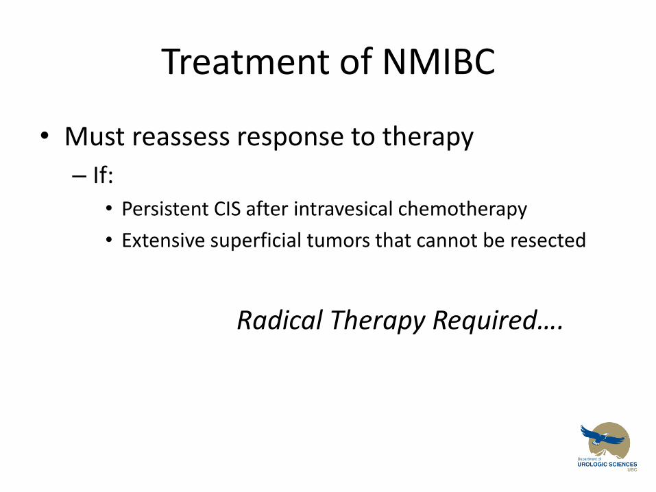
Treatment of NMIBC
• Must reassess response to therapy
– If:
• Persistent CIS after intravesical chemotherapy
• Extensive superficial tumors that cannot be resected
Radical Therapy Required….
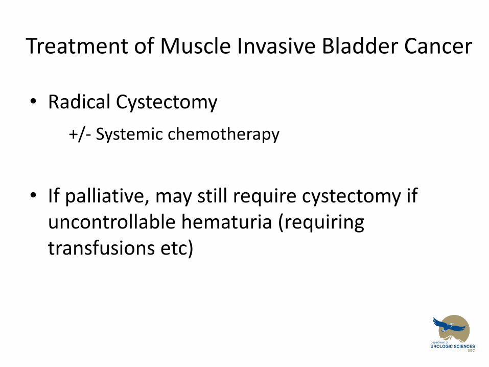
Treatment of Muscle Invasive Bladder Cancer
• Radical Cystectomy
+/- Systemic chemotherapy
• If palliative, may still require cystectomy if uncontrollable hematuria (requiring transfusions etc)
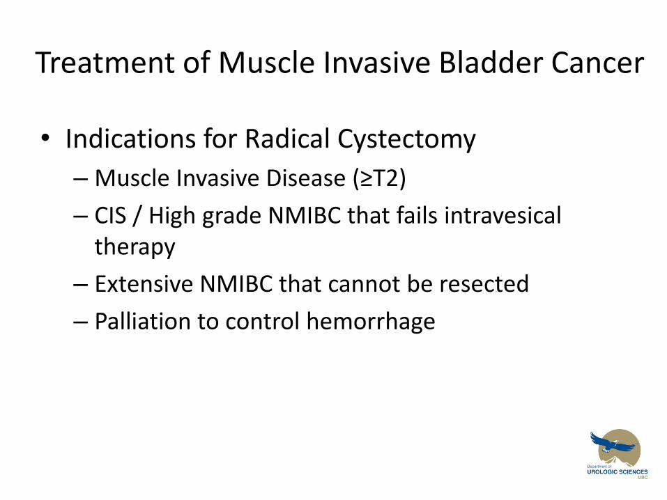
Treatment of Muscle Invasive Bladder Cancer
• Indications for Radical Cystectomy
– Muscle Invasive Disease (≥T2)
– CIS / High grade NMIBC that fails intravesical therapy
– Extensive NMIBC that cannot be resected
– Palliation to control hemorrhage
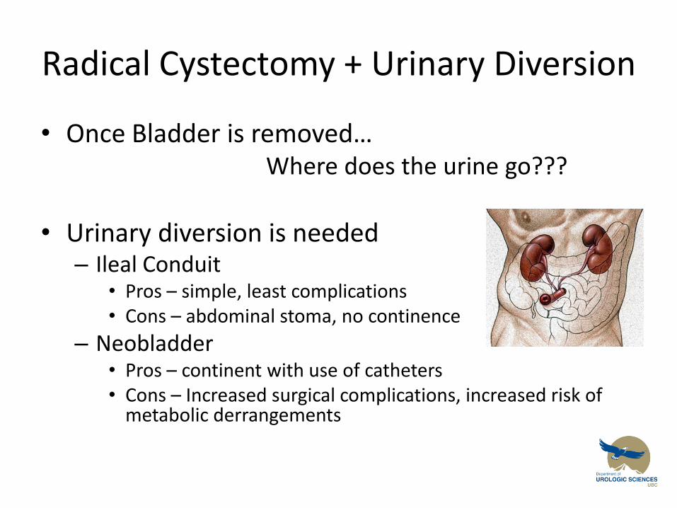
Radical Cystectomy + Urinary Diversion
• Once Bladder is removed… Where does the urine go???
• Urinary diversion is needed
– Ileal Conduit • Pros – simple, least complications • Cons – abdominal stoma, no continence
– Neobladder • Pros – continent with use of catheters • Cons – Increased surgical complications, increased risk of
metabolic derrangements
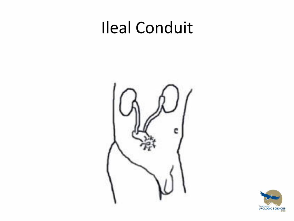
Ileal Conduit
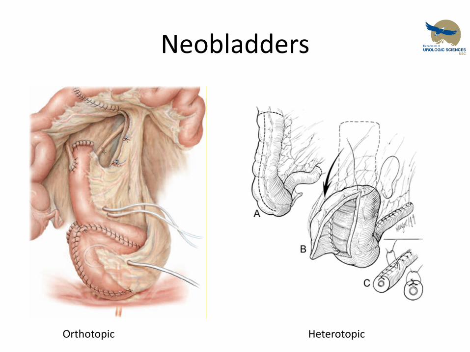
Neobladders
Orthotopic Heterotopic
Chemotherapy for Urothelial Carcinoma
• Gemcitabine / Cisplatin most common
• MVAC (methotrexate / vinblastine / adriamycin / cisplatin)
• 5% Survival benefit at 5 years if given neoadjuvant
• Adjuvant benefit less clear
Objectives
Urothelial Carcinoma:
1. To provide a framework for you to understand the initial workup and management of patients diagnosed with urothelial malignancies
2. To discuss the classification of urothelial tumours by histological grade and stage, and the implications this has for treatment interventions
Renal Mass
Objectives
Renal Mass
1. Give a differential diagnosis for a solid mass in the kidney
2. Describe the evaluation of a patient with a suspected renal cell carcinoma
3. Give three indications for a partial nephrectomy rather than a radical nephrectomy for renal cell carcinoma
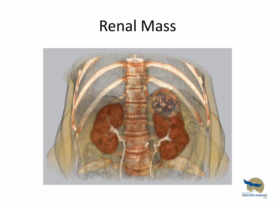
Renal Mass
• 65 year old male presents with a 2 day history of gross painless hematuria. He said he had some vague flank pain a few weeks ago. He has never smoked, and works as an accountant
UA: (+) RBC, (-) nitrites, (-) leuks
Urine culture negative
Urine cytology normal
Ultrasound showed a mass in left kidney
Next Step….
Renal Mass
• Presentation:
– Typically incidental finding!
– Classic Triad:
• Flank pain, hematuria, palpable mass (uncommon)
• How do you ‘work-up’ a Renal mass?
Need to think about your differential diagnosis…
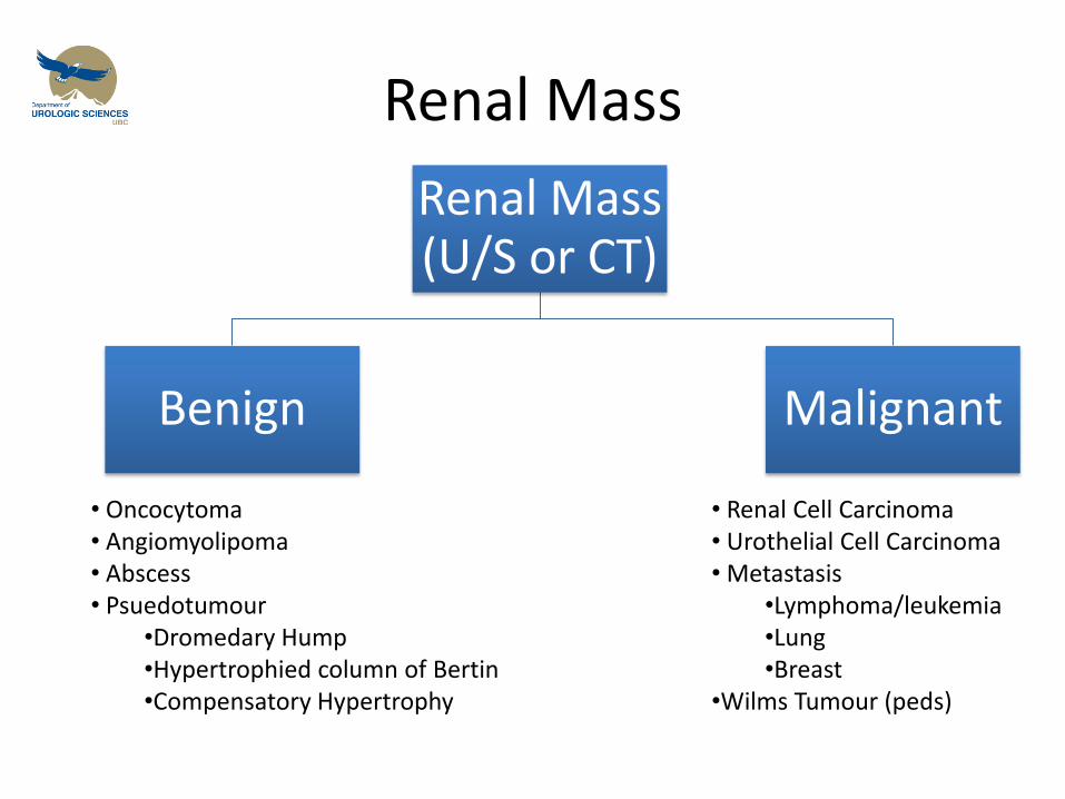
Renal Mass
Renal Mass (U/S or CT)
Benign Malignant
• Oncocytoma • Angiomyolipoma • Abscess • Psuedotumour
•Dromedary Hump •Hypertrophied column of Bertin •Compensatory Hypertrophy
• Renal Cell Carcinoma • Urothelial Cell Carcinoma • Metastasis
•Lymphoma/leukemia •Lung •Breast
•Wilms Tumour (peds)
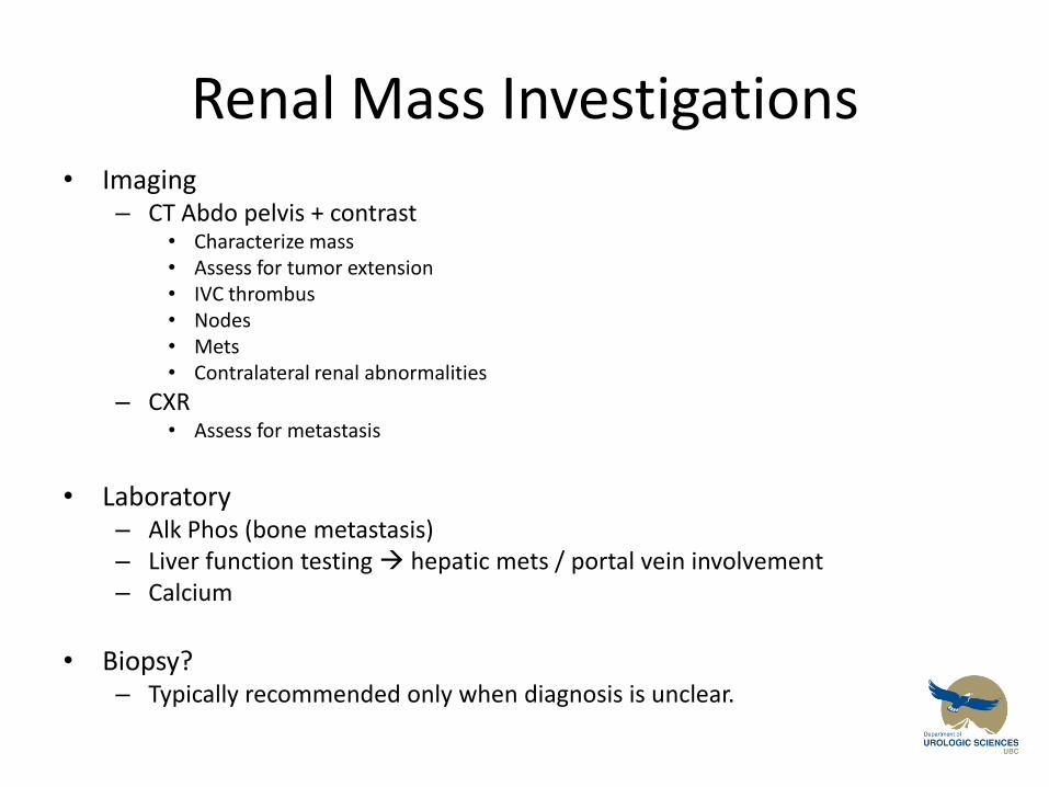
Renal Mass Investigations • Imaging
– CT Abdo pelvis + contrast • Characterize mass • Assess for tumor extension • IVC thrombus • Nodes • Mets • Contralateral renal abnormalities
– CXR • Assess for metastasis
• Laboratory
– Alk Phos (bone metastasis) – Liver function testing hepatic mets / portal vein involvement – Calcium
• Biopsy?
– Typically recommended only when diagnosis is unclear.
Why Investigate Calcium?
• Bone Mets or Paraneoplastic syndrome! – 20-30% of RCC have Paraneoplastic Syndrome
• Increased ESR
• Wt loss, cachexia
• Fever
• Anemia
• Hypertension (Due to increased Renin)
• Hypercalcemia (PTH-like Substance)
• Increased AlkPhos
• Polycythemia (increased EPO production)
• Stauffer’s syndrome – abnormal liver enzymes - reversible
Benign Renal Masses
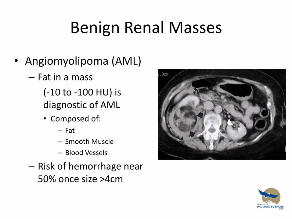
• Angiomyolipoma (AML)
– Fat in a mass
(-10 to -100 HU) is diagnostic of AML
• Composed of:
– Fat
– Smooth Muscle
– Blood Vessels
– Risk of hemorrhage near 50% once size >4cm
Benign Tumors
• Know that they exist.
• DDx: – Oncocytoma – angiomyolipoma (1-2% malignant) – papillary adenoma – pseudotumors etc….
• Differentiating pseudotumors from real tumors.
– DMSA scan • Pseudotumors will have normal uptake, tumors will be decreased
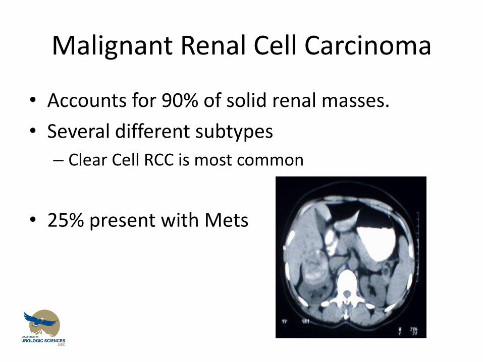
Malignant Renal Cell Carcinoma
• Accounts for 90% of solid renal masses.
• Several different subtypes
– Clear Cell RCC is most common
• 25% present with Mets
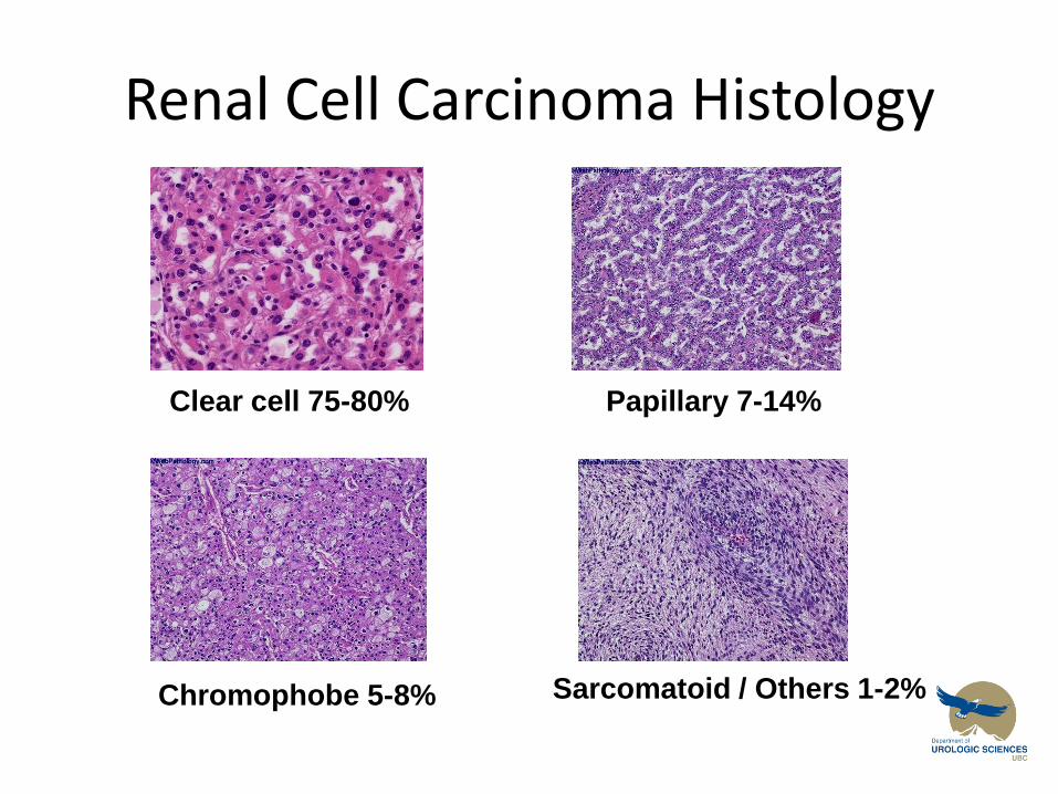
Renal Cell Carcinoma Histology
Sarcomatoid / Others 1-2%
Papillary 7-14%
Chromophobe 5-8%
Clear cell 75-80%
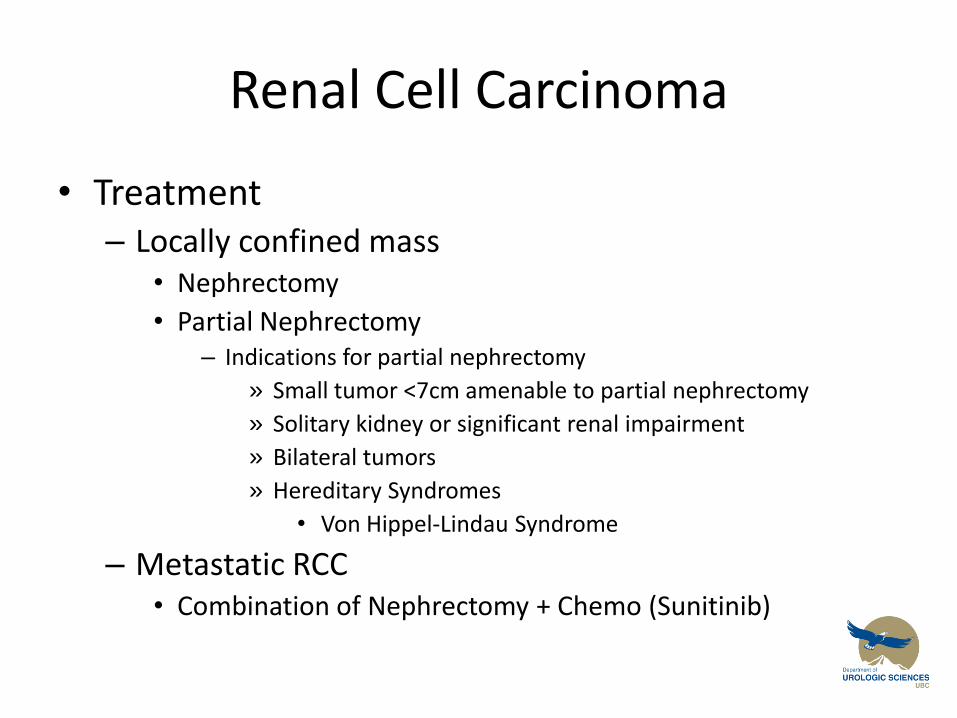
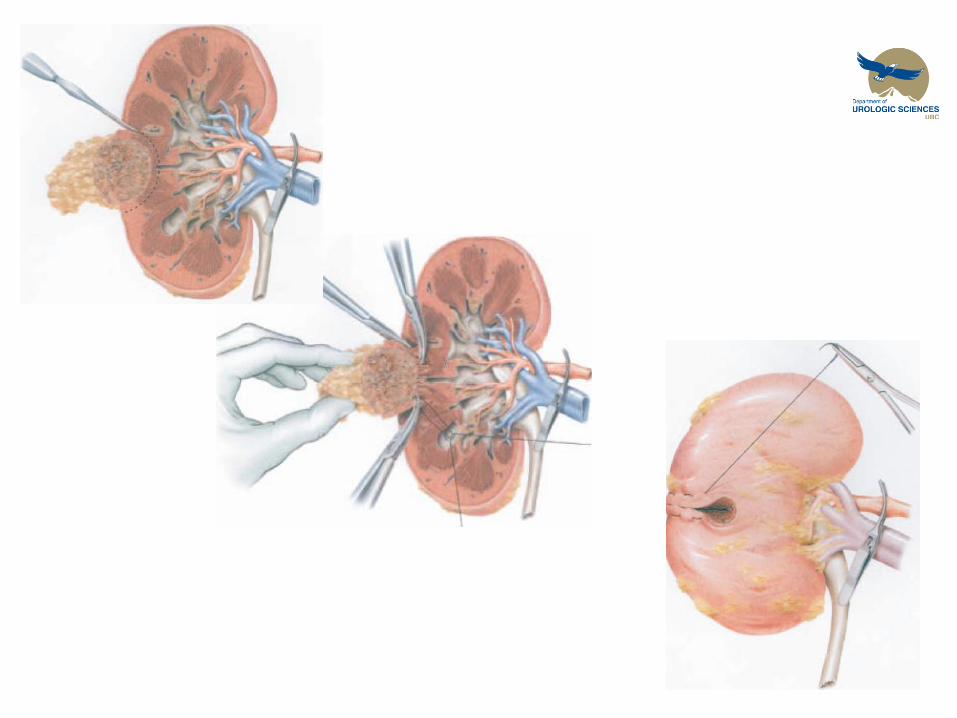
Renal Cell Carcinoma
• Treatment – Locally confined mass
• Nephrectomy
• Partial Nephrectomy – Indications for partial nephrectomy
» Small tumor <7cm amenable to partial nephrectomy
» Solitary kidney or significant renal impairment
» Bilateral tumors
» Hereditary Syndromes
• Von Hippel-Lindau Syndrome
– Metastatic RCC • Combination of Nephrectomy + Chemo (Sunitinib)
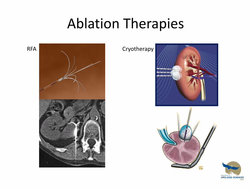
Ablation Therapies
RFA Cryotherapy
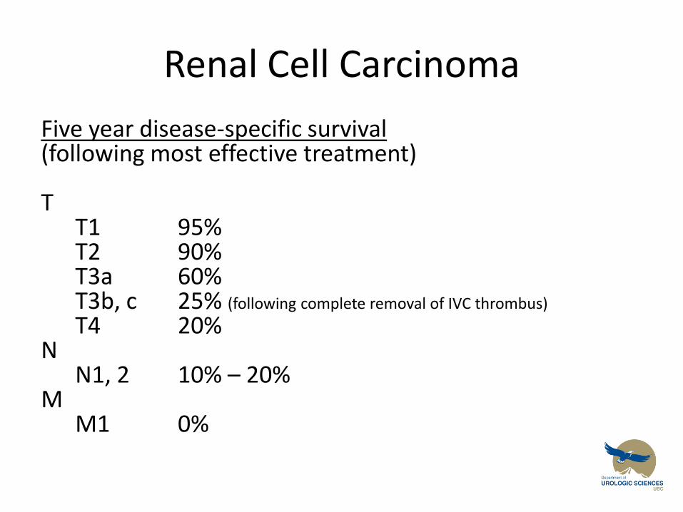
Renal Cell Carcinoma
Five year disease-specific survival (following most effective treatment) T T1 95% T2 90% T3a 60% T3b, c 25% (following complete removal of IVC thrombus)
T4 20% N N1, 2 10% – 20% M M1 0%
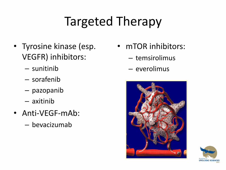
Targeted Therapy
• Tyrosine kinase (esp. VEGFR) inhibitors:
– sunitinib
– sorafenib
– pazopanib
– axitinib
• Anti-VEGF-mAb:
– bevacizumab
• mTOR inhibitors:
– temsirolimus
– everolimus
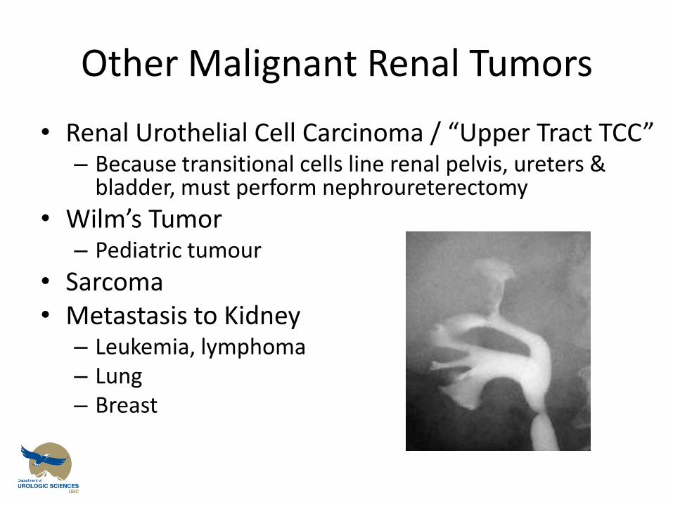
Other Malignant Renal Tumors
• Renal Urothelial Cell Carcinoma / “Upper Tract TCC” – Because transitional cells line renal pelvis, ureters &
bladder, must perform nephroureterectomy
• Wilm’s Tumor – Pediatric tumour
• Sarcoma • Metastasis to Kidney
– Leukemia, lymphoma – Lung – Breast
Objectives
Renal Mass
1. Give a differential diagnosis for a solid mass in the kidney
2. Describe the evaluation of a patient with a suspected renal cell carcinoma
3. Give three indications for a partial nephrectomy rather than a radical nephrectomy for renal cell carcinoma
Renal Mass
• Learning Resources – Canadian Consensus: Management of kidney
cancer: Canadian kidney cancer forum 2008 Consensus statement
https://www.kidneycancercanada.ca/media/files/81.pdf
– Canadian Consensus: Management of advanced kidney cancer: Canadian kidney cancer forum 2013 Consensus Update
https://www.kidneycancercanada.ca/media/886673/KCRNC%20mRCC%20Consensus%202013%20CUAJ%202013.pdf
Stones
Renal Colic
Objectives
1. Give a differential diagnosis for acute flank pain including two life-threatening conditions
2. Describe the laboratory and radiologic evaluation of a patient with renal colic
3. Know 4 different kinds of kidney stones and the risk factors for stone formation
4. Know 3 indications for emergency drainage of an obstructed kidney
Renal Colic
• 65 year old male presents with a 2 day history of gross hematuria with significant left sided flank pain. He has never smoked, and works as an accountant
UA: (+) RBC, (-) nitrites, (+) leuks
Urine culture negative
Next Step….
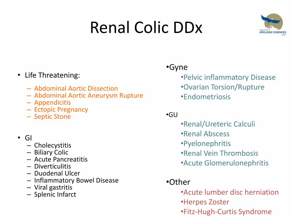
Renal Colic DDx
• Life Threatening: – Abdominal Aortic Dissection – Abdominal Aortic Aneurysm Rupture – Appendicitis – Ectopic Pregnancy – Septic Stone
• GI – Cholecystitis – Biliary Colic – Acute Pancreatitis – Diverticulitis – Duodenal Ulcer – Inflammatory Bowel Disease – Viral gastritis – Splenic Infarct
•Gyne •Pelvic inflammatory Disease •Ovarian Torsion/Rupture •Endometriosis
•GU
•Renal/Ureteric Calculi •Renal Abscess •Pyelonephritis •Renal Vein Thrombosis •Acute Glomerulonephritis
•Other •Acute lumber disc herniation •Herpes Zoster •Fitz-Hugh-Curtis Syndrome
Renal Colic Investigations
What investigations would you like to order….
Acute Renal Colic Investigations
• CBC – WBC – increased indicates inflammation or
infection
• Creatinine – Assess for impaired renal function (obstruction)
• Urine Microscopy – Bacteriuria, pyuria, pH
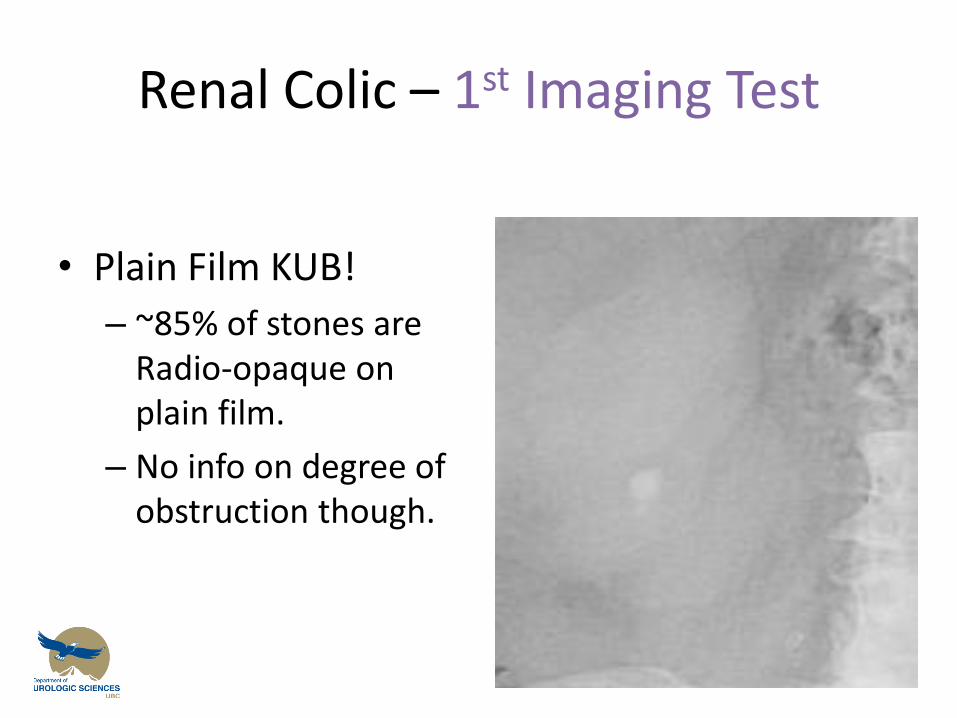
Renal Colic – 1st Imaging Test
• Plain Film KUB!
– ~85% of stones are Radio-opaque on plain film.
– No info on degree of obstruction though.
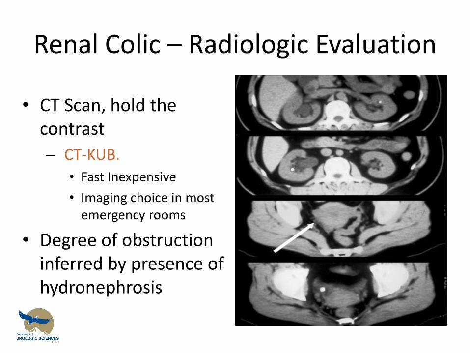
Renal Colic – Radiologic Evaluation
• CT Scan, hold the contrast
– CT-KUB.
• Fast Inexpensive
• Imaging choice in most emergency rooms
• Degree of obstruction inferred by presence of hydronephrosis
Stones - Factoids
• They are common! – Lifetime risk in North American Male is 1 in 8
– M:F ratio is 3:1
• Presenting complaint – Renal colic caused by acute obstruction of ureter by stone
• Initial Evaluation – Focuses on excluding other potential causes of abdominal
or flank pain
• Non-obstructing stones – Should not cause pain unless they are associated with
Urinary tract infection
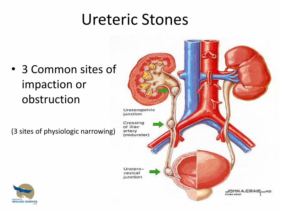
Ureteric Stones
• 3 Common sites of impaction or obstruction
(3 sites of physiologic narrowing)
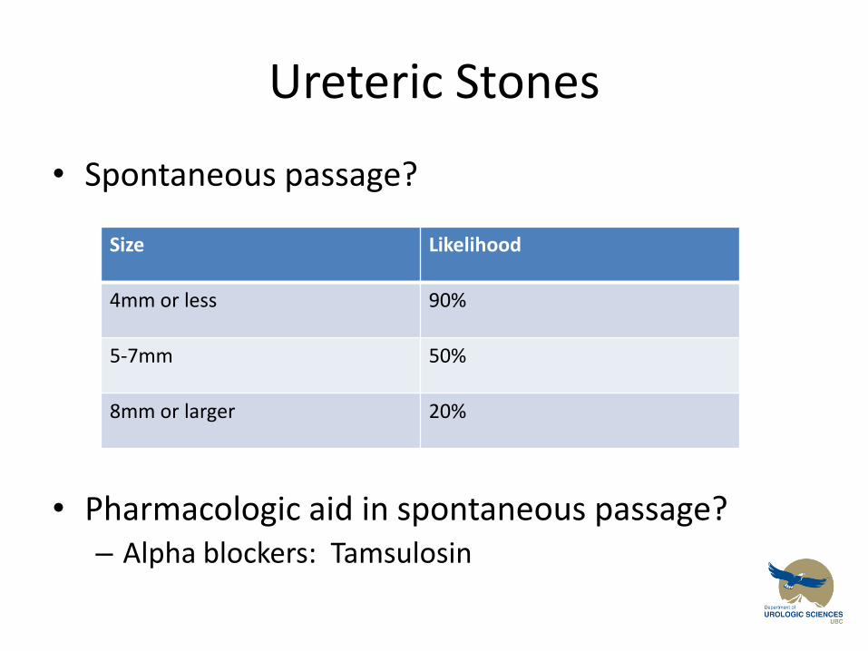
Ureteric Stones
• Spontaneous passage?
• Pharmacologic aid in spontaneous passage? – Alpha blockers: Tamsulosin
Size Likelihood
4mm or less 90%
5-7mm 50%
8mm or larger 20%
Renal and Ureteric Stones
• So you have established that there is a stone
– When is ‘immediate’ referral to a urologist necessary?
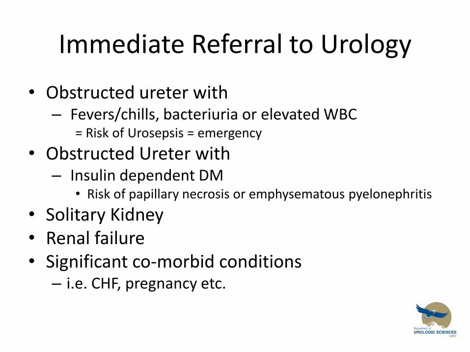
Immediate Referral to Urology
• Obstructed ureter with – Fevers/chills, bacteriuria or elevated WBC
= Risk of Urosepsis = emergency
• Obstructed Ureter with – Insulin dependent DM
• Risk of papillary necrosis or emphysematous pyelonephritis
• Solitary Kidney • Renal failure • Significant co-morbid conditions
– i.e. CHF, pregnancy etc.
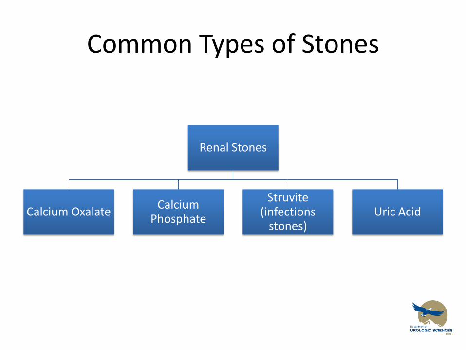
Common Types of Stones
Renal Stones
Calcium Oxalate Calcium
Phosphate
Struvite (infections
stones) Uric Acid
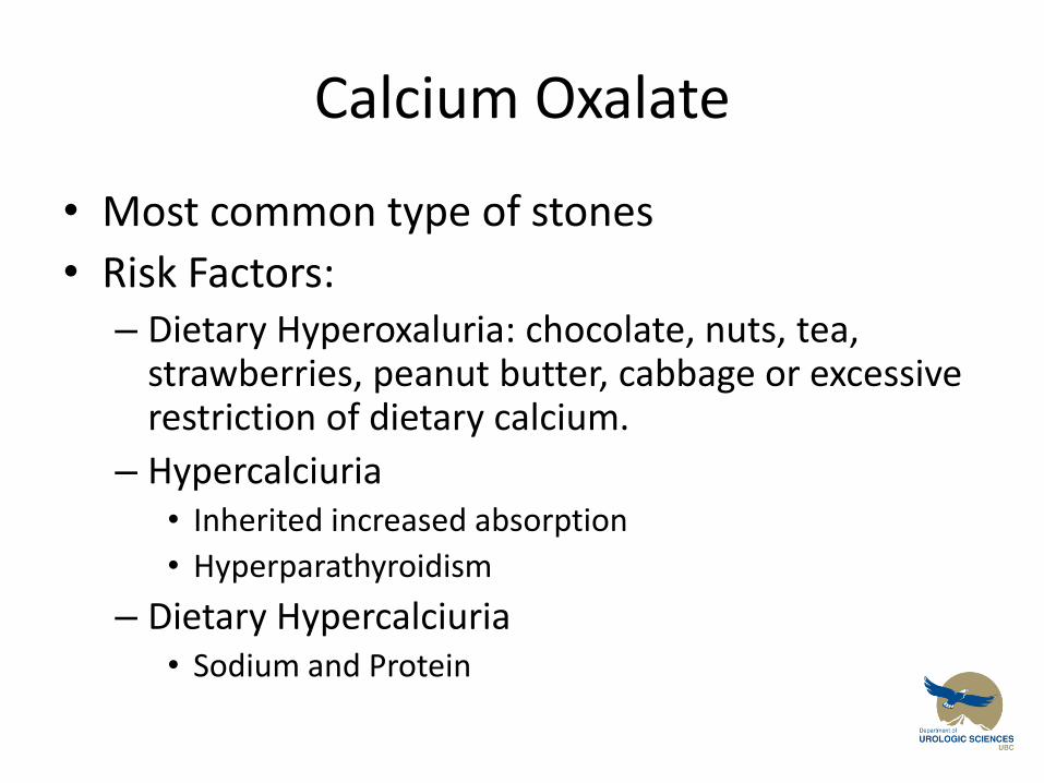
Calcium Oxalate
• Most common type of stones
• Risk Factors: – Dietary Hyperoxaluria: chocolate, nuts, tea,
strawberries, peanut butter, cabbage or excessive restriction of dietary calcium.
– Hypercalciuria • Inherited increased absorption
• Hyperparathyroidism
– Dietary Hypercalciuria • Sodium and Protein
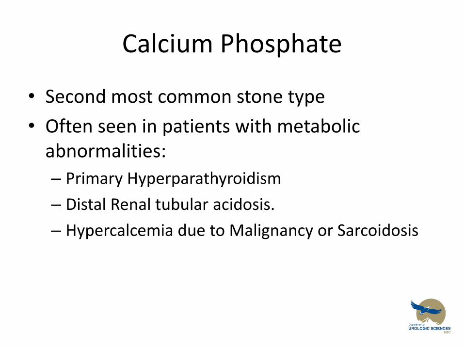
Calcium Phosphate
• Second most common stone type
• Often seen in patients with metabolic abnormalities:
– Primary Hyperparathyroidism
– Distal Renal tubular acidosis.
– Hypercalcemia due to Malignancy or Sarcoidosis
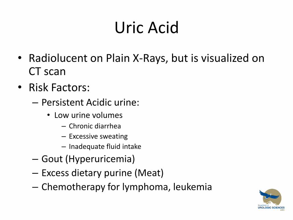
Uric Acid
• Radiolucent on Plain X-Rays, but is visualized on CT scan
• Risk Factors: – Persistent Acidic urine:
• Low urine volumes – Chronic diarrhea
– Excessive sweating
– Inadequate fluid intake
– Gout (Hyperuricemia)
– Excess dietary purine (Meat)
– Chemotherapy for lymphoma, leukemia
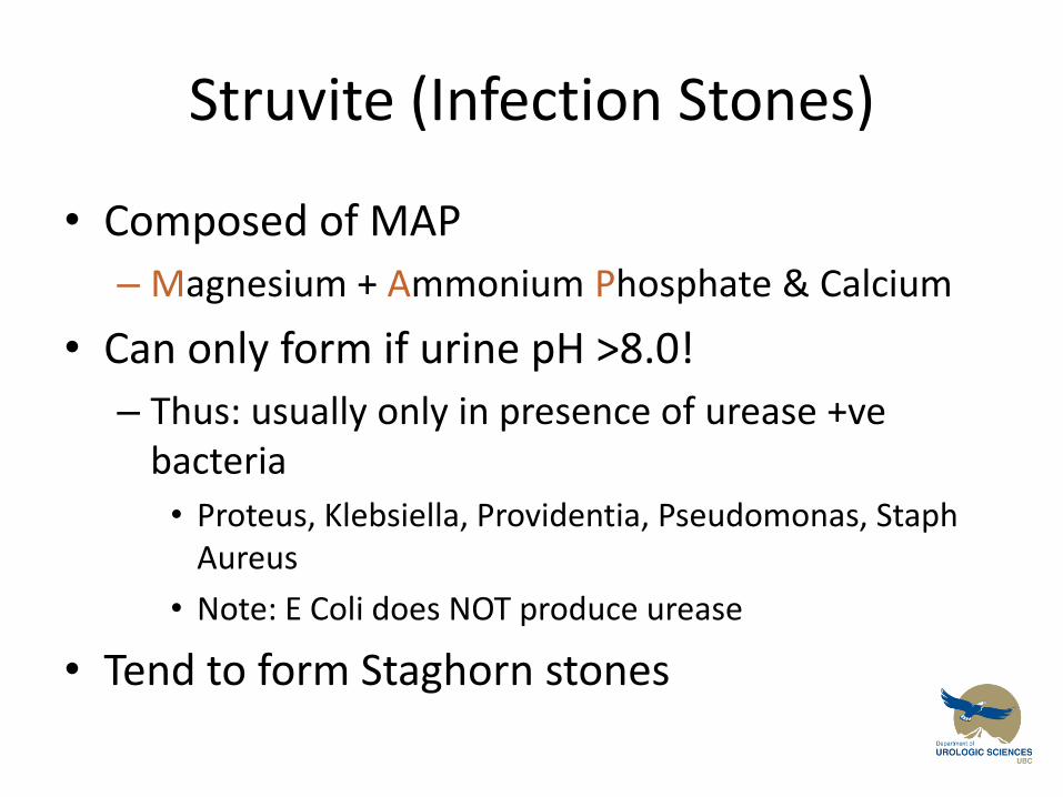
Struvite (Infection Stones)
• Composed of MAP
– Magnesium + Ammonium Phosphate & Calcium
• Can only form if urine pH >8.0!
– Thus: usually only in presence of urease +ve bacteria
• Proteus, Klebsiella, Providentia, Pseudomonas, Staph Aureus
• Note: E Coli does NOT produce urease
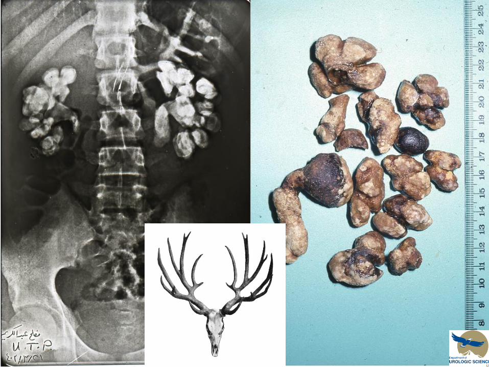
• Tend to form Staghorn stones
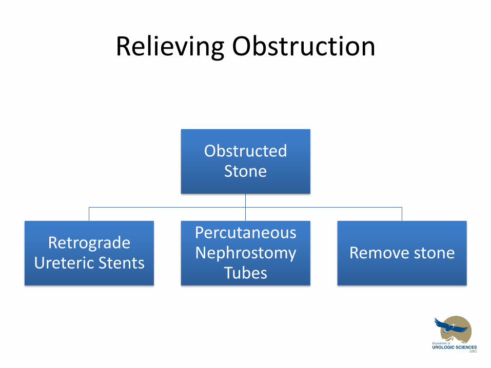
Relieving Obstruction
Obstructed Stone
Retrograde Ureteric Stents
Percutaneous Nephrostomy
Tubes Remove stone
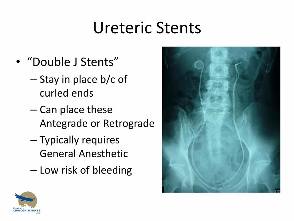
Ureteric Stents
• “Double J Stents”
– Stay in place b/c of curled ends
– Can place these Antegrade or Retrograde
– Typically requires General Anesthetic
– Low risk of bleeding
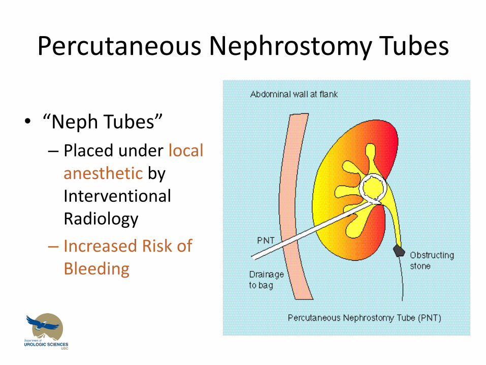
Percutaneous Nephrostomy Tubes
• “Neph Tubes”
– Placed under local anesthetic by Interventional Radiology
– Increased Risk of Bleeding
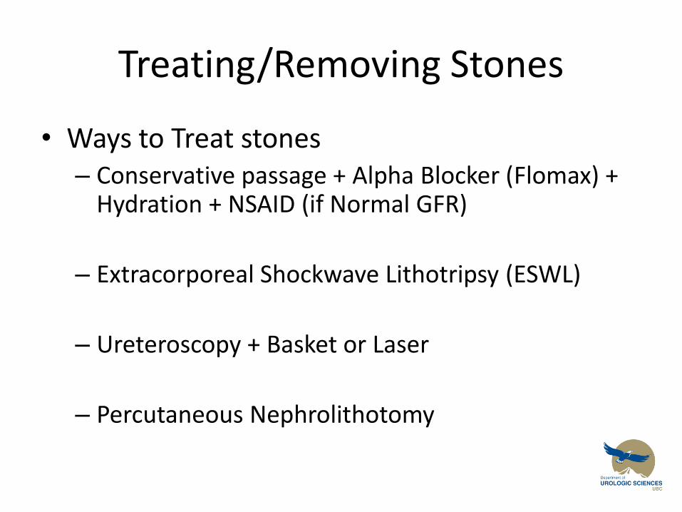
Treating/Removing Stones
• Ways to Treat stones – Conservative passage + Alpha Blocker (Flomax) +
Hydration + NSAID (if Normal GFR)
– Extracorporeal Shockwave Lithotripsy (ESWL)
– Ureteroscopy + Basket or Laser
– Percutaneous Nephrolithotomy
Treating Stones
• Conservative passage + Alpha Blocker (Flomax) + Hydration + NSAID (if Normal GFR)
– Indications
• Pain can be controlled with NSAID + Narcotic
• No renal impairment
• No Intractable Vomiting (aka pt not hypovolemic)
• No sign of infection
• No previous failed trials of conservative passage
ESWL
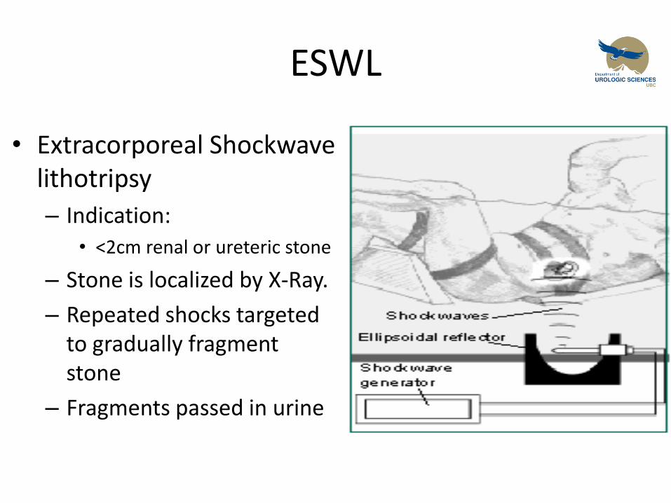
• Extracorporeal Shockwave lithotripsy
– Indication:
• <2cm renal or ureteric stone
– Stone is localized by X-Ray.
– Repeated shocks targeted to gradually fragment stone
– Fragments passed in urine

Extracorporeal Shock Wave Lithotripsy (ESWL)
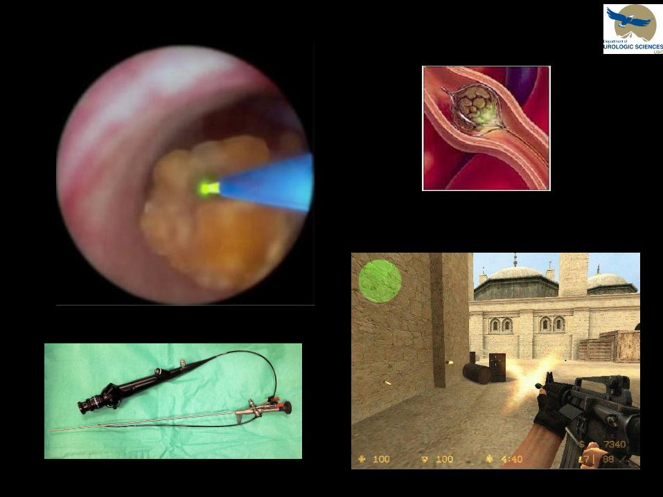
Treating Stones
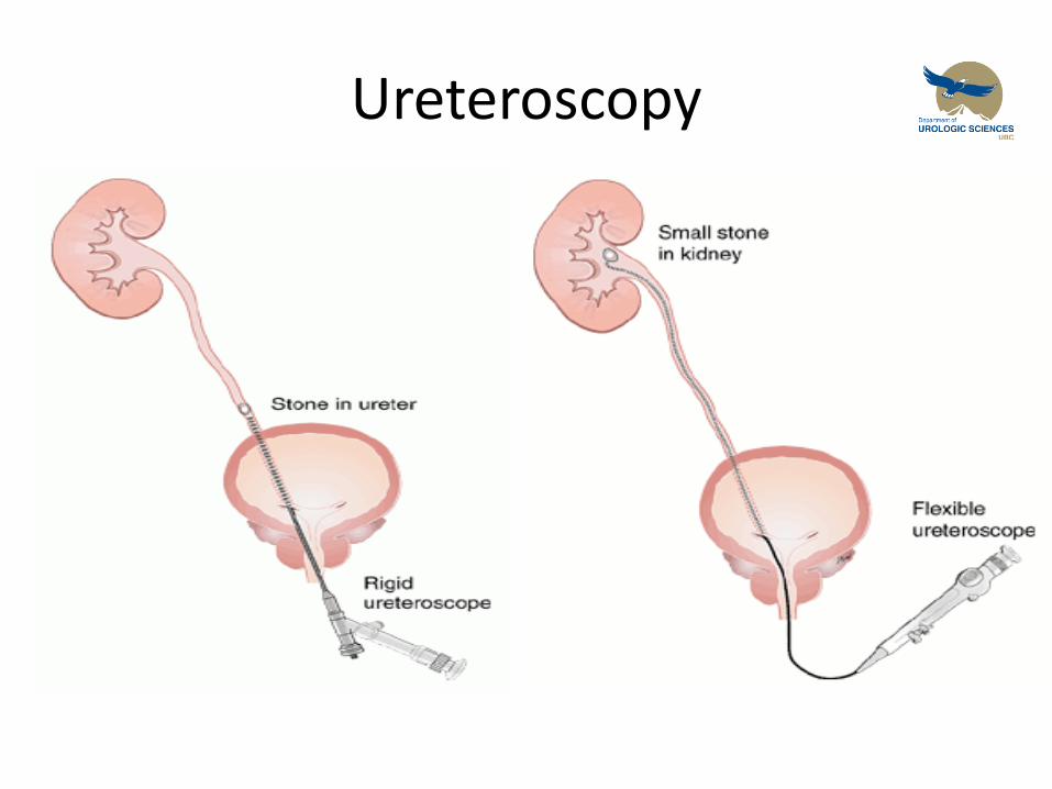
• Ureteroscopy
– + Basket
• If stone is small enough to adequately remove by basket
– + Holmium Laser
• If stone is ‘impacted’ or too large to basket out
Ureteroscopy
Treating Stones
• Percutaneous Nephrolithotomy
– Indications
• Large Proximal ureteric or renal calculi >~1-1.5cm
• Treatment of Staghorn Calculi
– Risks:
• Bleeding
• Renal Perforation or Avulsion
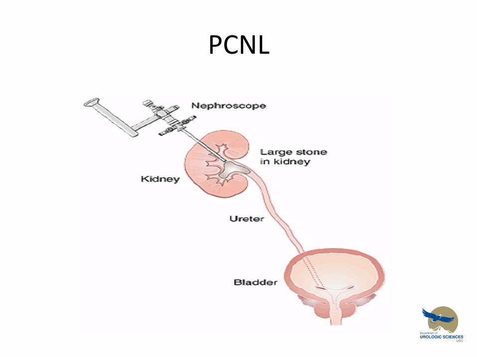
PCNL

Renal Colic
Objectives
1. Give a differential diagnosis for acute flank pain including two life-threatening conditions
2. Describe the laboratory and radiologic evaluation of a patient with renal colic
3. Know 4 different kinds of kidney stones and the risk factors for stone formation
4. Know 3 indications for emergency drainage of an obstructed kidney
Urolithiasis
• Kidney Stone Diagnosis and Treatment Learning Resources
Evaluation and Medical Management of the Kidney Stone Patient
http://www.cua.org/userfiles/files/guidelines/ksm_2011_en.pdf
Student Resources and Materials urology.med.ubc.ca