Embed Size (px)
Citation preview
Histomorphological Aspects of Vascular Anastomosis Established by Laser
A. AHMADI a B. Z IMMERMANN b, M. BOHM c aDepartment of Orthopedic Surgery and Outpatient Clinic, Oskar-Helene-Heim, Freie Universit~t, D-1000 Berlin 33, Germany ~lnstitute of Toxicology and Embryonal Pharmacology, Freie Universit#t, Berlin, Germany cCenter for Laser Medicine
Correspondence to Dr A wni Ahmadi
Abstract . The central problem in microsurgery is the reconstruction of small vessels. The long operating time, foreign body granuloma formation around the suture material as well as aneurys- mal alterations of the vessel wall after conventional suture technique make the search for alterna- tives indispensable. Some of these disadvantages can be avoided as demonstrated by our animal experiments and histological examinations in laser-assisted anastomosing. The aim of this study is to show these aspects in connection with laser application and compare them with conventional suture techniques.
INTRODUCTION
As early as 1759, Hallwell (1) in England suc- cessfully treated a lesion of the brachial ar tery by suture of the vessel. This was probably the first reconstructive intervention in a vessel. However, it was forgotten again. Vascular lesions continued to be treated by muti lat ing ligatures. This type of t rea tment was often responsible for severe circulatory disorders and loss of a limb or even life (aortic ligature).
The first experimental studies of vessel- suturing techniques were reported by Jasi- movsky (2), Murphy (3), DSrfler (4) and Jensen (5). Various suturing and reconstructive tech- niques of vessel stumps have been reported in the course of the development of vascular surgery.
For small vessels, Dombrowolskaya had already recommended in 1912 an oblique sec- tion of the vessel stumps for placement of an end-to-end anastomosis. Attempts were made to create an anastomosis mechanically or without a suture. The use of adhesive plastic material such as 2-methyl-cyanacrylate and ethyl-cyanacrylate for connecting the vessel ends was limited to experimental exami- nations (6, 7). Disadvantages of these rapidly polymerizing materials are aflexibility and considerable irritation of tissue to the point of necrosis.
The decisive impulses for the expansion of vascular surgery were given after the Second
World War by the general development in surgery.
Jacobson (8) introduced the microsurgical technique for small vessels. Fur ther advances in the development of microsurgical tech- niques in the past 30 years made it possible to solve, largely, the problem of rejoining the stumps of very small vessels with diameter of 0 .3 -2mm. Here too various at tempts were made to establish anastomosis of small vessels by different approaches.
Sealing the stumps, suture apparatuses, clips and tubuli were resorted to (9-11). However, none of these methods were gener- ally adopted. Medionecrosis, poor tensile strength, narrowing of the lumen but also re- tention of foreign bodies in the lumen proved to be disadvantages.
Continuous improvements in surgical tech- niques, refinement of the instruments and su- ture material , however, apart from some dis- advantages, led, in the conventional suture technique, to an increase in the success rate after revascularization.
Histomorphological alterations in vascular anastomoses have been described by several authors in animal experiments at different stages of the healing process. These results on the time of complete epithelialization of the anastomosis are subject to great fluctuations. While Buck (12) reports that epithelialization is completed after 24h, Murray et al (13) de- tected this alteration only after 2 weeks. Poole
Lasers in Medical Science Vol 6:363 1991 �9 Bailliere Tindall
364 A. Ahmadi, B. Zimmermann, M. B6hm
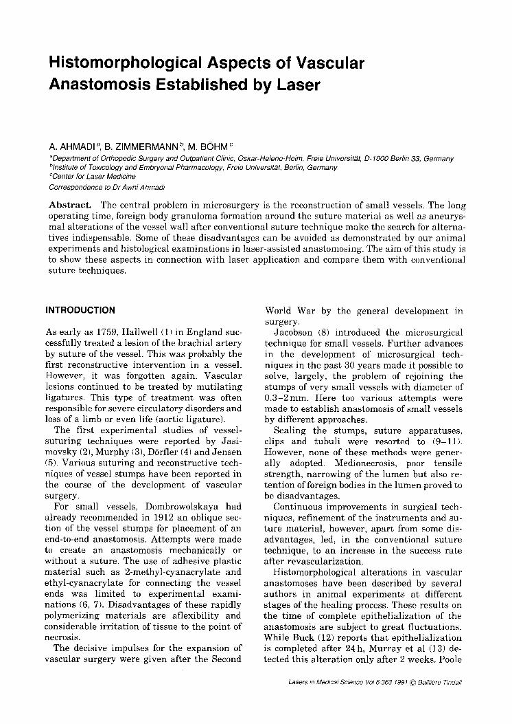
Fig. 1. Lightmicroscopic histology of the vessel wall immediately after laser fusion. TE: thrombocyte-erythrocyte conglomerate, LE: lamina elastica internal, L: lumen; x 165.
et al (14) failed to detect complete re- endothelialization of the anastomosed aorta of the rat even after the 33rd week.
The media plays a major role in the healing of the anastomosis (15). Medionecrosis and subintimal hyperplasia are characteristic al- terations found in microvascular anatomoses. The extent of this alteration depends on the degree to which the vascular wall has been traumatized, either by suture material, clamps, inappropriate preparation or by tech- nical errors (16-18).
Despite the technical progress made in refin- ing the inst rumentat ion and microscopes, or improving the tolerance of the suture material, some of the disadvantages can only be influ- enced up to a certain degree, but they cannot be completely eliminated.
A vascular anastomosis is considered ideal if it does not take too much time and is created without t raumatizat ion of the vascular wall, does not have any foreign body granulomas around the suture material and shows no tendency to form aneurysms.
Some of the above-mentioned disadvantages can be partially or completely avoided by the application of laser for establishing anasto- moses of small vessels. We carried out animal experiments on the femoral artery of the rabbit in order to make use of the advantages of a laser-assisted creation of vascular anatomoses, emphasize them and try to standardize the
possible parameters. We used different types of laser for fusioning of the anastomosis. The re- sults with the CO2 laser are already available.
Before fusioning, the exposed and severed vessel stumps were joined by two or three adaptation sutures to neutralize the muscular traction of the vessel wall. The gaps between the sutures were irradiated by a CO2 laser in the cw mode at 40mW and a focal size of 0.5mm. Duration of exposure was 10s each and power density was 25 mW mm 2.
Twenty-four vascular anastomoses were ex- amined by light and electron microscopy. Eight anastomoses were removed and histologically examined immediately after their establish- ment, the same number after 8 and after 30 days.
The anastomoses examined on the same day exhibit a clearly visible gap between the vessel stumps (Fig. 1). The gap is filled with thrombo- and erythrocyte aggregates. Signs of an exten- sive t rauma in the vicinity of the fusion area could not be visualized either by light- or elec- tronmicroscopy. Damaged cells or necroses were only seldom seen in the area.
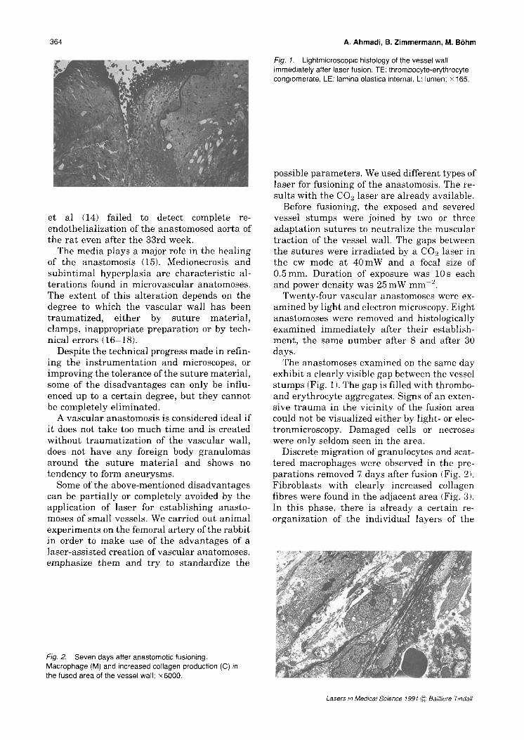
Discrete migration of granulocytes and scat- tered macrophages were observed in the pre- parations removed 7 days after fusion (Fig. 2). Fibroblasts with clearly increased collagen fibres were found in the adjacent area (Fig. 3). In this phase, there is already a certain re- organization of the individual layers of the
Fig. 2. Seven days after anastomotic fusioning. Macrophage (M) and increased collagen production (C) in the fused area of the vessel wall; x6000.
Lasers in Medical Science 1991 @ Bailliere Tindall
Histomorphological Aspects of Vascular Anastomosis 365
Fig. 3. Seven days anastomotic fusioning. F: fibroblast, TE: thrombo- and erythrocyte residues in the anastomosis gap, G: granulocyte, Le: lamina elastica interna; •
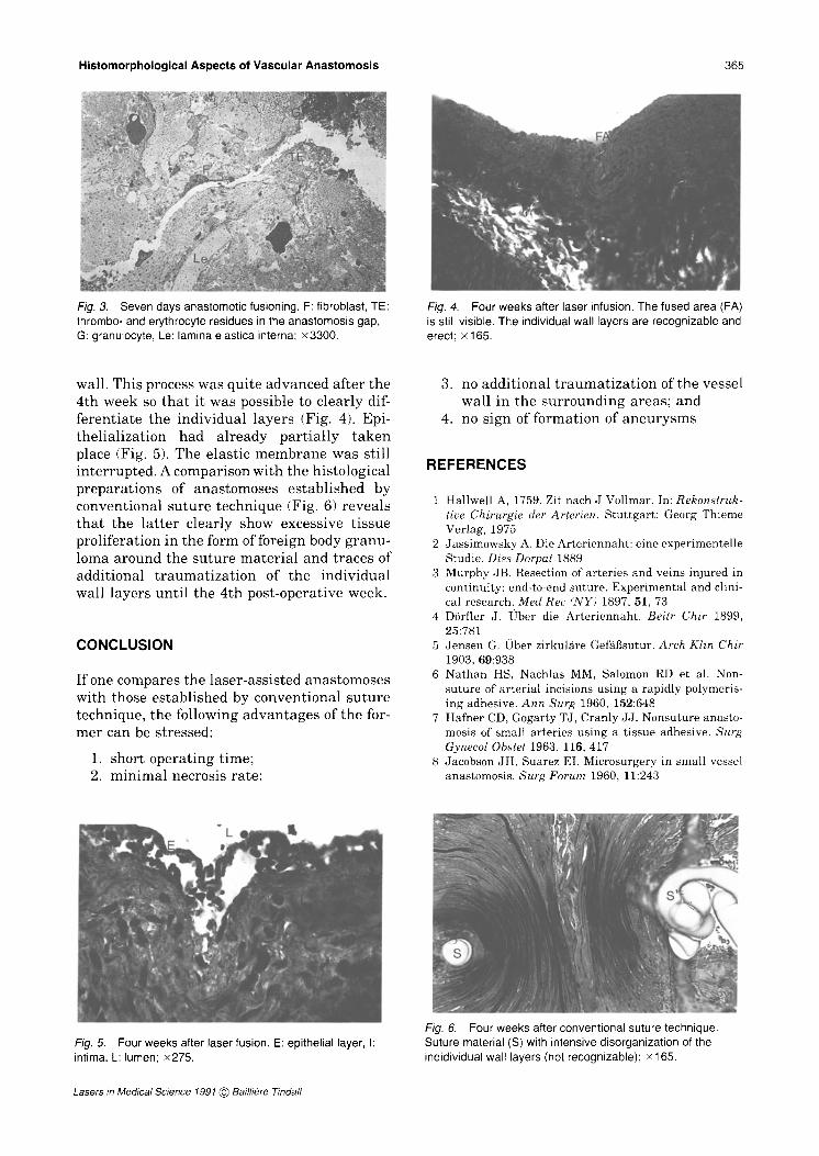
Fig. 4. Four weeks after laser infusion. The fused area (FA) is still visible. The individual wall layers are recognizable and erect; • 165.
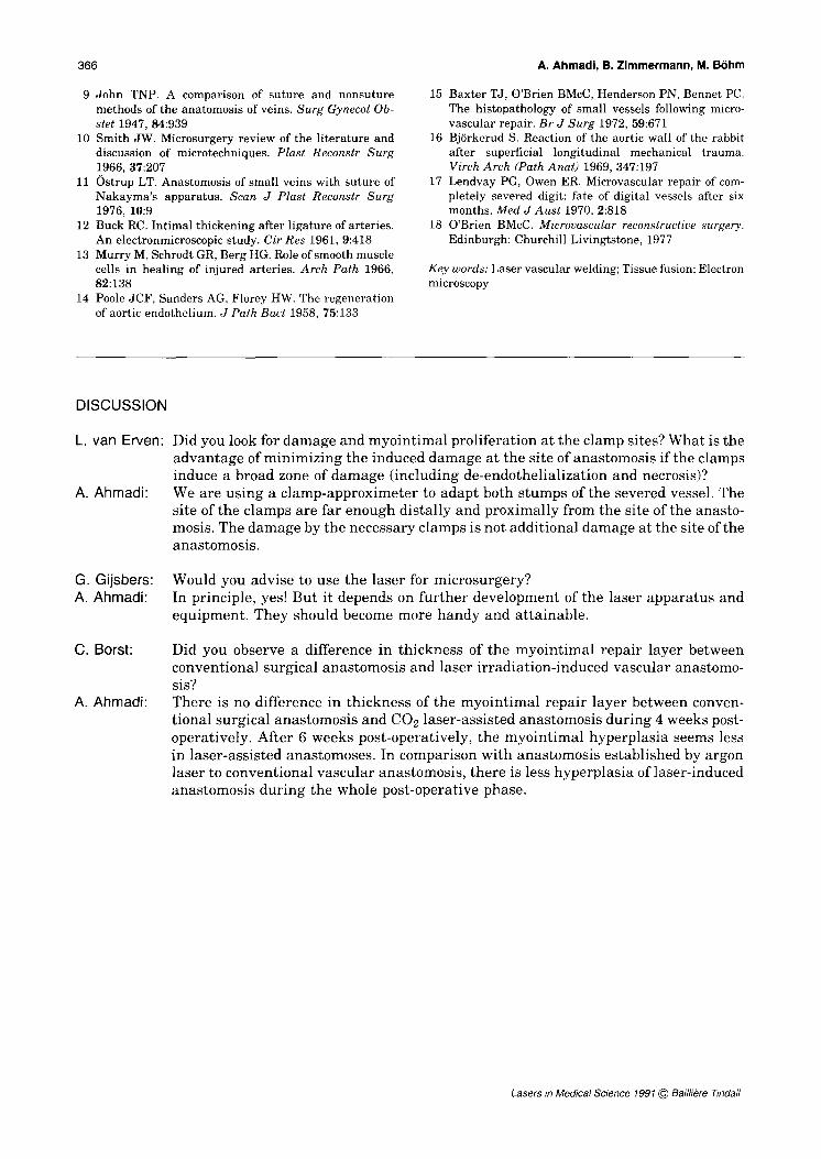
wall. This process was quite advanced after the 4th week so that it was possible to clearly dif- ferentiate the individual layers (Fig. 4). Epi- thelialization had already partially taken place (Fig. 5). The elastic membrane was still interrupted. A comparison with the histological preparations of anastomoses established by conventional suture technique (Fig. 6) reveals that the latter clearly show excessive tissue proliferation in the form of foreign body granu- loma around the suture material and traces of additional t raumatizat ion of the individual wall layers until the 4th post-operative week.
CONCLUSION
If one compares the laser-assisted anastomoses with those established by conventional suture technique, the following advantages of the for- mer can be stressed:
1. short operating time; 2. minimal necrosis rate:
3. no additional t raumatizat ion of the vessel wall in the surrounding areas; and
4. no sign of formation of aneurysms
REFERENCES
1 Hallwell A, 1759. Zit nach J Vollmar. In: Rekonstruk- tire Chirurgie der Arterien. Stut tgar t : Georg Thieme Verlag, 1975
2 Jassimowsky A. Die Ar ter iennaht : eine experimentel le Studie. Diss Dorpat 1889
3 Murphy JB. Resection of arteries and veins injured in continuity: end-to-end suture. Exper imental and clini- cal research. Med Rec (NY) 1897, 51, 73
4 D6rfler J. lJber die Ar ter iennaht . Beitr Chir 1899, 25:781
5 Jensen G. Ober zirkulgre Gei'fil3sutur. Arch Klin Chir 1903, 69:938
6 N a t h a n HS, Nachlas MM, Salomon RD et ah Non- suture of ar ter ial incisions using a rapidly polymeris- ing adhesive. Ann Surg 1960, 152:648
7 Hafner CD, Gogarty TJ, Cranly JJ . Nonsuture anasto- mosis of small ar teries using a t issue adhesive. Surg Gynecol Obstet 1963, 116, 417
8 Jacobson JH, Suarez EI. Microsurgery in small vessel anastomosis. Surg Forum 1960, 11:243
Fig. 5. Four weeks after laser fusion. E: epithelial layer, I: intima, L: lumen; x275.
Fig. 6. Four weeks after conventional suture technique. Suture material (S) with intensive disorganization of the indidividual wall layers (not recognizable); • 165.
Lasers in Medical Science 1991 @ Bailliore Tindall
366
9 John TNP. A comparison of suture and nonsuture methods of the anatomosis of veins. Surg Gynecol Ob- stet 1947, 84:939
10 Smith JW. Microsurgery review of the literature and discussion of microtechniques. Plast Reconstr Surg 1966, 37:207
11 Ostrup LT. Anastomosis of small veins with suture of Nakayma's apparatus. Scan J Plast Reconstr Surg 1976, 10:9
12 Buck RC. Intimal thickening after ligature of arteries. An electronmicroscopic study. Cir Res 1961, 9:418
13 Murry M, Schrodt GR, Berg HG. Role of smooth muscle cells in healing of injured arteries. Arch Path 1966, 82:138
14 Poole JCF, Sanders AG, Florey HW. The regeneration of aortic endothelium. J Path Bact 1958, 75:133
A. Ahmadi, B. Zimmermann, M. B6hm
15 Baxter TJ, O'Brien BMcC, Henderson PN, Bennet PC. The histopathology of small vessels following micro- vascular repair. Br J Surg 1972, 59:671
16 Bj6rkerud S. Reaction of the aortic wall of the rabbit after superficial longitudinal mechanical trauma. Virch Arch (Path Anat) 1969, 347:197
17 Lendvay PG, Owen ER. Microvascular repair of com- pletely severed digit: fate of digital vessels after six months. Med J Aus t 1970, 2:818
18 O'Brien BMcC. Microvascular reconstructive surgery. Edinburgh: Churchill Livingtstone, 1977
Key words: Laser vascular welding; Tissue fusion; Electron microscopy
DISCUSSION
L. van Erven:
A. Ahmadi:
G. Gijsbers: A. Ahmadi:
C. Borst:
A. Ahmadi:
Did you look for damage and myointimal proliferation at the clamp sites? What is the advantage of minimizing the induced damage at the site of anastomosis if the clamps induce a broad zone of damage (including de-endothelialization and necrosis)? We are using a clamp-approximeter to adapt both stumps of the severed vessel. The site of the clamps are far enough distally and proximally from the site of the anasto- mosis. The damage by the necessary clamps is not additional damage at the site of the anastomosis.
Would you advise to use the laser for microsurgery? In principle, yes! But it depends on further development of the laser apparatus and equipment. They should become more handy and attainable.
Did you observe a difference in thickness of the myointimal repair layer between conventional surgical anastomosis and laser irradiation-induced vascular anastomo- sis? There is no difference in thickness of the myointimal repair layer between conven- tional surgical anastomosis and CO2 laser-assisted anastomosis during 4 weeks post- operatively. After 6 weeks post-operatively, the myointimal hyperplasia seems less in laser-assisted anastomoses. In comparison with anastomosis established by argon laser to conventional vascular anastomosis, there is less hyperplasia of laser-induced anastomosis during the whole post-operative phase.
Lasers in Medical Science 1991 (~ Bailliere Tindall