Embed Size (px)
Citation preview
Histone Deacetylase Inhibitor Upregulates PeroxisomalFatty Acid Oxidation and Inhibits Apoptotic Cell Death inAbcd1-Deficient Glial CellsJaspreet Singh1, Mushfiquddin Khan1, Aurora Pujol2,3,4, Mauhamad Baarine1, Inderjit Singh1*
1 Department of Pediatrics, Darby Children Research Institute, Medical University of South Carolina, Charleston, South Carolina, United States of America,
2 Neurometabolic Diseases Laboratory, Institute of Neuropathology, Bellvitge Institute for Biomedical Research (IDIBELL), Hospitalet de Llobregat, Barcelona, Spain,
3 Center for Biomedical Research on Rare Diseases (CIBERER), Barcelona, Spain, 4 Catalan Institution for Research and Advanced Studies (ICREA), Barcelona, Spain
Abstract
In X-ALD, mutation/deletion of ALD gene (ABCD1) and the resultant very long chain fatty acid (VLCFA) derangement hasdramatically opposing effects in astrocytes and oligodendrocytes. While loss of Abcd1 in astrocytes produces a robustinflammatory response, the oligodendrocytes undergo cell death leading to demyelination in X-linked adrenoleukodys-trophy (X-ALD). The mechanisms of these distinct pathways in the two cell types are not well understood. Here, weinvestigated the effects of Abcd1-knockdown and the subsequent alteration in VLCFA metabolism in human U87 astrocytesand rat B12 oligodendrocytes. Loss of Abcd1 inhibited peroxisomal b-oxidation activity and increased expression of VLCFAsynthesizing enzymes, elongase of very long chain fatty acids (ELOVLs) (1 and 3) in both cell types. However, higherinduction of ELOVL’s in Abcd1-deficient B12 oligodendrocytes than astrocytes suggests that ELOVL pathway may play aprominent role in oligodendrocytes in X-ALD. While astrocytes are able to maintain the cellular homeostasis of anti-apoptotic proteins, Abcd1-deletion in B12 oligodendrocytes downregulated the anti-apototic (Bcl-2 and Bcl-xL) and cellsurvival (phospho-Erk1/2) proteins, and upregulated the pro-apoptotic proteins (Bad, Bim, Bax and Bid) leading to cell loss.These observations provide insights into different cellular signaling mechanisms in response to Abcd1-deletion in twodifferent cell types of CNS. The apoptotic responses were accompanied by activation of caspase-3 and caspase-9 suggestingthe involvement of mitochondrial-caspase-9-dependent mechanism in Abcd1-deficient oligodendrocytes. Treatment withhistone deacetylase (HDAC) inhibitor suberoylanilide hydroxamic acid (SAHA) corrected the VLCFA derangement both invitro and in vivo, and inhibited the oligodendrocytes loss. These observations provide a proof-of principle that HDACinhibitor SAHA may have a therapeutic potential for X-ALD.
Citation: Singh J, Khan M, Pujol A, Baarine M, Singh I (2013) Histone Deacetylase Inhibitor Upregulates Peroxisomal Fatty Acid Oxidation and Inhibits ApoptoticCell Death in Abcd1-Deficient Glial Cells. PLoS ONE 8(7): e70712. doi:10.1371/journal.pone.0070712
Editor: Ralf Andreas Linker, Friedrich-Alexander University Erlangen, Germany
Received August 28, 2012; Accepted June 26, 2013; Published July 26, 2013
Copyright: � 2013 Singh et al. This is an open-access article distributed under the terms of the Creative Commons Attribution License, which permitsunrestricted use, distribution, and reproduction in any medium, provided the original author and source are credited.
Funding: This work was supported in part by grants from the National Institutes of Health: NS-22576, NS-37766, C06 RR018823, C06 RR015455, and VA meritaward BX1072-01 to IS, and also by grants of the Autonomous Government of Catalonia [2009SGR85] and the Spanish Institute for Health Carlos III (FIS PI11/01043) to AP. The funders had no role in study design, data collection and analysis, decision to publish, or preparation of the manuscript.
Competing Interests: The authors have declared that no competing interests exist.
* E-mail: [email protected]
Introduction
The ALD gene (ABCD1), identified by positional cloning [1],
encodes a protein ALDP that is related to the peroxisomal ATP-
binding cassette (ABCD) transmembrane transporter proteins
[2,3]. Loss of ABCD1 function results in defective b-oxidation of
very long chain fatty acids (VLCFA) [4] resulting in accumulation
of VLCFA, the biochemical ‘‘hallmark’’ of X-ALD, in plasma and
tissues, most notably in brain and adrenal cortex [5]. Frequently
clinically distinct phenotypes ranging from a fatal childhood
cerebral ALD (cALD) to relatively benign adult disease of
adrenomyeloneuropathy (AMN) occur within the same family
with no phenotype-genotype correlation having been established
so far. The molecular events that trigger the transition from the
metabolic derangement, common to all forms of X-ALD, to
neuroinflammation and demyelination in cALD or to axonal
degeneration in spinal cords in AMN are largely unknown. Recent
studies from our laboratory [6,7] and others [8] show a correlation
between VLCFA accumulation caused by silencing of peroxisomal
transporters in neural tissue in X-ALD and glial cells to redox
imbalance, and changes in membrane lipid composition
[6,7,9,10,11] leading to astrocytic inflammatory response and loss
of oligodendrocytes and myelin [10,12].
Abcd1 knockout mouse does not develop demyelination
characteristic of cALD, although myelin disturbances are
evident starting at 15-month in sciatic nerve and spinal cord
tissue [13], although it does show nuclear factor-kB (NFkB) pro-
inflammatory cytokine induction [14]. In active lesions of X-
ALD brain, astrocytes expressed large amounts of tumor
necrosis factor-a (TNF-a) [12] and inducible nitric oxide
synthase (iNOS) [15]. Since acute glial death is reported to
promote neuronal death [16], the glial loss in X-ALD probably
plays a role in the progression of neurodegeneration in X-ALD.
The recently reported differential accumulation of VLCFA in
induced pluripotent stem cell (iPSC)-derived oligodendrocytes
from X-ALD and AMN fibroblasts [17] suggests that Abcd1 loss
may induce different cellular signaling or metabolic derange-
ments in these cell types.
PLOS ONE | www.plosone.org 1 July 2013 | Volume 8 | Issue 7 | e70712
In addition to b-oxidation defect, increased expression of
elongases (ELOVLs) also contributes to higher VLCFA levels [18].
However, the effect of Abcd1-deletion on ELOVLs in astrocytes
and oligodendrocytes has not been explored. Moreover, inflam-
matory mediators (TNF-a and IL-1b) downregulate peroxisomal
b-oxidation function [19]. Accordingly, different degrees of
VLCFA accumulation were observed in different areas (inflam-
matory, plaque and non-inflammatory) of X-ALD brain. In X-
ALD CNS, therefore, altered activities of ELOVLs and peroxi-
somal b-oxidation as well as the secondary effects of inflammatory
mediators may contribute towards the observed pathognomic
levels of VLCFA. Hence, an effective therapy should be able to
correct the metabolic derangements as well as attenuate the
inflammatory responses.
Current treatment options for X-ALD are limited [5]. The
concept of therapeutic induction of functionally redundant
Abcd2/Abcd3 has initiated intense investigations aiming to
modulate the ABCD gene expression as a novel therapeutic
strategy for X-ALD. Previous studies from our laboratory and
others have demonstrated that Abcd2 expression could be
upregulated in rodents by various therapeutic compounds
[17,20,21,22], but no induction was found in brain for various
reasons [23]. Long-term treatment with 4-phenylbutyrate in
Abcd1-KO mouse model lead to a reduced drug response and
VLCFA levels returned to pretreatment levels [24]. Valproic acid
(VPA) induced the Abcd2 expression but was unable to lower the
levels of VLCFA [20]. We have formerly shown that lovastatin
and sodium phenylacetate, can enhance VLCFA b-oxidation and
reduce VLCFA levels in human skin fibroblasts [25], lymphoblasts
[26] and plasma of X-ALD patients [27].
Using U87 astrocytes and B12 oligodendrocytes stably silenced
for Abcd1 using lentiviral vectors, this study describes the astrocyte
vs. oligodendrocyte-specific VLCFA-mediated derangements and
activation of mitochondrial cell death pathways. We also evaluated
the effect of SAHA treatment in these cells. Treatment with SAHA
corrected the metabolic derangements as well as inhibited the
Abcd1-deficiency-induced apoptotic response. Additionally, the
effect of SAHA was investigated in hippocampal slice cultures
from patients suffering from drug-resistant epilepsy that were
scheduled for hippocampal resection, where it upregulated the
ABCD2 expression. Remarkably, Abcd1-KO mice treated with
SAHA in diet had significantly lower VLCFA levels in the brain
providing the first pre-clinical proof-of-principal for testing SAHA
as therapy for X-ALD.
Experimental Procedures
Ethics StatementHippocampal specimens were derived from three patients
submitted to epilepsy surgery at the Erlangen Epilepsy Center.
The patients suffered from drug-resistant epilepsy and were
scheduled for hippocampal resection. For scientific use of tissue
specimens, written informed consent was obtained from each
patient with the approval of the local ethics committee of the
University Hospital of Erlangen.
ReagentsDulbecco’s modified Eagle’s medium (DMEM) and Hanks’
balanced salt solution (HBSS) were purchased from Invitrogen
Life Technologies; Fetal bovine serum (FBS), TNF-a and IL-1bwere purchased from BioAbChem Inc. (Ladson, SC). ALDP
antibody was from Chemicon International Inc. (Temecula, CA).
ALDRP antibody was custom-made from ANASPEC against the
mouse 20-residue c-terminal sequence: 722 CKILGEDSVLK-
TIQTPEKTS 741. 5-LOX antibody was purchased from
Cayman Chemical (Ann Arbor, Michigan). Na+K+ATPase anti-
body was purchased from Santa Cruz Biotechnology (Santa Cruz,
CA). All other antibodies were from Cell Signalling Technology
Inc. (MA, USA) unless otherwise mentioned. ECL and nitrocel-
lulose membranes were purchased from Amersham Biosciences.
Fatty acid methyl ester (FAME) standards were obtained from
Supelco (Bellefonte, Pennsylvania). [l-14C] Lignoceric acid was
prepared as described earlier [28]. [1-14C] Palmitic acid and 125I-
labeled protein A were obtained from ICN (Cleveland, Ohio).
Stable Lentiviral vector-mediated gene silencing ofAbcd1
A set of 3 human (SK-009605-00-10) and rat (SK-098142-00-
10) specific SMART vector 2.0 lentiviral shRNA particles
(108 TU/ml) for Abcd1 were purchased from Thermo Fisher
Scientific Dharmacon (CO, USA). The vector had an hCMV
promoter, a TurboGFP reporter gene and a puromycin selection
gene. SMART vector 2.0 non-targeting shRNA control particles
(NT) (108 TU/ml, Thermo Fisher Scientific Dharmacon, CO,
USA) designed so that no known gene in human, mouse or rat will
be targeted were used as negative controls.
Human U87 astrocytes and rat B12 oligodendrocytes were
cultured in DMEM with 10% FBS in the presence of antibiotic,
and viral particles (Abcd1 and non-targeting control) were added
with a multiplicity of infection (MOI) of 2.5 and 3.0 respectively
for U87 astrocytes and B12 oligodendrocytes. TurboGFP-expres-
sion was analysed using microscopy and GFP-positive cells were
selected using puromycin (3.0 mg/ml for U87 astrocytes and
2.0 mg/ml for B12 oligodendrocytes). The selected cells were
maintained in culture media containing puromycin at 0.5 mg/ml
until further use. Abcd1 silencing was observed by Western blot
and mRNA quatification.
In vivo SAHA treatment in Abcd1-knockout (Abcd1-KO)mice
Animal procedure was approved by the MUSC Animal Review
Committee, and all animals received humane care in compliance
with MUSC’s experimental guidelines and the National Research
Council’s criteria (Guide for the Care and Use of Laboratory
Animals).
Abcd1-KO (C57BL6) and control (normal) (C57BL6) breeding
pairs were purchased from Jackson Laboratory (Ban Habor, ME,
USA) and maintained at the institution’s animal facility. The
genotypes of all newborns from Abcd1-KO were determined by a
PCR-based method as described (Forss-Petter et al. 1997) using the
primers suggested in data sheets from Jackson Laboratory
(http://jaxmice.jax.org). 5-CACAGCCTCTCTCCTTAAGACC-3
(oIMR1120), 5-CTCGTTGTCTAGGCAACTGG-3 (oIMR1121)
and 5-CTTCTATCGCCTTCTTGACG-3 (oIMR1122). Two
primers detect disrupted allele Abcd1-KO. Presence of the wild-
type allele was revealed in the same reaction by using a third primer
(oIMR1121). PCR consisted of a 10 min heating step at 95uC,
followed by 35 cycles of denaturation at 95uC for 1 min, annealing
at 54uC for 1 min, and extension at 72uC for 1 min. The PCR
products were then subjected to agarose gel electrophoresis.
Vorinostat or suberoylanilide hydroxamic acid (SAHA) (Cayman
chemical, Michigan, USA) therapy was performed on 60–65 day-
old Abcd1-KO male mice. Animals were daily treated with SAHA
(50 mg/kg body weight) in diet (prepared by Research Diet Inc.,
New Brunswick, NJ) for 62 days. Brain tissue, (harvested from
cortex area) from control (normal), Abcd1-KO and SAHA-treated
Abcd1-KO mice was homegenized in RIPA buffer in a ratio of 1:10
SAHA Attenuates Oligodendrocyte Cell Death
PLOS ONE | www.plosone.org 2 July 2013 | Volume 8 | Issue 7 | e70712
(weight/volume). After centrifugation of samples at 1000 rpm for
2 min to remove all debris, supernatant was collected and protein
amount was estimated. 200 mL supernatant (,1 mg of protein) was
used for quantification of VLCFA by GC-MS.
Fatty Acid b-OxidationThe peroxisomal oxidation of fatty acids in control, Abcd1-
deficient and SAHA-treated Abcd1-deficient astrocytes and
oligodendrocytes was determined in 6-well plates as described
previously [7]. b-oxidation of fatty acids to acetate (water soluble
product) was determined using [1-14C]-labeled fatty acids as
substrate (C24:0, lignoceric acid or C16:0, palmitic acid (ARC, St.
Louis, MO)) as described previously [29]. Cells grown in parallel
in same plate were used to determine the protein present in the
assays. Experiments were performed in triplicate.
Lipid Extraction and Fatty Acid AnalysisTotal lipids were extracted from control, Abcd1-deficient and
SAHA-treated cells as described previously [30]. FAME were
analyzed by GC (Shimadzu chromatograph GC-15A attached to a
Shimadzu chromatopac C-R3A integrator) using a fused silica
capillary column (25 M 007 series methyl silicone, 0.25-mm
internal diameter, 0.25-mm film thickness) from Quadrex Corpo-
ration (Woodbridge, CT) in a gas chromatograph GC-17A
connected with a flame ionization detector from Shimadzu
Corporation.
Preparation of carbonate membranesCells were harvested with sucrose buffer (0.25 M sucrose, 1 mM
EDTA, 3 mM imidazole buffer, pH 7.4), and subjected to
sonication (10 sec at 8–9 watts output power). The homogenate
was centrifuged at 2,500 rpm for 5 min to remove unbroken cells.
To isolate carbonate membranes (membrane sheets containing
integral membrane proteins), equal protein amount from control
and X-ALD cell homogenates were diluted with an ice-cold
solution of 0.1 M sodium carbonate (pH 11.5) containing 30 mM
iodoacetamide. After 30 minutes of incubation at 4uC, carbonate
membranes were sedimented by centrifugation at 35,000 rpm for
1 hour in a 70Ti rotor (Beckman-Coulter, Fullerton, CA). The
sedimented membranes were washed twice with cold water,
lyophilized and stored at 270uC until use.
Western Blot Analysis40 mg of total cellular protein was resolved by electrophoresis on
4–20% polyacrylamide gels. After incubation with antiserum
raised against ALDP, ALDRP, peroxisomal membrane protein 70
(PMP70), 5-lipoxygenase (5-LOX), B-cell lymphoma 2 (Bcl-2), B
cell lymphoma-X long (Bcl-xL), Bcl-2-associated X (Bax), and Bcl-
2-antagonist/killer (Bak), Na+K+ATPase and cleaved caspase-9
and 3, the membranes were then incubated with horseradish
peroxidase-conjugated anti-rabbit or mouse IgG for 1 h. The
membranes were detected by autoradiography using ECL-plus
(Amersham Biosciences) after washing with TBST buffer.
cDNA Synthesis and Real-time PCRFollowing total RNA extraction using TRIzol (Invitrogen) per
the manufacturer’s protocol, single-stranded cDNA was synthe-
sized from total RNA as described previously [7]. Real time PCR
was conducted using Bio-Rad iCycler (iCycler iQ Multi-Color
Real Time PCR Detection System; Bio-Rad). Primers for human
and rat Abcd1, Abcd2, Abcd3, ELOVL1, and ELOVL3 were
purchased from Qiagen. Thermal cycling conditions were as
follows: activation of DNA polymerase at 95uC for 10 min,
followed by 40 cycles of amplification at 95uC for 30 s and 60uCfor 30 s. The normalized expression of target gene with respect to
glyceraldehyde-3-phosphate dehydrogenase or 18S RNA was
computed for all samples using Microsoft Excel data spreadsheet.
Organotypic hippocampal slice culture (OHSC)Hippocampal specimens were derived from three patients
submitted to epilepsy surgery at the Erlangen Epilepsy Center.
The patients suffered from drug-resistant epilepsy and were
scheduled for hippocampal resection. VPA treatment was stopped
several weeks prior to surgery. For scientific use of tissue
specimens, written informed consent was obtained from each
patient with the approval of the local ethics committee of the
University Hospital of Erlangen. Surgical specimens were
prepared and processed as described for rat OHSCs [31]. After
dissection of the frontal pole of the hemispheres and the
cerebellum, the brains were cut in 350-mm thick horizontal slices
on a vibratome (Leica Microsystems, Wetzlar, Germany). For each
experiment, three slices were transferred into culture plate insert
membranes (BD Biosciences, San Jose, CA, USA) and thereafter
into six-well culture dishes (BD Biosciences) containing 1.2 mL
culture medium as described in detail by Eyupoglu et al. [32]. One
day after preparation, the culture medium was changed, and
OHSCs were exposed to the test compound for 48 h and snap-
frozen in liquid nitrogen, as described previously [32].
Statistical analysisUsing the Student Newman-Keuls test and ANOVA, p values
were determined for the respective experiments from three
identical experiments using GraphPad software (GraphPad
Software Inc. San Diego, CA). The criterion for statistical
significance was p,0.05.
Results
Stable silencing of Abcd1 in U87 astrocytes and B12oligodendrocytes using lentiviral-shRNA vector
The observed astrocytic inflammatory activity and demyelin-
ation in X-ALD brain [10,15,33] suggests that VLCFA-induced
inflammatory activity may participate in X-ALD pathology.
Therefore, to understand the role of Abcd1 loss in astrocyte and
oligodendrocyte functions, we stably silenced the U87 astrocytes
and B12 oligodendrocytes for Abcd1 using lentiviral-shRNA.
Following transduction, successfully transduced cells were selected
with puromycin (3.0 mg/ml for U87 astrocytoma (Fig. 1A) and
1.5 mg/ml for B12 oligodendroglia (Fig. 1E)). Lentiviral-mediated
silencing of Abcd1 in U87 astrocytes (Fig. 1A) and B12-
oligodendrocytes (Fig. 1E) was highly successful (as seen with
GFP fluorescence). Subsequently the cultures were maintained in
0.5 mg/ml puromycin. Western analysis using antibody against
ALDP on carbonate membranes (membrane preparation contain-
ing integral membrane proteins) from control, non-targeting (NT)
and Abcd1-deficient U87 astrocytes (Fig. 1B-i) and B12
oligodendrocytes (Fig. 1F-i) shows almost complete loss of Abcd1
protein in Abcd1-lentiviral silenced cells (Fig. 1B-i and 1F-i).Real time PCR with primers against human and rat ALDP also
showed a stable significant (P,0.001) downregulation of Abcd1
expression in U87 astrocytes (Fig. 1B-ii) and B12 oligodendro-
cytes (Fig. 1F-ii) respectively. Since overexpression of Abcd2 and
Abcd3 can compensate for loss of Abcd1 [34,35], we investigated
the effect of Abcd1 silencing on the expression of Abcd2 and
Abcd3 in U87 astrocytes and B12 oligodendrocytes. Abcd1
silencing had no effect on Abcd2 (Fig. 1C and 1G) and Abcd3
(1D and 1H) either at protein (i) or mRNA (ii) levels in U87
SAHA Attenuates Oligodendrocyte Cell Death
PLOS ONE | www.plosone.org 3 July 2013 | Volume 8 | Issue 7 | e70712
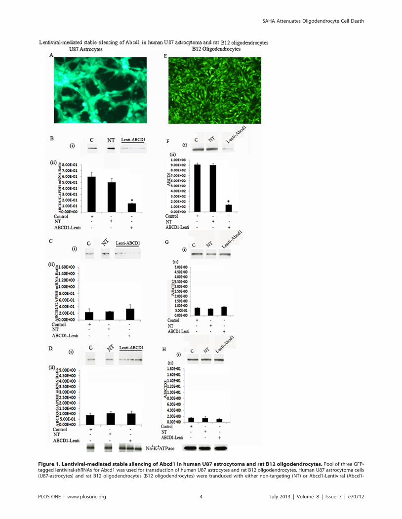
Figure 1. Lentiviral-mediated stable silencing of Abcd1 in human U87 astrocytoma and rat B12 oligodendrocytes. Pool of three GFP-tagged lentiviral-shRNAs for Abcd1 was used for transduction of human U87 astrocytes and rat B12 oligodendrocytes. Human U87 astrocytoma cells(U87-astrocytes) and rat B12 oligodendrocytes (B12 oligodendrocytes) were tranduced with either non-targeting (NT) or Abcd1-Lentiviral (Abcd1-
SAHA Attenuates Oligodendrocyte Cell Death
PLOS ONE | www.plosone.org 4 July 2013 | Volume 8 | Issue 7 | e70712
astrocytes and B12 oligodendrocytes. This, along with the
observation that NT sequence had no effect on levels of Abcd1
as well as Abcd2 and Abcd3 documents that the silencing was
specific for Abcd1.
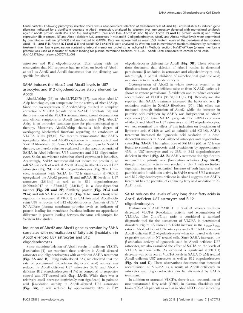
SAHA induces the Abcd2 and Abcd3 levels in U87astrocytes and B12 oligodendrocytes stably silenced forAbcd1
Abcd2/Aldrp [36] or Abcd3/PMP70 [37], two close Abcd1/
Aldp homologues, can compensate for the activity of Abcd1/Aldp.
Since the overexpression of Abcd2/Aldrp resulted in complete
correction of VLCFA b-oxidation in X-ALD fibroblasts [38] and
the prevention of the VLCFA accumulation, axonal degeneration
and clinical symptoms in Abcd1 knockout mice [34], Abcd2/
Aldrp is an attractive candidate gene for pharmacological gene
therapy. Moreover, Abcd2/Aldrp shares a high degree of
overlapping biochemical functions regarding the catabolism of
VLCFA in vivo [39,40]. We recently demonstrated that SAHA
upregulates Abcd2 and Abcd3 expression in human control and
X-ALD fibroblasts [35]. Since CNS is the target organ for X-ALD
therapy, we therefore further evaluated the therapeutic potential of
SAHA in Abcd1-silenced U87 astrocytes and B12 oligodendro-
cytes. So far, no evidence exists that Abcd1 expression is inducible.
Accordingly, SAHA treatment did not induce the protein (i) or
mRNA (ii) levels of residual Abcd1 (if any) in Abcd1-silenced U87
astrocytes (Fig. 2A) and B12 oligodendrocytes (Fig. 2E). How-
ever, treatment with SAHA for 72 h significantly (P,0.001)
upregulated the Abcd2 protein (i) and mRNA (ii) levels in U87
astrocytes (3.6-fold) as well as in B12 oligodendrocytes
(0.90960.047 to 4.5760.15) ((5.0-fold) in a dose-dependent
manner (Fig. 2B and 2F). Similarly, protein (Fig. 2C-i and2G-i) and mRNA levels of Abcd3 (Fig. 2C-ii and 2G-ii) were
significantly increased (P,0.001) in SAHA-treated Abcd1-defi-
ceint U87 astrocytes and B12 oligodendrocytes. Analysis of Na+/
K+-ATPase (plasma membrane protein) levels as indicator of
protein loading for membrane fractions indicate no appreciable
difference in protein loading between the same cell samples for
Western blot studies.
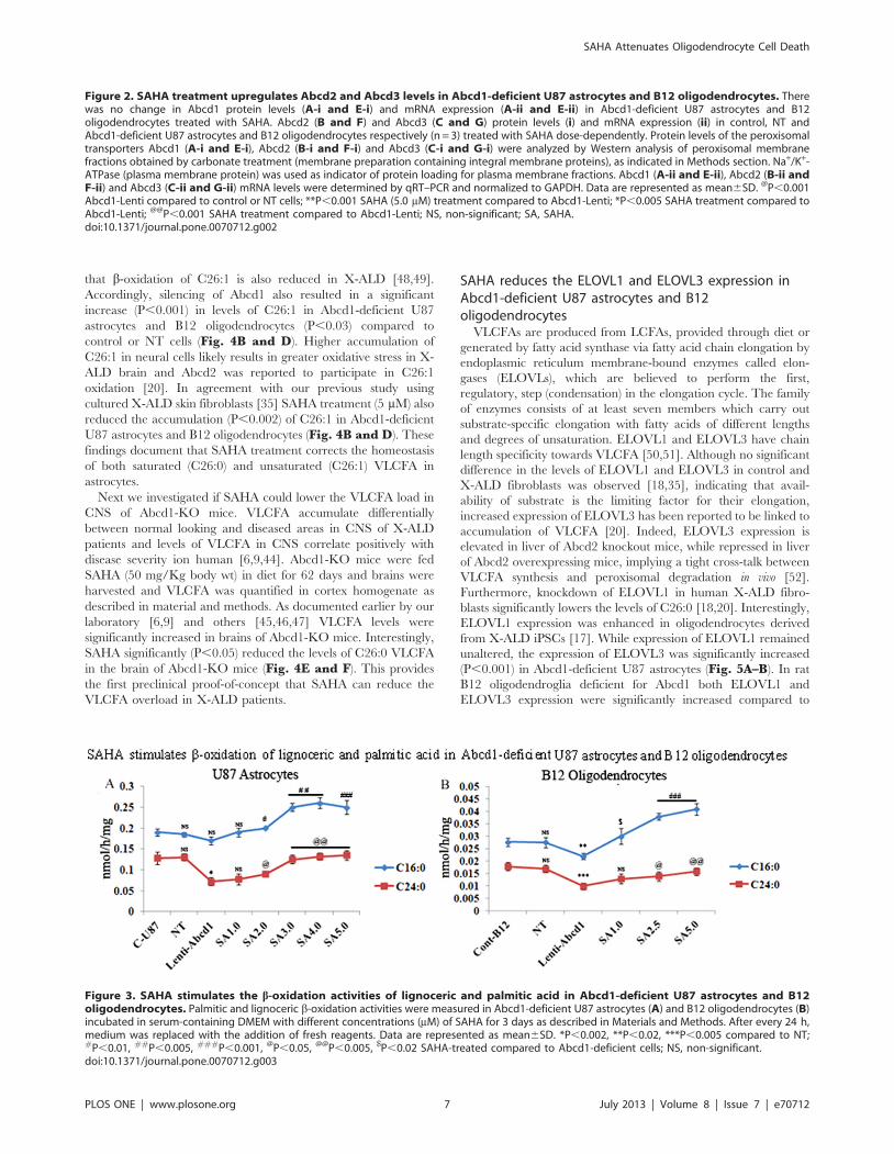
Induction of Abcd2 and Abcd3 gene expression by SAHAcorrelates with normalization of fatty acid b-oxidation inAbcd1-silenced U87 astrocytes and B12oligodendrocytes
Since mutation/deletion of Abcd1 results in deficient VLCFA
b-oxidation [4], we examined these activities in Abcd1-silenced
astrocytes and oligodendrocytes with or without SAHA treatment
(Fig. 3A and B). Using radiolabeled FAs, we observed that the
rate of peroxisomal b-oxidation (lignoceric acid) activity was
reduced in Abcd1-deficient U87 astrocytes (46%) and Abcd1-
deficient B12 oligodendrocytes (41%) as compared to respective
control and NT-treated cells (Fig. 3A–B). While there was a
relatively small decrease (statistically non-significant) in palmitic
acid b-oxidation activity in Abcd1-silenced U87 astrocytes
(Fig. 3A), it was reduced by approximately 20% in B12
oligodendrocytes deficient for Abcd1 (Fig. 3B). These observa-
tions document that deletion of Abcd1 results in decreased
peroxisomal b-oxidation in astrocytes and oligodendrocytes and,
interestingly, a partial inhibition of mitochondrial (palmitic acid)
oxidation activity in oligodendrocytes.
Overexpression of Abcd2 in whole nervous system or in
fibroblasts from Abcd1-deficient mice or from X-ALD patients is
shown to restore peroxisomal b-oxidation and to reduce excessive
accumulation of VLCFA [34,38,40,41,42,43]. Recently we also
reported that SAHA treatment increased the lignoceric acid b-
oxidation activity in X-ALD fibroblasts [35]. This effect was
mediated through induction of Abcd2 while the increase in
palmitic acid oxidation by SAHA was independent of Abcd2
expression [7,35]. Since SAHA upregulated the mRNA expression
of Abcd2 and Abcd3 in U87 astrocytes and B12 oligodendrocytes,
we next examined the effect of this induction on b-oxidation of
lignoceric acid (C24:0) as well as palmitic acid (C16:0). SAHA
treatment increased the lignoceric acid oxidation in a dose-
dependent manner in Abcd1-silenced astrocytes and oligodendro-
cytes (Fig. 3A–B). The highest dose of SAHA (5 mM) at 72 h was
found to stimulate lignoceric acid b-oxidation by approximately
45% in U87 astrocytes and by 38% in B12 oligodendrocytes
deficient in Abcd1 (Fig. 3A–B). SAHA treatment also significantly
increased the palmitic acid b-oxidation activities (Fig. 3A–B),
though maximum activity was reached at day 3 with no further
significant increase in oxidation. Increase of both lignoceric and
palmitic acids b-oxidation activity in SAHA treated U87 astrocytes
and B12 oligodendrocytes deficient in Abcd1 suggests that SAHA
treatment has the potential of enhancing fatty acid oxidation in X-
ALD brain.
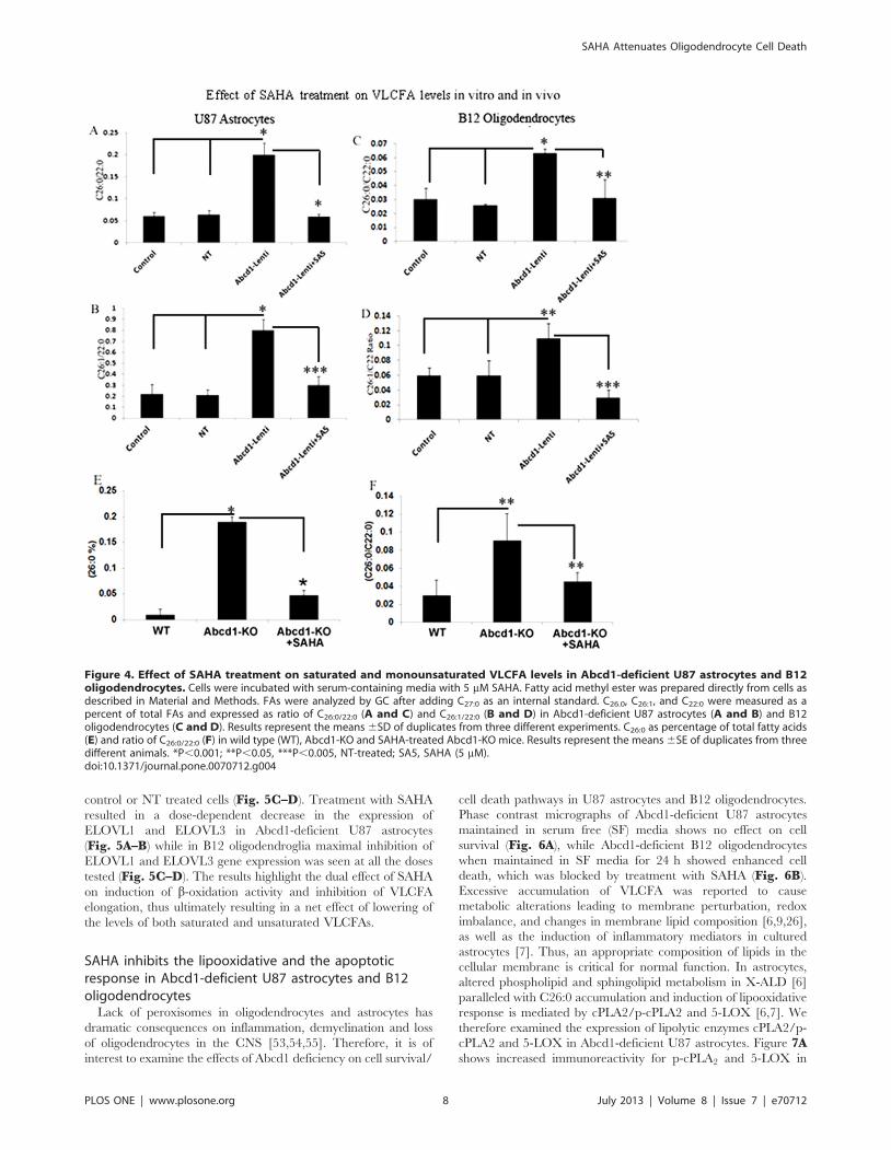
SAHA reduces the levels of very long chain fatty acids inAbcd1-deficient U87 astrocytes and B-12oligodendrocytes
Dysfunction of ALDP/ABCD1 in X-ALD patients results in
decreased VLCFA b-oxidation activity and accumulation of
VLCFAs. The C26:0/C22:0 ratio is considered a standard
diagnostic tool for the assessment of VLCFA in peroxisomal
disorders. Figure 4A shows a 3.1-fold increase in the C26:0/C22:0
ratio in Abcd1-deficient U87 astrocytes and a 3.15 fold increase in
Abcd1-deficient B12 oligodendrocytes when compared with their
respective control or NT-treated cells. Since SAHA increased the
b-oxidation activity of lignoceric acid in Abcd1-deficient U87
astrocytes, we also examined the effect of SAHA on the levels of
VLCFA in these cells. As expected a significant (P,0.001)
decrease was observed in VLCFA levels in SAHA (5 mM) treated
Abcd1-deficient U87 astrocytes as well as B12 oligodendrocytes
(Fig. 4A and C). These observations document that increased
accumulation of VLCFA as a result of Abcd1-deficiency in
astrocytes and oligodendrocytes can be attenuated by SAHA
treatment.
In addition to saturated VLCFA, there is also accumulation of
monounsaturated fatty acids (C26:1) in plasma, fibroblasts and
brain of X-ALD patients as well as in Abcd1-KO mouse indicating
Lenti) particles. Following puromycin selection there was a near-complete selection of transduced cells (A and E). Lentiviral-shRNAs induced genesilencing, indicated by a significant decrease in Abcd1 expression, analyzed by Western blot immunoassay detected with monoclonal antibodyagainst Abcd1 protein levels (B-i and F-i) and qRT-PCR (B-ii and F-ii). Abcd2 (C and G) and Abcd3 (D and H) protein levels (i) and mRNAexpression (ii) in control, NT and Abcd1-deficient U87 astrocytes (n = 3) and B12 oligodendrocytes. Abcd2 and Abcd3 mRNA levels were determinedby quantitative realtime qRT–PCR and normalized to GAPDH. Data are represented as mean6SD. Protein levels of the peroxisomal transportersAbcd1 (B-i and F-i), Abcd2 (C-i and G-i) and Abcd3 (D-i and H-i) were analyzed by Western blot in membranes fractions obtained by carbonatetreatment (membrane preparation containing integral membrane proteins), as indicated in Methods section. Na+/K+-ATPase (plasma membraneprotein) was used as indicator of protein loading for plasma membrane fractions. *P,0.001 Abcd1-Lenti compared to control or NT cells.doi:10.1371/journal.pone.0070712.g001
SAHA Attenuates Oligodendrocyte Cell Death
PLOS ONE | www.plosone.org 5 July 2013 | Volume 8 | Issue 7 | e70712
SAHA Attenuates Oligodendrocyte Cell Death
PLOS ONE | www.plosone.org 6 July 2013 | Volume 8 | Issue 7 | e70712
that b-oxidation of C26:1 is also reduced in X-ALD [48,49].
Accordingly, silencing of Abcd1 also resulted in a significant
increase (P,0.001) in levels of C26:1 in Abcd1-deficient U87
astrocytes and B12 oligodendrocytes (P,0.03) compared to
control or NT cells (Fig. 4B and D). Higher accumulation of
C26:1 in neural cells likely results in greater oxidative stress in X-
ALD brain and Abcd2 was reported to participate in C26:1
oxidation [20]. In agreement with our previous study using
cultured X-ALD skin fibroblasts [35] SAHA treatment (5 mM) also
reduced the accumulation (P,0.002) of C26:1 in Abcd1-deficient
U87 astrocytes and B12 oligodendrocytes (Fig. 4B and D). These
findings document that SAHA treatment corrects the homeostasis
of both saturated (C26:0) and unsaturated (C26:1) VLCFA in
astrocytes.
Next we investigated if SAHA could lower the VLCFA load in
CNS of Abcd1-KO mice. VLCFA accumulate differentially
between normal looking and diseased areas in CNS of X-ALD
patients and levels of VLCFA in CNS correlate positively with
disease severity ion human [6,9,44]. Abcd1-KO mice were fed
SAHA (50 mg/Kg body wt) in diet for 62 days and brains were
harvested and VLCFA was quantified in cortex homogenate as
described in material and methods. As documented earlier by our
laboratory [6,9] and others [45,46,47] VLCFA levels were
significantly increased in brains of Abcd1-KO mice. Interestingly,
SAHA significantly (P,0.05) reduced the levels of C26:0 VLCFA
in the brain of Abcd1-KO mice (Fig. 4E and F). This provides
the first preclinical proof-of-concept that SAHA can reduce the
VLCFA overload in X-ALD patients.
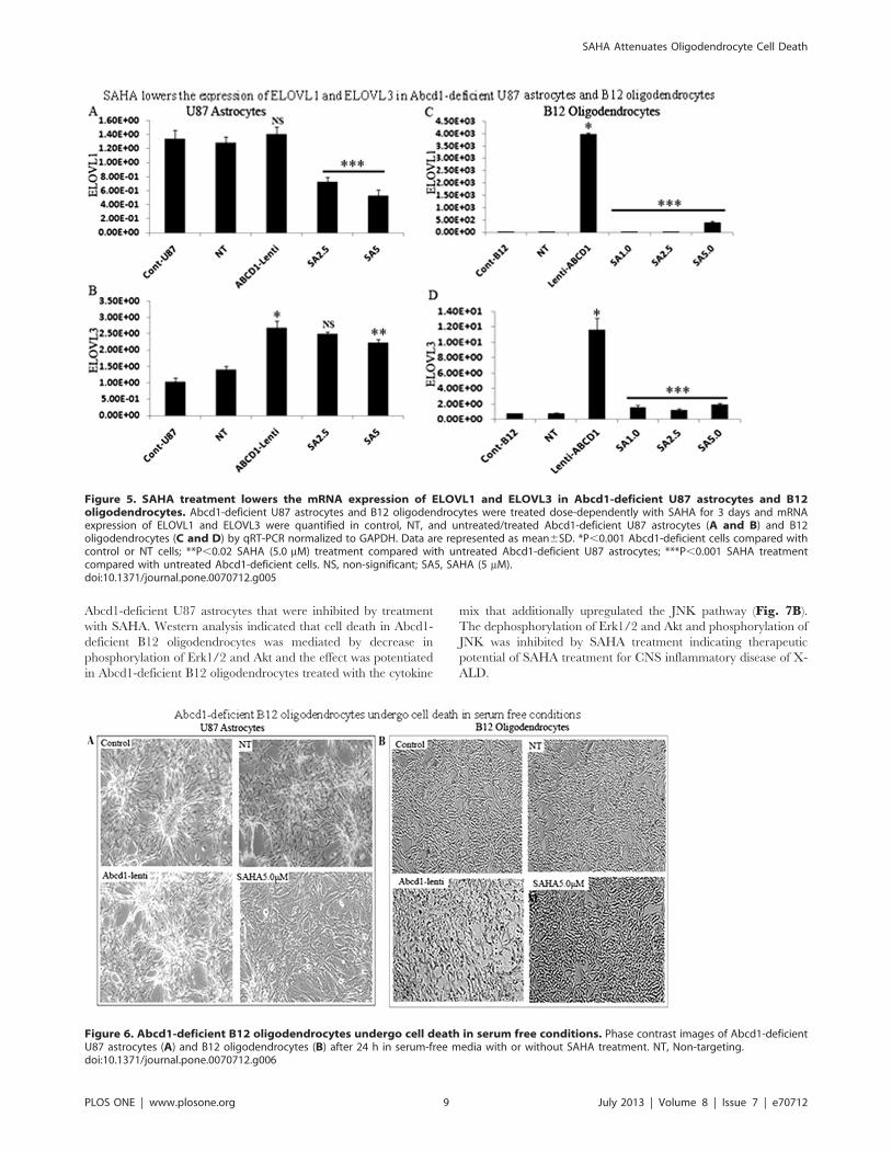
SAHA reduces the ELOVL1 and ELOVL3 expression inAbcd1-deficient U87 astrocytes and B12oligodendrocytes
VLCFAs are produced from LCFAs, provided through diet or
generated by fatty acid synthase via fatty acid chain elongation by
endoplasmic reticulum membrane-bound enzymes called elon-
gases (ELOVLs), which are believed to perform the first,
regulatory, step (condensation) in the elongation cycle. The family
of enzymes consists of at least seven members which carry out
substrate-specific elongation with fatty acids of different lengths
and degrees of unsaturation. ELOVL1 and ELOVL3 have chain
length specificity towards VLCFA [50,51]. Although no significant
difference in the levels of ELOVL1 and ELOVL3 in control and
X-ALD fibroblasts was observed [18,35], indicating that avail-
ability of substrate is the limiting factor for their elongation,
increased expression of ELOVL3 has been reported to be linked to
accumulation of VLCFA [20]. Indeed, ELOVL3 expression is
elevated in liver of Abcd2 knockout mice, while repressed in liver
of Abcd2 overexpressing mice, implying a tight cross-talk between
VLCFA synthesis and peroxisomal degradation in vivo [52].
Furthermore, knockdown of ELOVL1 in human X-ALD fibro-
blasts significantly lowers the levels of C26:0 [18,20]. Interestingly,
ELOVL1 expression was enhanced in oligodendrocytes derived
from X-ALD iPSCs [17]. While expression of ELOVL1 remained
unaltered, the expression of ELOVL3 was significantly increased
(P,0.001) in Abcd1-deficient U87 astrocytes (Fig. 5A–B). In rat
B12 oligodendroglia deficient for Abcd1 both ELOVL1 and
ELOVL3 expression were significantly increased compared to
Figure 2. SAHA treatment upregulates Abcd2 and Abcd3 levels in Abcd1-deficient U87 astrocytes and B12 oligodendrocytes. Therewas no change in Abcd1 protein levels (A-i and E-i) and mRNA expression (A-ii and E-ii) in Abcd1-deficient U87 astrocytes and B12oligodendrocytes treated with SAHA. Abcd2 (B and F) and Abcd3 (C and G) protein levels (i) and mRNA expression (ii) in control, NT andAbcd1-deficient U87 astrocytes and B12 oligodendrocytes respectively (n = 3) treated with SAHA dose-dependently. Protein levels of the peroxisomaltransporters Abcd1 (A-i and E-i), Abcd2 (B-i and F-i) and Abcd3 (C-i and G-i) were analyzed by Western analysis of peroxisomal membranefractions obtained by carbonate treatment (membrane preparation containing integral membrane proteins), as indicated in Methods section. Na+/K+-ATPase (plasma membrane protein) was used as indicator of protein loading for plasma membrane fractions. Abcd1 (A-ii and E-ii), Abcd2 (B-ii andF-ii) and Abcd3 (C-ii and G-ii) mRNA levels were determined by qRT–PCR and normalized to GAPDH. Data are represented as mean6SD. @P,0.001Abcd1-Lenti compared to control or NT cells; **P,0.001 SAHA (5.0 mM) treatment compared to Abcd1-Lenti; *P,0.005 SAHA treatment compared toAbcd1-Lenti; @@P,0.001 SAHA treatment compared to Abcd1-Lenti; NS, non-significant; SA, SAHA.doi:10.1371/journal.pone.0070712.g002
Figure 3. SAHA stimulates the b-oxidation activities of lignoceric and palmitic acid in Abcd1-deficient U87 astrocytes and B12oligodendrocytes. Palmitic and lignoceric b-oxidation activities were measured in Abcd1-deficient U87 astrocytes (A) and B12 oligodendrocytes (B)incubated in serum-containing DMEM with different concentrations (mM) of SAHA for 3 days as described in Materials and Methods. After every 24 h,medium was replaced with the addition of fresh reagents. Data are represented as mean6SD. *P,0.002, **P,0.02, ***P,0.005 compared to NT;#P,0.01, ##P,0.005, ###P,0.001, @P,0.05, @@P,0.005, $P,0.02 SAHA-treated compared to Abcd1-deficient cells; NS, non-significant.doi:10.1371/journal.pone.0070712.g003
SAHA Attenuates Oligodendrocyte Cell Death
PLOS ONE | www.plosone.org 7 July 2013 | Volume 8 | Issue 7 | e70712
control or NT treated cells (Fig. 5C–D). Treatment with SAHA
resulted in a dose-dependent decrease in the expression of
ELOVL1 and ELOVL3 in Abcd1-deficient U87 astrocytes
(Fig. 5A–B) while in B12 oligodendroglia maximal inhibition of
ELOVL1 and ELOVL3 gene expression was seen at all the doses
tested (Fig. 5C–D). The results highlight the dual effect of SAHA
on induction of b-oxidation activity and inhibition of VLCFA
elongation, thus ultimately resulting in a net effect of lowering of
the levels of both saturated and unsaturated VLCFAs.
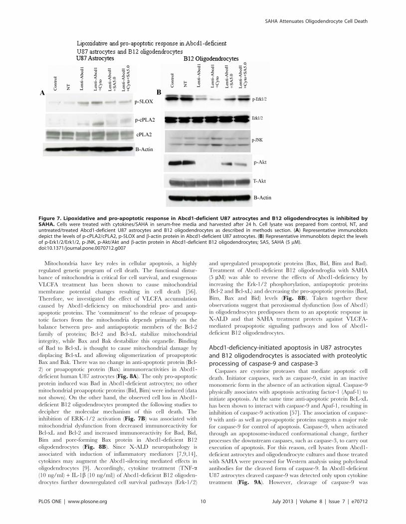
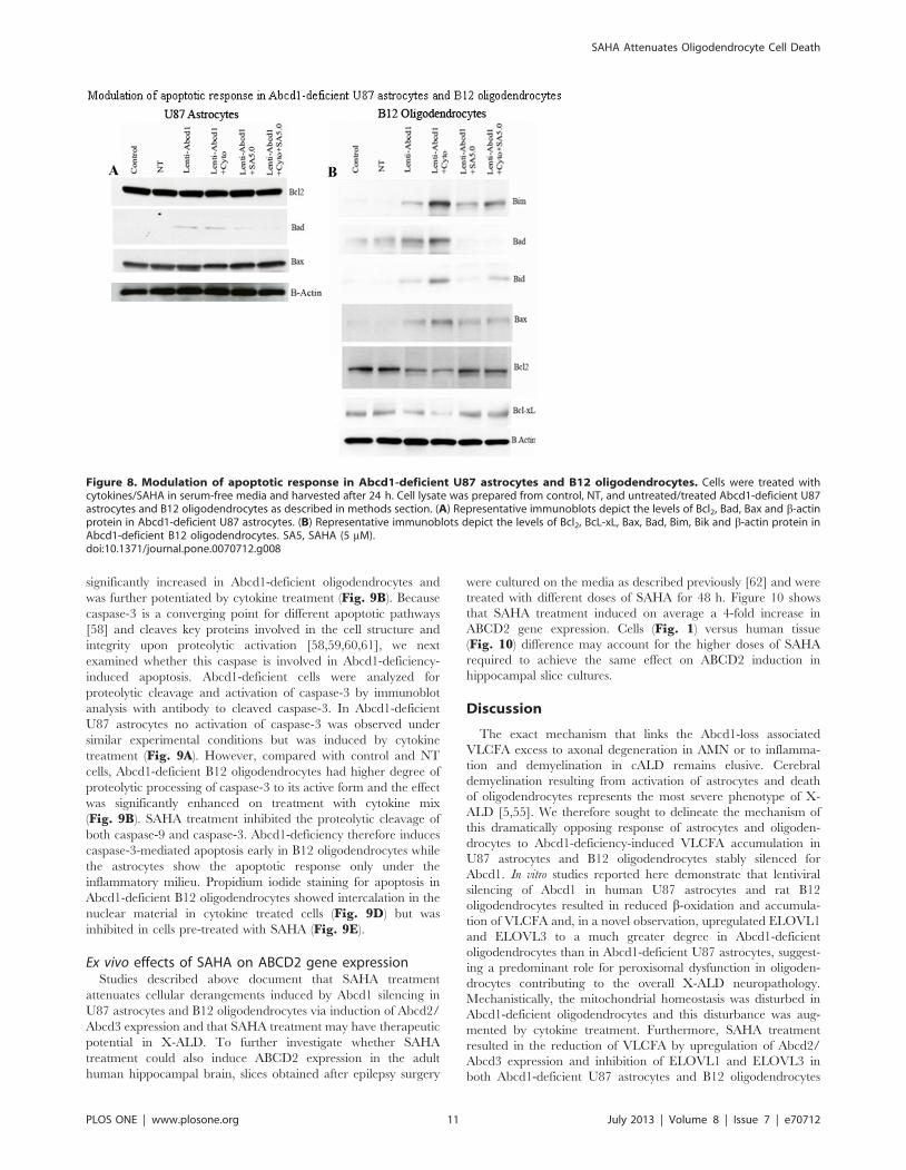
SAHA inhibits the lipooxidative and the apoptoticresponse in Abcd1-deficient U87 astrocytes and B12oligodendrocytes
Lack of peroxisomes in oligodendrocytes and astrocytes has
dramatic consequences on inflammation, demyelination and loss
of oligodendrocytes in the CNS [53,54,55]. Therefore, it is of
interest to examine the effects of Abcd1 deficiency on cell survival/
cell death pathways in U87 astrocytes and B12 oligodendrocytes.
Phase contrast micrographs of Abcd1-deficient U87 astrocytes
maintained in serum free (SF) media shows no effect on cell
survival (Fig. 6A), while Abcd1-deficient B12 oligodendrocytes
when maintained in SF media for 24 h showed enhanced cell
death, which was blocked by treatment with SAHA (Fig. 6B).
Excessive accumulation of VLCFA was reported to cause
metabolic alterations leading to membrane perturbation, redox
imbalance, and changes in membrane lipid composition [6,9,26],
as well as the induction of inflammatory mediators in cultured
astrocytes [7]. Thus, an appropriate composition of lipids in the
cellular membrane is critical for normal function. In astrocytes,
altered phospholipid and sphingolipid metabolism in X-ALD [6]
paralleled with C26:0 accumulation and induction of lipooxidative
response is mediated by cPLA2/p-cPLA2 and 5-LOX [6,7]. We
therefore examined the expression of lipolytic enzymes cPLA2/p-
cPLA2 and 5-LOX in Abcd1-deficient U87 astrocytes. Figure 7Ashows increased immunoreactivity for p-cPLA2 and 5-LOX in
Figure 4. Effect of SAHA treatment on saturated and monounsaturated VLCFA levels in Abcd1-deficient U87 astrocytes and B12oligodendrocytes. Cells were incubated with serum-containing media with 5 mM SAHA. Fatty acid methyl ester was prepared directly from cells asdescribed in Material and Methods. FAs were analyzed by GC after adding C27:0 as an internal standard. C26.0, C26:1, and C22:0 were measured as apercent of total FAs and expressed as ratio of C26:0/22:0 (A and C) and C26:1/22:0 (B and D) in Abcd1-deficient U87 astrocytes (A and B) and B12oligodendrocytes (C and D). Results represent the means 6SD of duplicates from three different experiments. C26:0 as percentage of total fatty acids(E) and ratio of C26:0/22:0 (F) in wild type (WT), Abcd1-KO and SAHA-treated Abcd1-KO mice. Results represent the means 6SE of duplicates from threedifferent animals. *P,0.001; **P,0.05, ***P,0.005, NT-treated; SA5, SAHA (5 mM).doi:10.1371/journal.pone.0070712.g004
SAHA Attenuates Oligodendrocyte Cell Death
PLOS ONE | www.plosone.org 8 July 2013 | Volume 8 | Issue 7 | e70712
Abcd1-deficient U87 astrocytes that were inhibited by treatment
with SAHA. Western analysis indicated that cell death in Abcd1-
deficient B12 oligodendrocytes was mediated by decrease in
phosphorylation of Erk1/2 and Akt and the effect was potentiated
in Abcd1-deficient B12 oligodendrocytes treated with the cytokine
mix that additionally upregulated the JNK pathway (Fig. 7B).
The dephosphorylation of Erk1/2 and Akt and phosphorylation of
JNK was inhibited by SAHA treatment indicating therapeutic
potential of SAHA treatment for CNS inflammatory disease of X-
ALD.
Figure 5. SAHA treatment lowers the mRNA expression of ELOVL1 and ELOVL3 in Abcd1-deficient U87 astrocytes and B12oligodendrocytes. Abcd1-deficient U87 astrocytes and B12 oligodendrocytes were treated dose-dependently with SAHA for 3 days and mRNAexpression of ELOVL1 and ELOVL3 were quantified in control, NT, and untreated/treated Abcd1-deficient U87 astrocytes (A and B) and B12oligodendrocytes (C and D) by qRT-PCR normalized to GAPDH. Data are represented as mean6SD. *P,0.001 Abcd1-deficient cells compared withcontrol or NT cells; **P,0.02 SAHA (5.0 mM) treatment compared with untreated Abcd1-deficient U87 astrocytes; ***P,0.001 SAHA treatmentcompared with untreated Abcd1-deficient cells. NS, non-significant; SA5, SAHA (5 mM).doi:10.1371/journal.pone.0070712.g005
Figure 6. Abcd1-deficient B12 oligodendrocytes undergo cell death in serum free conditions. Phase contrast images of Abcd1-deficientU87 astrocytes (A) and B12 oligodendrocytes (B) after 24 h in serum-free media with or without SAHA treatment. NT, Non-targeting.doi:10.1371/journal.pone.0070712.g006
SAHA Attenuates Oligodendrocyte Cell Death
PLOS ONE | www.plosone.org 9 July 2013 | Volume 8 | Issue 7 | e70712
Mitochondria have key roles in cellular apoptosis, a highly
regulated genetic program of cell death. The functional distur-
bance of mitochondria is critical for cell survival, and exogenous
VLCFA treatment has been shown to cause mitochondrial
membrane potential changes resulting in cell death [56].
Therefore, we investigated the effect of VLCFA accumulation
caused by Abcd1-deficiency on mitochondrial pro- and anti-
apoptotic proteins. The ‘commitment’ to the release of proapop-
totic factors from the mitochondria depends primarily on the
balance between pro- and antiapoptotic members of the Bcl-2
family of proteins; Bcl-2 and Bcl-xL stabilize mitochondrial
integrity, while Bax and Bak destabilize this organelle. Binding
of Bad to Bcl-xL is thought to cause mitochondrial damage by
displacing Bcl-xL and allowing oligomerization of proapoptotic
Bax and Bak. There was no change in anti-apoptotic protein (Bcl-
2) or proapoptotic protein (Bax) immunoreactivities in Abcd1-
deficient human U87 astrocytes (Fig. 8A). The only pro-apoptotic
protein induced was Bad in Abcd1-deficient astrocytes; no other
mitochondrial proapoptotic proteins (Bid, Bim) were induced (data
not shown). On the other hand, the observed cell loss in Abcd1-
deficient B12 oligodendrocytes prompted the following studies to
decipher the molecular mechanism of this cell death. The
inhibition of ERK-1/2 activation (Fig. 7B) was associated with
mitochondrial dysfunction from decreased immunoreactivity for
Bcl-xL and Bcl-2 and increased immunoreactivity for Bad, Bid,
Bim and pore-forming Bax protein in Abcd1-deficient B12
oligodendrocytes (Fig. 8B). Since X-ALD neuropathology is
associated with induction of inflammatory mediators [7,9,14],
cytokines may augment the Abcd1-silencing mediated effects in
oligodendrocytes [9]. Accordingly, cytokine treatment (TNF-a(10 ng/ml) + IL-1b (10 ng/ml)) of Abcd1-deficient B12 oligoden-
drocytes further downregulated cell survival pathways (Erk-1/2)
and upregulated proapoptotic proteins (Bax, Bid, Bim and Bad).
Treatment of Abcd1-deficient B12 oligodendroglia with SAHA
(5 mM) was able to reverse the effects of Abcd1-deficiency by
increasing the Erk-1/2 phosphorylation, antiapoptotic proteins
(Bcl-2 and Bcl-xL) and decreasing the pro-apoptotic proteins (Bad,
Bim, Bax and Bid) levels (Fig. 8B). Taken together these
observations suggest that peroxisomal dysfunction (loss of Abcd1)
in oligodendrocytes predisposes them to an apoptotic response in
X-ALD and that SAHA treatment protects against VLCFA-
mediated proapoptotic signaling pathways and loss of Abcd1-
deficient B12 oligodendrocytes.
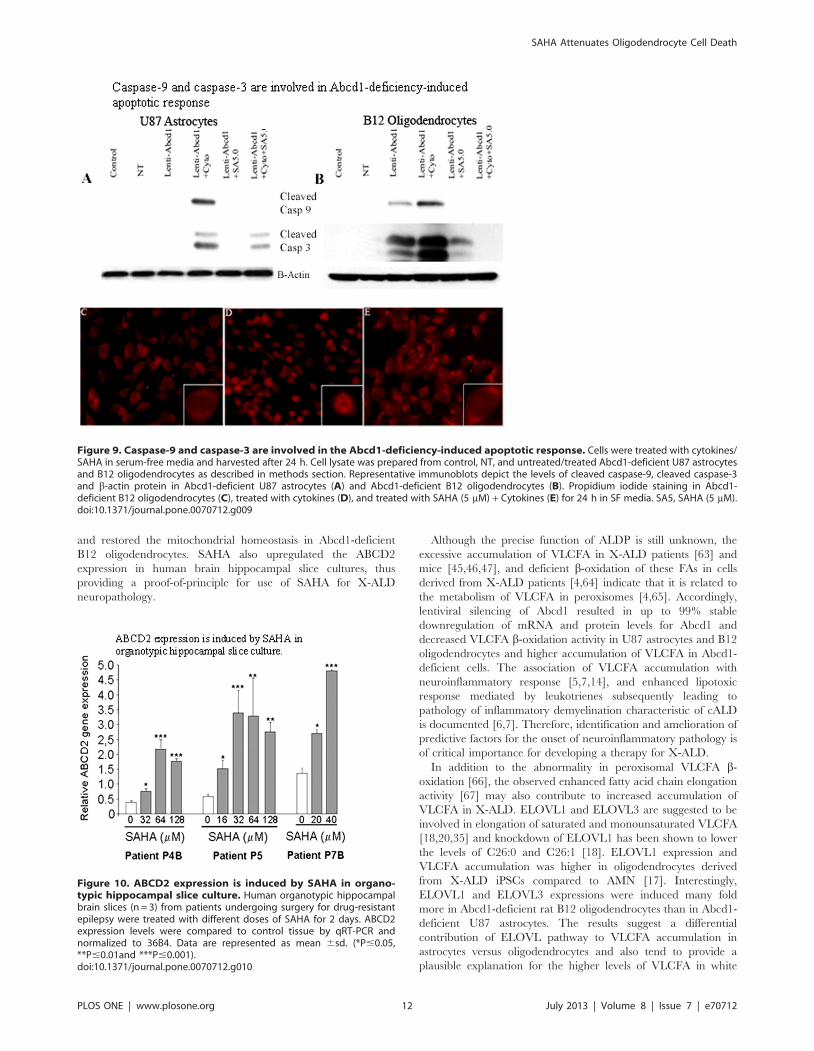
Abcd1-deficiency-initiated apoptosis in U87 astrocytesand B12 oligodendrocytes is associated with proteolyticprocessing of caspase-9 and caspase-3
Caspases are cysteine proteases that mediate apoptotic cell
death. Initiator caspases, such as caspase-9, exist in an inactive
monomeric form in the absence of an activation signal. Caspase-9
physically associates with apoptosis activating factor-1 (Apaf-1) to
initiate apoptosis. At the same time anti-apoptotic protein BcL-xL
has been shown to interact with caspase-9 and Apaf-1, resulting in
inhibition of caspase-9 activation [57]. The association of caspase-
9 with anti- as well as pro-apoptotic proteins suggests a major role
for caspase-9 for control of apoptosis. Caspase-9, when activated
through an apoptosome-induced conformational change, further
processes the downstream caspases, such as caspase-3, to carry out
execution of apoptosis. For this reason, cell lysates from Abcd1-
deficient astrocytes and oligodendrocyte cultures and those treated
with SAHA were processed for Western analysis using polyclonal
antibodies for the cleaved form of caspase-9. In Abcd1-deficient
U87 astrocytes cleaved caspase-9 was detected only upon cytokine
treatment (Fig. 9A). However, cleavage of caspase-9 was
Figure 7. Lipoxidative and pro-apoptotic response in Abcd1-deficient U87 astrocytes and B12 oligodendrocytes is inhibited bySAHA. Cells were treated with cytokines/SAHA in serum-free media and harvested after 24 h. Cell lysate was prepared from control, NT, anduntreated/treated Abcd1-deficient U87 astrocytes and B12 oligodendrocytes as described in methods section. (A) Representative immunoblotsdepict the levels of p-cPLA2/cPLA2, p-5LOX and b-actin protein in Abcd1-deficient U87 astrocytes. (B) Representative immunoblots depict the levelsof p-Erk1/2/Erk1/2, p-JNK, p-Akt/Akt and b-actin protein in Abcd1-deficient B12 oligodendrocytes; SA5, SAHA (5 mM).doi:10.1371/journal.pone.0070712.g007
SAHA Attenuates Oligodendrocyte Cell Death
PLOS ONE | www.plosone.org 10 July 2013 | Volume 8 | Issue 7 | e70712
significantly increased in Abcd1-deficient oligodendrocytes and
was further potentiated by cytokine treatment (Fig. 9B). Because
caspase-3 is a converging point for different apoptotic pathways
[58] and cleaves key proteins involved in the cell structure and
integrity upon proteolytic activation [58,59,60,61], we next
examined whether this caspase is involved in Abcd1-deficiency-
induced apoptosis. Abcd1-deficient cells were analyzed for
proteolytic cleavage and activation of caspase-3 by immunoblot
analysis with antibody to cleaved caspase-3. In Abcd1-deficient
U87 astrocytes no activation of caspase-3 was observed under
similar experimental conditions but was induced by cytokine
treatment (Fig. 9A). However, compared with control and NT
cells, Abcd1-deficient B12 oligodendrocytes had higher degree of
proteolytic processing of caspase-3 to its active form and the effect
was significantly enhanced on treatment with cytokine mix
(Fig. 9B). SAHA treatment inhibited the proteolytic cleavage of
both caspase-9 and caspase-3. Abcd1-deficiency therefore induces
caspase-3-mediated apoptosis early in B12 oligodendrocytes while
the astrocytes show the apoptotic response only under the
inflammatory milieu. Propidium iodide staining for apoptosis in
Abcd1-deficient B12 oligodendrocytes showed intercalation in the
nuclear material in cytokine treated cells (Fig. 9D) but was
inhibited in cells pre-treated with SAHA (Fig. 9E).
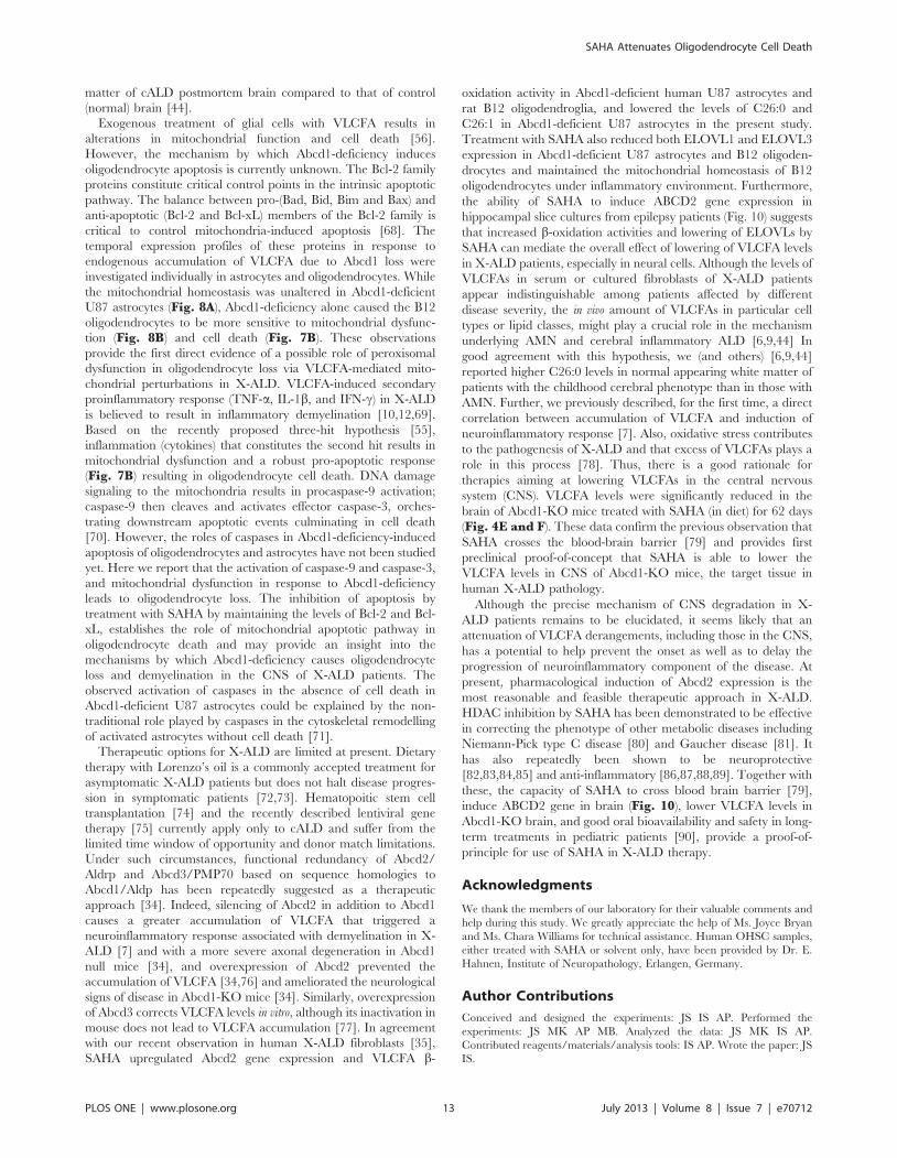
Ex vivo effects of SAHA on ABCD2 gene expressionStudies described above document that SAHA treatment
attenuates cellular derangements induced by Abcd1 silencing in
U87 astrocytes and B12 oligodendrocytes via induction of Abcd2/
Abcd3 expression and that SAHA treatment may have therapeutic
potential in X-ALD. To further investigate whether SAHA
treatment could also induce ABCD2 expression in the adult
human hippocampal brain, slices obtained after epilepsy surgery
were cultured on the media as described previously [62] and were
treated with different doses of SAHA for 48 h. Figure 10 shows
that SAHA treatment induced on average a 4-fold increase in
ABCD2 gene expression. Cells (Fig. 1) versus human tissue
(Fig. 10) difference may account for the higher doses of SAHA
required to achieve the same effect on ABCD2 induction in
hippocampal slice cultures.
Discussion
The exact mechanism that links the Abcd1-loss associated
VLCFA excess to axonal degeneration in AMN or to inflamma-
tion and demyelination in cALD remains elusive. Cerebral
demyelination resulting from activation of astrocytes and death
of oligodendrocytes represents the most severe phenotype of X-
ALD [5,55]. We therefore sought to delineate the mechanism of
this dramatically opposing response of astrocytes and oligoden-
drocytes to Abcd1-deficiency-induced VLCFA accumulation in
U87 astrocytes and B12 oligodendrocytes stably silenced for
Abcd1. In vitro studies reported here demonstrate that lentiviral
silencing of Abcd1 in human U87 astrocytes and rat B12
oligodendrocytes resulted in reduced b-oxidation and accumula-
tion of VLCFA and, in a novel observation, upregulated ELOVL1
and ELOVL3 to a much greater degree in Abcd1-deficient
oligodendrocytes than in Abcd1-deficient U87 astrocytes, suggest-
ing a predominant role for peroxisomal dysfunction in oligoden-
drocytes contributing to the overall X-ALD neuropathology.
Mechanistically, the mitochondrial homeostasis was disturbed in
Abcd1-deficient oligodendrocytes and this disturbance was aug-
mented by cytokine treatment. Furthermore, SAHA treatment
resulted in the reduction of VLCFA by upregulation of Abcd2/
Abcd3 expression and inhibition of ELOVL1 and ELOVL3 in
both Abcd1-deficient U87 astrocytes and B12 oligodendrocytes
Figure 8. Modulation of apoptotic response in Abcd1-deficient U87 astrocytes and B12 oligodendrocytes. Cells were treated withcytokines/SAHA in serum-free media and harvested after 24 h. Cell lysate was prepared from control, NT, and untreated/treated Abcd1-deficient U87astrocytes and B12 oligodendrocytes as described in methods section. (A) Representative immunoblots depict the levels of Bcl2, Bad, Bax and b-actinprotein in Abcd1-deficient U87 astrocytes. (B) Representative immunoblots depict the levels of Bcl2, BcL-xL, Bax, Bad, Bim, Bik and b-actin protein inAbcd1-deficient B12 oligodendrocytes. SA5, SAHA (5 mM).doi:10.1371/journal.pone.0070712.g008
SAHA Attenuates Oligodendrocyte Cell Death
PLOS ONE | www.plosone.org 11 July 2013 | Volume 8 | Issue 7 | e70712
and restored the mitochondrial homeostasis in Abcd1-deficient
B12 oligodendrocytes. SAHA also upregulated the ABCD2
expression in human brain hippocampal slice cultures, thus
providing a proof-of-principle for use of SAHA for X-ALD
neuropathology.
Although the precise function of ALDP is still unknown, the
excessive accumulation of VLCFA in X-ALD patients [63] and
mice [45,46,47], and deficient b-oxidation of these FAs in cells
derived from X-ALD patients [4,64] indicate that it is related to
the metabolism of VLCFA in peroxisomes [4,65]. Accordingly,
lentiviral silencing of Abcd1 resulted in up to 99% stable
downregulation of mRNA and protein levels for Abcd1 and
decreased VLCFA b-oxidation activity in U87 astrocytes and B12
oligodendrocytes and higher accumulation of VLCFA in Abcd1-
deficient cells. The association of VLCFA accumulation with
neuroinflammatory response [5,7,14], and enhanced lipotoxic
response mediated by leukotrienes subsequently leading to
pathology of inflammatory demyelination characteristic of cALD
is documented [6,7]. Therefore, identification and amelioration of
predictive factors for the onset of neuroinflammatory pathology is
of critical importance for developing a therapy for X-ALD.
In addition to the abnormality in peroxisomal VLCFA b-
oxidation [66], the observed enhanced fatty acid chain elongation
activity [67] may also contribute to increased accumulation of
VLCFA in X-ALD. ELOVL1 and ELOVL3 are suggested to be
involved in elongation of saturated and monounsaturated VLCFA
[18,20,35] and knockdown of ELOVL1 has been shown to lower
the levels of C26:0 and C26:1 [18]. ELOVL1 expression and
VLCFA accumulation was higher in oligodendrocytes derived
from X-ALD iPSCs compared to AMN [17]. Interestingly,
ELOVL1 and ELOVL3 expressions were induced many fold
more in Abcd1-deficient rat B12 oligodendrocytes than in Abcd1-
deficient U87 astrocytes. The results suggest a differential
contribution of ELOVL pathway to VLCFA accumulation in
astrocytes versus oligodendrocytes and also tend to provide a
plausible explanation for the higher levels of VLCFA in white
Figure 9. Caspase-9 and caspase-3 are involved in the Abcd1-deficiency-induced apoptotic response. Cells were treated with cytokines/SAHA in serum-free media and harvested after 24 h. Cell lysate was prepared from control, NT, and untreated/treated Abcd1-deficient U87 astrocytesand B12 oligodendrocytes as described in methods section. Representative immunoblots depict the levels of cleaved caspase-9, cleaved caspase-3and b-actin protein in Abcd1-deficient U87 astrocytes (A) and Abcd1-deficient B12 oligodendrocytes (B). Propidium iodide staining in Abcd1-deficient B12 oligodendrocytes (C), treated with cytokines (D), and treated with SAHA (5 mM) + Cytokines (E) for 24 h in SF media. SA5, SAHA (5 mM).doi:10.1371/journal.pone.0070712.g009
Figure 10. ABCD2 expression is induced by SAHA in organo-typic hippocampal slice culture. Human organotypic hippocampalbrain slices (n = 3) from patients undergoing surgery for drug-resistantepilepsy were treated with different doses of SAHA for 2 days. ABCD2expression levels were compared to control tissue by qRT-PCR andnormalized to 36B4. Data are represented as mean 6sd. (*P#0.05,**P#0.01and ***P#0.001).doi:10.1371/journal.pone.0070712.g010
SAHA Attenuates Oligodendrocyte Cell Death
PLOS ONE | www.plosone.org 12 July 2013 | Volume 8 | Issue 7 | e70712
matter of cALD postmortem brain compared to that of control
(normal) brain [44].
Exogenous treatment of glial cells with VLCFA results in
alterations in mitochondrial function and cell death [56].
However, the mechanism by which Abcd1-deficiency induces
oligodendrocyte apoptosis is currently unknown. The Bcl-2 family
proteins constitute critical control points in the intrinsic apoptotic
pathway. The balance between pro-(Bad, Bid, Bim and Bax) and
anti-apoptotic (Bcl-2 and Bcl-xL) members of the Bcl-2 family is
critical to control mitochondria-induced apoptosis [68]. The
temporal expression profiles of these proteins in response to
endogenous accumulation of VLCFA due to Abcd1 loss were
investigated individually in astrocytes and oligodendrocytes. While
the mitochondrial homeostasis was unaltered in Abcd1-deficient
U87 astrocytes (Fig. 8A), Abcd1-deficiency alone caused the B12
oligodendrocytes to be more sensitive to mitochondrial dysfunc-
tion (Fig. 8B) and cell death (Fig. 7B). These observations
provide the first direct evidence of a possible role of peroxisomal
dysfunction in oligodendrocyte loss via VLCFA-mediated mito-
chondrial perturbations in X-ALD. VLCFA-induced secondary
proinflammatory response (TNF-a, IL-1b, and IFN-c) in X-ALD
is believed to result in inflammatory demyelination [10,12,69].
Based on the recently proposed three-hit hypothesis [55],
inflammation (cytokines) that constitutes the second hit results in
mitochondrial dysfunction and a robust pro-apoptotic response
(Fig. 7B) resulting in oligodendrocyte cell death. DNA damage
signaling to the mitochondria results in procaspase-9 activation;
caspase-9 then cleaves and activates effector caspase-3, orches-
trating downstream apoptotic events culminating in cell death
[70]. However, the roles of caspases in Abcd1-deficiency-induced
apoptosis of oligodendrocytes and astrocytes have not been studied
yet. Here we report that the activation of caspase-9 and caspase-3,
and mitochondrial dysfunction in response to Abcd1-deficiency
leads to oligodendrocyte loss. The inhibition of apoptosis by
treatment with SAHA by maintaining the levels of Bcl-2 and Bcl-
xL, establishes the role of mitochondrial apoptotic pathway in
oligodendrocyte death and may provide an insight into the
mechanisms by which Abcd1-deficiency causes oligodendrocyte
loss and demyelination in the CNS of X-ALD patients. The
observed activation of caspases in the absence of cell death in
Abcd1-deficient U87 astrocytes could be explained by the non-
traditional role played by caspases in the cytoskeletal remodelling
of activated astrocytes without cell death [71].
Therapeutic options for X-ALD are limited at present. Dietary
therapy with Lorenzo’s oil is a commonly accepted treatment for
asymptomatic X-ALD patients but does not halt disease progres-
sion in symptomatic patients [72,73]. Hematopoitic stem cell
transplantation [74] and the recently described lentiviral gene
therapy [75] currently apply only to cALD and suffer from the
limited time window of opportunity and donor match limitations.
Under such circumstances, functional redundancy of Abcd2/
Aldrp and Abcd3/PMP70 based on sequence homologies to
Abcd1/Aldp has been repeatedly suggested as a therapeutic
approach [34]. Indeed, silencing of Abcd2 in addition to Abcd1
causes a greater accumulation of VLCFA that triggered a
neuroinflammatory response associated with demyelination in X-
ALD [7] and with a more severe axonal degeneration in Abcd1
null mice [34], and overexpression of Abcd2 prevented the
accumulation of VLCFA [34,76] and ameliorated the neurological
signs of disease in Abcd1-KO mice [34]. Similarly, overexpression
of Abcd3 corrects VLCFA levels in vitro, although its inactivation in
mouse does not lead to VLCFA accumulation [77]. In agreement
with our recent observation in human X-ALD fibroblasts [35],
SAHA upregulated Abcd2 gene expression and VLCFA b-
oxidation activity in Abcd1-deficient human U87 astrocytes and
rat B12 oligodendroglia, and lowered the levels of C26:0 and
C26:1 in Abcd1-deficient U87 astrocytes in the present study.
Treatment with SAHA also reduced both ELOVL1 and ELOVL3
expression in Abcd1-deficient U87 astrocytes and B12 oligoden-
drocytes and maintained the mitochondrial homeostasis of B12
oligodendrocytes under inflammatory environment. Furthermore,
the ability of SAHA to induce ABCD2 gene expression in
hippocampal slice cultures from epilepsy patients (Fig. 10) suggests
that increased b-oxidation activities and lowering of ELOVLs by
SAHA can mediate the overall effect of lowering of VLCFA levels
in X-ALD patients, especially in neural cells. Although the levels of
VLCFAs in serum or cultured fibroblasts of X-ALD patients
appear indistinguishable among patients affected by different
disease severity, the in vivo amount of VLCFAs in particular cell
types or lipid classes, might play a crucial role in the mechanism
underlying AMN and cerebral inflammatory ALD [6,9,44] In
good agreement with this hypothesis, we (and others) [6,9,44]
reported higher C26:0 levels in normal appearing white matter of
patients with the childhood cerebral phenotype than in those with
AMN. Further, we previously described, for the first time, a direct
correlation between accumulation of VLCFA and induction of
neuroinflammatory response [7]. Also, oxidative stress contributes
to the pathogenesis of X-ALD and that excess of VLCFAs plays a
role in this process [78]. Thus, there is a good rationale for
therapies aiming at lowering VLCFAs in the central nervous
system (CNS). VLCFA levels were significantly reduced in the
brain of Abcd1-KO mice treated with SAHA (in diet) for 62 days
(Fig. 4E and F). These data confirm the previous observation that
SAHA crosses the blood-brain barrier [79] and provides first
preclinical proof-of-concept that SAHA is able to lower the
VLCFA levels in CNS of Abcd1-KO mice, the target tissue in
human X-ALD pathology.
Although the precise mechanism of CNS degradation in X-
ALD patients remains to be elucidated, it seems likely that an
attenuation of VLCFA derangements, including those in the CNS,
has a potential to help prevent the onset as well as to delay the
progression of neuroinflammatory component of the disease. At
present, pharmacological induction of Abcd2 expression is the
most reasonable and feasible therapeutic approach in X-ALD.
HDAC inhibition by SAHA has been demonstrated to be effective
in correcting the phenotype of other metabolic diseases including
Niemann-Pick type C disease [80] and Gaucher disease [81]. It
has also repeatedly been shown to be neuroprotective
[82,83,84,85] and anti-inflammatory [86,87,88,89]. Together with
these, the capacity of SAHA to cross blood brain barrier [79],
induce ABCD2 gene in brain (Fig. 10), lower VLCFA levels in
Abcd1-KO brain, and good oral bioavailability and safety in long-
term treatments in pediatric patients [90], provide a proof-of-
principle for use of SAHA in X-ALD therapy.
Acknowledgments
We thank the members of our laboratory for their valuable comments and
help during this study. We greatly appreciate the help of Ms. Joyce Bryan
and Ms. Chara Williams for technical assistance. Human OHSC samples,
either treated with SAHA or solvent only, have been provided by Dr. E.
Hahnen, Institute of Neuropathology, Erlangen, Germany.
Author Contributions
Conceived and designed the experiments: JS IS AP. Performed the
experiments: JS MK AP MB. Analyzed the data: JS MK IS AP.
Contributed reagents/materials/analysis tools: IS AP. Wrote the paper: JS
IS.
SAHA Attenuates Oligodendrocyte Cell Death
PLOS ONE | www.plosone.org 13 July 2013 | Volume 8 | Issue 7 | e70712
References
1. Moser HW (1993) Lorenzo oil therapy for adrenoleukodystrophy: a prematurely
amplified hope. Annals of neurology 34: 121–122.
2. Contreras M, Mosser J, Mandel JL, Aubourg P, Singh I (1994) The protein
coded by the X-adrenoleukodystrophy gene is a peroxisomal integral membrane
protein. FEBS letters 344: 211–215.
3. Contreras M, Sengupta TK, Sheikh F, Aubourg P, Singh I (1996) Topology of
ATP-binding domain of adrenoleukodystrophy gene product in peroxisomes.
Archives of biochemistry and biophysics 334: 369–379.
4. Singh I, Moser AE, Goldfischer S, Moser HW (1984) Lignoceric acid is oxidized
in the peroxisome: implications for the Zellweger cerebro-hepato-renal
syndrome and adrenoleukodystrophy. Proceedings of the National Academy of
Sciences of the United States of America 81: 4203–4207.
5. Moser HW SK, Watkins PA, Powers J, Moser AB (2001) X-linked
adrenoleukodystrophy. In: CR Scriver AB, Sly WS, Valle D, The Metabolic
and Molecular Bases of Inherited Disease. NewYork: McGraw Hill. 3257–3301.
6. Khan M, Singh J, Gilg AG, Uto T, Singh I (2010) Very long-chain fatty acid
accumulation causes lipotoxic response via 5-lipoxygenase in cerebral adreno-
leukodystrophy. Journal of lipid research 51: 1685–1695.
7. Singh J, Khan M, Singh I (2009) Silencing of Abcd1 and Abcd2 genes sensitizes
astrocytes for inflammation: implication for X-adrenoleukodystrophy. Journal of
lipid research 50: 135–147.
8. Di Benedetto R, Denti MA, Salvati S, Attorri L, Di Biase A (2009) PMP70
knock-down generates oxidative stress and pro-inflammatory cytokine produc-
tion in C6 glial cells. Neurochemistry international 54: 37–42.
9. Khan M, Singh J, Singh I (2008) Plasmalogen deficiency in cerebral
adrenoleukodystrophy and its modulation by lovastatin. Journal of neurochem-
istry 106: 1766–1779.
10. Paintlia AS, Gilg AG, Khan M, Singh AK, Barbosa E, et al. (2003) Correlation
of very long chain fatty acid accumulation and inflammatory disease progression
in childhood X-ALD: implications for potential therapies. Neurobiology of
disease 14: 425–439.
11. Fourcade S, Lopez-Erauskin J, Galino J, Duval C, Naudi A, et al. (2008) Early
oxidative damage underlying neurodegeneration in X-adrenoleukodystrophy.
Human molecular genetics 17: 1762–1773.
12. Powers JM, Liu Y, Moser AB, Moser HW (1992) The inflammatory
myelinopathy of adreno-leukodystrophy: cells, effector molecules, and pathoge-
netic implications. Journal of neuropathology and experimental neurology 51:
630–643.
13. Pujol A, Hindelang C, Callizot N, Bartsch U, Schachner M, et al. (2002) Late
onset neurological phenotype of the X-ALD gene inactivation in mice: a mouse
model for adrenomyeloneuropathy. Human molecular genetics 11: 499–505.
14. Schluter A, Espinosa L, Fourcade S, Galino J, Lopez E, et al. (2012) Functional
genomic analysis unravels a metabolic-inflammatory interplay in adrenoleuko-
dystrophy. Human molecular genetics 21: 1062–1077.
15. Gilg AG, Singh AK, Singh I (2000) Inducible nitric oxide synthase in the central
nervous system of patients with X-adrenoleukodystrophy. Journal of neuropa-
thology and experimental neurology 59: 1063–1069.
16. Takuma K, Baba A, Matsuda T (2004) Astrocyte apoptosis: implications for
neuroprotection. Progress in neurobiology 72: 111–127.
17. Jang J, Kang HC, Kim HS, Kim JY, Huh YJ, et al. (2011) Induced pluripotent
stem cell models from X-linked adrenoleukodystrophy patients. Annals of
neurology 70: 402–409.
18. Ofman R, Dijkstra IM, van Roermund CW, Burger N, Turkenburg M, et al.
(2010) The role of ELOVL1 in very long-chain fatty acid homeostasis and X-
linked adrenoleukodystrophy. EMBO molecular medicine 2: 90–97.
19. Khan M, Pahan K, Singh AK, Singh I (1998) Cytokine-induced accumulation of
very long-chain fatty acids in rat C6 glial cells: implication for X-
adrenoleukodystrophy. Journal of neurochemistry 71: 78–87.
20. Fourcade S, Ruiz M, Guilera C, Hahnen E, Brichta L, et al. (2010) Valproic
acid induces antioxidant effects in X-linked adrenoleukodystrophy. Human
molecular genetics 19: 2005–2014.
21. Gondcaille C, Depreter M, Fourcade S, Lecca MR, Leclercq S, et al. (2005)
Phenylbutyrate up-regulates the adrenoleukodystrophy-related gene as a
nonclassical peroxisome proliferator. The Journal of cell biology 169: 93–104.
22. Singh I, Khan M, Key L, Pai S (1998) Lovastatin for X-linked adrenoleuko-
dystrophy. The New England journal of medicine 339: 702–703.
23. Berger J, Albet S, Bentejac M, Netik A, Holzinger A, et al. (1999) The four
murine peroxisomal ABC-transporter genes differ in constitutive, inducible and
developmental expression. European journal of biochemistry/FEBS 265: 719–
727.
24. McGuinness MC, Zhang HP, Smith KD (2001) Evaluation of pharmacological
induction of fatty acid beta-oxidation in X-linked adrenoleukodystrophy.
Molecular genetics and metabolism 74: 256–263.
25. Singh I, Pahan K, Khan M (1998) Lovastatin and sodium phenylacetate
normalize the levels of very long chain fatty acids in skin fibroblasts of X-
adrenoleukodystrophy. FEBS letters 426: 342–346.
26. Uto T, Contreras MA, Gilg AG, Singh I (2008) Oxidative imbalance in
nonstimulated X-adrenoleukodystrophy-derived lymphoblasts. Developmental
neuroscience 30: 410–418.
27. Pai GS, Khan M, Barbosa E, Key LL, Craver JR, et al. (2000) Lovastatin
therapy for X-linked adrenoleukodystrophy: clinical and biochemical observa-tions on 12 patients. Molecular genetics and metabolism 69: 312–322.
28. Hoshi M, Kishimoto Y (1973) Synthesis of cerebronic acid from lignoceric acid
by rat brain preparation. Some properties and distribution of the -hydroxylation
system. The Journal of biological chemistry 248: 4123–4130.
29. Singh I, Paintlia AS, Khan M, Stanislaus R, Paintlia MK, et al. (2004) Impairedperoxisomal function in the central nervous system with inflammatory disease of
experimental autoimmune encephalomyelitis animals and protection bylovastatin treatment. Brain research 1022: 1–11.
30. Wilson R, Sargent JR (1993) Lipid and fatty acid composition of brain tissue
from adrenoleukodystrophy patients. Journal of neurochemistry 61: 290–297.
31. Stoppini L, Buchs PA, Muller D (1991) A simple method for organotypiccultures of nervous tissue. J Neurosci Methods 37: 173–182.
32. Eyupoglu IY, Hahnen E, Buslei R, Siebzehnrubl FA, Savaskan NE, et al. (2005)
Suberoylanilide hydroxamic acid (SAHA) has potent anti-glioma properties invitro, ex vivo and in vivo. J Neurochem 93: 992–999.
33. Aubourg P, Dubois-Dalcq M (2000) X-linked adrenoleukodystrophy enigma:
how does the ALD peroxisomal transporter mutation affect CNS glia? Glia 29:
186–190.
34. Pujol A, Ferrer I, Camps C, Metzger E, Hindelang C, et al. (2004) Functionaloverlap between ABCD1 (ALD) and ABCD2 (ALDR) transporters: a
therapeutic target for X-adrenoleukodystrophy. Human molecular genetics 13:2997–3006.
35. Singh J, Khan M, Singh I (2011) HDAC inhibitor SAHA normalizes the levels
of VLCFAs in human skin fibroblasts from X-ALD patients and downregulatesthe expression of proinflammatory cytokines in Abcd1/2-silenced mouse
astrocytes. Journal of lipid research 52: 2056–2069.
36. Holzinger A, Kammerer S, Berger J, Roscher AA (1997) cDNA cloning and
mRNA expression of the human adrenoleukodystrophy related protein(ALDRP), a peroxisomal ABC transporter. Biochemical and biophysical
research communications 239: 261–264.
37. Kamijo K, Kamijo T, Ueno I, Osumi T, Hashimoto T (1992) Nucleotidesequence of the human 70 kDa peroxisomal membrane protein: a member of
ATP-binding cassette transporters. Biochimica et biophysica acta 1129: 323–327.
38. Kemp S, Wei HM, Lu JF, Braiterman LT, McGuinness MC, et al. (1998) Gene
redundancy and pharmacological gene therapy: implications for X-linked
adrenoleukodystrophy. Nature medicine 4: 1261–1268.
39. Ferrer I, Kapfhammer JP, Hindelang C, Kemp S, Troffer-Charlier N, et al.(2005) Inactivation of the peroxisomal ABCD2 transporter in the mouse leads to
late-onset ataxia involving mitochondria, Golgi and endoplasmic reticulumdamage. Human molecular genetics 14: 3565–3577.
40. Fourcade S, Ruiz M, Camps C, Schluter A, Houten SM, et al. (2009) A key role
for the peroxisomal ABCD2 transporter in fatty acid homeostasis. Americanjournal of physiology Endocrinology and metabolism 296: E211–221.
41. Albet S, Bentejac M, Savary S, Gondcaille C, Netik A, et al. (2001) Rat
adrenoleukodystrophy-related (ALDR) gene: full-length cDNA sequence and
new insight in expression. Biochimica et biophysica acta 1517: 257–269.42. Flavigny E, Sanhaj A, Aubourg P, Cartier N (1999) Retroviral-mediated
adrenoleukodystrophy-related gene transfer corrects very long chain fatty acid
metabolism in adrenoleukodystrophy fibroblasts: implications for therapy. FEBSletters 448: 261–264.
43. Netik A, Forss-Petter S, Holzinger A, Molzer B, Unterrainer G, et al. (1999)
Adrenoleukodystrophy-related protein can compensate functionally for adreno-leukodystrophy protein deficiency (X-ALD): implications for therapy. Human
molecular genetics 8: 907–913.
44. Asheuer M, Bieche I, Laurendeau I, Moser A, Hainque B, et al. (2005)Decreased expression of ABCD4 and BG1 genes early in the pathogenesis of X-
linked adrenoleukodystrophy. Human molecular genetics 14: 1293–1303.
45. Forss-Petter S, Werner H, Berger J, Lassmann H, Molzer B, et al. (1997)
Targeted inactivation of the X-linked adrenoleukodystrophy gene in mice.Journal of neuroscience research 50: 829–843.
46. Kobayashi T, Shinnoh N, Kondo A, Yamada T (1997) Adrenoleukodystrophy
protein-deficient mice represent abnormality of very long chain fatty acidmetabolism. Biochemical and biophysical research communications 232: 631–
636.
47. Lu JF, Lawler AM, Watkins PA, Powers JM, Moser AB, et al. (1997) A mousemodel for X-linked adrenoleukodystrophy. Proceedings of the National
Academy of Sciences of the United States of America 94: 9366–9371.
48. Sharp P, Johnson D, Poulos A (1991) Molecular species of phosphatidylcholine
containing very long chain fatty acids in human brain: enrichment in X-linkedadrenoleukodystrophy brain and diseases of peroxisome biogenesis brain.
Journal of neurochemistry 56: 30–37.
49. Valianpour F, Selhorst JJ, van Lint LE, van Gennip AH, Wanders RJ, et al.(2003) Analysis of very long-chain fatty acids using electrospray ionization mass
spectrometry. Molecular genetics and metabolism 79: 189–196.
50. Tvrdik P, Asadi A, Kozak LP, Nedergaard J, Cannon B, et al. (1997) Cig30, amouse member of a novel membrane protein gene family, is involved in the
recruitment of brown adipose tissue. The Journal of biological chemistry 272:
31738–31746.
SAHA Attenuates Oligodendrocyte Cell Death
PLOS ONE | www.plosone.org 14 July 2013 | Volume 8 | Issue 7 | e70712
51. Tvrdik P, Westerberg R, Silve S, Asadi A, Jakobsson A, et al. (2000) Role of a
new mammalian gene family in the biosynthesis of very long chain fatty acidsand sphingolipids. The Journal of cell biology 149: 707–718.
52. Brolinson A, Fourcade S, Jakobsson A, Pujol A, Jacobsson A (2008) Steroid
hormones control circadian Elovl3 expression in mouse liver. Endocrinology149: 3158–3166.
53. Bottelbergs A, Verheijden S, Hulshagen L, Gutmann DH, Goebbels S, et al.(2010) Axonal integrity in the absence of functional peroxisomes from projection
neurons and astrocytes. Glia 58: 1532–1543.
54. Kassmann CM, Lappe-Siefke C, Baes M, Brugger B, Mildner A, et al. (2007)Axonal loss and neuroinflammation caused by peroxisome-deficient oligoden-
drocytes. Nature genetics 39: 969–976.55. Singh I, Pujol A (2010) Pathomechanisms underlying X-adrenoleukodystrophy:
a three-hit hypothesis. Brain pathology 20: 838–844.56. Hein S, Schonfeld P, Kahlert S, Reiser G (2008) Toxic effects of X-linked
adrenoleukodystrophy-associated, very long chain fatty acids on glial cells and
neurons from rat hippocampus in culture. Human molecular genetics 17: 1750–1761.
57. Hu Y, Benedict MA, Wu D, Inohara N, Nunez G (1998) Bcl-XL interacts withApaf-1 and inhibits Apaf-1-dependent caspase-9 activation. Proceedings of the
National Academy of Sciences of the United States of America 95: 4386–4391.
58. Nicholson DW, Ali A, Thornberry NA, Vaillancourt JP, Ding CK, et al. (1995)Identification and inhibition of the ICE/CED-3 protease necessary for
mammalian apoptosis. Nature 376: 37–43.59. Tewari M, Quan LT, O’Rourke K, Desnoyers S, Zeng Z, et al. (1995) Yama/
CPP32 beta, a mammalian homolog of CED-3, is a CrmA-inhibitable proteasethat cleaves the death substrate poly(ADP-ribose) polymerase. Cell 81: 801–809.
60. Casciola-Rosen L, Nicholson DW, Chong T, Rowan KR, Thornberry NA, et al.
(1996) Apopain/CPP32 cleaves proteins that are essential for cellular repair: afundamental principle of apoptotic death. The Journal of experimental medicine
183: 1957–1964.61. Song Q, Lees-Miller SP, Kumar S, Zhang Z, Chan DW, et al. (1996) DNA-
dependent protein kinase catalytic subunit: a target for an ICE-like protease in
apoptosis. The EMBO journal 15: 3238–3246.62. Eyupoglu IY, Hahnen E, Buslei R, Siebzehnrubl FA, Savaskan NE, et al. (2005)
Suberoylanilide hydroxamic acid (SAHA) has potent anti-glioma properties invitro, ex vivo and in vivo. Journal of neurochemistry 93: 992–999.
63. Moser HW, Powers JM, Smith KD (1995) Adrenoleukodystrophy: moleculargenetics, pathology, and Lorenzo’s oil. Brain pathology 5: 259–266.
64. Singh I, Moser HW, Moser AB, Kishimoto Y (1981) Adrenoleukodystrophy:
impaired oxidation of long chain fatty acids in cultured skin fibroblasts anadrenal cortex. Biochemical and biophysical research communications 102:
1223–1229.65. van Roermund CW, Visser WF, Ijlst L, Waterham HR, Wanders RJ (2011)
Differential substrate specificities of human ABCD1 and ABCD2 in peroxisomal
fatty acid beta-oxidation. Biochimica et biophysica acta 1811: 148–152.66. Singh I, Moser AE, Moser HW, Kishimoto Y (1984) Adrenoleukodystrophy:
impaired oxidation of very long chain fatty acids in white blood cells, culturedskin fibroblasts, and amniocytes. Pediatric research 18: 286–290.
67. Kemp S, Valianpour F, Denis S, Ofman R, Sanders RJ, et al. (2005) Elongationof very long-chain fatty acids is enhanced in X-linked adrenoleukodystrophy.
Molecular genetics and metabolism 84: 144–151.
68. Ren D, Tu HC, Kim H, Wang GX, Bean GR, et al. (2010) BID, BIM, andPUMA are essential for activation of the BAX- and BAK-dependent cell death
program. Science 330: 1390–1393.69. McGuinness MC, Griffin DE, Raymond GV, Washington CA, Moser HW, et
al. (1995) Tumor necrosis factor-alpha and X-linked adrenoleukodystrophy.
Journal of neuroimmunology 61: 161–169.70. Kurokawa M, Kornbluth S (2009) Caspases and kinases in a death grip. Cell
138: 838–854.71. Acarin L, Villapol S, Faiz M, Rohn TT, Castellano B, et al. (2007) Caspase-3
activation in astrocytes following postnatal excitotoxic damage correlates with
cytoskeletal remodeling but not with cell death or proliferation. Glia 55: 954–965.
72. Asano J, Suzuki Y, Yajima S, Inoue K, Shimozawa N, et al. (1994) Effects oferucic acid therapy on Japanese patients with X-linked adrenoleukodystrophy.
Brain & development 16: 454–458.
73. Aubourg P, Adamsbaum C, Lavallard-Rousseau MC, Rocchiccioli F, Cartier N,
et al. (1993) A two-year trial of oleic and erucic acids (‘‘Lorenzo’s oil’’) astreatment for adrenomyeloneuropathy. The New England journal of medicine
329: 745–752.
74. Shapiro E, Krivit W, Lockman L, Jambaque I, Peters C, et al. (2000) Long-termeffect of bone-marrow transplantation for childhood-onset cerebral X-linked
adrenoleukodystrophy. Lancet 356: 713–718.75. Cartier N, Hacein-Bey-Abina S, Bartholomae CC, Veres G, Schmidt M, et al.
(2009) Hematopoietic stem cell gene therapy with a lentiviral vector in X-linked
adrenoleukodystrophy. Science 326: 818–823.76. Genin EC, Geillon F, Gondcaille C, Athias A, Gambert P, et al. (2011) Substrate
specificity overlap and interaction between adrenoleukodystrophy protein(ALDP/ABCD1) and adrenoleukodystrophy-related protein (ALDRP/ABCD2).
The Journal of biological chemistry 286: 8075–8084.77. Silva-Zolezzi I, Hebron K, Mihalik S, Thomas G, Valle D, Jimenez-
Sanchez G (2003) Abcd3-/- mice have a non-shivering thermogenesis defect
78. Galea E, Launay N, Portero-Otin M, Ruiz M, Pamplona R, et al. (2012)Oxidative stress underlying axonal degeneration in adrenoleukodystrophy: A
paradigm for multifactorial neurodegenerative diseases? Biochimica et biophy-
sica acta.79. Hockly E, Richon VM, Woodman B, Smith DL, Zhou X, et al. (2003)
Suberoylanilide hydroxamic acid, a histone deacetylase inhibitor, amelioratesmotor deficits in a mouse model of Huntington’s disease. Proceedings of the
National Academy of Sciences of the United States of America 100: 2041–2046.80. Pipalia NH, Cosner CC, Huang A, Chatterjee A, Bourbon P, et al. (2011)
Histone deacetylase inhibitor treatment dramatically reduces cholesterol
accumulation in Niemann-Pick type C1 mutant human fibroblasts. Proceedingsof the National Academy of Sciences of the United States of America 108: 5620–
5625.81. Lu J, Yang C, Chen M, Ye DY, Lonser RR, et al. (2011) Histone deacetylase
inhibitors prevent the degradation and restore the activity of glucocerebrosidase
in Gaucher disease. Proceedings of the National Academy of Sciences of theUnited States of America 108: 21200–21205.
82. Chen SH, Wu HM, Ossola B, Schendzielorz N, Wilson BC, et al. (2012)Suberoylanilide hydroxamic acid, a histone deacetylase inhibitor, protects
dopaminergic neurons from neurotoxin-induced damage. British journal ofpharmacology 165: 494–505.
83. Cenik B, Sephton CF, Dewey CM, Xian X, Wei S, et al. (2011) Suberoylanilide
hydroxamic acid (vorinostat) up-regulates progranulin transcription: rationaltherapeutic approach to frontotemporal dementia. The Journal of biological
chemistry 286: 16101–16108.84. Riessland M, Ackermann B, Forster A, Jakubik M, Hauke J, et al. (2010) SAHA
ameliorates the SMA phenotype in two mouse models for spinal muscular
atrophy. Human molecular genetics 19: 1492–1506.85. Hashioka S, Klegeris A, McGeer PL (2012) The histone deacetylase inhibitor
suberoylanilide hydroxamic acid attenuates human astrocyte neurotoxicityinduced by interferon-gamma. Journal of neuroinflammation 9: 113.
86. Leoni F, Zaliani A, Bertolini G, Porro G, Pagani P, et al. (2002) The antitumorhistone deacetylase inhibitor suberoylanilide hydroxamic acid exhibits antiin-
flammatory properties via suppression of cytokines. Proceedings of the National
Academy of Sciences of the United States of America 99: 2995–3000.87. Bode KA, Schroder K, Hume DA, Ravasi T, Heeg K, et al. (2007) Histone
deacetylase inhibitors decrease Toll-like receptor-mediated activation ofproinflammatory gene expression by impairing transcription factor recruitment.
Immunology 122: 596–606.
88. Li Y, Liu B, Zhao H, Sailhamer EA, Fukudome EY, et al. (2009) Protectiveeffect of suberoylanilide hydroxamic acid against LPS-induced septic shock in
rodents. Shock 32: 517–523.89. Bai G, Wei D, Zou S, Ren K, Dubner R (2010) Inhibition of class II histone
deacetylases in the spinal cord attenuates inflammatory hyperalgesia. Molecular
pain 6: 51.90. Fouladi M, Park JR, Stewart CF, Gilbertson RJ, Schaiquevich P, et al. (2010)
Pediatric phase I trial and pharmacokinetic study of vorinostat: a Children’sOncology Group phase I consortium report. Journal of clinical oncology: official
journal of the American Society of Clinical Oncology 28: 3623–3629.
SAHA Attenuates Oligodendrocyte Cell Death
PLOS ONE | www.plosone.org 15 July 2013 | Volume 8 | Issue 7 | e70712
related to a disturbance in fasting fuel homeostasis; Los Angeles, California.The American Society of Human Genetics. 199.