Embed Size (px)
Citation preview
How animals get their skin patterns:
fish pigment pattern as a live Turing wave
SHIGERU KONDO*,1, MOTOKO IWASHITA2 and MOTOOMI YAMAGUCHI3
1Graduate School of Frontier Biosciences, Osaka University, Suita, Osaka, Japan, 2Unit on Neural Circuitsand Adaptive Behaviors, Genes, Cognition and Psychosis Program, National Institute of Mental Health /
NIH, Bethesda, MD, USA and 3Center of Developmental Biology, Riken, Chuo-ku, Japan
ABSTRACT It is more than fifty years since Alan Turing first presented the reaction-diffusion (RD)
model, to account for the mechanism of biological pattern formation. In the paper entitled "The
chemical basis of morphogenesis", Turing concluded that spatial patterns autonomously made in
the embryo are generated as the stationary wave of the chemical (cellular) reactions. Although
this novel idea was paid little attention by experimental biologists, recent experimental data are
suggesting that the RD mechanism really functions in some of the course of animal development.
Among the phenomena in which involvement of the RD mechanism is suspected, the striped
pigment pattern of zebrafish has been highlighted as an ideal model system for the following
reasons: the stationary wave made by the RD mechanism stays alive and can be observed only in
the fish skin; and in zebrafish, we can utilize genomic information and molecular genetic
techniques to clarify the molecular basis of pattern formation. In this review, we summarize recent
progresses in the study of zebrafish pigment pattern formation that is uncovering how the RD
wave is made and maintained in the skin.
KEY WORDS: pattern formation, reaction-diffusion system, zebrafish, pigment cell, Turing
Mechanisms of biological pattern formation
One of the major issues in developmental biology is howpositional information is laid down in the tissues and bodies ofanimals. Research in molecular genetics during the past threedecades has proved that eggs already possess a substantialdegree of positional information as evidenced by the localizeddistribution of key molecules proteins or ribonucleic acids (RNAs).(Gilbert, 2003; Wolpert, 2006) Cells in an embryo are able to readtheir position from the concentration of such molecules, and arethereby induced to undergo position-specific differentiation. Thedifferentiated cell can produce signaling molecules that are usedas secondary positional information. By repeating this inductioncascade, it is theoretically possible to determine positional infor-mation in later stages.(Wolpert, 1969; Wolpert, 1989) This simplemechanism, generally called “pre-pattern theory” or “morphogentheory,” has been experimentally proved in many morphogeneticevents in early development, and is widely accepted as a mecha-
Int. J. Dev. Biol. 53: 851-856 (2009)doi: 10.1387/ijdb.072502sk
THE INTERNATIONAL JOURNAL OF
DEVELOPMENTAL
BIOLOGYwww.intjdevbiol.com
*Address correspondence to: Dr. Shigeru Kondo. Graduate School of Frontier Biosciences, Osaka University, 1-3 Yamadaoka, Suita, Osaka 565-0871, Japan.Fax: :+81-6-6879-7977. e-mail: [email protected] or [email protected]
Published online: 18 June 2009.
ISSN: Online 1696-3547, Print 0214-6282© 2009 UBC PressPrinted in Spain
Abbreviations used in this paper: RD, reaction-diffusion; RNA, ribonucleic acid.
nistic principle that functions when spatial patterns are laid downin the embryos. However, this simple mechanism is apparentlynot fully capable of accounting for the complex structures of theadult body because its complexity far exceeds the positionalinformation of the egg.
Moreover, many examples have shown that animal develop-ment is quite robust against the artificial disturbance of primarystructures. For example, hydra can regenerate correct structuresfrom an aggregate of dispersed cells.(Bode, 2003; Guder et al.,2006; Wolpert et al., 1971) When a planarian is cut into pieces, thecomplete structure is regenerated from each piece(Agata et al.,2007; Best and Morita, 1982), suggesting that there is no fixedorigin of a “morphogen.” This amazing robustness of animaldevelopment implies the existence of the second principle ofspatial pattern formation, which is independent of the pre-existing
852 S. Kondo et al.
positional information that is established in the egg.
RD mechanism: an autonomous mechanism that cangenerate the spatial pattern
There are several theoretical mechanisms that are able togenerate spatial patterns autonomously without any pre-pattern(Meinhardt, 1982; Murray and Oster, 1984). Among them, one ofthe most plausible in biological systems is the reaction-diffusion(RD) mechanism which was first presented by A. Turing in 1952(Gierer and Meinhardt, 1972; Turing, 1952), and mathematicallyrefined by mathematical biologists (Meinhardt and Gierer, 2000a;Murray, 2003). In the model, the spatial pattern is made asstationary waves generated by the interactive RD of putativechemical substances. According to mathematical modelling usingcomputer simulation, an RD system is able to generate stable andevenly-spaced patterns when the whole network satisfies a con-dition of “local activation and long range inhibition.”(Meinhardtand Gierer, 2000a) The spatial patterns made by the system(e.g.,”RD pattern” or “Turing pattern”) do not need any pre-pattern, and autonomously regenerate when artificially disturbed.In the 1970s, extensive computational studies showed that theRD model can reproduce a variety of morphogenetic phenomenaof animal development, those that the pre-pattern model cannotexplain. (Meinhardt, 2003; Murray, 2003)
However, in spite of its theoretical importance, until veryrecently, Turing's theory was not widely accepted by experimen-tal biologists for two major reasons. First, the main concept of thetheory, namely that the pattern is made by a wave, is quiteunfamiliar to many experimental biologists. Second, it is difficultto prove the existence of such a wave by some experiment. Inorder to prove that the RD mechanism functions in a particularmorphogenetic event, we need to show that the pattern pos-sesses the dynamic nature of the RD wave.
Morphological phenomena in which the involvement ofthe RD mechanism is suspected
For about 40 years after Turing’s original work, studies of RDmechanisms were almost exclusively theoretical, and there waslittle convincing experimental evidence for Turing patterns in anysystem, biological or otherwise. The first clear experimentalsupport for this mechanism came in chemical experiments in theearly 1990s. Castets et.al. and Ouyang et. al. succeeded inmaking the Turing pattern using complex chemical reactions.(Castets et al., 1990; Ouyang and Swinney, 1991) These worksencouraged biologists to study the theoretical mechansim, andKondo and Asai found that the pigmentation pattern on the skin ofan angel fish moved exactly in the manner that Turing’s theorypredicted (Kondo and Asai, 1995).
They recorded the pattern change of the pigment pattern of amarine angelfish, Pomacanthus imperator, and suggested thatthe time course of the pattern change is identical to that predictedby Turing’s theory. The involvement of the Turing mechanism isnow seriously investigated experimentally in several morphologi-cal events; hair pattern of mammals (Jung et al., 1998; Nagorcka,1983; Sick et al., 2006), feather patterns of birds (Harris et al.,2005; Jiang et al., 2004; Prum and Williamson, 2002), regenera-tion of hydra (Bode, 2003; Gierer et al., 1972; Technau et al.,
2000) and right-left determination in the vertebrates (Hamada etal., 2001; Hamada et al., 2002; Nakamura et al., 2006). In thesecases, candidates for the core functional molecules of patternformation were proposed.
For example, in the case of the mouse hair pattern, Sick et. al.suggested that the WNT and DKK proteins play the role of theputative activator and inhibitor in the reaction-diffusion system(Sick et al., 2006). Moreover, by artificially changing the param-eters of the interactions, it is possible to induce the pattern changethat is predicted from the simulation, suggesting that an RDmechanism underlies the determination of the hair distribution(Sick et al., 2006). However, most of these morphogenetic eventsare irreversible, and the patterns that we can observe are com-pleted and fixed ones. Therefore, it is usually impossible todirectly detect the existence of the ”wave” in the course of the
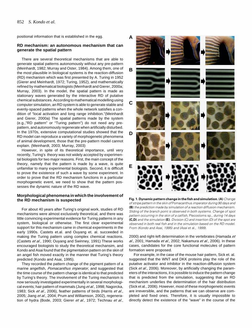
Fig. 1. Dynamic pattern change in the fish and simulation. (A) Changeof stripe pattern in the skin of Pomacanthus imperator during 90 days and(B) the prediction made by simulation of a reaction diffusion mechanism.Sliding of the branch point is observed in both systems. Change of spotpattern occurring in the skin of a catfish, Plecostoms sp., during 14 days(C,D) and the simulation (E). Division (C) and insertion (D) of the spot areobserved in both real fish and in the simulation based on the RD model.From (Kondo and Asai, 1995) and (Asai et al., 1999).
B
C
D
E
A
Turing patterns in fish skin 853
pattern-forming event.
Characteristic movement of the RD wave is visible inthe animal skin
To date, pigmentation patterns in animal skins (Murray et al.,1990; Murray and Oster, 1984), feathers of birds (Harris et al.,2005; Prum and Williamson, 2002), and shells of the snails(Meinhardt, 2003) are the only examples in which we can detectthe dynamic nature of Turing waves as a time course of the patternchange. Especially, the 2D skin pattern of the fish is quiteconvenient to study because waves are sometimes active evenwhen the fish has reached adulthood.
For example, when a striped angel fish (Pomacanthusimperator) grows, the branching points of the stripes slide hori-zontally as the zip opens, and add a number of stripes; eventuallythe spacing between the stripes remains stable (Fig. 1A) (Kondoand Asai, 1995). In the case of spotted catfish (Plecostoms sp.),both division of the spots and insertion of the new spots occur toretain the density and size of the spots (Fig. 1B) (Asai et al., 1999).Both stripes and the spots are the most typical 2D patternsgenerated by the RD mechanism, and the time course of thepattern change possesses the characteristics of the dynamics ofRD waves, strongly suggesting that the RD mechanism underliesthe process of pigment-pattern formation of fish.
Zebrafish as a model system for studying patterningmechanisms
In order to understand the principles of autonomous patternformation controlled by the dynamic mechanism, it is important toidentify the molecular-level network that functions in skin patternformation of fish where the wave is active. Observation on thedynamics of the molecules related to the RD pattern formationwould dramatically contribute to our understanding of how ani-mals keep the stable structure under an environment that is full ofdisturbances. Fortunately, zebrafish, a small fish species withvery clear stripes in their trunk and fins, was selected as a modelanimalÄfor biological studies, and for the use of the genomicinformation and molecular-genetic technologies that are avail-able to identify the molecular mechanism of pigment-patternformation.
The pigment pattern of zebrafish retains the dynamicnature of the RD wave during the post embryonic stage
The pattern of skin pigmentation in zebrafish is composed ofthree types of pigment cells distributed in the hypodermis: mel-anophores, the main component of dark stripes; xanthophores,the main component of light stripes; and iridophores (Hirata et al.,2003; Kelsh, 2004). Although different from the stripes ofPomacanthus imperator, the stripes of zebrafish do not becomerearranged during normal growth, artificial disturbance of thepattern can induce the characteristic pattern change that isspecific to the RD mechanism. Kirschbaum (Kirschbaum, 1975a)cut a piece of skin from the trunk and re-planted it at an angle toits original orientation. The stripes on the graft were terminated atthe edge of the transplant when the operation was completed.But, he observed that the ends of the stripes moved and rejoined
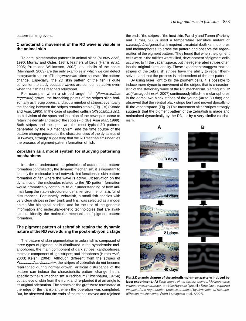
Fig. 2.Dynamic change of the zebrafish pigment pattern induced by
laser experiment. (A) Time course of the pattern change. Melanophoresin upper two black stripes are killed by laser light. (B) Time-lapse capturedimages of the regeneration process produced by simulation of reaction-diffusion mechanisms. From Yamaguchi et al. (2007).
3 days
14 days
21 days
40 days
BA
the end of the stripes of the host skin. Parichy and Turner (Parichyand Turner, 2003) used a temperature sensitive mutant ofpanther(c-fms) gene, that is required to maintain both xanthophoresand melanophores, to erase the pattern and observe the regen-eration of the pigment pattern. They found that when the pigmentcells were in the tail fins were‘killed, development of pigment cellsoccurred to fill the vacant space, but the regenerated stripes oftenlost the original directionality. These experiments suggest that thestripes of the zebrafish stripes have the ability to repair them-selves. and that the process is independent of the pre-pattern.
By using laser light to kill the pigment cells, it is possible toinduce more dynamic movement of the stripes that is character-istic of the stationary wave of the RD mechanism. Yamaguchi etal. (Yamaguchi et al., 2007) continuously killed the melanophoresin the dorsal two black stripes of the young (40 to 80 day) andobserved that the ventral black stripe bent and moved dorsally tofill the vacant space. (Fig. 2) This movement of the stripes stronglysuggests that the pigment pattern of the zebrafish is made andmaintained dynamically by the RD, or by a very similar mecha-nism.
854 S. Kondo et al.
Another important suggestion which arises from these experi-ments is that the pigment cells do not simply make the hiddenpattern visible, but are the major players in the pattern formation.Therefore, to uncover the mechanism, the most critical stepshould be the identification of the interactions between the twotypes of pigment cells (melanophores and xanthophores).
Mutants that affect both development of pigment cellsand the resulting pattern
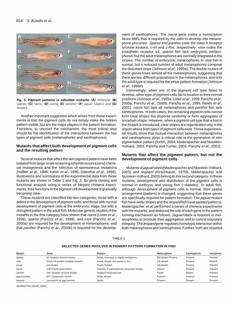
Several mutants that affect the skin pigment pattern have beenisolated from large-scale screening ethylnitrosourea (enu) chemi-cal mutagenesis and the selection of spontaneous mutations(Haffter et al., 1996; Kelsh et al., 1996; Odenthal et al., 1996).Illustrations and summaries of the experimental data from thesemutants are shown in Table 1 and Fig. 3. By gene cloning andfunctional analysis using a series of elegant chimera experi-ments, their functions in the pigment cell development is graduallybecoming clear.
These mutants are classified into two categories: those with adefect in the development of pigment cells; and those with normaldevelopment of pigment cells at the embryonic stage, but with adisrupted pattern in the adult fish. Molecular genetic studies of themutants in the first category have shown that nacre (Lister et al.,1999), sparse (Parichy et al., 1999), and rose (Parichy et al.,2000a) are required for the development of melanophores, andthat panther (Parichy et al., 2000b) is required for the develop-
ment of xanthophores. The nacre gene codes a transcriptionfactor Mitf1 that is required by the cells to develop into melano-phore precursor. Sparse and panther code the class II receptortyrosine kinases, c-kit and c-fms, respectively. rose codes theendothelin receptor b1. sparse fish lack embryonic melano-phores, but the adult melanophores are normally integrated in thestripes. The number of embryonic melanophores in rose fish isnormal, but a reduced number of adult melanophores composethe disturbed stripe (Johnson et al., 1995b). The double mutant ofthese genes loses almost all the melanophores, suggesting thatthere are two different populations in the melanophores, and onlythe adult type is required for the stripe pattern formation (Johnsonet al., 1995b).
Interestingly, when one of the pigment cell type failed todevelop, other type of pigment cells fail to localize to their normalpositions (Johnson et al., 1995a; Lister et al., 1999; Parichy et al.,2000a; Parichy et al., 2000b; Parichy et al., 1999; Rawls et al.,2001). nacre fish lack all melanophores and panther fish lackxanthophores. In both cases, the remaining pigment cells cannotform clear stripes but disperse randomly or form aggregates ofuncertain shape. However, when a pigment cell type that is lost inthe mutant is introduced, clear stripes are regenerated only in theregion where both types of pigment cells exist. These experimen-tal results show that mutual interaction between melanophoresand xanthophores plays a critical role in the generation of skinpigmentation pattern (Kelsh, 2004; Maderspacher and Nusslein-Volhard, 2003; Parichy and Turner, 2003; Parichy et al., 2003).
Mutants that affect the pigment pattern, but not thedevelopment of pigment cells
Mutants of jaguar/obelix(Maderspacher and Nusslein-Volhard,2003) and leopard (Kirschbaum, 1975b; Maderspacher andNusslein-Volhard, 2003) belong to this second category. In thesemutants, development and distribution of the pigment cells isnormal in embryos and young fish (~4weeks). In adult fish,although development of pigment cells is normal, their spatialarrangement (pattern) is changed, suggesting that these genesare specifically required for pattern formation. The jaguar mutantfish have wider stripes and the leopard fish have spotted patterns.Maderspacher et al. performed a series of chimera experimentswith the mutants, and deduced the role of each gene in the patternforming mechanism as follows. Jaguar/obelx is required in mel-anophores to promote their aggregation and to control boundaryinteguity. The leopard gene regulates homotypic interaction withinboth melanophores and xanthophores. Further, both are required
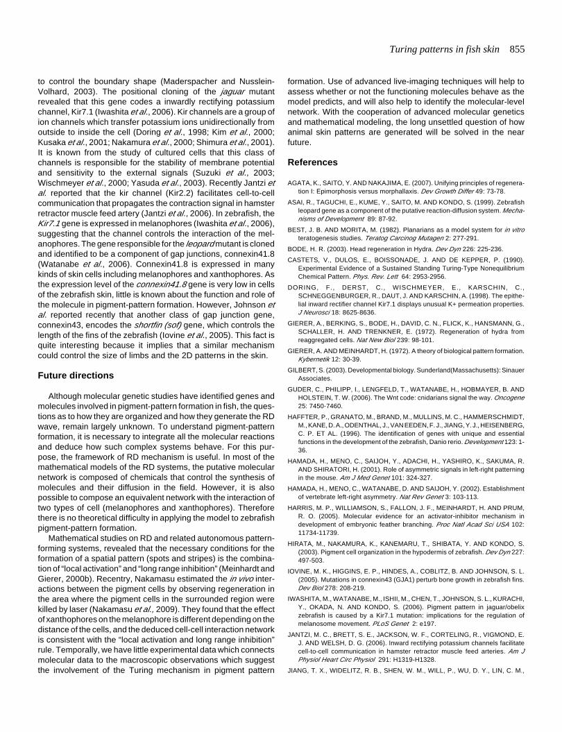
Mutant Gene Pattern Melanophore Xanthophore iridophore
sparse kit /receptor tyrosine kinase Stripe, boundary is slightly ambiguous EM absent Present Present Present
rose ednrb1/G-protein coupled receptor Fewer stripes, but normal in fins LM absent Present Absent
puma not cloned Purely formed LM absent Present Present
nacre mitf1/transcription factor Clusters of xanthophores, but purely striped Absent Present Present
panther fms /receptor tyrosine kinase Scatterd melanophores Fewer Absent Present
jaguar(obelix) kir7.1/potassium chnnel Wider stripes Present Present Present
leopard connexin41.8/ gap junction Spots Present Present Present
TABLE 1
SELECTED GENES INVOLVED IN PIGMENT PATTERN FORMATION IN FISH
Modified from (Kelsh, 2004)
Fig. 3. Pigment patterns in zebrafish mutants. (A) Wildtype; (B)
sparse; (C) nacre; (D) puma; (E) panther; (F) jaguar (obelix) and (G)
leopard.
D
G
B
C
E
F
H
A
Turing patterns in fish skin 855
to control the boundary shape (Maderspacher and Nusslein-Volhard, 2003). The positional cloning of the jaguar mutantrevealed that this gene codes a inwardly rectifying potassiumchannel, Kir7.1 (Iwashita et al., 2006). Kir channels are a group ofion channels which transfer potassium ions unidirectionally fromoutside to inside the cell (Doring et al., 1998; Kim et al., 2000;Kusaka et al., 2001; Nakamura et al., 2000; Shimura et al., 2001).It is known from the study of cultured cells that this class ofchannels is responsible for the stability of membrane potentialand sensitivity to the external signals (Suzuki et al., 2003;Wischmeyer et al., 2000; Yasuda et al., 2003). Recently Jantzi etal. reported that the kir channel (Kir2.2) facilitates cell-to-cellcommunication that propagates the contraction signal in hamsterretractor muscle feed artery (Jantzi et al., 2006). In zebrafish, theKir7.1 gene is expressed in melanophores (Iwashita et al., 2006),suggesting that the channel controls the interaction of the mel-anophores. The gene responsible for the leopard mutant is clonedand identified to be a component of gap junctions, connexin41.8(Watanabe et al., 2006). Connexin41.8 is expressed in manykinds of skin cells including melanophores and xanthophores. Asthe expression level of the connexin41.8 gene is very low in cellsof the zebrafish skin, little is known about the function and role ofthe molecule in pigment-pattern formation. However, Johnson etal. reported recently that another class of gap junction gene,connexin43, encodes the shortfin (sof) gene, which controls thelength of the fins of the zebrafish (Iovine et al., 2005). This fact isquite interesting because it implies that a similar mechanismcould control the size of limbs and the 2D patterns in the skin.
Future directions
Although molecular genetic studies have identified genes andmolecules involved in pigment-pattern formation in fish, the ques-tions as to how they are organized and how they generate the RDwave, remain largely unknown. To understand pigment-patternformation, it is necessary to integrate all the molecular reactionsand deduce how such complex systems behave. For this pur-pose, the framework of RD mechanism is useful. In most of themathematical models of the RD systems, the putative molecularnetwork is composed of chemicals that control the synthesis ofmolecules and their diffusion in the field. However, it is alsopossible to compose an equivalent network with the interaction oftwo types of cell (melanophores and xanthophores). Thereforethere is no theoretical difficulty in applying the model to zebrafishpigment-pattern formation.
Mathematical studies on RD and related autonomous pattern-forming systems, revealed that the necessary conditions for theformation of a spatial pattern (spots and stripes) is the combina-tion of “local activation” and “long range inhibition” (Meinhardt andGierer, 2000b). Recentry, Nakamasu estimated the in vivo inter-actions between the pigment cells by observing regeneration inthe area where the pigment cells in the surrounded region werekilled by laser (Nakamasu et al., 2009). They found that the effectof xanthophores on the melanophore is different depending on thedistance of the cells, and the deduced cell-cell interaction networkis consistent with the “local activation and long range inhibition”rule. Temporally, we have little experimental data which connectsmolecular data to the macroscopic observations which suggestthe involvement of the Turing mechanism in pigment pattern
formation. Use of advanced live-imaging techniques will help toassess whether or not the functioning molecules behave as themodel predicts, and will also help to identify the molecular-levelnetwork. With the cooperation of advanced molecular geneticsand mathematical modeling, the long unsettled question of howanimal skin patterns are generated will be solved in the nearfuture.
References
AGATA, K., SAITO, Y. AND NAKAJIMA, E. (2007). Unifying principles of regenera-tion I: Epimorphosis versus morphallaxis. Dev Growth Differ 49: 73-78.
ASAI, R., TAGUCHI, E., KUME, Y., SAITO, M. AND KONDO, S. (1999). Zebrafishleopard gene as a component of the putative reaction-diffusion system. Mecha-nisms of Development 89: 87-92.
BEST, J. B. AND MORITA, M. (1982). Planarians as a model system for in vitroteratogenesis studies. Teratog Carcinog Mutagen 2: 277-291.
BODE, H. R. (2003). Head regeneration in Hydra. Dev Dyn 226: 225-236.
CASTETS, V., DULOS, E., BOISSONADE, J. AND DE KEPPER, P. (1990).Experimental Evidence of a Sustained Standing Turing-Type NonequilibriumChemical Pattern. Phys. Rev. Lett 64: 2953-2956.
DORING, F., DERST, C., WISCHMEYER, E., KARSCHIN, C.,SCHNEGGENBURGER, R., DAUT, J. AND KARSCHIN, A. (1998). The epithe-lial inward rectifier channel Kir7.1 displays unusual K+ permeation properties.J Neurosci 18: 8625-8636.
GIERER, A., BERKING, S., BODE, H., DAVID, C. N., FLICK, K., HANSMANN, G.,SCHALLER, H. AND TRENKNER, E. (1972). Regeneration of hydra fromreaggregated cells. Nat New Biol 239: 98-101.
GIERER, A. AND MEINHARDT, H. (1972). A theory of biological pattern formation.Kybernetik 12: 30-39.
GILBERT, S. (2003). Developmental biology. Sunderland(Massachusetts): SinauerAssociates.
GUDER, C., PHILIPP, I., LENGFELD, T., WATANABE, H., HOBMAYER, B. ANDHOLSTEIN, T. W. (2006). The Wnt code: cnidarians signal the way. Oncogene25: 7450-7460.
HAFFTER, P., GRANATO, M., BRAND, M., MULLINS, M. C., HAMMERSCHMIDT,M., KANE, D. A., ODENTHAL, J., VAN EEDEN, F. J., JIANG, Y. J., HEISENBERG,C. P. ET AL. (1996). The identification of genes with unique and essentialfunctions in the development of the zebrafish, Danio rerio. Development 123: 1-36.
HAMADA, H., MENO, C., SAIJOH, Y., ADACHI, H., YASHIRO, K., SAKUMA, R.AND SHIRATORI, H. (2001). Role of asymmetric signals in left-right patterningin the mouse. Am J Med Genet 101: 324-327.
HAMADA, H., MENO, C., WATANABE, D. AND SAIJOH, Y. (2002). Establishmentof vertebrate left-right asymmetry. Nat Rev Genet 3: 103-113.
HARRIS, M. P., WILLIAMSON, S., FALLON, J. F., MEINHARDT, H. AND PRUM,R. O. (2005). Molecular evidence for an activator-inhibitor mechanism indevelopment of embryonic feather branching. Proc Natl Acad Sci USA 102:11734-11739.
HIRATA, M., NAKAMURA, K., KANEMARU, T., SHIBATA, Y. AND KONDO, S.(2003). Pigment cell organization in the hypodermis of zebrafish. Dev Dyn 227:497-503.
IOVINE, M. K., HIGGINS, E. P., HINDES, A., COBLITZ, B. AND JOHNSON, S. L.(2005). Mutations in connexin43 (GJA1) perturb bone growth in zebrafish fins.Dev Biol 278: 208-219.
IWASHITA, M., WATANABE, M., ISHII, M., CHEN, T., JOHNSON, S. L., KURACHI,Y., OKADA, N. AND KONDO, S. (2006). Pigment pattern in jaguar/obelixzebrafish is caused by a Kir7.1 mutation: implications for the regulation ofmelanosome movement. PLoS Genet 2: e197.
JANTZI, M. C., BRETT, S. E., JACKSON, W. F., CORTELING, R., VIGMOND, E.J. AND WELSH, D. G. (2006). Inward rectifying potassium channels facilitatecell-to-cell communication in hamster retractor muscle feed arteries. Am JPhysiol Heart Circ Physiol 291: H1319-H1328.
JIANG, T. X., WIDELITZ, R. B., SHEN, W. M., WILL, P., WU, D. Y., LIN, C. M.,
856 S. Kondo et al.
JUNG, H. S. AND CHUONG, C. M. (2004). Integument pattern formationinvolves genetic and epigenetic controls: feather arrays simulated by digitalhormone models. Int J Dev Biol 48: 117-135.
JOHNSON, S. L., AFRICA, D., HORNE, S. AND POSTLETHWAIT, J. H. (1995a).Half-tetrad analysis in zebrafish: mapping the ros mutation and the centromereof linkage group I. Genetics 139: 1727-1735.
JOHNSON, S. L., AFRICA, D., WALKER, C. AND WESTON, J. A. (1995b). Geneticcontrol of adult pigment stripe development in zebrafish. Dev Biol 167: 27-33.
JUNG, H. S., FRANCIS-WEST, P. H., WIDELITZ, R. B., JIANG, T. X., TING-BERRETH, S., TICKLE, C., WOLPERT, L. AND CHUONG, C. M. (1998). Localinhibitory action of BMPs and their relationships with activators in featherformation: implications for periodic patterning. Dev Biol 196: 11-23.
KELSH, R. N. (2004). Genetics and evolution of pigment patterns in fish. PigmentCell Res 17: 326-336.
KELSH, R. N., BRAND, M., JIANG, Y. J., HEISENBERG, C. P., LIN, S., HAFFTER,P., ODENTHAL, J., MULLINS, M. C., VAN EEDEN, F. J., FURUTANI-SEIKI, M.ET AL. (1996). Zebrafish pigmentation mutations and the processes of neuralcrest development. Development 123: 369-389.
KIM, S. J., KERST, G., SCHREIBER, R., PAVENSTADT, H., GREGER, R., HUG,M. J. AND BLEICH, M. (2000). Inwardly rectifying K+ channels in the basolateralmembrane of rat pancreatic acini. Pflugers Arch 441: 331-340.
KIRSCHBAUM, F. (1975a). Untersuchnugen uber das Farbmuster der ZebrabarbeBrachydanio rerio. Roux’s Arch. Dev. Biol. 177: 129-152.
KIRSCHBAUM, F. (1975b). Untersuchungen ueber das Farbmuster der ZebrabarbeBrachydanio rerio (Cyprinidae, Teleostei). Wilhelm Roux’s Arch 177: 129-152.
KONDO, S. AND ASAI, R. (1995). A reaction-diffusion wave on the skin of themarine angelfish Pomacanthus. Nature 376: 765-768.
KUSAKA, S., INANOBE, A., FUJITA, A., MAKINO, Y., TANEMOTO, M.,MATSUSHITA, K., TANO, Y. AND KURACHI, Y. (2001). Functional Kir7.1channels localized at the root of apical processes in rat retinal pigmentepithelium. J Physiol 531: 27-36.
LISTER, J. A., ROBERTSON, C. P., LEPAGE, T., JOHNSON, S. L. AND RAIBLE,D. W. (1999). nacre encodes a zebrafish microphthalmia-related protein thatregulates neural-crest-derived pigment cell fate. Development 126: 3757-3767.
MADERSPACHER, F. AND NUSSLEIN-VOLHARD, C. (2003). Formation of theadult pigment pattern in zebrafish requires leopard and obelix dependent cellinteractions. Development 130: 3447-3457.
MEINHARDT, H. (1982). Models of biological pattern formation. Academic press,London.
MEINHARDT, H. (2003). The Algorithmic Beauty of Sea Shells. Springer, Berlin.
MEINHARDT, H. AND GIERER, A. (2000a). Pattern formation by local self-activation and lateral inhibition. Bioessays 22: 753-760.
MEINHARDT, H. AND GIERER, A. (2000b). Pattern formation by local self-activation and lateral inhibition.[see comment]. Bioessays 22: 753-760.
MURRAY, J. (2003). Mathematical BIology. Springer, Berlin.
MURRAY, J. D., DEEMING, D. C. AND FERGUSON, M. W. (1990). Size-dependentpigmentation-pattern formation in embryos of Alligator mississippiensis: time ofinitiation of pattern generation mechanism. Proc R Soc Lond B Biol Sci 239:279-293.
MURRAY, J. D. AND OSTER, G. F. (1984). Generation of biological pattern andform. IMA J Math Appl Med Biol 1: 51-75.
NAGORCKA, B. N. (1983). Evidence for a reaction-diffusion system as a mecha-nism controlling mammalian hair growth. Biosystems 16: 323-332.
NAKAMASU, A., TAKAHASHI, G., KANBE, A. and KONDO, S. (2009) Interactionsbetween the zebrafish pigment cells responsible for the generation of Turingpattern. Proc. Natl. Acad. Sci. USA 106: 8429-8434.
NAKAMURA, N., SUZUKI, Y., IKEDA, Y., NOTOYA, M. AND HIROSE, S. (2000).Complex structure and regulation of expression of the rat gene for inwardrectifier potassium channel Kir7.1. J Biol Chem 275: 28276-28284.
NAKAMURA, T., MINE, N., NAKAGUCHI, E., MOCHIZUKI, A., YAMAMOTO, M.,YASHIRO, K., MENO, C. AND HAMADA, H. (2006). Generation of robust left-right asymmetry in the mouse embryo requires a self-enhancement and lateral-inhibition system. Dev Cell 11: 495-504.
ODENTHAL, J., ROSSNAGEL, K., HAFFTER, P., KELSH, R. N., VOGELSANG, E.,
BRAND, M., VAN EEDEN, F. J., FURUTANI-SEIKI, M., GRANATO, M.,HAMMERSCHMIDT, M. ET AL. (1996). Mutations affecting xanthophore pig-mentation in the zebrafish, Danio rerio. Developmentt 123: 391-398.
OUYANG, Q. AND SWINNEY, H. (1991). Transition from a uniform state tohexagonal and striped Turing patterns. Nature 352: 610-612.
PARICHY, D. M., MELLGREN, E. M., RAWLS, J. F., LOPES, S. S., KELSH, R. N.AND JOHNSON, S. L. (2000a). Mutational analysis of endothelin receptor b1(rose) during neural crest and pigment pattern development in the zebrafishDanio rerio. Dev. Biol. 227: 294-306.
PARICHY, D. M., RANSOM, D. G., PAW, B., ZON, L. I. AND JOHNSON, S. L.(2000b). An orthologue of the kit-related gene fms is required for developmentof neural crest-derived xanthophores and a subpopulation of adult melanocytesin the zebrafish, Danio rerio. Development 127: 3031-3044.
PARICHY, D. M., RAWLS, J. F., PRATT, S. J., WHITFIELD, T. T. AND JOHNSON,S. L. (1999). Zebrafish sparse corresponds to an orthologue of c-kit and isrequired for the morphogenesis of a subpopulation of melanocytes, but is notessential for hematopoiesis or primordial germ cell development. Development126: 3425-3436.
PARICHY, D. M. AND TURNER, J. M. (2003). Temporal and cellular requirementsfor Fms signaling during zebrafish adult pigment pattern development. Devel-opment 130: 817-833.
PARICHY, D. M., TURNER, J. M. AND PARKER, N. B. (2003). Essential role forpuma in development of postembryonic neural crest-derived cell lineages inzebrafish. Dev Biol 256: 221-241.
PRUM, R. O. AND WILLIAMSON, S. (2002). Reaction-diffusion models of within-feather pigmentation patterning. Proc Biol Sci 269: 781-792.
RAWLS, J., EM., M. AND SL., J. (2001). How the zebrafish gets its stripes. Dev Biol240: 301-314.
SHIMURA, M., YUAN, Y., CHANG, J. T., ZHANG, S., CAMPOCHIARO, P. A.,ZACK, D. J. AND HUGHES, B. A. (2001). Expression and permeation propertiesof the K(+) channel Kir7.1 in the retinal pigment epithelium. J Physiol 531: 329-346.
SICK, S., REINKER, S., TIMMER, J. AND SCHLAKE, T. (2006). WNT and DKKdetermine hair follicle spacing through a reaction-diffusion mechanism. Science314: 1447-1450.
SUZUKI, Y., YASUOKA, Y., SHIMOHAMA, T., NISHIKITANI, M., NAKAMURA, N.,HIROSE, S. AND KAWAHARA, K. (2003). Expression of the K+ channel Kir7.1in the developing rat kidney: role in K+ excretion. Kidney Int 63: 969-975.
TECHNAU, U., CRAMER VON LAUE, C., RENTZSCH, F., LUFT, S., HOBMAYER,B., BODE, H. R. AND HOLSTEIN, T. W. (2000). Parameters of self-organizationin Hydra aggregates. Proc Natl Acad Sci USA 97: 12127-12131.
TURING, A. (1952). The chemical basis of morphogenesis. Philos Trans R SocLond B 237: 37-72.
WATANABE, M., IWASHITA, M., ISHII, M., KURACHI, Y., KAWAKAMI, A., KONDO,S. AND OKADA, N. (2006). Spot pattern of leopard Danio is caused by mutationin the zebrafish connexin41.8 gene. EMBO Reports 7: 893-897.
WISCHMEYER, E., DORING, F. AND KARSCHIN, A. (2000). Stable cation coor-dination at a single outer pore residue defines permeation properties in Kirchannels. FEBS Lett 466: 115-120.
WOLPERT, L. (1969). Positional information and the spatial pattern of cellulardifferentiation. J Theor Biol 25: 1-47.
WOLPERT, L. (1989). Positional information revisited. Development 107 SUPPL:3-12.
WOLPERT, L. (2006). Principles of Development. Oxford University Press, NewYork.
WOLPERT, L., HICKLIN, J. AND HORNBRUCH, A. (1971). Positional informationand pattern regulation in regeneration of hydra. Symp Soc Exp Biol 25: 391-415.
YAMAGUCHI, M., YOSHIMOTO, E. AND KONDO, S. (2007). Pattern regulation inthe stripe of zebrafish suggests an underlying dynamic and autonomousmechanism. Proc Natl Acad Sci USA 104: 4790-4793.
YASUDA, K., SHIMURA, M., NAKAZAWA, T., SATO, H., TOMITA, H., SUGANO,E. AND TAMAI, M. (2003). Expression and functional properties of uniqueinward rectifier K+ channel Kir7.1 in the porcine iris and retinal pigmentepithelium. Curr Eye Res 27: 279-287.

Turing patterns in fish skin 857
Further Related Reading, published previously in the Int. J. Dev. Biol.
See our Special Issue Developmental Morphodynamics edited by Lev Beloussov and Richard Gordon at:http://www.ijdb.ehu.es/web/contents.php?vol=50&issue=2-3
See our Special Issue Fertilization, in honor of David L. Garbers and edited by Paul M. Wassarman and Victor D. Vacquier at:http://www.ijdb.ehu.es/web/contents.php?vol=52&issue=5-6
Analyses of regenerative wave patterns in adult hair follicle populations reveal macro-environmental regulation of stem cell activityMaksim V. Plikus, Randall B. Widelitz, Rob Maxson and Cheng-Ming ChuongInt. J. Dev. Biol. in press
Expression of the novel gene Ened during mouse and Xenopus embryonic developmentRenata Meszaros, Ina Strate, Edgar M. Pera and Madeleine DurbeejInt. J. Dev. Biol. (2008) 52: 1119-1122
Drosophila retinal pigment cell death is regulated in a position-dependent manner by acell memory geneNicolas Dos-Santos, Thomas Rubin, Fabienne Chalvet, Pierre Gandille, Frederic Cremazy,Jacqueline Leroy, E. Boissonneau and Laurent ThéodoreInt. J. Dev. Biol. (2008) 52: 21-31
Pax7 identifies neural crest, chromatophore lineages and pigment stem cells duringzebrafish developmentAna M Lacosta, Jesús Canudas, Cristina González, Pedro Muniesa, Manuel Sarasa and LuisDomínguezInt. J. Dev. Biol. (2007) 51: 327-331
Before programs: The physical origination of multicellular formsStuart A. Newman, Gabor Forgacs and Gerd B. MüllerInt. J. Dev. Biol. (2006) 50: 289-299
The dynamic geometry of mass cell movements in animal morphogenesisVladimir G. CherdantsevInt. J. Dev. Biol. (2006) 50: 169-182
From observations to paradigms; the importance of theories and models. An interviewwith Hans MeinhardtRichard Gordon and Lev BeloussovInt. J. Dev. Biol. (2006) 50: 103-111
Involvement of Hex in the initiation of feather morphogenesisAkiko Obinata and Yoshihiro AkimotoInt. J. Dev. Biol. (2005) 49: 953-960
Expression of Hex during feather bud developmentAkiko Obinata and Yoshihiro AkimotoInt. J. Dev. Biol. (2005) 49: 885-890
Integument pattern formation involves genetic and epigenetic controls: feather arrayssimulated by digital hormone models.Ting-Xin Jiang, Randall B Widelitz, Wei-Min Shen, Peter Will, Da-Yu Wu, Chih-Min Lin, Han-Sung Jung and Cheng-Ming ChuongInt. J. Dev. Biol. (2004) 48: 117-135
5 yr ISI Impact Factor (2008) = 3.271