Embed Size (px)
Citation preview
Human Biology: Respiratory System
Lesson 2: Processes of the Respiratory System
(Inquiry into Life pg. 289-294)
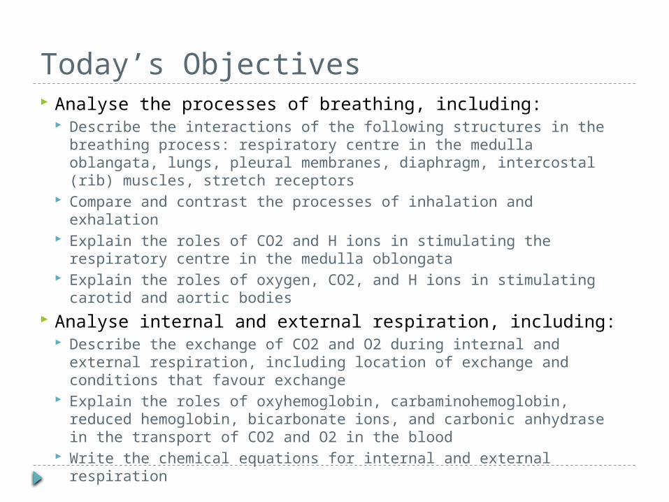
Today’s Objectives Analyse the processes of breathing, including:
Describe the interactions of the following structures in the breathing process: respiratory centre in the medulla oblangata, lungs, pleural membranes, diaphragm, intercostal (rib) muscles, stretch receptors
Compare and contrast the processes of inhalation and exhalation Explain the roles of CO2 and H ions in stimulating the respiratory
centre in the medulla oblongata Explain the roles of oxygen, CO2, and H ions in stimulating carotid
and aortic bodies Analyse internal and external respiration, including:
Describe the exchange of CO2 and O2 during internal and external respiration, including location of exchange and conditions that favour exchange
Explain the roles of oxyhemoglobin, carbaminohemoglobin, reduced hemoglobin, bicarbonate ions, and carbonic anhydrase in the transport of CO2 and O2 in the blood
Write the chemical equations for internal and external respiration
Processes of the Respiratory System The respiratory system supplies the
body with oxygen for its energy production
Without oxygen, the body shuts down in minutes
The respiratory system works closely with the circulatory system
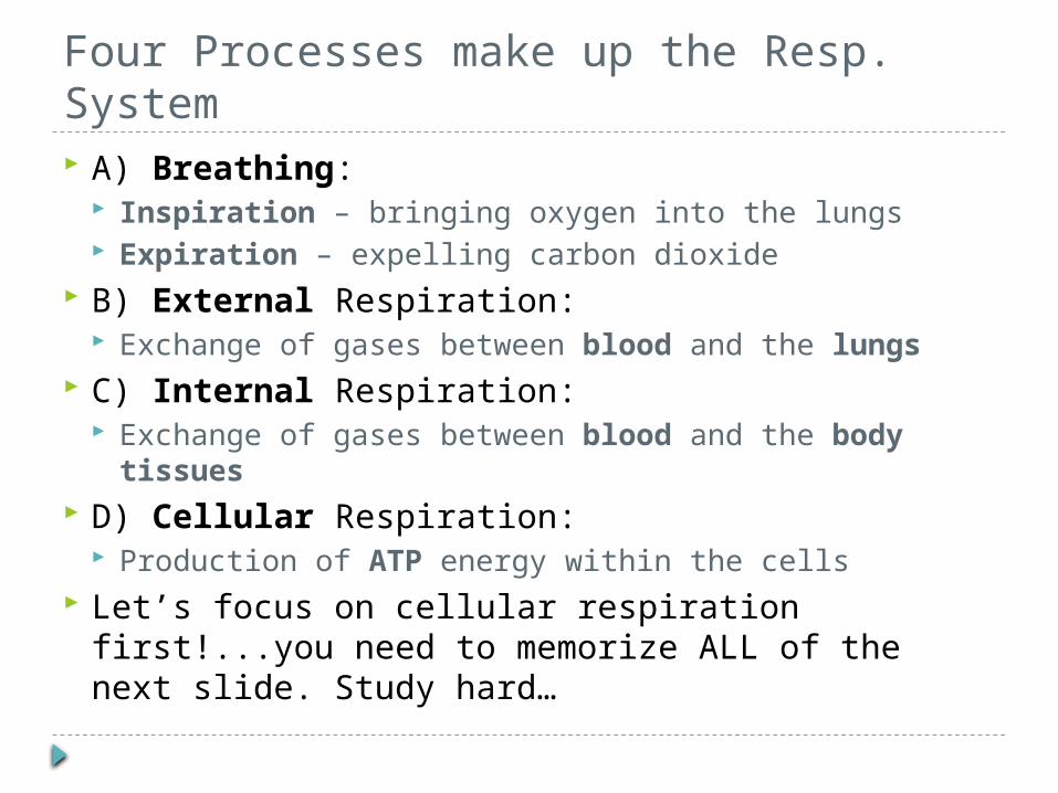
Four Processes make up the Resp. System A) Breathing:
Inspiration – bringing oxygen into the lungs Expiration – expelling carbon dioxide
B) External Respiration: Exchange of gases between blood and the lungs
C) Internal Respiration: Exchange of gases between blood and the body
tissues D) Cellular Respiration:
Production of ATP energy within the cells Let’s focus on cellular respiration first!...you need
to memorize ALL of the next slide. Study hard…
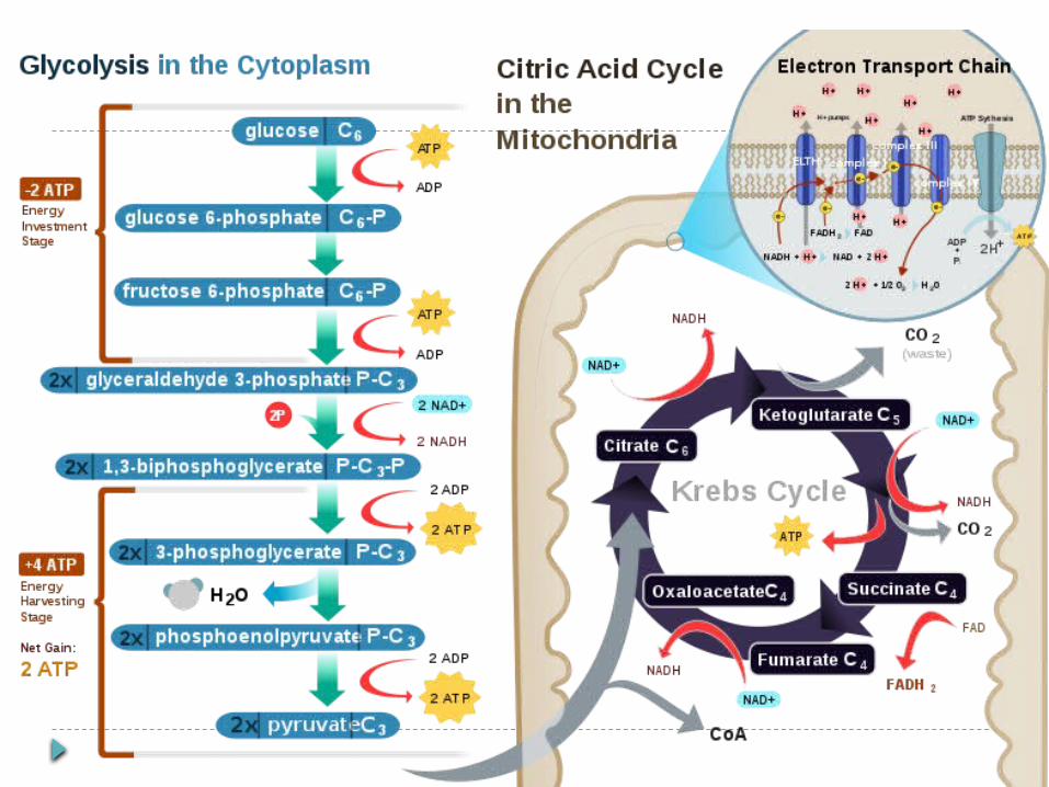

Just kidding ;)
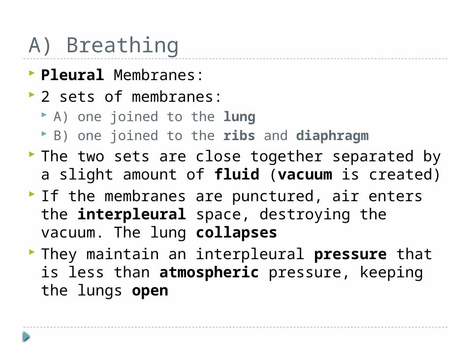
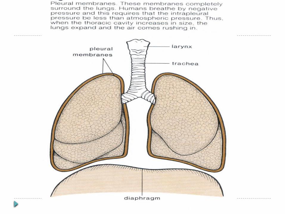
A) Breathing Pleural Membranes: 2 sets of membranes:
A) one joined to the lung B) one joined to the ribs and diaphragm
The two sets are close together separated by a slight amount of fluid (vacuum is created)
If the membranes are punctured, air enters the interpleural space, destroying the vacuum. The lung collapses
They maintain an interpleural pressure that is less than atmospheric pressure, keeping the lungs open
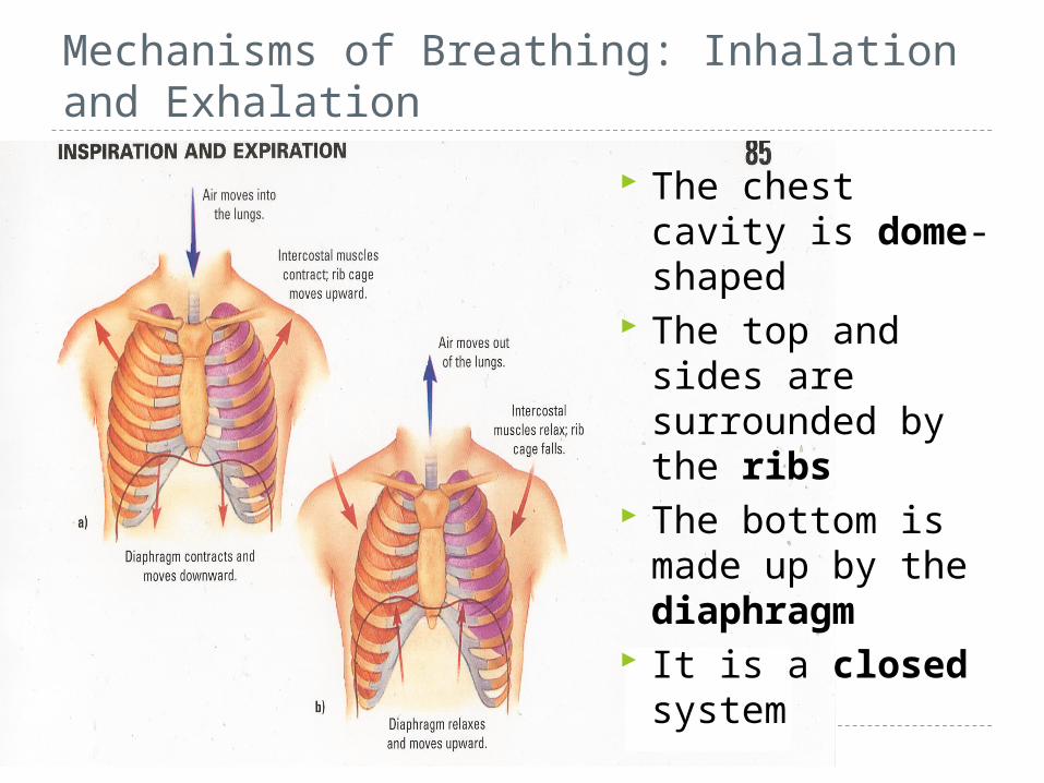
Mechanisms of Breathing: Inhalation and Exhalation
The chest cavity is dome-shaped
The top and sides are surrounded by the ribs
The bottom is made up by the diaphragm
It is a closed system
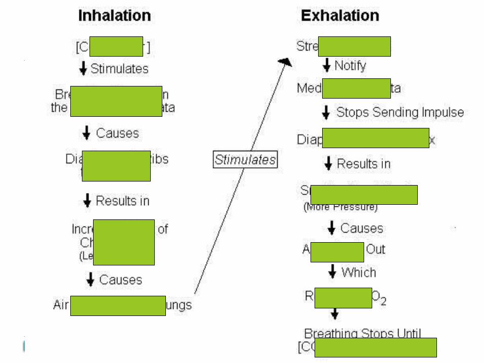
Inhalation 1) CO2 concentration and H+ concentration are
the PRIMARY STIMULI that cause us to breathe When CO2 and/or H+ concentration gets too high, the
breathing center in the Medulla Oblongata is stimulated
2) A nerve impulse is sent from the Medulla Oblongata to the diaphragm and rib cage
3) The diaphragm contracts and lowers; the intercostal (rib) muscles contract and raise the ribs
4) A partial vacuum is created in the lungs (air pressure in the lungs is reduced) due to the increased volume of the chest cavity
Inhalation 5) Air rushes into the lungs from
outside in order to rebalance the pressure This is the process of inspiration Note: air comes in because the lungs
have already opened. The air does not force the lungs open. This is why it is said we breathe by negative pressure (low pressure sucks the air into our lungs)
Note: The lungs themselves have no muscles
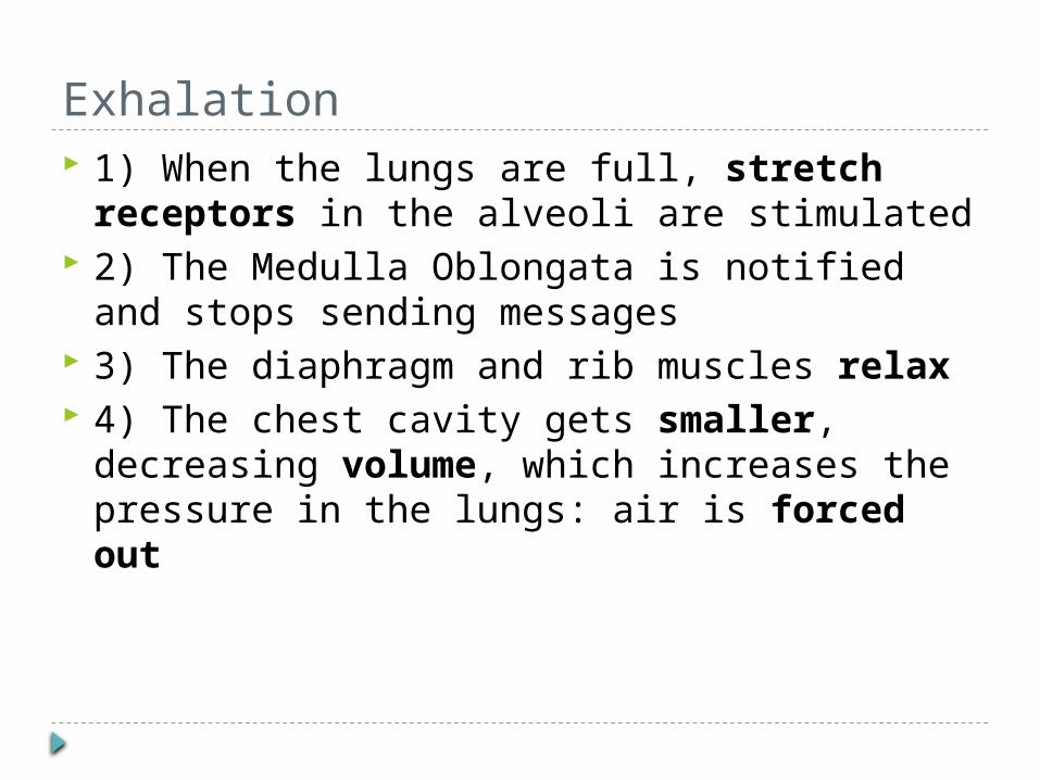
Exhalation 1) When the lungs are full, stretch receptors
in the alveoli are stimulated 2) The Medulla Oblongata is notified and stops
sending messages 3) The diaphragm and rib muscles relax 4) The chest cavity gets smaller, decreasing
volume, which increases the pressure in the lungs: air is forced out
Other Receptors In addition to the respiratory center in the
Medulla Oblongata, there are other receptors that can respond to stimuli: A) carotid bodies – in the carotid artery B) aortic bodies – in the aorta
These respond to low oxygen concentration but can also respond to levels of CO2 and H+ ions in the blood
Who has the largest lung capacity? We are going to go to the 1st floor Bio lab and
find out our lung capacity using a Spirometer We take as deep a breath as possible, then
blow allllll of the air in our lungs into the mouthpiece of the spirometer
The spirometer has a gauge on the side that indicates your lung capacity
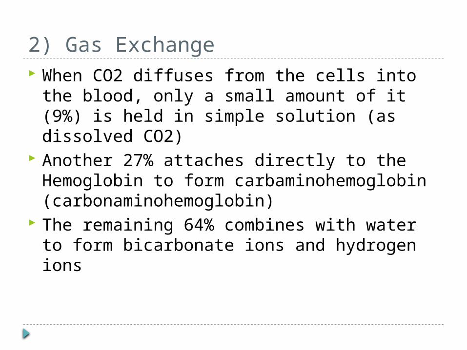
2) Gas Exchange When CO2 diffuses from the cells into the
blood, only a small amount of it (9%) is held in simple solution (as dissolved CO2)
Another 27% attaches directly to the Hemoglobin to form carbaminohemoglobin (carbonaminohemoglobin)
The remaining 64% combines with water to form bicarbonate ions and hydrogen ions
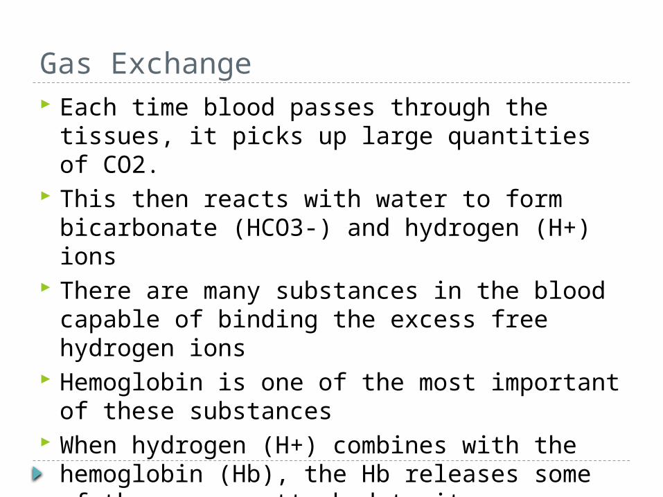
Gas Exchange Each time blood passes through the tissues, it
picks up large quantities of CO2. This then reacts with water to form
bicarbonate (HCO3-) and hydrogen (H+) ions There are many substances in the blood
capable of binding the excess free hydrogen ions
Hemoglobin is one of the most important of these substances
When hydrogen (H+) combines with the hemoglobin (Hb), the Hb releases some of the oxygen attached to it
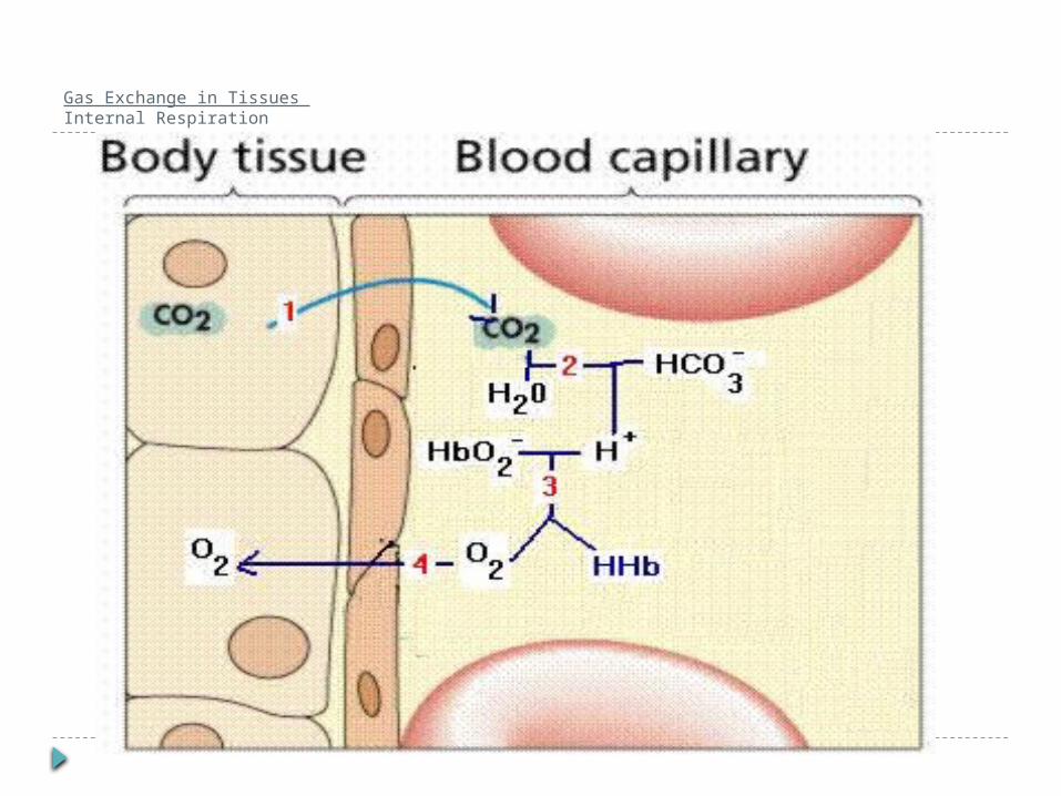
Gas Exchange in Tissues Internal Respiration
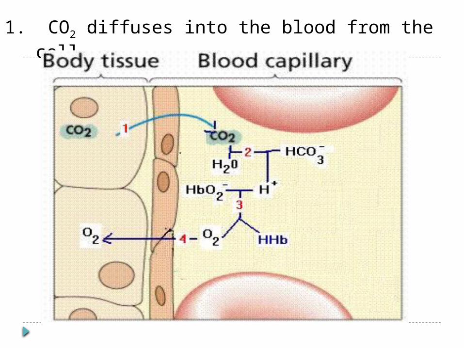
1. CO2 diffuses into the blood from the cell
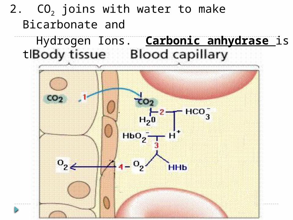
2. CO2 joins with water to make Bicarbonate and
Hydrogen Ions. Carbonic anhydrase is the enzyme that runs this reaction
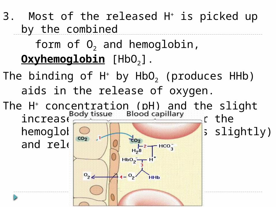
3. Most of the released H+ is picked up by the combined
form of O2 and hemoglobin, Oxyhemoglobin [HbO2].
The binding of H+ by HbO2 (produces HHb) aids in the release of oxygen.
The H+ concentration (pH) and the slight increase in temperature alter the hemoglobin (protein denatures slightly) and releases oxygen easily.
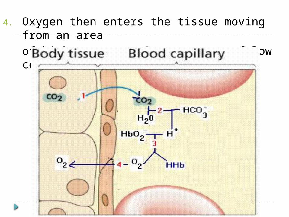
4. Oxygen then enters the tissue moving from an area of high concentration to areas of low concentration.
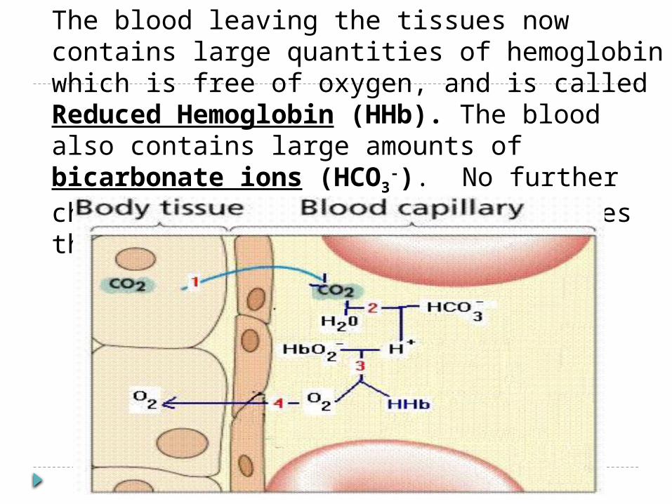
The blood leaving the tissues now contains large quantities of hemoglobin which is free of oxygen, and is called Reduced Hemoglobin (HHb). The blood also contains large amounts of bicarbonate ions (HCO3
-). No further changes occur until the blood reaches the lungs.
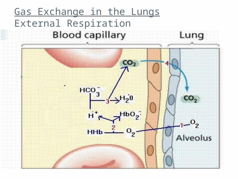
Gas Exchange in the Lungs External Respiration
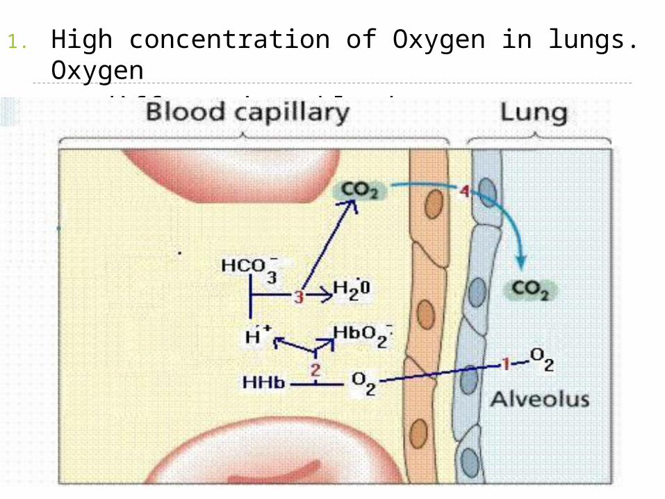
1. High concentration of Oxygen in lungs. Oxygen
diffuses into blood. .
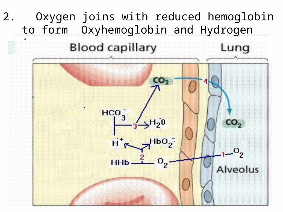
2. Oxygen joins with reduced hemoglobin to form Oxyhemoglobin and Hydrogen ions.
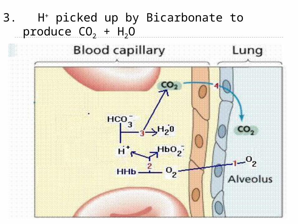
3. H+ picked up by Bicarbonate to produce CO2 + H2O
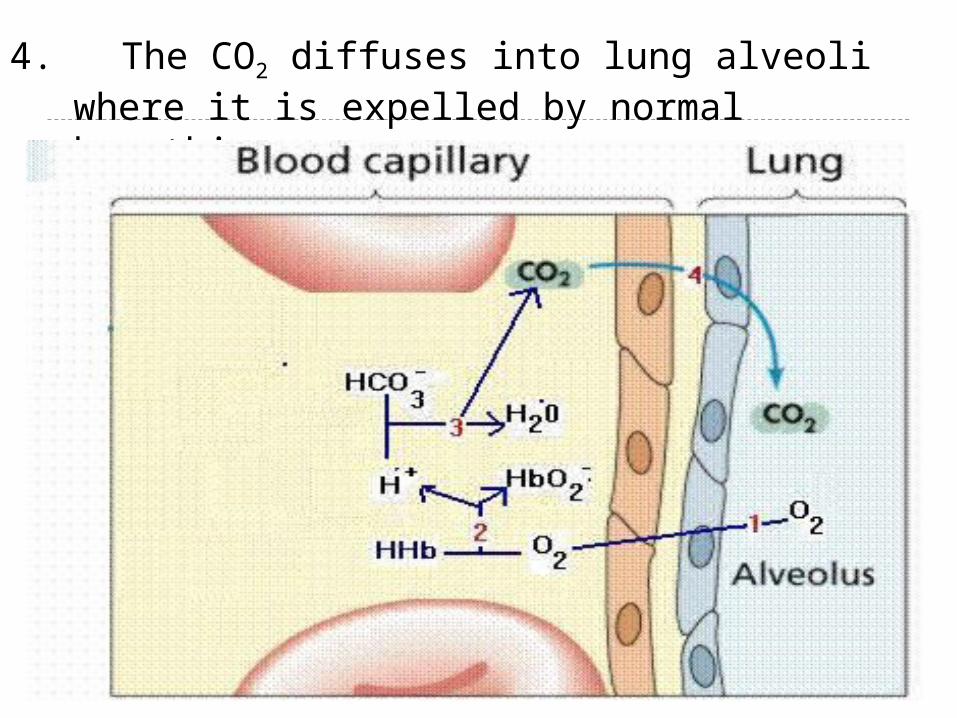
4. The CO2 diffuses into lung alveoli where it is expelled by normal breathing.
External Respiration Note: H+ does not accumulate because as
soon as it is released from HHb, it combines with HCO3- to release CO2
Hemoglobin is essential in the blood because it serves as a carrier for O2, CO2, and H+ ions (acts like a buffer)
H+ concentration (pH) and decrease in temperature in lungs allows hemoglobin to regain its original shape, allowing it to bind with oxygen easier
Respiratory Rap! Song lyrics sheet:
Due Monday, April 22 Videos/Presentations:
In class on Friday, April 26
*Groups of 3 to 5List group members on your lyrics sheet


![Respiratory System [โหมดความเข้ากันได้] · PATHOLOGY OF RESPIRATORY SYSTEM นพ. อรรณพ นาคะป ท Respiratory system U it](https://img.pdfslide.net/doc/110x75/5fa578efd4e80f055f6b3401/respiratory-system-aaaaaaaaaaaaaaaaaa-pathology.jpg)


![Respiratory system roadmap.pptx [Repaired] - Loginanatomical-sciences.health.wits.ac.za/roadmaps/Respiratory system... · DIVISION OF THE RESPIRATORY SYSTEM CONDUCTING PORTION Nasal](https://img.pdfslide.net/doc/110x75/5a78c3d87f8b9ae6228c9db0/respiratory-system-repaired-loginanatomical-scienceshealthwitsaczaroadmapsrespiratory.jpg)