Embed Size (px)
Citation preview
LETTER TO THE EDITOR
Journal of Dermatological Science (2006) 42, 75—77
www.intl.elsevierhealth.com/journals/jods
Human endothelial progenitor cells in-duce capillary-like tube formation in skinequivalents
KEYWORDSSkin equivalents;Endothelial progenitorcells;Angiotensin II;Vascular endothelialgrowth factor
To the Editor,
Neovascularization comprises angiogenesis andvasculogenesis. Angiogenesis, new capillary forma-tion from preexisting blood vessels, is a morphoge-netic process. It includes the degradation ofbasement membrane and extracellular matrix bya variety of proteolytic enzymes. On the otherhand, vasculogenesis is de novo formation of capil-lary-like tubes by the differentiation of endothelialprogenitor cells (EPCs) [1].
EPCs arise from bone marrow and other tissuesincluding skeletal muscle and vascular parenchyma.In addition, small numbers of EPCs are found in theperipheral blood and umbilical cord blood [2]. Bycytokine stimulation, EPCs move from bone marrowinto the circulation and then into ischemic tissuewhere they form new blood vessels [3]. In the recon-struction of skin equivalents (SEs), blood vessel for-mation is as important as the formation of other skinorgans, such as sweat and sebaceous glands and hair.Therefore, human umbilical vein endothelial cells(HUVECs) hadbeenused to create vascular structuresin endothelialized reconstructed dermis [4]. Toimprove blood vessel formation, the use of EPCscan be considered. However, EPCs have not beenused for this purpose although they are known toinduce capillary-like tubes by de novo formation. Inthe present study, we investigated the capillary-liketube formation abilities of EPCs during the recon-struction of SEs. Furthermore, there are important
0923-1811/$30.00 # 2006 Japanese Society for Investigative Dermadoi:10.1016/j.jdermsci.2006.01.002
angiogenic regulatory factors. In particular, it hasbeen reported that vascular endothelial growth fac-tor (VEGF) and angiotensin II (Ang II) can induceneovascularization [2]. Based on these reports, theeffects of VEGF and Ang II were also investigated.
Human keratinocytes and dermal fibroblasts wereisolated from human foreskins obtained during cir-cumcision. Whereas EPCs, isolated from human per-ipheral blood,were kindly provided by Drs. DukkyungKim and Wonhee Suh (Sungkyunkwan University,Seoul, South Korea) [5]. All samples were obtainedwith informed consent. Keratinocytes were culturedin keratinocyte growth medium (KGM, Clonetics, SanDiego, CA), fibroblasts in Dulbecco’s modified Eagle’smedium (DMEM) supplemented with 10% fetal bovineserum (FBS), and EPCs in epithelium progenitor cellgrowth medium (EGM-2, Clonetics, San Diego, CA).SEs were prepared as described with minor modifica-tions [6]. In this study, collagenousdermal substituteswere prepared using only fibroblasts (1 � 106, pas-sage number 5) or by combination of fibroblasts andEPCs (5 � 105, passage number 5 and 5 � 105, pas-sage number 5, respectively). To reconstruct SEs, amixture of DMEM and Ham’s F12 (3:1), supplementedwith 5% FBS, 0.4 mg/ml hydrocortisone, 1 mM isopro-terenol, 5 mg/ml insulin, 10 ng/ml epidermal growthfactor (Invitrogen Co., Carlsbad, CA), 1 ng/ml bFGF(Sigma Chemical Co., St. Louis, USA) and 25 mg/mlascorbic acidwasused. Inorder to study theeffects ofVEGF and Ang II, 20 ng/ml VEGF (R&D Systems Inc.,Minneapolis, USA) and/or100 nM Ang II (R&D SystemsInc., Minneapolis, USA) were added. SEs were sub-merged for a day, and then air—liquid exposed for anadditional 12 days. The medium containing VEGFand/or Ang II was changed every other day.
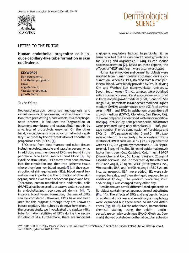
Results showed awell-differentiated epidermis onfibroblast-containing collagenous dermal substitutes(Fig. 1A). The effects of EPCs and angiogenic factorson epidermal thickness and keratinocytemorphologywere examined but there were no marked differ-ences (Fig. 1B—E). On the other hand, immunohisto-chemical staining using the avidin— biotin—peroxidase complex technigue (DAKO,Glostrup,Den-mark) showed platelet-endothelial cellular adhesion
tology. Published by Elsevier Ireland Ltd. All rights reserved.
76 Letter to the Editor
Fig. 1 Hematoxylin/eosin staining and CD31 and integrin b1 immunohistochemical staining. SEs were reconstructedusing fibroblasts (A, F and K) or with fibroblasts plus EPCs (B—E, G—J and L—O). Sections of SEs were hematoxylin/eosinstained (A—E), or immunostained for CD31 (F—J) and integrin b1 (K—O). Original magnification: 400� in A—O.
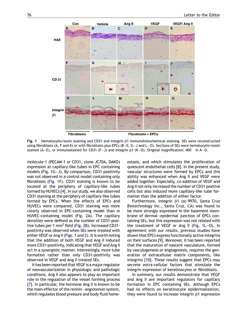
molecule-1 (PECAM-1 or CD31, clone JC70A, DAKO)expression at capillary-like tubes in EPC containingmodels (Fig. 1G—J). By comparison, CD31-positivitywas not observed in a control model containing onlyfibroblasts (Fig. 1F). CD31 staining is known to belocated at the periphery of capillary-like tubesformed by HUVECs [4]. In our study, we also observedCD31 staining at the periphery of capillary-like tubesformed by EPCs. When the effects of EPCs andHUVECs were compared, CD31 staining was moreclearly observed in EPC-containing model than inHUVEC-containing model (Fig. 2A). The capillarydensities were defined as the number of CD31-posi-tive tubes per 1 mm2 field (Fig. 2B). Increased CD31-positivity was observed when SEs were treated witheither VEGF or Ang II (Figs. 1 and 2). It is worth notingthat the addition of both VEGF and Ang II inducedmore CD31-positivity, indicating that VEGF and Ang IIact in a synergistic manner. Interestingly, more tubeformation rather than only CD31-positivity wasobserved in VEGF and Ang II treated SEs.
It has been reported that VEGF is amajor regulatorof neovascularization in physiologic and pathologicconditions. Ang II also appears to play an importantrole in the regulation of the vessel forming process[7]. In particular, the hormone Ang II is known to bethe main effector of the rennin—angiotensin system,which regulates blood pressure and body fluid home-
ostasis, and which stimulates the proliferation ofquiescent endothelial cells [8]. In the present study,vascular structures were formed by EPCs and thisability was enhanced when Ang II and VEGF wereadded together. Especially, co-addition of VEGF andAng II not only increased the number of CD31 positivecells but also induced more capillary-like tube for-mation than the addition of either factor.
Furthermore, integrin b1 (sc-9970, Santa CruzBiotechnology Inc., Santa Cruz, CA) was found tobe more strongly expressed in the basement mem-brane of dermal—epidermal junction of EPCs con-taining SEs, but this expression was not related withthe treatment of VEGF or Ang II (Fig. 1L—O). Inagreement with our results, previous studies haveshown that EPCs express functionally active integrinson their surfaces [9]. Moreover, it has been reportedthat the maturation of nascent vasculature, formedby vasculogenesis or angiogenesis, requires the gen-eration of extracellular matrix components, likeintegrins [10]. These results suggest that EPCs maysecrete extra-cellular factors that stimulate theintegrin expression of keratinocytes or fibroblasts.
In summary, our results demonstrate that VEGFand Ang II are important regulators for capillaryformation in EPC containing SEs. Although EPCshad no effects on keratinocyte epidermalization,they were found to increase integrin b1 expression
Letter to the Editor 77
Fig. 2 Capillary-like tube density in the dermis of SEs. (A)SEs were reconstructed using fibroblasts or with fibroblastsplus HUVECs or with fibroblasts plus EPCs. Sections of SEswere immunostained for CD31. Original magnification:400�. (B) Values are expressed as means (�S.D.) of num-bers of capillary-like tubes per 1 mm2 field. Results wereobtained from 10 random fields per tissue section. Statis-tical significancewasevaluatedby t-test in the SPSS release12.0 (SPSS Inc.). **P < 0.01 compared to the vehicle-trea-ted SE. Experiments were repeated at least in duplicate.
in basement membrane, which is an importantrequirement for SE reconstruction.
Acknowledgements
This researchwas supported by a grant (SC3260) fromthe Stem Cell Research Center of the 21st CenturyFrontier Program funded by the Ministry of Scienceand Technology, Republic of Korea. We also wish toacknowledge the support of the Pacific Corporation.We gratefully acknowledge the assistance of Drs.Dukkyung Kim and Wonhee Suh (SungkyunkwanUniv., Seoul, Korea) who kindly donated EPCs.
References
[1] Bauer SM, Bauer RJ, Liu ZJ, Chen H, Goldstein L, VelazquezOC. Vascular endothelial growth factor-C promotes vascu-
logenesis, angiogenesis, and collagen constriction in three-dimensional collagen gels. J Vasc Surg 2005;41:699—707.
[2] Zammaretti P, Zisch AH. Adult ‘endothelial progenitor cells’.Renewing vasculature. Int J Biochem Cell Biol 2005;37:493—503.
[3] Choi JH, Hur J, Yoon CH, Kim JH, Lee CS, Youn SW, et al.Augmentation of therapeutic angiogenesis using geneticallymodified human endothelial progenitor cells with alteredglycogen synthase kinase-3beta activity. J Biol Chem2004;279:49430—8.
[4] Hudon V, Berthod F, Black AF, Damour O, Germain L, AugerFA. A tissue-engineered endothelialized dermis to study themodulation of angiogenic and angiostatic molecules oncapillary-like tube formation in vitro. Br J Dermatol2003;148:1094—104.
[5] Choi JH, Kim KL, Huh W, Kim B, Byun J, Suh W, et al.Decreased number and impaired angiogenic function ofendothelial progenitor cells in patients with chronic renalfailure. Arterioscler Thromb Vasc Biol 2004;24:1246—52.
[6] Auger FA, Lopez Valle CA, Guignard R, Tremblay N, Noel B,Goulet F, et al. Skin equivalent produced with human col-lagen. In Vitro Cell Dev Biol Anim 1995;31:432—9.
[7] Michel F, Ambroisine ML, Duriez M, Delcayre C, Levy BI,Silvestre JS. Aldosterone enhances ischemia-induced neo-vascularization through angiotensin II-dependent pathway.Circulation 2004;109:1933—7.
[8] Walther T, Menrad A, Orzechowski HD, Siemeister G, Paul M,Schirner M. Differential regulation of in vivo angiogenesis byangiotensin II receptors. FASEB J 2003;17:2061—7.
[9] Chavakis E, Aicher A, Heeschen C, Sasaki K, Kaiser R, ElMakhfi N, et al. Role of beta2-integrins for homing andneovascularization capacity of endothelial progenitor cells.J Exp Med 2005;201:63—72.
[10] Kelm JM, Diaz Sanchez-Bustamante C, Ehler E, Hoerstrup SP,Djonov V, Ittner L, et al. VEGF profiling and angiogenesis inhuman microtissues. J Biotechnol 2005;118:213—29.
Hyun-Joo ChoDong-Seok KimEun-Sang Park
Kyoung-Chan Park*Department of Dermatology,
Seoul National University College of Medicine,28 Yongon-Dong, Chongno-Gu, Seoul 110-744,
Republic of Korea
*Corresponding author. Present address:Department of Dermatology,
Seol National University Bundang Hospital,300 Gumi-Dong, Bundang-Gu,
Kyoungki-Do 463-707, Republic of Korea.Tel.: +82 31 787 7311; fax: +82 2 3675 1187
E-mail address: [email protected]
13 September 2005


![Effect of vitamin D on endothelial progenitor cells function · vitamin D on EPCs function. Aim ... immune cells and endothelial cells [16]). Additional studies suggest a favorable](https://img.pdfslide.net/doc/110x75/60c10a1fa60e3e04a118fdb0/effect-of-vitamin-d-on-endothelial-progenitor-cells-function-vitamin-d-on-epcs-function.jpg)