Embed Size (px)
Citation preview
weiteren Forschungsbemühungen zielen darauf hin, dieses neue Testverfahren als Screeningmethode einer breiten Öffent-lichkeit zugänglich zu machen.
KorrespondenzadresseDr. R. HerwigUrologische Klinik, Medizinische UniversitätWähringer Gürtel 18–20, 1090 WienÖ[email protected]
Literatur
1. Hsing AW, Chokkalingam AP (2006) Prostate can-cer epidemiology. Front Biosci 11: 1388–1413
2. Chodak G (2006) Prostate cancer: epidemiology, screening, and biomarkers. Rev Urol 8(Suppl 2): 3–8
3. Sinha AA, Wilson MJ, Gleason DF (1987) Immuno-electron microscopic localization of prostatic-spe-cific antigen in human prostate by the protein A-gold complex. Cancer 60: 1288–1293
4. Hamdy FC, Lawry J, Anderson JB et al. (1992) Cir-culating prostate specific antigen-positive cells correlate with metastatic prostate cancer. Br J Urol 69: 392–396
5. Fadlon EJ, Rees RC, McIntyre C et al. (1996) Detec-tion of circulating prostate-specific antigen-posi-tive cells in patients with prostate cancer by flow cytometry and reverse transcription polymerase chain reaction. Br J Cancer 74: 400–405
6. Nockher WA, Scherberich JE (1998) Expanded CD14+ CD16+ monocyte subpopulation in pati-ents with acute and chronic infections undergoing hemodialysis. Infect Immun 66: 2782–2790
7. Cheung DL, Hamilton JA (1992) Regulation of hu-man monocyte DNA synthesis by colony-stimula-ting factors, cytokines, and cyclic adenosine mo-nophosphate. Blood 79: 1972–1981
8. Finnin M, Hamilton JA, Moss ST (1999) Direct com-parison of the effects of CSF-1 (M-CSF) and GM-CSF on human monocyte DNA synthesis and CSF receptor expression. J Interferon Cytokine Res 19: 417–423
9. Finnin M, Hamilton JA, Moss ST (1999) Characteri-zation of a CSF-induced proliferating subpopulati-on of human peripheral blood monocytes by sur-face marker expression and cytokine production. J Leukoc Biol 66: 953–960
10. Moss ST, Hamilton JA (2000) Proliferation of a sub-population of human peripheral blood monocytes in the presence of colony stimulating factors may contribute to the inflammatory process in diseases such as rheumatoid arthritis. Immunobiology 202: 18–25
11. Bitterman PB, Saltzman LE, Adelberg S et al. (1984) Alveolar macrophage replication. One mechanism for the expansion of the mononuclear phagocyte population in the chronically inflamed lung. J Clin Invest 74: 460–469
12. Hamilton JA (1993) Rheumatoid arthritis: oppo-sing actions of haemopoietic growth factors and slow-acting anti-rheumatic drugs. Lancet 342: 536–539
13. Henderson B, Glynn LE, Bitensky L, Chayen J (1981) Evidence for cell division in synoviocytes in acutely inflamed rabbit joints. Ann Rheum Dis 40: 177–181
Urologe 2007 · 46:1070–1071 · DOI 10.1007/s00120-007-1420-8 · Online publiziert: 1. Juli 2007
© Springer Medizin Verlag 2007
A. Rabien1 · G. Kristiansen2 · E.P. Diamandis3 · K. Jung1 · C. Stephan1
1 Klinik für Urologie, Charité - Universitätsmedizin Berlin,Campus Charité Mitte, Berlin2 Institut für Pathologie, Charité - Universitätsmedizin Berlin, Campus Charité Mitte, Berlin3 Department of Pathology and Laboratory Medicine, Mount Sinai Hospital, Toronto
Humane Kallikreine als TumormarkerValidierung potenzieller Marker des Prostatakarzinoms in Serum und Gewebe
14. Jutila MA, Banks KL (1988) Increased macrophage division in the synovial fluid of goats infected with caprine arthritis-encephalitis virus. J Infect Dis 157: 1193–1202
15. Hornell TM, Burster T, Jahnsen FL et al. (2006) Hu-man dendritic cell expression of HLA-DO is subset specific and regulated by maturation. J Immunol 176: 3536–3547
16. Quadbeck B, Stucke M, Eckstein AK et al. (2006) Dysregulation of TNF/TNFR superfamily members: A systemic link between intra- and extrathyroidal manifestations in Graves‘ disease. Scand J Immu-nol 64: 523–530
17. Tacke F, Randolph GJ (2006) Migratory fate and dif-ferentiation of blood monocyte subsets. Immuno-biology 211: 609–618
18. Gille C, Spring B, Tewes L et al. (2006) A new me-thod to quantify phagocytosis and intracellular de-gradation using green fluorescent protein-labeled Escherichia coli: comparison of cord blood macro-phages and peripheral blood macrophages of healthy adults. Cytometry A 69: 152–154
19. Herwig R, Horninger W, Rehder P et al. (2005) Abi-lity of PSA-positive circulating macrophages to detect prostate cancer. Prostate 62: 290–298
20. Herwig R, Pelzer A, Horninger W et al. (2004) Measurement of intracellular versus extracellu-lar prostate-specific antigen levels in peripheral macrophages: a new approach to noninvasive di-agnosis of prostate cancer. Clin Prostate Cancer 3: 184–188
21. Herwig R, Djavan B, Kramer G et al. (2006) Diffe-rentiation enhancement of circulating immune cells containing intracellular PSA: A new method for discrimination between benign and malignant prostatic disease. J Urol 175: 82
22. Barry MJ (2001) Clinical practice. Prostate-specific-antigen testing for early diagnosis of prostate can-cer. N Engl J Med 344: 1373–1377
23. Ornstein DK, Kang J (2001) How to improve pros-tate biopsy detection of prostate cancer. Curr Urol Rep 2: 218–223
Die humanen Kallikrein-ähnlichen Pepti-dasen (KLK, alt: Kallikreine), zu denen auch das prostataspezifische Antigen (PSA) gehört, sind sekretorische Proteine, die sowohl an Prozessen des Zellwachs-tums, der Migration und Angiogenese als auch an Invasion und Metastasierung be-teiligt sind [1]. Da unsere Untersuchungen der beiden jüngsten Mitglieder der KLK-Familie, KLK14 und KLK15, erste Hinwei-se auf eine tumorspezifische Regulation beim Prostatakarzinom lieferten [2, 4, 5], wurden ausführliche Expressionsprofile für KLK14 und KLK15 in Gewebe von Prostataadenokarzinomen nach radikaler Prostatektomie erstellt und mit klinisch-pathologischen bzw. Verlaufsdaten kom-
biniert. Die Analyse 25 lasermikrodisse-zierter Gewebepaare (normal/Tumor) mittels quantitativer RT-PCR (reverse transcriptase polymerase chain reaction) zeigte keine tumorrelevanten Ergebnisse auf Transkriptionsebene.
Die umfangreiche immunhistoche-mische Analyse der KLK14- und KLK15-Proteine an 237 Paraffinschnitten mit im Labor Diamandis generierten Antikörpern [3, 5] stellte jedoch die prognostische Be-deutung der Kallikreine heraus. Die Ex-pression beider Proteine korrelierte positiv mit dem pathologischen Tumorstadium (Rangtest nach Spearman). Eine höhere KLK15-Expression ging mit einem höheren Gleason-Grad des Tumors einher (exakter
1070 | Der Urologe 9 · 2007
Prostatakarzinom
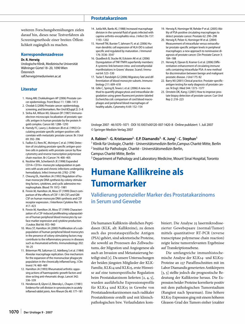
Test nach Fisher). Kaplan-Meier-Analysen zeigten, dass ein höheres KLK14- bzw. KLK15-Niveau mit einer kürzeren rezidiv-freien postoperativen Verlaufszeit assozi-iert war. Rezidive wurden dabei durch den Anstieg der PSA-Werte in der Nachbeob-achtungszeit definiert (. Abb. 1).
Die multivariate Analyse dichoto-misierter Daten nach dem „cox propor-tional hazards regression model“ stell-te KLK15 als unabhängigen prognosti-schen Faktor im Vergleich mit den rele-vanten klinisch-pathologischen Parame-tern präoperativer PSA-Wert, Tumorsta-dium, Tumorgrad nach Gleason und Sta-tus des Operationsrandes heraus (rela-tives Risiko =1,739; 95%-Konfidenzinter-vall =1,033–2,928; p=0,037).
Mit im Labor Diamandis kürzlich ent-wickelten hochspezifischen ELISA- (en-zyme-linked immunosorbent assay-)Tests für KLK14 und KLK15 konnten die Protei-ne bereits im Prostatagewebe und Samen-plasma nachgewiesen werden [2, 5]. Ein Vergleich gesunder Probanden mit Prosta-takarzinompatienten im KLK14-ELISA zeigte ein höheres KLK14-Niveau im Serum der Krebspatienten [2]. Aufgrund dieser viel versprechenden Daten sollen ausge-dehnte Serumtests erfolgen, um die Eig-nung der Kallikreine KLK14 und KLK15 als prognostische Marker in Kombination mit den bisher nicht ausreichenden PSA-Wer-ten bewerten zu können.
1.0 P = 0.002 P = 0.002
wenig KLK14wenig KLK15
viel KLK14viel KLK15
0.8
0.6
Prog
ress
ions
feie
Übe
rlebe
nsra
te
0.4
0.2
0.0
0 50 100 150 200
Monate
1.0
0.8
0.6
0.4
0.2
0.0
0 50 100 150 200
Monate
Abb. 1 8 Kaplan-Meier-Analysen der KLK14- (links) bzw. KLK15-Expression (rechts) von 193 Prostata-karzinomen mit signifikant kürzerer progressionsfreier Überlebensrate für Fälle mit höherer KLK-Ex-pression [zensierte Fälle sind markiert (+); postoperative Beobachtungszeit 10–188 Monate, Median 60 Monate]
Gefördert durch die Deutsche Forschungsge-meinschaft (JU365/6–1/2) und die SONNEN-FELD-Stiftung, Berlin.
KorrespondenzadresseA. RabienKlinik für Urologie, Charité - Universitätsmedizin Berlin,Campus Charité MitteCharitéplatz 1, 10117 [email protected]
Literatur
1. Borgono CA, Diamandis EP (2004) The emerging roles of human tissue kallikreins in cancer. Nat Rev Cancer 4: 876–890
2. Borgono CA, Michael IP, Shaw JL et al. (2007) Ex-pression and functional characterization of the cancer-related serine protease, human tissue kalli-krein 14. J Biol Chem 282: 2405–2422
3. Fritzsche F, Gansukh T, Borgono CA et al. (2006) Ex-pression of human Kallikrein 14 (KLK14) in bre-ast cancer is associated with higher tumour grades and positive nodal status. Br J Cancer 94: 540–547
4. Paliouras M, Borgono C, Diamandis EP (2007) Hu-man tissue kallikreins: The cancer biomarker fami-ly. Cancer Lett 249: 61–79
5. Shaw JL, Grass L, Sotiropoulou G, Diamandis EP (2007) Development of an immunofluorometric assay for human kallikrein 15 (KLK15) and identifi-cation of KLK15 in tissues and biological fluids. Clin Biochem 40: 104–110
Urologe 2007 · 46:1071–1077 · DOI 10.1007/s00120-007-1422-6 · Online publiziert: 14. Juli 2007
© Springer Medizin Verlag 2007
L. Rinnab1 · M. Cronauer2 · D. Spindler2 · R.E. Hautmann1 · R. Küfer1
1 Urologische Universitätsklinik, Ulm2 Institut für Allgemeine Zoologie und Endokrinologie, Universität, Ulm
Forschungsaktivitäten an der Universität Ulm um das Prostatakarzinom
Urologische Universitätsklinik Ulm (Prof. Dr. Dr. h.c. R. Hautmann, Arbeitsgruppe PD Dr. R. Küfer)
Schwerpunkt der Forschungsaktivitäten um das Prostatakarzinom an der Urolo-gischen Universitätsklinik in Ulm sind die Identifikation von Biomarkern sowohl für die Diagnostik als auch solche mit pro-gnostischem Wert oder als potentielle Therapietargets. Mit der Identifikation von Zielstrukturen für eine Therapie ist die Forschung auf dem Gebiet der Pathways
verknüpft. Im Nachfolgenden sollen in Form eines Berichts die Hauptaktivitäten auf diesem Gebiet skizziert werden.
Zur Differenzierung eines harmlosen von einem aggressiven Prostatakarzinom (PCA) sowie zur Prognoseeinschätzung besteht großer Bedarf der Identifikation spezifischer Marker. Idealerweise sollten solche Marker sowohl diagnostisch wert-voll sein, als auch die Biologie des Tu-mors unabhängig von klinischen Para-metern reflektieren und einen prognosti-schen Wert besitzen. Eine auf Biomarker gestützte Klassifikation könnte z. B. nicht
1071Der Urologe 9 · 2007 |