Embed Size (px)
Citation preview
The IAC Standards and Guidelines for Cardiac Electrophysiology
Accreditation
2 IAC Standards and Guidelines for Cardiac Electrophysiology Accreditation (Published 8/15/2017, Revised 4/5/2018) ©2018 Intersocietal Accreditation Commission. All Rights Reserved.
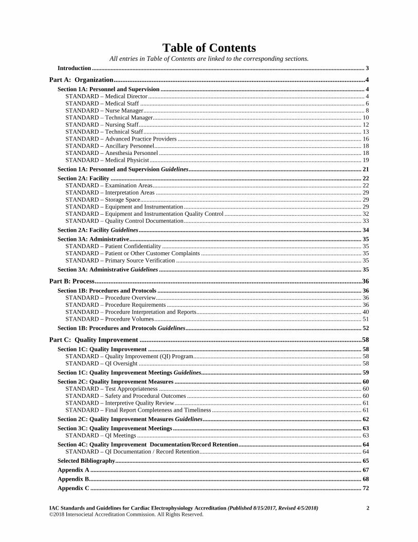
Table of Contents All entries in Table of Contents are linked to the corresponding sections.
Introduction ............................................................................................................................................................................... 3 Part A: Organization .................................................................................................................................................. 4
Section 1A: Personnel and Supervision ................................................................................................................................... 4 STANDARD – Medical Director ........................................................................................................................................... 4 STANDARD – Medical Staff ................................................................................................................................................ 6 STANDARD – Nurse Manager .............................................................................................................................................. 8 STANDARD – Technical Manager...................................................................................................................................... 10 STANDARD – Nursing Staff ............................................................................................................................................... 12 STANDARD – Technical Staff ............................................................................................................................................ 13 STANDARD – Advanced Practice Providers ...................................................................................................................... 16 STANDARD – Ancillary Personnel ..................................................................................................................................... 18 STANDARD – Anesthesia Personnel .................................................................................................................................. 18 STANDARD – Medical Physicist ........................................................................................................................................ 19
Section 1A: Personnel and Supervision Guidelines ............................................................................................................... 21 Section 2A: Facility ................................................................................................................................................................. 22
STANDARD – Examination Areas ...................................................................................................................................... 22 STANDARD – Interpretation Areas .................................................................................................................................... 29 STANDARD – Storage Space .............................................................................................................................................. 29 STANDARD – Equipment and Instrumentation .................................................................................................................. 29 STANDARD – Equipment and Instrumentation Quality Control ........................................................................................ 32 STANDARD – Quality Control Documentation .................................................................................................................. 33
Section 2A: Facility Guidelines ............................................................................................................................................... 34 Section 3A: Administrative ..................................................................................................................................................... 35
STANDARD – Patient Confidentiality ................................................................................................................................ 35 STANDARD – Patient or Other Customer Complaints ....................................................................................................... 35 STANDARD – Primary Source Verification ....................................................................................................................... 35
Section 3A: Administrative Guidelines .................................................................................................................................. 35 Part B: Process ........................................................................................................................................................... 36
Section 1B: Procedures and Protocols ................................................................................................................................... 36 STANDARD – Procedure Overview .................................................................................................................................... 36 STANDARD – Procedure Requirements ............................................................................................................................. 36 STANDARD – Procedure Interpretation and Reports .......................................................................................................... 40 STANDARD – Procedure Volumes ..................................................................................................................................... 51
Section 1B: Procedures and Protocols Guidelines ................................................................................................................. 52 Part C: Quality Improvement ................................................................................................................................. 58
Section 1C: Quality Improvement ......................................................................................................................................... 58 STANDARD – Quality Improvement (QI) Program ............................................................................................................ 58 STANDARD – QI Oversight ............................................................................................................................................... 58
Section 1C: Quality Improvement Meetings Guidelines ....................................................................................................... 59 Section 2C: Quality Improvement Measures ........................................................................................................................ 60
STANDARD – Test Appropriateness .................................................................................................................................. 60 STANDARD – Safety and Procedural Outcomes ................................................................................................................ 60 STANDARD – Interpretive Quality Review ........................................................................................................................ 61 STANDARD – Final Report Completeness and Timeliness ................................................................................................ 61
Section 2C: Quality Improvement Measures Guidelines ...................................................................................................... 62 Section 3C: Quality Improvement Meetings ......................................................................................................................... 63
STANDARD – QI Meetings ................................................................................................................................................ 63 Section 4C: Quality Improvement Documentation/Record Retention ............................................................................... 64
STANDARD – QI Documentation / Record Retention ........................................................................................................ 64 Selected Bibliography .............................................................................................................................................................. 65 Appendix A .............................................................................................................................................................................. 67 Appendix B............................................................................................................................................................................... 68 Appendix C .............................................................................................................................................................................. 72
IAC Standards and Guidelines for Cardiac Electrophysiology Accreditation (Published 8/15/2017, Revised 4/5/2018) 3 ©2018 Intersocietal Accreditation Commission. All Rights Reserved. Return to Table of Contents»
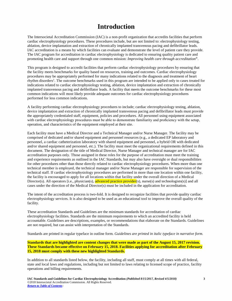
Introduction The Intersocietal Accreditation Commission (IAC) is a non-profit organization that accredits facilities that perform cardiac electrophysiology procedures. These procedures include, but are not limited to: electrophysiology testing, ablation, device implantation and extraction of chronically implanted transvenous pacing and defibrillator leads. IAC accreditation is a means by which facilities can evaluate and demonstrate the level of patient care they provide. The IAC program for accreditation in cardiac electrophysiology is dedicated to ensuring quality patient care and promoting health care and support through one common mission: Improving health care through accreditation®.
This program is designed to accredit facilities that perform cardiac electrophysiology procedures by ensuring that the facility meets benchmarks for quality based on resources, training and outcomes. Cardiac electrophysiology procedures may be appropriately performed for many indications related to the diagnosis and treatment of heart rhythm disorders1. The outcome benchmarks used in this program are intended to be applied only to cases treated for indications related to cardiac electrophysiology testing, ablation, device implantation and extraction of chronically implanted transvenous pacing and defibrillator leads. A facility that meets the outcome benchmarks for these most common indications will most likely provide adequate outcomes for cardiac electrophysiology procedures performed for less common indications.
A facility performing cardiac electrophysiology procedures to include; cardiac electrophysiology testing, ablation, device implantation and extraction of chronically implanted transvenous pacing and defibrillator leads must provide the appropriately credentialed staff, equipment, policies and procedures. All personnel using equipment associated with cardiac electrophysiology procedures must be able to demonstrate familiarity and proficiency with the setup, operation, and characteristics of the equipment employed at their site.
Each facility must have a Medical Director and a Technical Manager and/or Nurse Manager. The facility may be comprised of dedicated and/or shared equipment and personnel resources (e.g., a dedicated EP laboratory and personnel, a cardiac catheterization laboratory with shared equipment and personnel, a hybrid OR with dedicated and/or shared equipment and personnel, etc.). The facility must meet the organizational requirements defined in this document. The designation of the title of Medical Director, Nurse Manager and technical manager are for IAC accreditation purposes only. Those assigned in these roles for the purpose of accreditation must meet the training and experience requirements as outlined in the IAC Standards, but may also have oversight or dual responsibilities for other procedures other than those directly related to cardiac electrophysiology procedures. When more than one technical member is employed, the technical manager and/or Nurse Manager are responsible for supervision of the technical staff. If cardiac electrophysiology procedures are performed in more than one location within one facility, the facility is encouraged to apply for all locations within that facility under the overall direction of a Medical Director(s). All operators [i.e., physician(s), advanced practice provider(s), nurse(s) and technologists(s)] and all cases under the direction of the Medical Director(s) must be included in the application for accreditation.
The intent of the accreditation process is two-fold. It is designed to recognize facilities that provide quality cardiac electrophysiology services. It is also designed to be used as an educational tool to improve the overall quality of the facility.
These accreditation Standards and Guidelines are the minimum standards for accreditation of cardiac electrophysiology facilities. Standards are the minimum requirements to which an accredited facility is held accountable. Guidelines are descriptions, examples, or recommendations that elaborate on the Standards. Guidelines are not required, but can assist with interpretation of the Standards.
Standards are printed in regular typeface in outline form. Guidelines are printed in italic typeface in narrative form.
Standards that are highlighted are content changes that were made as part of the August 15, 2017 revision. These Standards became effective on February 15, 2018. Facilities applying for accreditation after February 15, 2018 must comply with these new highlighted Standards.
In addition to all standards listed below, the facility, including all staff, must comply at all times with all federal, state and local laws and regulations, including but not limited to laws relating to licensed scope of practice, facility operations and billing requirements.
IAC Standards and Guidelines for Cardiac Electrophysiology Accreditation (Published 8/15/2017, Revised 4/5/2018) 4 ©2018 Intersocietal Accreditation Commission. All Rights Reserved. Return to Table of Contents»
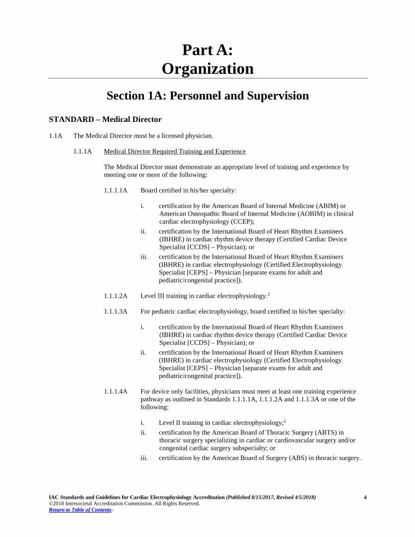
Part A: Organization
Section 1A: Personnel and Supervision
STANDARD – Medical Director 1.1A The Medical Director must be a licensed physician.
1.1.1A Medical Director Required Training and Experience
The Medical Director must demonstrate an appropriate level of training and experience by meeting one or more of the following: 1.1.1.1A Board certified in his/her specialty:
i. certification by the American Board of Internal Medicine (ABIM) or
American Osteopathic Board of Internal Medicine (AOBIM) in clinical cardiac electrophysiology (CCEP);
ii. certification by the International Board of Heart Rhythm Examiners (IBHRE) in cardiac rhythm device therapy (Certified Cardiac Device Specialist [CCDS] – Physician); or
iii. certification by the International Board of Heart Rhythm Examiners (IBHRE) in cardiac electrophysiology (Certified Electrophysiology Specialist [CEPS] – Physician [separate exams for adult and pediatric/congenital practice]).
1.1.1.2A Level III training in cardiac electrophysiology.2
1.1.1.3A For pediatric cardiac electrophysiology, board certified in his/her specialty:
i. certification by the International Board of Heart Rhythm Examiners
(IBHRE) in cardiac rhythm device therapy (Certified Cardiac Device Specialist [CCDS] – Physician); or
ii. certification by the International Board of Heart Rhythm Examiners (IBHRE) in cardiac electrophysiology (Certified Electrophysiology Specialist [CEPS] – Physician [separate exams for adult and pediatric/congenital practice]).
1.1.1.4A For device only facilities, physicians must meet at least one training experience
pathway as outlined in Standards 1.1.1.1A, 1.1.1.2A and 1.1.1.3A or one of the following:
i. Level II training in cardiac electrophysiology;2 ii. certification by the American Board of Thoracic Surgery (ABTS) in
thoracic surgery specializing in cardiac or cardiovascular surgery and/or congenital cardiac surgery subspecialty; or
iii. certification by the American Board of Surgery (ABS) in thoracic surgery.
IAC Standards and Guidelines for Cardiac Electrophysiology Accreditation (Published 8/15/2017, Revised 4/5/2018) 5 ©2018 Intersocietal Accreditation Commission. All Rights Reserved. Return to Table of Contents»
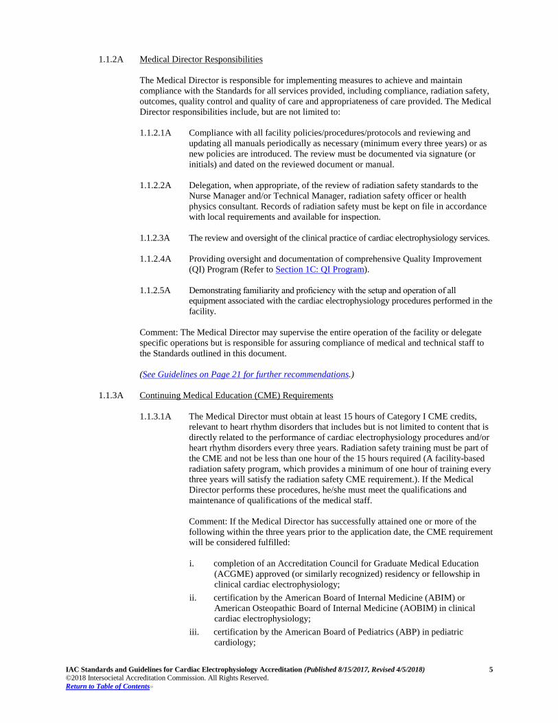
1.1.2A Medical Director Responsibilities The Medical Director is responsible for implementing measures to achieve and maintain compliance with the Standards for all services provided, including compliance, radiation safety, outcomes, quality control and quality of care and appropriateness of care provided. The Medical Director responsibilities include, but are not limited to: 1.1.2.1A Compliance with all facility policies/procedures/protocols and reviewing and
updating all manuals periodically as necessary (minimum every three years) or as new policies are introduced. The review must be documented via signature (or initials) and dated on the reviewed document or manual.
1.1.2.2A Delegation, when appropriate, of the review of radiation safety standards to the Nurse Manager and/or Technical Manager, radiation safety officer or health physics consultant. Records of radiation safety must be kept on file in accordance with local requirements and available for inspection.
1.1.2.3A The review and oversight of the clinical practice of cardiac electrophysiology services.
1.1.2.4A Providing oversight and documentation of comprehensive Quality Improvement
(QI) Program (Refer to Section 1C: QI Program).
1.1.2.5A Demonstrating familiarity and proficiency with the setup and operation of all equipment associated with the cardiac electrophysiology procedures performed in the facility.
Comment: The Medical Director may supervise the entire operation of the facility or delegate specific operations but is responsible for assuring compliance of medical and technical staff to the Standards outlined in this document. (See Guidelines on Page 21 for further recommendations.)
1.1.3A Continuing Medical Education (CME) Requirements
1.1.3.1A The Medical Director must obtain at least 15 hours of Category I CME credits,
relevant to heart rhythm disorders that includes but is not limited to content that is directly related to the performance of cardiac electrophysiology procedures and/or heart rhythm disorders every three years. Radiation safety training must be part of the CME and not be less than one hour of the 15 hours required (A facility-based radiation safety program, which provides a minimum of one hour of training every three years will satisfy the radiation safety CME requirement.). If the Medical Director performs these procedures, he/she must meet the qualifications and maintenance of qualifications of the medical staff. Comment: If the Medical Director has successfully attained one or more of the following within the three years prior to the application date, the CME requirement will be considered fulfilled: i. completion of an Accreditation Council for Graduate Medical Education
(ACGME) approved (or similarly recognized) residency or fellowship in clinical cardiac electrophysiology;
ii. certification by the American Board of Internal Medicine (ABIM) or American Osteopathic Board of Internal Medicine (AOBIM) in clinical cardiac electrophysiology;
iii. certification by the American Board of Pediatrics (ABP) in pediatric cardiology;
IAC Standards and Guidelines for Cardiac Electrophysiology Accreditation (Published 8/15/2017, Revised 4/5/2018) 6 ©2018 Intersocietal Accreditation Commission. All Rights Reserved. Return to Table of Contents»
iv. certification by the International Board of Heart Rhythm Examiners (IBHRE) in cardiac rhythm device therapy (Certified Cardiac Device Specialist [CCDS] – Physician); or
v. certification by the International Board of Heart Rhythm Examiners (IBHRE) in cardiac electrophysiology (Certified Electrophysiology Specialist [CEPS] – Physician [separate exams for adult and pediatric/congenital practice]).
1.1.3.2A Documentation of CME credits must be kept on file and available for inspection.
STANDARD – Medical Staff 1.2A All members of the medical staff must be licensed physicians.
1.2.1A Medical Staff Required Training and Experience
The medical staff must demonstrate an appropriate level of training and experience by meeting one or more of the following:
1.2.1.1A Board certified in his/her specialty:
i. completion of an Accreditation Council for Graduate Medical Education
(ACGME) approved (or similarly recognized) residency or fellowship in clinical cardiac electrophysiology;
ii. certification by the American Board of Internal Medicine (ABIM) or American Osteopathic Board of Internal Medicine (AOBIM) in clinical cardiac electrophysiology;
iii. certification by the International Board of Heart Rhythm Examiners (IBHRE) in cardiac rhythm device therapy (Certified Cardiac Device Specialist [CCDS] – Physician); or
iv. certification by the International Board of Heart Rhythm Examiners (IBHRE) in cardiac electrophysiology (Certified Electrophysiology Specialist [CEPS] – Physician [separate exams for adult and pediatric/congenital practice]).
1.2.1.2A Level III training in cardiac electrophysiology.28
1.2.1.3A For pediatric cardiac electrophysiology, all medical staff should be board certified in his/her specialty: i. certification by the International Board of Heart Rhythm Examiners
(IBHRE) in cardiac rhythm device therapy (Certified Cardiac Device Specialist [CCDS] – Physician); or
ii. certification by the International Board of Heart Rhythm Examiners (IBHRE) in cardiac electrophysiology (Certified Electrophysiology Specialist [CEPS] – Physician [separate examinations for adult and pediatric/congenital practice]).
Comment: The facility must have a plan in place for all non-certified medical staff to obtain an appropriate certification prior to the next accreditation cycle.
1.2.1.4A For device implantation only facilities, physicians must meet at least one training
experience pathway as outlined in Standards 1.1.1.1A, 1.1.1.2A and 1.1.1.3A or one of the following:
IAC Standards and Guidelines for Cardiac Electrophysiology Accreditation (Published 8/15/2017, Revised 4/5/2018) 7 ©2018 Intersocietal Accreditation Commission. All Rights Reserved. Return to Table of Contents»
i. Level II training in cardiac electrophysiology;2 ii. certification by the American Board of Thoracic Surgery (ABTS) in
thoracic surgery specializing in cardiac or cardiovascular surgery and/or congenital cardiac surgery subspecialty; or
iii. certification by the American Board of Surgery (ABS) in thoracic surgery.
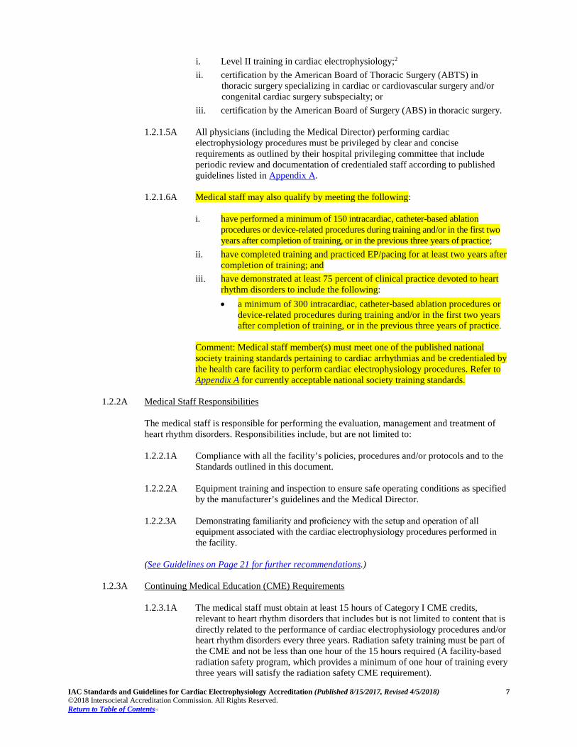
1.2.1.5A All physicians (including the Medical Director) performing cardiac electrophysiology procedures must be privileged by clear and concise requirements as outlined by their hospital privileging committee that include periodic review and documentation of credentialed staff according to published guidelines listed in Appendix A.
1.2.1.6A Medical staff may also qualify by meeting the following:
i. have performed a minimum of 150 intracardiac, catheter-based ablation procedures or device-related procedures during training and/or in the first two years after completion of training, or in the previous three years of practice;
ii. have completed training and practiced EP/pacing for at least two years after completion of training; and
iii. have demonstrated at least 75 percent of clinical practice devoted to heart rhythm disorders to include the following: • a minimum of 300 intracardiac, catheter-based ablation procedures or
device-related procedures during training and/or in the first two years after completion of training, or in the previous three years of practice.
Comment: Medical staff member(s) must meet one of the published national society training standards pertaining to cardiac arrhythmias and be credentialed by the health care facility to perform cardiac electrophysiology procedures. Refer to Appendix A for currently acceptable national society training standards.
1.2.2A Medical Staff Responsibilities
The medical staff is responsible for performing the evaluation, management and treatment of heart rhythm disorders. Responsibilities include, but are not limited to: 1.2.2.1A Compliance with all the facility’s policies, procedures and/or protocols and to the
Standards outlined in this document.
1.2.2.2A Equipment training and inspection to ensure safe operating conditions as specified by the manufacturer’s guidelines and the Medical Director.
1.2.2.3A Demonstrating familiarity and proficiency with the setup and operation of all
equipment associated with the cardiac electrophysiology procedures performed in the facility.
(See Guidelines on Page 21 for further recommendations.)
1.2.3A Continuing Medical Education (CME) Requirements
1.2.3.1A The medical staff must obtain at least 15 hours of Category I CME credits,
relevant to heart rhythm disorders that includes but is not limited to content that is directly related to the performance of cardiac electrophysiology procedures and/or heart rhythm disorders every three years. Radiation safety training must be part of the CME and not be less than one hour of the 15 hours required (A facility-based radiation safety program, which provides a minimum of one hour of training every three years will satisfy the radiation safety CME requirement).
IAC Standards and Guidelines for Cardiac Electrophysiology Accreditation (Published 8/15/2017, Revised 4/5/2018) 8 ©2018 Intersocietal Accreditation Commission. All Rights Reserved. Return to Table of Contents»
Comment: If the medical staff member has successfully attained one or more of the following within the three years prior to the application date, the CME requirement will be considered fulfilled: i. completion of an Accreditation Council for Graduate Medical Education
(ACGME) approved (or similarly recognized) residency or fellowship; ii. attaining certification by the American Board of Internal Medicine (ABIM)
or American Osteopathic Board of Internal Medicine (AOBIM) in clinical cardiac electrophysiology;
iii. certification by the International Board of Heart Rhythm Examiners (IBHRE) in cardiac rhythm device therapy (Certified Cardiac Device Specialist [CCDS] – Physician); or
iv. certification by the International Board of Heart Rhythm Examiners (IBHRE) in cardiac electrophysiology (Certified Electrophysiology Specialist [CEPS] – Physician [separate examinations for adult and pediatric/congenital practice]).
1.2.3.2A Documentation of CME credits must be kept on file and available for inspection.
STANDARD – Nurse Manager 1.3A The manager of the technical and nursing staff must be an appropriately credentialed technologist (1.4A)
and/or nurse and meet the required training and experience qualifications as outlined below. 1.3.1A Nurse Manager Required Training and Experience
1.3.1.1A The Nurse Manager must be licensed and demonstrate an appropriate level of
training and experience by meeting at least one of the following criteria: i. Registered Nurse (RN) ii. Advanced Practice Nurse (APRN) iii. Advanced health care degree or Bachelor of Science in Nursing (BSN)
preferred iv. Certification in interventional nursing specialty such as Cardiac Nurse
Practitioner (NP-C), Cardiovascular Clinical Nurse Specialist (CNS), Cardiac Vascular Nursing (CVRN), Certified Radiology Nurse (CRN)
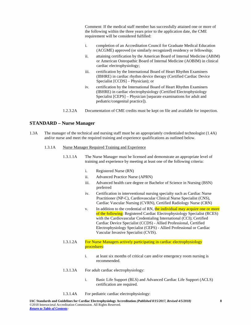
v. In addition to the credential of RN, the individual may acquire one or more of the following: Registered Cardiac Electrophysiology Specialist (RCES) with the Cardiovascular Credentialing International (CCI); Certified Cardiac Device Specialist (CCDS) - Allied Professional, Certified Electrophysiology Specialist (CEPS) - Allied Professional or Cardiac Vascular Invasive Specialist (CVIS).
1.3.1.2A For Nurse Managers actively participating in cardiac electrophysiology
procedures: i. at least six months of critical care and/or emergency room nursing is
recommended.
1.3.1.3A For adult cardiac electrophysiology:
i. Basic Life Support (BLS) and Advanced Cardiac Life Support (ACLS) certification are required.
1.3.1.4A For pediatric cardiac electrophysiology:
IAC Standards and Guidelines for Cardiac Electrophysiology Accreditation (Published 8/15/2017, Revised 4/5/2018) 9 ©2018 Intersocietal Accreditation Commission. All Rights Reserved. Return to Table of Contents»
i. Basic Life Support (BLS) and Pediatric Advanced Life Support (PALS) are
required.
1.3.2A Nurse Manager Responsibilities The Nurse Manager responsibilities may include, but are not limited to: 1.3.2.1A the day-to-day operations of the facility;
1.3.2.2A management of pre- and post-procedural care areas;
1.3.2.3A direct participation in the observation and care of patients undergoing cardiac
electrophysiology procedures;
1.3.2.4A application of institutional guidelines for patient monitoring, medication administration, procedural sedation and patient safety;
1.3.2.5A managing staff competencies and proficiency in performing tasks required before,
during, and after the procedure;
1.3.2.6A the delegation, when necessary, of specific responsibilities to the technical and/or nursing staff and/or ancillary staff;
1.3.2.7A verification of documentation of proper training and, at least annually, assessment
of the competence of technical, nursing staff and/or any ancillary staff who report to the Nurse Manager; and
1.3.2.8A demonstrating familiarity and proficiency with the setup and operation of all
equipment associated with the cardiac electrophysiology procedures performed in the facility.
(See Guidelines on Page 21 for further recommendations.)
1.3.3A Continuing Education (CE) Requirements
1.3.3.1A The Nurse Manager must obtain at least 15 hours of accredited CE relevant to heart
rhythm disorders that includes, but is not limited to, content that is directly related to the performance of cardiac electrophysiology procedures, heart rhythm disorders, cardiovascular assessment and/or patient management every three years. Radiation safety training must be part of the CE and not be less than one hour of the 15 hours required (A facility-based radiation safety program, which provides a minimum of one hour of training every three years will satisfy the radiation safety CME requirement.).
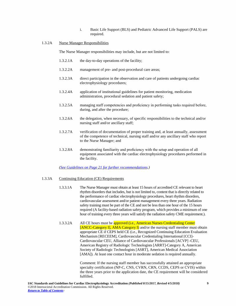
1.3.3.2A All CE hours must be approved (i.e., American Nurses Credentialing Center [ANCC-Category I], AMA Category I) and/or the nursing staff member must obtain appropriate CE if CEPS held CE (i.e., Recognized Continuing Education Evaluation Mechanism [RECEEM], Cardiovascular Credentialing International [CCI]-Cardiovascular CEU, Alliance of Cardiovascular Professionals [ACVP] -CEU, American Registry of Radiologic Technologists [ARRT]-Category A, American Society of Radiologic Technologists [ASRT], American Medical Association [AMA]). At least one contact hour in moderate sedation is required annually.
Comment: If the nursing staff member has successfully attained an appropriate specialty certification (NP-C, CNS, CVRN, CRN, CCDS, CEPS or CVIS) within the three years prior to the application date, the CE requirement will be considered fulfilled.
IAC Standards and Guidelines for Cardiac Electrophysiology Accreditation (Published 8/15/2017, Revised 4/5/2018) 10 ©2018 Intersocietal Accreditation Commission. All Rights Reserved. Return to Table of Contents»
1.3.3.3A Documentation of CE credits must be kept on file and available for inspection.
STANDARD – Technical Manager 1.4A The manager of the technical and nursing staff must be an appropriately credentialed technologist and/or
nurse (1.3A) and meet the required training and experience qualifications as outlined below. 1.4.1A Technical Manager Required Training and Experience
The Technical Manager must be licensed (where applicable) and demonstrate an appropriate level of training and experience by meeting one the following criteria: 1.4.1.1A A registered specialist with the Cardiovascular Credentialing International (CCI)
meeting at least one of the following criteria: i. Registered Cardiac Electrophysiology Specialist (RCES) with the
Cardiovascular Credentialing International (CCI); ii. Registered Cardiovascular Invasive Specialist (RCIS) with the
Cardiovascular Credentialing International (CCI); iii. Certified Electrophysiology Specialist (CEPS) Allied Professional with the
International Board of Heart Rhythm Examiners (IBHRE); or iv. Certified Cardiac Device Specialist (CCDS) Allied Professional with
International Board of Heart Rhythm Examiners (IBHRE).
1.4.1.2A A Registered Radiologic Technologist [RT(R)] with the American Registry of Radiologic Technologists (ARRT) meeting one or more of the following criteria: i. Cardiovascular-Interventional Radiography RT (CV); ii. Cardiac-Interventional Radiography RT (CI); iii. Certified Electrophysiology Specialist (CEPS) Allied Professional with the
International Board of Heart Rhythm Examiners (IBHRE); or iv. Certified Cardiac Device Specialist (CCDS) Allied Professional with
International Board of Heart Rhythm Examiners (IBHRE).
1.4.1.3A A Registered Technologist in Radiological Technology (RTR) with the Canadian Association of Medical Radiation Technologists (CAMRT) meeting one or more of the following criteria: i. Certified Electrophysiology Specialist (CEPS) Allied Professional with the
International Board of Heart Rhythm Examiners (IBHRE); or ii. Certified Cardiac Device Specialist (CCDS) Allied Professional with
International Board of Heart Rhythm Examiners (IBHRE).
1.4.1.4A An allied professional meeting one or more of the following criteria: i. Certified Electrophysiology Specialist (CEPS) Allied Professional with the
International Board of Heart Rhythm Examiners (IBHRE); or ii. Certified Cardiac Device Specialist (CCDS) Allied Professional with
International Board of Heart Rhythm Examiners (IBHRE).
1.4.1.5A A registered specialist with the Cardiovascular Credentialing International (CCI) or a Registered Radiologic Technologist [RT(R)] with American Registry of Radiologic Technologists (ARRT) or a Registered Technologist in Radiological Technology (RTR) with the Canadian Association of Medical Radiation Technologists (CAMRT)

IAC Standards and Guidelines for Cardiac Electrophysiology Accreditation (Published 8/15/2017, Revised 4/5/2018) 11 ©2018 Intersocietal Accreditation Commission. All Rights Reserved. Return to Table of Contents»
with a minimum of five years of experience performing cardiac electrophysiology procedures. A letter from the Medical Director or supervising physician verifying the training, experience and competency in performance and supervision of cardiac electrophysiology procedures is required. Comment: If the Technical Manager applying under pathway 1.4.1.5A no longer works in this capacity, it is a recommendation the newly appointed Technical Manager meet one of the following training pathways: 1.4.1.1A, 1.4.1.2A, 1.4.1.3A or 1.4.1.4A.
1.4.2A Technical Manager Responsibilities The Technical Manager responsibilities may include, but are not limited to: 1.4.2.1A the day-to-day operations of the facility;
1.4.2.2A management of pre- and post-procedural care areas;
1.4.2.3A direct participation in the observation and care of patients undergoing cardiac
electrophysiology procedures;
1.4.2.4A application of institutional guidelines for patient monitoring, medication administration, procedural sedation and patient safety;
1.4.2.5A managing staff competencies and proficiency in performing tasks required before,
during and after the procedure;
1.4.2.6A the delegation, when necessary, of specific responsibilities to the technical and/or nursing staff and/or ancillary staff;
1.4.2.7A verification of documentation of proper training and, at least annually, assessment
of the competence of technical and/or nursing staff and/or any ancillary staff who report to the Technical Manager; and
1.4.2.8A demonstrating familiarity and proficiency with the setup and operation of all equipment
associated with the cardiac electrophysiology procedures performed in the facility. (See Guidelines on Page 21 for further recommendations.)
1.4.3A Continuing Education (CE) Requirements 1.4.3.1A The Technical Manager must obtain at least 15 hours of accredited CE relevant to
heart rhythm disorders that includes, but is not limited to, content that is directly related to the performance of cardiac electrophysiology procedures, heart rhythm disorders and/or patient management every three years. Comment: Radiation safety training must be part of the CE and not be less than one hour of the 15 hours required (A facility-based radiation safety program, which provides a minimum of one hour of training every three years will satisfy the radiation safety CME requirement.).
1.4.3.2A All CE hours must be approved (i.e., Recognized Continuing Education Evaluation Mechanism [RECEEM], Cardiovascular Credentialing International [CCI]-Cardiovascular CEU, Alliance of Cardiovascular Professionals [ACVP]-CEU, American Registry of Radiologic Technologists [ARRT]-Category A, American Society of Radiologic Technologists [ASRT], American Medical Association [AMA], American Nurses Credentialing Center [ANCC]-Category I).
IAC Standards and Guidelines for Cardiac Electrophysiology Accreditation (Published 8/15/2017, Revised 4/5/2018) 12 ©2018 Intersocietal Accreditation Commission. All Rights Reserved. Return to Table of Contents»
Comment: If the Technical Manager has successfully attained an appropriate technical credential [CCDS, CEPS, RCES, RCIS, RT (CI) or RT (CV)] within the three years, prior to the application date, the CE requirement hours will be considered fulfilled.
1.4.3.3A Documentation of CE credits must be kept on file and available for inspection.
STANDARD – Nursing Staff
1.5A Nurse(s) at the facility must meet the following qualifications: 1.5.1A Nurse(s) Required Training and Experience
1.5.1.1A The nurse(s) must be licensed and meet at least one of the following criteria:
i. Registered Nurse (RN) ii. Advanced Practice Nurse (APRN) iii. Advanced health care degree or Bachelor of Science in Nursing (BSN)
preferred iv. Certification in interventional nursing specialty such as Cardiac Nurse
Practitioner (NP-C), Cardiovascular Clinical Nurse Specialist (CNS), Cardiac Vascular Nursing (CVRN), Certified Radiology Nurse (CRN)
v. In addition to the credential of RN: Registered Cardiac Electrophysiology Specialist (RCES) with the Cardiovascular Credentialing International (CCI); Certified Cardiac Device Specialist (CCDS) - Allied Professional, Certified Electrophysiology Specialist (CEPS) - Allied Professional or Cardiac Vascular Invasive Specialist (CVIS).
1.5.1.2A At least six months of critical care or emergency room nursing is recommended.
1.5.1.3A Basic Life Support (BLS) and Advanced Cardiac Life Support (ACLS)
certification are required.
1.5.1.4A For pediatric cardiac electrophysiology:
i. Basic Life Support (BLS) and Pediatric Advanced Life Support (PALS) are required.
1.5.2A Nurse(s) Responsibilities
The nurse(s) responsibilities may include, but are not limited to: 1.5.2.1A reporting to the Nurse Manager and/or Technical Manager;
1.5.2.2A administering and monitoring moderate sedation;
1.5.2.3A performing cardiovascular assessment;
1.5.2.4A knowing relevant radiation safety;
1.5.2.5A monitoring and assessing clinical status of patient;
1.5.2.6A cardiovascular and hemodynamic monitoring and management;
1.5.2.7A monitoring, assessing and managing of emergency care to include Advanced
Cardiac Life Support (ACLS) and/or Pediatric Advanced Life Support (PALS) in facilities performing pediatric cardiac electrophysiology procedures;
IAC Standards and Guidelines for Cardiac Electrophysiology Accreditation (Published 8/15/2017, Revised 4/5/2018) 13 ©2018 Intersocietal Accreditation Commission. All Rights Reserved. Return to Table of Contents»
1.5.2.8A advising patient care team and treating patient appropriately; and
1.5.2.9A demonstrating familiarity and proficiency with the setup and operation of all equipment
associated with the cardiac electrophysiology procedures performed in the facility.
(See Guidelines on Page 21 for further recommendations.)
1.5.3A Continuing Education (CE) Requirements 1.5.3.1A The nursing staff must obtain at least 15 hours of accredited CE relevant to heart
rhythm disorders that includes, but is not limited to, content that is directly related to the performance of cardiac electrophysiology procedures, heart rhythm disorders, cardiovascular assessment and/or patient management every three years. Comment: Radiation safety training must be part of the CE and not be less than one hour of the 15 hours required (A facility-based radiation safety program, which provides a minimum of one hour of training every three years will satisfy the radiation safety CME requirement.).
1.5.3.2A All CE hours must be American Nurses Credentialing Center (ANCC) approved and/or obtain appropriate CE if CEPS held CE (i.e., Recognized Continuing Education Evaluation Mechanism [RECEEM], Cardiovascular Credentialing International [CCI]-Cardiovascular CEU, Alliance of Cardiovascular Professionals [ACVP]-CEU, American Registry of Radiologic Technologists [ARRT]-Category A, American Society of Radiologic Technologists [ASRT], American Medical Association [AMA]). For nursing staff who administer sedation, at least one contact hour in moderate sedation is required annually.
Comment: If the nursing staff member has successfully attained an appropriate specialty certification (NP-C, CNS, CVRN, CRN, RCES, CCDS, CEPS or CVIS) within the three years prior to the application date, the CE requirement will be considered fulfilled.
1.5.3.3A Documentation of CE credits must be kept on file and available for inspection.
STANDARD – Technical Staff 1.6A Technologist(s) at the facility must meet the following qualifications:
1.6.1A Technologist(s) Required Training and Experience
The technologist(s) must be licensed (where applicable) and meet one or more of the following criteria: 1.6.1.1A A registered specialist with the Cardiovascular Credentialing International (CCI)
meeting at least one of the following criteria: i. Registered Cardiac Electrophysiology Specialist (RCES) with the
Cardiovascular Credentialing International (CCI); ii. Registered Cardiovascular Invasive Specialist (RCIS) with the
Cardiovascular Credentialing International (CCI); iii. Certified Electrophysiology Specialist (CEPS) Allied Professional with the
International Board of Heart Rhythm Examiners (IBHRE); or iv. Certified Cardiac Device Specialist (CCDS) Allied Professional with
International Board of Heart Rhythm Examiners (IBHRE).
IAC Standards and Guidelines for Cardiac Electrophysiology Accreditation (Published 8/15/2017, Revised 4/5/2018) 14 ©2018 Intersocietal Accreditation Commission. All Rights Reserved. Return to Table of Contents»
1.6.1.2A A Registered Radiologic Technologist [RT(R)] with the American Registry of
Radiologic Technologists (ARRT) meeting one or more of the following criteria: i. Cardiovascular-Interventional Radiography RT (CV); ii. Cardiac-Interventional Radiography RT (CI); iii. Certified Electrophysiology Specialist (CEPS) Allied Professional with the
International Board of Heart Rhythm Examiners (IBHRE); or iv. Certified Cardiac Device Specialist (CCDS) Allied Professional with
International Board of Heart Rhythm Examiners (IBHRE).
1.6.1.3A A Registered Technologist in Radiological Technology (RT[R]) with the Canadian Association of Medical Radiation Technologists (CAMRT) meeting one or more of the following criteria: i. Certified Electrophysiology Specialist (CEPS) Allied Professional with the
International Board of Heart Rhythm Examiners (IBHRE); or ii. Certified Cardiac Device Specialist (CCDS) Allied Professional with
International Board of Heart Rhythm Examiners (IBHRE).
1.6.1.4A An allied professional meeting one or more of the following criteria: i. Certified Electrophysiology Specialist (CEPS) Allied Professional with the
International Board of Heart Rhythm Examiners (IBHRE); or ii. Certified Cardiac Device Specialist (CCDS) Allied Professional with
International Board of Heart Rhythm Examiners (IBHRE).
1.6.1.5A A registered specialist with the Cardiovascular Credentialing International (CCI) or a Registered Radiologic Technologist [RT(R)] with American Registry of Radiologic Technologists (ARRT) or a Registered Technologist in Radiological Technology (RTR) with the Canadian Association of Medical Radiation Technologists (CAMRT) with a minimum of one year of full-time equivalent experience as a cardiac electrophysiology technologist/specialist under the direct supervision of personnel meeting pathway 1.6.1.1A or 1.6.1.2A or 1.6.1.3A as indicated above. A clinical rotation in interventional, cardiology, or invasive procedures as part of their educational program may be counted for up to six months of clinical experience.
1.6.1.6A Completion of 12 months full-time (35 hours/week) clinical cardiac electrophysiology experience assisting in cardiac electrophysiology procedures plus one of the following:
i. completion of a formal two-year program in another allied health
profession; ii. completion of a bachelor’s degree unrelated to a Commission on
Accreditation of Allied Health Education Programs (CAAHEP), Joint Review Committee on Education in Radiologic Technology (JRCERT), Accrediting Bureau of Health Education Schools (ABHES) or Canadian Medical Association (CMA) accredited program or bachelor’s degree in cardiovascular technology, cardiac electrophysiology or minor in some aspect of cardiovascular technology, which is unrelated to a CAAHEP, JRCERT, ABHES or CMA accredited program.
1.6.2A Technologist(s) Responsibilities The technologist(s) responsibilities may include, but are not limited to:

IAC Standards and Guidelines for Cardiac Electrophysiology Accreditation (Published 8/15/2017, Revised 4/5/2018) 15 ©2018 Intersocietal Accreditation Commission. All Rights Reserved. Return to Table of Contents»
1.6.2.1A reporting to the Technical Manager and/or Nurse Manager;
1.6.2.2A reviewing and/or recording pertinent patient history and supporting clinical data;
1.6.2.3A obtaining a record of anatomical, pathological and/or physiological data for
interpretation by the physician;
1.6.2.4A positioning of the patient, selection of radiation exposure parameters, imaging of the patient and archiving of the images;
1.6.2.5A maintaining a high degree of awareness of all radiation and patient safety issues
involved with any invasive procedure;
1.6.2.6A demonstrating a thorough understanding and working knowledge of normal and abnormal anatomy, physiology, radiation safety, interventional supplies and equipment operation;
1.6.2.7A recognizing and resolving equipment problems and discrepancies, anticipating
patient needs and concerns and communicating the appropriate care needed;
1.6.2.8A using professional judgment and critical thinking when assisting procedures;
1.6.2.9A scrubbing in and assisting the physician in the procedure when necessary;
1.6.2.10A circulating within the procedure room and procuring equipment needed for any given procedure;
1.6.2.11A performing other procedures and duties, as assigned;
1.6.2.12A familiar with equipment and be able to troubleshoot;
1.6.2.13A certified in Basic Life Support (BLS);
1.6.2.14A certification in Advanced Cardiac Life Support (ACLS) is recommended;
1.6.2.15A for pediatric cardiac electrophysiology:
i. certified in Basic Life Support (BLS); ii. certification in Pediatric Advanced Life Support (PALS) is recommended.
1.6.2.16A demonstrating familiarity and proficiency with the setup and operation of all
equipment associated with the cardiac electrophysiology procedures performed in the facility.
(See Guidelines on Page 21 for further recommendations.)
1.6.3A Continuing Education (CE) Requirements
1.6.3.1A The technologist staff must obtain at least 15 hours of accredited CE relevant to
heart rhythm disorders that includes but is not limited to content that is directly related to the performance of cardiac electrophysiology procedures, heart rhythm disorders and/or patient management every three years. Radiation safety training must be part of the CE and not be less than one hour of the 15 hours required (A facility-based radiation safety program, which provides a minimum of one hour of training every three years will satisfy the radiation safety CME requirement.).
IAC Standards and Guidelines for Cardiac Electrophysiology Accreditation (Published 8/15/2017, Revised 4/5/2018) 16 ©2018 Intersocietal Accreditation Commission. All Rights Reserved. Return to Table of Contents»
1.6.3.2A All CE hours must be approved (i.e., Recognized Continuing Education Evaluation Mechanism [RECEEM], Cardiovascular Credentialing International [CCI]-Cardiovascular CEU, Alliance of Cardiovascular Professionals [ACVP]-CEU, American Registry of Radiologic Technologists [ARRT]-Category A, American Society of Radiologic Technologists [ASRT], American Medical Association [AMA], American Nurses Credentialing Center (ANCC]). Comment: If the technologist staff member has successfully attained an appropriate technical credential [CCDS, CEPS, RCES, RCIS, RT(CI) or RT(CV)] within the three years prior to the application date, the CE requirement will be considered fulfilled.
1.6.3.3A Documentation of CE credits must be kept on file and available for inspection.
STANDARD – Advanced Practice Providers 1.7A An advanced practice provider(s) works under the direction of the Medical Director or medical staff
member who is listed in the application. The advanced practice provider must be a licensed professional who possesses knowledge in the treatment and performance of cardiac electrophysiology procedures and/or heart rhythm disorders and meets the required certification and experience qualifications as outlined in this document and the required certification and experience qualifications determined by local, state and/or federal regulations within the scope of practice of an advanced practice provider. 1.7.1A Advanced Practice Provider Required Training and Experience:
1.7.1.1A The advanced practice provider(s) must be licensed and meet one of the following
criteria for required certification and experience:
i. Physician Assistant (PA) ii. Doctor of Nursing Practice (DNP) iii. Cardiac Nurse Practitioner (NP-C) iv. Nurse Practitioner (NP)
1.7.1.2A The advanced practice provider must perform, under the supervision of a qualified
physician, evaluation of the minimum suggested volume of patients in the previous three years including obtaining a history, performing a physical examination and making medical decisions including the assessment of pertinent diagnostic studies and forming a treatment plan. i. If assisting cardiac electrophysiology testing and ablation procedures,
supervised participation in the active care of a minimum of 50 cases over the previous three years is suggested (but not required) and must be documented, if claimed.
ii. If assisting cardiac device implantation procedures, supervised participation in the active care of a minimum of 50 cases over the previous three years is suggested (but not required) and must be documented, if claimed.
iii. If assisting chronic lead extraction procedures, supervised participation in the active care of a minimum of 20 cases over the previous three years is suggested (but not required) and must be documented, if claimed. Comment: Active care means direct care of a patient that would include, at a minimum, gathering a history, performing a physical examination, assessing pertinent diagnostic studies, forming and carrying out a treatment plan and assisting in the performance of the procedure(s) if indicated, as well as documentation of patient outcomes.
(See Guidelines for Standard 1.11B on Pages 52-54 for further recommendations.)
IAC Standards and Guidelines for Cardiac Electrophysiology Accreditation (Published 8/15/2017, Revised 4/5/2018) 17 ©2018 Intersocietal Accreditation Commission. All Rights Reserved. Return to Table of Contents»
1.7.2A Advanced Practice Provider Responsibilities: 1.7.2.1A Advanced practice provider responsibilities may include, but are not limited to:
i. participation in cardiac electrophysiology safety practices including, but not
limited to, safe use of equipment and review of patient outcomes and complications;
ii. knowledge and maintenance of sterile technique; iii. knowledge regarding compression techniques and bandaging; iv. administering and monitoring moderate sedation; v. performing cardiovascular assessment; vi. knowledge of relevant radiation safety; vii. monitoring and assessing clinical status of patient; viii. cardiovascular and hemodynamic monitoring and management; ix. monitoring, assessing and managing of emergency care to include
Advanced Cardiac Life Support (ACLS) and/or Pediatric Advanced Life Support (PALS) in facilities performing Pediatric Cardiac Electrophysiology procedures;
x. advising patient care team and treating patient appropriately; xi. post-procedure discharge instructions; xii. patient education; xiii. assisting a staff physician with image-guided cardiac electrophysiology testing,
ablation, device implantation and chronic lead extraction (when required); xiv. performing other procedures and duties, as assigned; and xv. demonstrating familiarity and proficiency with the setup and operation of all
equipment associated with the cardiac electrophysiology procedures performed in the facility.
(See Guidelines on Page 21 for further recommendations.)
1.7.3A Provisional Advanced Practice Providers: 1.7.3.1A The Medical Director may appoint an advanced practice provider (s) as provisional
staff who meets all the above criteria with the exception of the direct participation in the active cardiac electrophysiology procedure case volumes as outlined. The Medical Director will be responsible for review of the provisional advanced practice provider including biannual review of the case log including outcomes. The provisional advanced practice provider must attain full advanced practice provider status within three years.
1.7.4A Continuing Education (CE) Requirements: 1.7.4.1A The advanced practice provider must obtain a minimum of 15 credit hours or dedicated
CME for advanced practice providers relevant to heart rhythm disorders that includes, but is not limited to, content that is directly related to the performance of cardiac electrophysiology procedures, heart rhythm disorders, cardiovascular assessment and/or patient management every three years. Comment: Radiation safety training must be part of the CE and not be less than one hour of the 15 hours required (A facility-based radiation safety program, which provides a minimum of one hour of training every three years will satisfy the radiation safety CME requirement.).
IAC Standards and Guidelines for Cardiac Electrophysiology Accreditation (Published 8/15/2017, Revised 4/5/2018) 18 ©2018 Intersocietal Accreditation Commission. All Rights Reserved. Return to Table of Contents»
Comment: If the advanced practice provider has completed formal training and successfully attained an appropriate advanced practice provider credential within the three years prior to the application date, the CE requirement hours will be considered fulfilled. For those who are appropriately credentialed and completed training prior to three years of the application date, the CE requirement hours will be considered fulfilled if the advanced practice provider has successfully attained a technical credential (i.e., CCDS, CEPS and/or RCES).
1.7.4.2A All CE hours must be approved (i.e., Recognized Continuing Education Evaluation Mechanism [RECEEM], Cardiovascular Credentialing International [CCI]-Cardiovascular CEU, Alliance of Cardiovascular Professionals [ACVP]-CEU, American Registry of Radiologic Technologists [ARRT]-Category A, American Society of Radiologic Technologists [ASRT], American Medical Association [AMA], American Nurses Credentialing Center [ANCC]).
1.7.4.3A Documentation of CE credits must be kept on file and available for inspection.
STANDARD – Ancillary Personnel 1.8A The facility must ensure that adequately trained and experienced ancillary personnel are available to
perform safe and effective patient care appropriate for the level of service as designated by the Medical Director, Nurse Manager or Technical Manager. The specific needs of a facility must be determined by an evaluation of the types and volumes of procedures as well as facility configuration. 1.8.1A Ancillary personnel may consist of, but are not limited to:
1.8.1.1A advance practice nurses (APRN);
1.8.1.2A technical assistants;
1.8.1.3A clerical and administrative assistants;
1.8.1.4A computer support staff; or
1.8.1.5A equipment support staff (i.e., biomedical, x-ray service).
1.8.2A All ancillary personnel within the department must be supervised by the Medical Director or a
qualified designee. The supervisor must document/verify proper training, at least annually and current competence of their ancillary personnel appropriate to the assigned duties.
STANDARD – Anesthesia Personnel 1.9A The facility must ensure that adequately trained and experienced anesthesia personnel are available to
perform safe and effective patient care appropriate for the level of service as designated by the Medical Director. The specific needs of a facility must be determined by an evaluation of the types and volumes of procedures as well as facility configuration. 1.9.1A Anesthesia personnel may consist of, but are not limited to:
1.9.1.1A Licensed physician board certified by the American Board of Anesthesiology
(ABA)
1.9.1.2A Certified Registered Nurse Anesthetist (CRNA)
1.9.1.3A Anesthesia assistants are permitted when under the direct supervision of a board-certified anesthesiologist or a CRNA.
IAC Standards and Guidelines for Cardiac Electrophysiology Accreditation (Published 8/15/2017, Revised 4/5/2018) 19 ©2018 Intersocietal Accreditation Commission. All Rights Reserved. Return to Table of Contents»
STANDARD – Medical Physicist 1.10A A qualified medical physicist must be appointed for the facility and meet the following qualifications:
1.10.1A Medical Physicist Required Training and Experience
The medical physicist(s) must meet one of the following criteria: 1.10.1.1A Board certification by the American Board of Radiology (ABR), the American
Board of Medical Physics (ABMP) or the Canadian College of Physicists in Medicine (CCPM) in a discipline that includes diagnostic imaging is recommended.
1.10.1.2A A physicist who has passed Part 2 of the ABR examination in a discipline of medical physics that includes diagnostic imaging is acceptable. Full certification by a recognized board as outlined above is required prior to the next accreditation cycle.
1.10.1.3A Licensed or certified in accordance with state and local regulations. Full
certification by a recognized board as outlined above is required prior to the next accreditation cycle. Individuals listed in the National QMP Registry maintained by the Conference of Radiation Control Program Directors (CRCPD) for a subspecialty of medical physics in diagnostic imaging are acceptable.
1.10.2A Medical Physicist Responsibilities
The medical physicist(s) responsibilities may include, but are not limited to: 1.10.2.1A Performing initial and annual surveys for equipment performance evaluation
including: i. radiation output measurements; ii. system quality control tests; iii. image quality performance measurements; iv. analyze all data with appropriate recommendations; v. appropriate shielding of rooms and areas of the room considered protected
from radiation; and vi. operation of collimators.
1.10.2.2A Providing a written summary of all assessment and evaluations performed.
1.10.2.3A Providing guidance for any patient and/or staff dosimetry issues.
1.10.2.4A Providing radiation training for facility physicians and staff as required.
1.10.2.5A Other personnel, deemed by the medical physicist as competent to perform the
assigned tasks, may assist the medical physicist in the collection of data under the direct supervision of the medical physicist. The medical physicist must review and approve all such data. The medical physicist remains personally responsible for the performance quality of the assigned tasks.
1.10.2.6A It is recommended that the medical physicist observe at least one (cardiac
electrophysiology) procedure with diagnostic imaging including fluoroscopy per year.
IAC Standards and Guidelines for Cardiac Electrophysiology Accreditation (Published 8/15/2017, Revised 4/5/2018) 20 ©2018 Intersocietal Accreditation Commission. All Rights Reserved. Return to Table of Contents»
1.10.3A Continuing Education (CE) Requirements 1.10.3.1A The medical physicist must obtain at least 15 credits hours of CE approved by the
Commission on Accreditation of Medical Physics Education Program (CAMPEP) in diagnostic imaging including fluoroscopy, every three years. i. The 15 CAMPEP hours should include education in radiation dosimetry,
radiation protection and equipment performance related to the use of fluoroscopy. The medical physicist should regularly perform a sufficient number of radiation measurements, dosimetry calculations and equipment performance evaluations of fluoroscopic equipment to maintain competence in the performance of these activities.
Comment: If the medical physicist has successfully attained board certification within the three years prior to the application date, the CE requirement will be considered fulfilled.
1.10.3.2A Documentation of CAMPEP credits must be kept on file and available for inspection.
IAC Standards and Guidelines for Cardiac Electrophysiology Accreditation (Published 8/15/2017, Revised 4/5/2018) 21 ©2018 Intersocietal Accreditation Commission. All Rights Reserved. Return to Table of Contents»
Section 1A: Personnel and Supervision Guidelines
1.1.2A, 1.2.2A, 1.3.2A, 1.4.2A, 1.5.2A, 1.6.2A and 1.7.2A Personnel performing and/or assisting
electrophysiology testing and ablation and /or device implantation and/or extraction of chronically implanted transvenous pacing and defibrillator leads should comply with training requirements as listed in the Heart Rhythm Society Expert Consensus Statement on Electrophysiology Laboratory Standards: Process, Protocols, Equipment, Personnel and Safety11 and Transvenous Lead Extraction: Heart Rhythm Society Expert Consensus on Facilities, Training, Indications, and Patient Management10.
IAC Standards and Guidelines for Cardiac Electrophysiology Accreditation (Published 8/15/2017, Revised 4/5/2018) 22 ©2018 Intersocietal Accreditation Commission. All Rights Reserved. Return to Table of Contents»
Section 2A: Facility
STANDARD – Examination Areas
2.1A Adequate facilities must be provided for all operations of the facility so that patient comfort, safety, dignity and privacy are ensured as well as staff comfort and safety. Procedure areas must have sufficient space, be well maintained and clean. There should be adequate space for the cardiac electrophysiology personnel to freely access the patient and for all staff to maintain the sterile field. Physical space requirements include, but are not limited to: (See Guidelines on Page 34 for further recommendations.) 2.1.1A waiting, reception and patient/staff bathrooms;
2.1.2A patient education, consultation and examination areas;
2.1.3A readily accessible handwashing/sanitation for staff;
2.1.4A performance of pre-test/post-procedures within appropriate proximity of the procedure area
including adequate space for performing resuscitation in case of emergency;
2.1.5A emergency cardiovascular surgical support must be immediately available in case of life-threatening bleeding complications from the extraction of chronic device leads and complex mapping/ablation procedures, particularly those requiring pericardial access;
2.1.5.1A For all other procedures, a facility must have a protocol in place for transferring the patient(s) to a tertiary facility.
Comment: Ablation and device procedures on pediatric patients, as well as patients of any age with complex congenital heart defects, should only be performed at centers with experienced cardiac surgical staff and the proper equipment to provide back-up for emergencies.10
2.1.6A adequate space, facility configuration and doorways for the emergency transport of patients
from patient care areas and for emergency exit of staff; and
2.1.7A the following procedure room type/area must comply with all Standards listed above (2.1.1A through 2.1.6A) and have or meet, but are not limited to the following:
(See Guidelines on Page 34 for further recommendations.)
2.1.7.1A Device only procedure room must have or meet, but are not limited to:
i. positive airflow; ii. high flow oxygen and vacuum for suctioning; iii. if procedure requires fluoroscopy, radiation shielded barriers that meet state
and federal requirements; iv. patient post-procedural care area(s); v. Room utilities: Adequate utilities based upon the types of procedures and
workload. These utilities include water taps, lighting, electrical outlets, emergency power, telephones, heating/cooling and ventilation.
vi. General room lighting: Overhead and task lighting must be adequate to perform cardiac electrophysiology procedures and for clinical evaluation and treatment of the patient. The overhead lighting must be able to be
IAC Standards and Guidelines for Cardiac Electrophysiology Accreditation (Published 8/15/2017, Revised 4/5/2018) 23 ©2018 Intersocietal Accreditation Commission. All Rights Reserved. Return to Table of Contents»
dimmed during fluoroscopy. It is recommended that the overhead lighting be controlled by a foot pedal used by the operating physician.
vii. Room power: The facility must have a plan that outlines the response to unexpected power loss or computer function, such as movement of the patient to another similarly capable procedure room in the immediate vicinity. • When normal power is not available, emergency power should be
capable of providing a minimum of 10 minutes of fluoroscopy, and at least one hour of backup power for the computers, monitoring equipment and ancillary equipment.
• For systems ordered after July 2011, there should be sufficient emergency power supply to run fluoroscopy for a duration of one hour and to run the remainder of the x-ray system components including lighting, for a minimum of 24 hours.
• Utilization of emergency power must be visible by the operator at the normal working position.
• An uninterruptible power supply for all computer equipment is required.
• X-ray equipment and computers should not require rebooting during transition between normal and emergency power or during power line instabilities.
viii. emergency equipment and supplies as required by Standard 2.4.2A; an ix. if fluoroscopy equipment is present, the equipment must comply with
requirements set by the Standards (Refer to Appendix A).
2.1.7.2A Special Procedure Rooms: Special procedures, such as, cardioversions, tilt table studies and noninvasive programmed stimulation defibrillation threshold testing require the following, but are not limited to: i. high flow oxygen and vacuum for suctioning; ii. if procedure requires fluoroscopy, radiation shielded barriers that meet state
and federal requirements; iii. patient post-procedural care area(s); iv. Room utilities: Adequate utilities based upon the types of procedures and
workload. These utilities include water taps, lighting, electrical outlets, emergency power, telephones, heating/cooling and ventilation.
v. General room lighting: Overhead and task lighting must be adequate to perform cardiac electrophysiology procedures and for clinical evaluation and treatment of the patient. The overhead lighting must be able to be dimmed during fluoroscopy. It is recommended that the overhead lighting be controlled by a foot pedal used by the operating physician.
vi. Room power: The facility must have a plan that outlines the response to unexpected power loss or computer function, such as movement of the patient to another similarly capable procedure room in the immediate vicinity. • When normal power is not available, emergency power should be
capable of providing a minimum of 10 minutes of fluoroscopy, and at least one hour of backup power for the computers, monitoring equipment and ancillary equipment.
• For systems ordered after July 2011, there should be sufficient emergency power supply to run fluoroscopy for a duration of one hour and to run the remainder of the x-ray system components including lighting, for a minimum of 24 hours.
IAC Standards and Guidelines for Cardiac Electrophysiology Accreditation (Published 8/15/2017, Revised 4/5/2018) 24 ©2018 Intersocietal Accreditation Commission. All Rights Reserved. Return to Table of Contents»
• Utilization of emergency power must be visible by the operator at the normal working position.
• An uninterruptible power supply for all computer equipment is required.
• X-ray equipment and computers should not require rebooting during transition between normal and emergency power or during power line instabilities.
vii. defibrillator; viii. procedure tables that have the capability for 70-degree head-up tilt; ix. an electrocardiogram (ECG) monitor; x. non-invasive blood pressure monitor; xi. supplies specific to the procedure(s) being performed; xii. emergency equipment and supplies as required by Standard 2.4.2A; and xiii. if fluoroscopy equipment is present, the equipment must comply with
requirements set by these Standards (Refer to Appendix A).
2.1.7.3A Dedicated Cardiac Electrophysiology Suite: Procedures may include cardiac electrophysiology testing, ablation, device implantation, chronic lead extraction, temporary pacemakers, three-dimensional (3D) mapping, intracardiac echocardiography (ICE), and use of robotics, which must provide for/include, but are not limited to:
i. positive airflow; ii. high flow oxygen and vacuum for suctioning; iii. medical gas availability; iv. substerile scrub area; v. patient post-procedural care area(s); vi. Room utilities: Adequate utilities based upon the types of procedures and
workload. These utilities include water taps, lighting, electrical outlets, emergency power, telephones, heating/cooling and ventilation;
vii. General room lighting: Overhead and task lighting must be adequate to perform cardiac electrophysiology procedures and for clinical evaluation and treatment of the patient. The overhead lighting must be able to be dimmed during fluoroscopy. It is recommended that the overhead lighting be controlled by a foot pedal used by the operating physician. • Additionally, the procedure room must have surgical lighting.
viii. Room power: The facility must have a plan that outlines the response to unexpected power loss or computer function, such as movement of the patient to another similarly capable procedure room in the immediate vicinity. • When normal power is not available, emergency power should be
capable of providing a minimum of 10 minutes of fluoroscopy, and at least one hour of backup power for the computers, monitoring equipment and ancillary equipment.
• For systems ordered after July 2011, there should be sufficient emergency power supply to run fluoroscopy for a duration of one hour and to run the remainder of the x-ray system components including lighting, for a minimum of 24 hours.
• Utilization of emergency power must be visible by the operator at the normal working position.
• An uninterruptible power supply for all computer equipment is required.
IAC Standards and Guidelines for Cardiac Electrophysiology Accreditation (Published 8/15/2017, Revised 4/5/2018) 25 ©2018 Intersocietal Accreditation Commission. All Rights Reserved. Return to Table of Contents»
• X-ray equipment and computers should not require rebooting during transition between normal and emergency power or during power line instabilities.
ix. cardiac electrophysiology specific equipment: • electrogram recording systems; • three-dimensional (3D) mapping systems; and • programmed stimulators.
x. defibrillator; xi. electrocardiogram and hemodynamic monitoring equipment capabilities as
described in Standard 2.4.3A; xii. non-invasive blood pressure monitor; xiii. supplies specific to the procedure(s) being performed; xiv. emergency equipment and supplies as required by Standard 2.4.2A; xv. if procedure requires fluoroscopy, radiation shielded barriers that meet state
and federal requirements; and xvi. if fluoroscopy equipment is present, the equipment must comply with
requirements set by the Standards (Refer to Appendix A). (See Guidelines on Page 34 for further recommendations.)
2.1.7.4A Combined Hybrid Laboratories/Hybrid Surgical Suites: These are operating
surgical rooms offering advanced mapping, ablation, device implantation capabilities and extraction of chronically implanted pacemaker or ICD leads, which must provide for/include, but are not limited to: i. positive airflow; ii. high flow oxygen and vacuum for suctioning; iii. medical gas availability; iv. substerile scrub area; v. patient post-procedural care area(s); vi. Room utilities: Adequate utilities based upon the types of procedures and
workload. These utilities include water taps, lighting, electrical outlets, emergency power, telephones, heating/cooling and ventilation.
vii. General room lighting: Overhead and task lighting must be adequate to perform cardiac electrophysiology procedures and for clinical evaluation and treatment of the patient. The overhead lighting must be able to be dimmed during fluoroscopy. It is recommended that the overhead lighting be controlled by a foot pedal used by the operating physician. • Additionally, the procedure room must have surgical lighting.
viii. Room power: The facility must have a plan that outlines the response to unexpected power loss or computer function, such as movement of the patient to another similarly capable procedure room in the immediate vicinity. • When normal power is not available, emergency power should be
capable of providing a minimum of 10 minutes of fluoroscopy, and at least one hour of backup power for the computers, monitoring equipment and ancillary equipment.
• For systems ordered after July 2011, there should be sufficient emergency power supply to run fluoroscopy for a duration of one hour and to run the remainder of the x-ray system components including lighting, for a minimum of 24 hours.
IAC Standards and Guidelines for Cardiac Electrophysiology Accreditation (Published 8/15/2017, Revised 4/5/2018) 26 ©2018 Intersocietal Accreditation Commission. All Rights Reserved. Return to Table of Contents»
• Utilization of emergency power must be visible by the operator at the normal working position.
• An uninterruptible power supply for all computer equipment is required.
• X-ray equipment and computers should not require rebooting during transition between normal and emergency power or during power line instabilities.
ix. cardiac electrophysiology specific equipment: • electrogram recording systems; • three-dimensional (3D) mapping systems; and • programmed stimulators.
x. defibrillator; xi. electrocardiogram and hemodynamic monitoring equipment capabilities as
described in Standard 2.4.3A; xii. non-invasive blood pressure monitor; xiii. supplies specific to the procedure(s) being performed; xiv. emergency equipment and supplies as required by Standard 2.4.2A; xv. if procedure requires fluoroscopy, radiation shielded barriers that meet state
and federal requirements; xvi. devices and tools used for chronic lead extraction; and xvii. if fluoroscopy equipment is present, the equipment must comply with
requirements set by Standards (Refer to Appendix A).
2.1.7.5A Pediatric EP Laboratory: Procedure rooms performing pediatric cardiac electrophysiology procedures have similar requirements as that of rooms performing adult/non-congenital procedures (refer to Standard 2.1.7.3A) with the exception of the following requirements, which must include, but are not limited to: i. pediatric resuscitation equipment; ii. pediatric appropriate medication dosages; iii. inventory of pediatric catheters; iv. inventory of pediatric basic supplies: and v. if fluoroscopy equipment is present, the equipment must comply with
requirements set by the Standards (Refer to Appendix A).
2.1.7.6A Lead Extraction Procedure Room: Procedures that require the removal of a lead that has been implanted for more than one year, or a lead regardless of duration of implant requiring the assistance of specialized equipment that is not included as part of the typical implant package, and/or removal of a lead from a route other than via the implant vein. Implantable Cardioverter Defibrillator (ICD) leads may require specialized extraction equipment even when implantation duration is less than one year. Procedures can be performed in either operating rooms, or procedural laboratories specifically designed for device implantation procedures. The room must be of adequate size to allow for emergent interventions, within 10 minutes, such as thoracotomy and sternotomy.10 The room must be equipped with a ventilation system designed to prevent surgical infections and to handle anesthetic gases. These procedures rooms must have or meet, but are not limited to:
i. positive airflow; ii. high flow oxygen and vacuum for suctioning; iii. medical gas availability;

IAC Standards and Guidelines for Cardiac Electrophysiology Accreditation (Published 8/15/2017, Revised 4/5/2018) 27 ©2018 Intersocietal Accreditation Commission. All Rights Reserved. Return to Table of Contents»
iv. substerile scrub area; v. patient post-procedural care area(s); vi. if procedure requires fluoroscopy, radiation shielded barriers that meet state
and federal requirements; vii. patient post-procedural care area(s); viii. Room utilities: Adequate utilities based upon the types of procedures and
workload. These utilities include water taps, lighting, electrical outlets (to include outlets appropriate for specialized equipment [e.g., laser generator, etc.]), emergency power, telephones, heating/cooling and ventilation.
ix. General room lighting: Overhead and task lighting must be adequate to perform cardiac electrophysiology procedures and for clinical evaluation and treatment of the patient. The overhead lighting must be able to be dimmed during fluoroscopy. It is recommended that the overhead lighting be controlled by a foot pedal used by the operating physician.
x. Room power: The facility must have a plan that outlines the response to unexpected power loss or computer function, such as movement of the patient to another similarly capable procedure room in the immediate vicinity. • When normal power is not available, emergency power should be
capable of providing a minimum of 10 minutes of fluoroscopy, and at least one hour of backup power for the computers, monitoring equipment and ancillary equipment.
• For systems ordered after July 2011, there should be sufficient emergency power supply to run fluoroscopy for a duration of one hour and to run the remainder of the x-ray system components including lighting, for a minimum of 24 hours.
• Utilization of emergency power must be visible by the operator at the normal working position.
• An uninterruptible power supply for all computer equipment is required.
• X-ray equipment and computers should not require rebooting during transition between normal and emergency power or during power line instabilities.
xi. emergency equipment and supplies as required Standard 2.4.2A; xii. surgical instruments;
• appropriate for transvenous lead extraction and device implantation; and • to perform vascular repairs, thoracotomy, sternotomy and cardio-
pulmonary by-pass. xiii. extraction tools:
• simple traction using tools typically supplied for lead implant; • traction devices; • mechanical sheaths; • laser sheaths; • electrosurgical sheaths; • rotating threaded tip sheath; • telescoping sheaths; • locking stylets; and • suture materials and/or ties.
xiv. extraction snares: • large sheaths with a hemostatic valve;
IAC Standards and Guidelines for Cardiac Electrophysiology Accreditation (Published 8/15/2017, Revised 4/5/2018) 28 ©2018 Intersocietal Accreditation Commission. All Rights Reserved. Return to Table of Contents»
• grasping device(s); and • snaring devices.
xv. Cardiovascular Implantable Electronic Device (CIED) implantation tools: • stylets; • wrenches; • fixation tools; • repair kits; • adapters, sterile sleeves for the programmer; • pin plugs; • lead anchoring sleeves; and • lead end caps.
xvi. standard implantation equipment to included, but not limited to: • introducer sheaths; • guide wires; and • venous entry needles.
xvii. transthoracic and transesophageal echocardiography must be immediately available;
xviii. anesthesia cart; xix. invasive and non-invasive arterial pressure monitoring; xx. oxygen saturation and CO2 monitoring; xxi. pericardiocentesis tray; xxii. water seal/vacuum containers for chest tube drainage; xxiii. temporary transvenous pacemakers and connectors; xxiv. transcutaneous temporary pacing; xxv. defibrillation equipment, intravenous contrast agents; xxvi. fluids, pressors and other emergency medications; xxvii. equipment for cardio-pulmonary bypass must be readily available; xxviii. open chest tray including sternal saw and surgical instruments for open
chest (refer to Standard 2.4.2.3A); and xxix. high-quality fluoroscopy equipment is present; the equipment must comply
with requirements set by the Standards (Refer to Appendix A). Comment: Lead extraction procedures must only be performed at centers with on-site cardiac surgery and cardiac catheterization programs. A cardiothoracic surgeon must be physically on site and capable of initiating an emergent procedure promptly. Facilities offering lead extraction and personnel participating in these programs must have a protocol for emergency response when the need arises. Comment: Facilities offering lead extraction and personnel participating in these programs must have a protocol for emergency response when the need arises. There must be a mechanism in place to activate a rapid operating room response team that is capable of performing emergency surgery. This “disaster plan” should be regularly tested on a scheduled basis so that each member of the team knows exactly what to do and how to accomplish their role. This plan must be recorded as part of the written standard operating procedure of every extraction laboratory or operating room. (See Guidelines on Page 34 for further recommendations.)
IAC Standards and Guidelines for Cardiac Electrophysiology Accreditation (Published 8/15/2017, Revised 4/5/2018) 29 ©2018 Intersocietal Accreditation Commission. All Rights Reserved. Return to Table of Contents»
2.1.8A The control room/area must have or meet: 2.1.8.1A if the procedure room is contiguous with the control room, a leaded wall with a
large leaded viewing window;
2.1.8.2A a full duplex intercom system;
2.1.8.3A desk space adequate to accommodate:
i. fluoroscopy monitors; ii. a mapping system; iii. a recording system; and iv. a stimulator.
(See Guidelines on Page 34 for further recommendations.)
2.1.9A The substerile entrance(s) must have or meet:
2.1.9.1A entrance with dedicated or shared between adjacent procedure rooms;
2.1.9.2A entrance for patient transport from the prep area to the EP laboratory; and
2.1.9.3A egress that connects to hallways leading to the hospital wards and other areas.
STANDARD – Interpretation Areas 2.2A Adequate space must be provided for the interpretation of examination results and preparation of reports.
STANDARD – Storage Space 2.3A Adequate space must be provided for:
2.3.1A the storage must ensure confidentiality of data and should be safe from fire/flood;
2.3.2A patient records, reports and digital data storage areas;
2.3.3A administration records and support areas; and
2.3.4A equipment/supply storage areas.
(See Guidelines on Page 34 for further recommendations.)
STANDARD – Equipment and Instrumentation 2.4A Equipment and instrumentation used in the performance of cardiac electrophysiology procedures must be
appropriate, in good working condition and should substantially comply with the International Electrotechnical Commission (IEC) 60601-2-43 interventional standard. (See Guidelines on Page 34 for further recommendations.)
2.4.1A All equipment and instrumentation must be routinely inspected for safety and proper
functionality and records of the inspections kept on file.
2.4.2A Emergency equipment and supplies (response cart or kit) must include, but not limited to:
IAC Standards and Guidelines for Cardiac Electrophysiology Accreditation (Published 8/15/2017, Revised 4/5/2018) 30 ©2018 Intersocietal Accreditation Commission. All Rights Reserved. Return to Table of Contents»
2.4.2.1A oxygen/suction;
2.4.2.2A biphasic external defibrillator with a backup defibrillator immediately accessible;
2.4.2.3A external pacemaker equipment;
2.4.2.4A emergency trays for pericardiocentesis, thoracentesis. If lead extraction is performed thoracotomy and sternotomy trays must be readily available; and
2.4.2.5A standard Advance Cardiac Life Support (ACLS) medications (including a master
list with verification of expiration date) to include, but not limited to:
i. epinephrine; ii. atropine; iii. dopamine; iv. vasopressin; v. adenosine; vi. amiodarone; vii. magnesium sulfate; viii. calcium chloride; ix. potassium chloride; x. sodium bicarbonate; and xi. sedative reversal agents:
• flumazenil • naloxone
2.4.2.6A endotracheal intubation equipment to include:
i. sedative(s); ii. paralytic agent(s); and iii. anesthetic agent.
2.4.2.7A resuscitator bag and mask;
2.4.2.8A non-rebreather mask;
2.4.2.9A arterial blood gas kits;
2.4.2.10A separate monitoring system for ECG and hemodynamics to include:
i. pressure transducer; and ii. end-tidal carbon dioxide monitor.
2.4.3A Monitoring equipment must be available to perform:
2.4.3.1A intravascular pressure;
2.4.3.2A non-invasive blood pressure;
2.4.3.3A pulse oximetry;
2.4.3.4A electrocardiogram (ECG):
i. 12-lead surface ECG;
IAC Standards and Guidelines for Cardiac Electrophysiology Accreditation (Published 8/15/2017, Revised 4/5/2018) 31 ©2018 Intersocietal Accreditation Commission. All Rights Reserved. Return to Table of Contents»
ii. 24 intracardiac electrogram channels for non-complex ablation procedures; and
iii. 64-128 intracardiac electrogram channels for complex ablation procedures.
2.4.3.5A capnography (CO2) monitoring is recommended for use with moderate sedation.
2.4.4A Procedure table(s) must be motorized and have the following capabilities: 2.4.4.1A height adjustable;
2.4.4.2A support more than 159kg/350 lbs;
2.4.4.3A 20-degree tilting capability (Trendelenburg and reverse Trendelenburg positions),
is recommended;
2.4.4.4A table rotation up to 180 degrees; and
2.4.4.5A length and width appropriate to accommodate the patient population being treated (e.g., pediatric, adult, bariatric).
2.4.5A Programmable electrical stimulators must:
2.4.5.1A be reliable, accurate and provide effective electrical stimulation;
2.4.5.2A have multiple output channels (ranging from two to four channels), which are
independent and isolated and accurately provide stimuli of adjustable amplitude and pulse duration;
2.4.5.3A provide burst pacing and delivery of one or more premature extra stimuli; and
2.4.5.4A for fully automated stimulators with preprogrammed stimulation protocols to
access physiological parameters to include:
i. thresholds; ii. sinus node recovery times; iii. refractory periods; and iv. Wenckebach periods.
2.4.6A Ablation systems must:
2.4.6.1A interface with cardiac electrophysiology monitoring and electroanatomic mapping
systems;
2.4.6.2A provide energy sources, which can be in the form of one or more of the following:
i. radiofrequency (RF) ablation; ii. irrigated RF ablation; iii. ultrasound ablation; iv. microwave ablation; v. laser ablation; vi. cryothermal ablation; and vii. other.
2.4.7A 3D electroanatomic mapping systems must:
IAC Standards and Guidelines for Cardiac Electrophysiology Accreditation (Published 8/15/2017, Revised 4/5/2018) 32 ©2018 Intersocietal Accreditation Commission. All Rights Reserved. Return to Table of Contents»
2.4.7.1A provide accurate and reproducible electrical and anatomic information and display in 3D;
2.4.7.2A include the following:
i. workstation computer; ii. local and bedside monitors; iii. fiber-optic cable; iv. amplifier; v. diagnostic and ablative catheters; and vi. patient interface unit that provides the central connection of the computer
system to catheters, cables and amplifier.
2.4.7.3A provide an interface with recording systems and the potential to integrate with intracardiac echocardiography (ICE), fluoroscopy, CT and MRI systems.
(See Guidelines on Page 34 for further recommendations.)
2.4.8A Additional systems and applications may be used during the course of performing a cardiac
electrophysiology procedure: 2.4.8.1A Intracardiac echocardiography (ICE) systems using a linear phased array
transducer that produces a 90-degree image longitudinal to the catheter and/or a rotational transducer to display a 360-degree image perpendicular to the catheter. (See Guidelines on Page 34 for further recommendations.)
2.4.8.2A robotic navigation systems; and
2.4.8.3A transthoracic echocardiography (TTE) and/or transesophageal echocardiography (TEE).
(See Guidelines on Page 34 for further recommendations.)
2.4.9A Low or iso osmolar contrast must be used for intravascular injections.
2.4.10A Adequate disposable supplies must be immediately available. These include U.S. Food and
Drug Administration (FDA) approved catheters, wires, stents, balloons and embolic protection devices. Non-FDA approved devices may also be used as permitted by law.
2.4.11A Ancillary equipment as appropriate (e.g., monitoring equipment, blood coagulation testing
equipment, workstations, picture archiving communication system (PACS), radiation protection for personnel (aprons and thyroid shields, portable shield either on wheels or suspended from ceiling).
STANDARD – Equipment and Instrumentation Quality Control 2.5A There must be a comprehensive Quality Improvement (QI) program to provide a standard of measurement
for system performance and the documentation of any variance thereof. 2.5.1A A QI Committee should be appointed to provide oversight to the equipment and instrumentation
quality control (QC).
2.6A Fluoroscopic system QC testing must include a comprehensive evaluation of the system components, image performance and radiation output limits as outlined in the FDA Code of Federal Regulations (CFR) Title 21 subchapter J, Parts 1010 and 1020 and applicable FDA guidance documents.11
IAC Standards and Guidelines for Cardiac Electrophysiology Accreditation (Published 8/15/2017, Revised 4/5/2018) 33 ©2018 Intersocietal Accreditation Commission. All Rights Reserved. Return to Table of Contents»
2.7A Image quality requirements, radiation output limits, and other fluoroscopic performance requirements must also comply with the health-code regulations of the state in which the facility is located.
2.8A The site-appointed qualified medical physicist must complete the performance evaluations at equipment
installation and annually, unless state regulations require more frequent testing. Equipment performance evaluations are recommended semi-annually to include radiation output measurements, system quality control tests and image quality performance measurements.
2.9A The site-appointed medical physicist must perform a radiation safety survey to ensure that occupational
workers and members of the public are shielded according to state regulation. This must be performed prior to installation of each new angiographic imaging system. A documented radiation safety survey of the cardiac electrophysiology suite and adjacent areas that has been accepted by the State Radiation Program fulfills this requirement.
2.10A A radiation safety survey must be performed on all renovated or newly constructed interventional/cardiac
electrophysiology suites and adjacent areas. This must be performed prior to first patient use. This survey must confirm that the levels of radiation protection are in conformance with the State Radiation Program.
2.11A All spaces outside the procedure rooms should provide adequate protection for full time occupancy by non-
radiation workers. This recommendation includes the control room. 2.12A Preventive maintenance (PM) on all cardiac electrophysiology equipment listed in Standards 2.4.4.A,
2.4.5A, 2.4.6A, 2.4.7A and 2.4.8A is required according to the manufacturer’s recommendations. 2.13A Preventive maintenance (PM) service is recommended periodically per the manufacturers’
recommendations for each angiographic system at the facility. 2.14A Ancillary equipment (e.g., hemodynamic monitoring equipment, blood gas and coagulation testing
equipment, workstations, PACS, lead aprons, suction, oxygen lines, etc.) should also be included in a PM program.
2.15A The emergency response cart or kit must be checked at least monthly, with documentation to assure that all
expected items are present and the supplies are not expired. 2.16A There must be a process to check inventory of disposable supplies (e.g., catheters, wires, balloons, stents,
embolic protection devices, contrast and portable oxygen tank) on a regular basis to assure that these supplies are readily available during a procedure.
STANDARD – Quality Control Documentation 2.17A All QC results must be documented and reviewed.
2.17.1A Documentation of the physicists’ evaluation, preventative maintenance and quality control tests
performed, and service records for all angiographic systems and ancillary equipment must be maintained at the facility and available for review. The reports must be signed and dated by the person(s) performing the tests.
2.17.2A Results of all QC tests must be documented, archived and stored on film, in digital format, or on other suitable media according to state requirements (if applicable).
IAC Standards and Guidelines for Cardiac Electrophysiology Accreditation (Published 8/15/2017, Revised 4/5/2018) 34 ©2018 Intersocietal Accreditation Commission. All Rights Reserved. Return to Table of Contents»
Section 2A: Facility Guidelines
2.1A The participation of an ergonomics expert in the planning should be considered as a measure to comply with Occupational Safety and Health Administration standards.
2.1.7A The Guidelines for Design and Construction of Hospitals and Health Care Facilities published by the American
Institute of Architects and the Facility Guidelines Institute provide space and functionality standards for EP laboratories with a goal to improve work flow in the EP environment.
The minimal procedural area of a complete EP laboratory (not including control room space) is 350 square feet of clear floor area.
There should be a minimum of 8 feet of clear space between the wall and the edges of each side of the patient table when it is positioned at the isocenter. Enough clearance at the head of the bed should be allocated for anesthesia equipment on either side and sterile access to jugular vein entry sites, if employed, while allowing for free range of movement of the fluoroscopy C-arm.
Current electrical system regulations for health care facilities should follow Article 517 of the National Electrical Code (NEC) Handbook.
The air flow/heating, ventilation, and air conditioning design should comply with the Guidelines for Environmental Infection Control in Health-Care Facilities Recommendations of the Centers for Disease Control and Prevention and the Healthcare Infection Control Practices Advisory Committee document. Lighting should include an overhead light on an articulating arm, 2 x 2 feet lighting squares to flood the main procedure area, and a dedicated workspace light for the nursing/anesthesia area.
2.1.7A and 2.1.8A The ideal sound/communication system is an always-on, full-duplex, two-way intercom system.
Network cabling and hardware should have a minimum capability of support for gigabit Ethernet speed.
2.1.8A An additional 45 inches of desk space is suggested for a two-monitor reading station or single-monitor
workstation. 2.1.7.1Ai and 2.1.7.3Ai and 2.1.7.6Ai For procedure areas where CIEDs are implanted; airflow should meet or exceed
the published CDC Healthcare Infection Control Practices Advisory Committee (HICPAC) guidelines.21
2.2A and 2.3A Electronic storage of cardiac electrophysiology data should be Health Insurance Portability
and Accountability Act (HIPAA) compliant. Data should be maintained for at least the minimum duration as determined by each state.
2.4A Integrated data display systems provide flexibility and efficiency in data display; it is advisable to have separate
backup monitors in case of failure. 2.4.7A Advanced mapping systems should be available for complex ablation procedures.
It is important to achieve the lowest possible noise signal with all recording systems.
2.4.8.1A Intracardiac Echocardiography (ICE) may be useful as an adjunctive imaging modality during complex
procedures. 2.4.8.3A Transthoracic echocardiography and transesophageal echocardiography should be readily available for
emergency use and for adjunctive imaging in selected cases.
IAC Standards and Guidelines for Cardiac Electrophysiology Accreditation (Published 8/15/2017, Revised 4/5/2018) 35 ©2018 Intersocietal Accreditation Commission. All Rights Reserved. Return to Table of Contents»
Section 3A: Administrative
STANDARD – Patient Confidentiality
3.1A All facility personnel must ascribe to professional principles of patient-physician confidentiality as legally required by federal, state, local or institutional policy or regulation.
STANDARD – Patient or Other Customer Complaints
3.2A There must be a policy in place outlining the process for patients or other customers to issue a complaint/grievance in reference to the care/services they received at the facility and how the facility handles complaints/grievances.
STANDARD – Primary Source Verification 3.3A There must be a policy in place identifying how the facility verifies the medical education, training,
appropriate licenses and certifications of all physicians as well as, the certification and training of all technical staff members and any other direct patient care providers.
Section 3A: Administrative Guidelines
Sample documents are available for each of the required policies listed in Section 3A on the IAC Cardiac Electrophysiology website at intersocietal.org/ep/seeking/sample_documents.htm.
IAC Standards and Guidelines for Cardiac Electrophysiology Accreditation (Published 8/15/2017, Revised 4/5/2018) 36 ©2018 Intersocietal Accreditation Commission. All Rights Reserved. Return to Table of Contents»
Part B: Process
Section 1B: Procedures and Protocols
STANDARD – Procedure Overview 1.1B The cardiac electrophysiology procedure overview described below is not intended to be a comprehensive
list of requirements to perform a case, nor does it list every step necessary for every patient. It represents an overview of the general steps to perform a typical elective case in order to provide a context for the overall requirements of this accreditation program. A facility may find it helpful to use this description to create an institutional template to be used as a reference when analyzing outcomes. 1.1.1B The facility must assure that appropriate staff members with BLS, ACLS and PALS
certification are present during the procedure.
1.1.2B Appropriate staff must be available to assist the patient should an adverse event occur during the procedure and/or during recovery.
1.1.3B All staff must observe adherence to:
1.1.3.1B standardized uniformly applied universal precautions in every aspect of patient care;
1.1.3.2B national patient safety goals as it applies to medication safety;
1.1.3.3B infection control measures consistent with CDC and OSHA guidelines. 1.1.4B When in the presence of ionizing radiation, all staff must observe proper radiation safety
techniques to include, but not limited to: wearing radiation protective garments; thyroid shield, vest with skirt or full-length apron or full-length jacket. Garments must meet a lead equivalent of 0.5mm with a weight per unit area of 7 kg/m2. Alternatively, staff may use a floor-mounted/portable radiation protection cabin and a ceiling- or gantry-mounted suspended radiation protection system. However, all staff using these systems must be able to completely fit behind these lead barriers whenever radiation is being used.
(See Guidelines on Pages 52-57 for further recommendations.)
STANDARD – Procedure Requirements 1.2B Prior to performance of the procedure:
1.2.1B An adequate supply of devices approved by the FDA for marketing or investigational use must
be available. This includes, but is not limited to: diagnostic catheters, therapeutic catheters and implantable devices.
1.2.2B Appropriate pharmacologic agents must be readily available for use during the procedure. The facility must have policy in place for the oversight of distribution for pharmacologic agents by a clinical pharmacist.
1.2.3B Proper identification of the patient and planned procedure must be carried out prior to puncture
according to national patient safety goals and the proper patient name or identification (ID) must
IAC Standards and Guidelines for Cardiac Electrophysiology Accreditation (Published 8/15/2017, Revised 4/5/2018) 37 ©2018 Intersocietal Accreditation Commission. All Rights Reserved. Return to Table of Contents»
be present on the imaging system.13 This must be performed immediately before the initiation of the procedure when all key personnel are present.
1.2.4B All procedures, performed with or without moderate sedation and/or with or without general
anesthesia, must be explained to the patient and/or the parents or guardians of those unable to give informed consent. Consent must be obtained in a manner consistent with the rules and regulations required by the hospital or facility. During the use of moderate sedation and/or general anesthesia there must be methods in place to assess the patient’s level of consciousness pre-procedure and throughout the procedure. Written policies must exist for the use of conscious sedation including, but not limited to:
1.2.4.1B type of sedatives and appropriate dosing; and
1.2.4.2B monitoring during and after the examination.
(See Guidelines on Pages 52-57 for further recommendations.)
1.2.5B A fire safety evaluation must be performed prior to the start of the procedure.14,15,16 This must be
performed immediately before the initiation of the procedure when all key personnel are present.
1.2.6B History and physical examination must be performed within 30 days and should be in the chart and include documentation of relevant laboratory testing, medications, allergies and bleeding disorders.
1.2.7B Document pre-procedural rhythm either in the form of a 12-lead ECG or rhythm strip.
1.2.7.1B For device implantation procedures; within one month prior to procedure.
1.2.7.2B For testing and ablation procedures; immediately prior to procedure. 1.2.8B Cardiovascular assessment, must be documented.
1.2.8.1B Patients undergoing a cardiac electrophysiology procedure will undergo cardiovascular assessment prior to and following the procedure to document pre-procedural status, post-procedural status and evaluate for any procedural complications. Cardiovascular assessment must include, but not limited to: i. pre-procedure assessment;
• heart rate and rhythm; • blood pressure; • symptoms; • comorbidity(s); • medications and allergies; and • other.
ii. post-procedure assessment; • heart rate and rhythm; • blood pressure; • symptoms; • complication(s); and • other.
1.2.9B When applicable, laboratory testing should be carried out and documented in the medical record
to include, but not limited to: electrolytes, blood urea nitrogen (BUN), creatinine, complete blood count (CBC), blood type and screen (if indicated, within 30 days of the procedure).
IAC Standards and Guidelines for Cardiac Electrophysiology Accreditation (Published 8/15/2017, Revised 4/5/2018) 38 ©2018 Intersocietal Accreditation Commission. All Rights Reserved. Return to Table of Contents»
Prothrombin time (INR), if taking warfarin and pregnancy test (in females of childbearing age) should be performed within 24 hours of procedure. If pre-procedure laboratory testing is performed outside the facility, the results of that testing must be included inside the facility’s medical record (e.g., intake history and physical). Positive blood cultures must also be documented in the facility’s medical record and interpreted by the responsible physician. 1.2.9.1B In the case of chronic lead extraction, four units (typed and cross-matched) should
be available.
1.2.10B When applicable, antithrombotic therapy should be administered prior to the procedure, during the procedure and after the procedure.
1.2.11B Self-adhesive external defibrillation pads must be place on the patient’s chest prior to the onset
of the procedure. 1.2.12B The facility must have a process to address procedural complications (Refer to Standard
3.1.2C). 1.2.13B For any surgical incisions, to include but not limited to: device implantation, chronic lead
extraction and generator change. Administration of an antibiotic, usually a first-generation cephalosporin, within one hour before implantation is required.
1.2.14B For patients with CIED-related infections, a plan for pre-, intra- and post-operative antibiotics
must be formulated, including the type, route and duration of antibiotics. The need for additional testing, such as transesophageal echocardiography to evaluate for the presence and/or size of vegetations, must be determined as this will help determine the most appropriate approach (transvenous or open surgical) for the extraction.
1.2.15B The operator must be aware of all device and lead hardware present, including those in use and
previously abandoned. 1.2.16B For chronic lead extraction, procedure preparation must also include:
1.2.16.1B large bore (18 gauge or larger) venous access;
1.2.16.2B continuous electrocardiographic monitoring;
1.2.16.3B blood pressure monitoring (invasive or non-invasive); and
1.2.16.4B skin prep to allow for emergent pericardiocentesis, thoracotomy, sternotomy and cardio-pulmonary bypass.
Comment: Recommendations for chronic lead extraction apply only to those patients in whom the benefits of lead removal outweigh the risks when assessed based on individualized patient factors and operator-specific experience and outcomes.
(See Guidelines on Pages 52-57 for further recommendations.)
1.3B During the performance of the procedure:
1.3.1B Cardiac pacing supplies and all necessary equipment, according to Standard 2.4A must be
available.
1.3.2B Standard Advance Cardiac Life Support (ACLS) and Pediatric Advanced Life Support (PALS) medications must be available, according to Standard 2.4.2.5A.
1.3.3B Physiologic monitoring must include continuous electrocardiographic monitoring:
IAC Standards and Guidelines for Cardiac Electrophysiology Accreditation (Published 8/15/2017, Revised 4/5/2018) 39 ©2018 Intersocietal Accreditation Commission. All Rights Reserved. Return to Table of Contents»
1.3.3.1B blood pressure monitoring (invasive or non-invasive);
1.3.3.2B pulse oximetry; and
1.3.3.3B capnography may be used (if appropriate). 1.3.4B Intravenous access for administration of fluids and medications must be in place.
1.3.5B Radiation must be monitored during the procedure.
1.3.5.1B Radiation use must be consistent with the “as low as reasonably achievable” principle or ALARA radiation safety guidelines.
1.3.6B Adequate anticoagulation should be monitored with activated clotting time (ACT) throughout the procedure.
1.4B Following the performance of the procedure: 1.4.1B Perform and document post-procedure basic cardiovascular evaluation to assess for new
complications prior to moving the patient off the table. 1.4.1.1B The facility must have a protocol in place to address post-procedure
complications.
1.4.2B Assessment of blood pressure and the status of the puncture site. 1.4.2.1B Blood pressure must be controlled post-procedure according to the facility
protocol.
1.4.2.2B The facility must have a protocol in place to address sheath removal and personnel appropriate to manage sheath removal.
1.4.3B A post-procedure note in the patient’s chart must be generated summarizing the procedure and
addressing any immediate complications and the patient’s status at the end of the procedure.23, 29, 31 1.4.3.1B Complications may include, but not limited to:
i. acute renal failure; ii. cardiac arrest; iii. cardiac perforation; iv. cardiac valve injury; v. conduction block; vi. coronary venous dissection; vii. hematoma; viii. hemothorax; ix. lead dislodgement; x. myocardial infarction:
• rise and fall of cardiac biomarkers; • ECG changes with or without symptoms; and • imaging evidence of regional loss of viable myocardium at rest in the
absence of a non-ischemic cause. xi. pericardial effusion; xii. peripheral embolus;
IAC Standards and Guidelines for Cardiac Electrophysiology Accreditation (Published 8/15/2017, Revised 4/5/2018) 40 ©2018 Intersocietal Accreditation Commission. All Rights Reserved. Return to Table of Contents»
xiii. pneumothorax; xiv. transient ischemic attach (TIA) or stroke; and xv. other.
(See Guidelines on Pages 52-57 for further recommendations.)
1.4.4B The patient must be moved to an appropriate setting such as a separate periprocedural area, the general cardiology floor, or a cardiac critical care/intensive care/step down unit with the equipment and trained personnel necessary to perform cardiovascular and hemodynamic monitoring and assessment. Continuous telemetry should be available for the evaluation of heart rhythm. The environment for post-procedural care should be appropriate for patient age and development. When appropriate, the nursing and physician staff should be experienced in the care of pediatric and congenital EP patients.
1.4.5B Document post-procedural rhythm either in the form of a 12-lead ECG or rhythm strip.
1.4.6B Document post-procedure cardiovascular assessment within approximately 24 hours and/or prior to discharge.
1.4.7B Document discharge instructions for patient and/or family. 1.4.8B Radiation usage as recorded by the angiographic system (i.e., fluoro time, DAP, mGy/cm)
during the procedure must be documented in the final procedure report as defined in Fluoroscopy: Equipment and Instrumentation and referenced in the NCDR Statement Number 11: Report 16817 (refer to Appendix B).
(See Guidelines on Pages 52-57 for further recommendations.)
STANDARD – Procedure Interpretation and Reports
1.5B Provisions must exist for the timely reporting of examination data. 1.5.1B There must be a policy in place for communicating critical results. 1.5.2B Preliminary reports and/or post-procedural note(s) can only be issued by a physician and/or
physician assistant or nurse practitioner under the direction of the interpreting physician. There must be a policy in place for communicating any significant changes between the preliminary and final reports.
1.5.3B Routine inpatient cardiac electrophysiology procedures must be interpreted by a qualified
physician within 24 hours of completion of the examination. Outpatient studies must be interpreted by the end of the next business day. The final verified (by the interpreting physician) signed report must be completed within 48 hours after interpretation or two business days for outpatient procedures.
1.6B Cardiac electrophysiology reporting must be standardized in the facility. Complete information regarding
all components of the procedure must be documented in the medical record, although the exact format of data reporting may vary among institutions. Generally, reporting is accomplished with a physician-authored procedure or operative note, a nursing or technical record, and an anesthesia or sedation record. In cases where procedural sedation is administered by non-anesthesia nursing staff, the sedation record may be included within the nursing record. 1.6.1B The nursing or technical record must include all technical aspects of the procedure, unless
recorded in the anesthesia record, to include but may not be limited to: 1.6.1.1B Demographics:
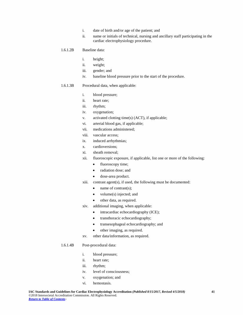
IAC Standards and Guidelines for Cardiac Electrophysiology Accreditation (Published 8/15/2017, Revised 4/5/2018) 41 ©2018 Intersocietal Accreditation Commission. All Rights Reserved. Return to Table of Contents»
i. date of birth and/or age of the patient; and ii. name or initials of technical, nursing and ancillary staff participating in the
cardiac electrophysiology procedure.
1.6.1.2B Baseline data:
i. height; ii. weight; iii. gender; and iv. baseline blood pressure prior to the start of the procedure.
1.6.1.3B Procedural data, when applicable:
i. blood pressure; ii. heart rate; iii. rhythm; iv. oxygenation; v. activated clotting time(s) (ACT), if applicable; vi. arterial blood gas, if applicable; vii. medications administered; viii. vascular access; ix. induced arrhythmias; x. cardioversions; xi. sheath removal; xii. fluoroscopic exposure, if applicable, list one or more of the following:
• fluoroscopy time; • radiation dose; and • dose-area product.
xiii. contrast agent(s), if used, the following must be documented: • name of contrast(s); • volume(s) injected; and • other data, as required.
xiv. additional imaging, when applicable: • intracardiac echocardiography (ICE); • transthoracic echocardiography; • transesophageal echocardiography; and • other imaging, as required.
xv. other data/information, as required.
1.6.1.4B Post-procedural data:
i. blood pressure; ii. heart rate; iii. rhythm; iv. level of consciousness; v. oxygenation; and vi. hemostasis.
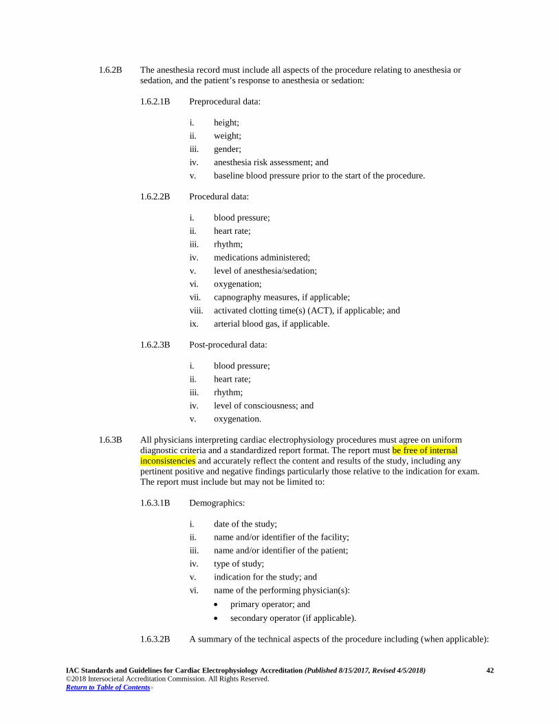
IAC Standards and Guidelines for Cardiac Electrophysiology Accreditation (Published 8/15/2017, Revised 4/5/2018) 42 ©2018 Intersocietal Accreditation Commission. All Rights Reserved. Return to Table of Contents»
1.6.2B The anesthesia record must include all aspects of the procedure relating to anesthesia or
sedation, and the patient’s response to anesthesia or sedation:
1.6.2.1B Preprocedural data:
i. height; ii. weight; iii. gender; iv. anesthesia risk assessment; and v. baseline blood pressure prior to the start of the procedure.
1.6.2.2B Procedural data:
i. blood pressure; ii. heart rate; iii. rhythm; iv. medications administered; v. level of anesthesia/sedation; vi. oxygenation; vii. capnography measures, if applicable; viii. activated clotting time(s) (ACT), if applicable; and ix. arterial blood gas, if applicable.
1.6.2.3B Post-procedural data:
i. blood pressure; ii. heart rate; iii. rhythm; iv. level of consciousness; and v. oxygenation.
1.6.3B All physicians interpreting cardiac electrophysiology procedures must agree on uniform
diagnostic criteria and a standardized report format. The report must be free of internal inconsistencies and accurately reflect the content and results of the study, including any pertinent positive and negative findings particularly those relative to the indication for exam. The report must include but may not be limited to:
1.6.3.1B Demographics:
i. date of the study; ii. name and/or identifier of the facility; iii. name and/or identifier of the patient; iv. type of study; v. indication for the study; and vi. name of the performing physician(s):
• primary operator; and • secondary operator (if applicable).
1.6.3.2B A summary of the technical aspects of the procedure including (when applicable):
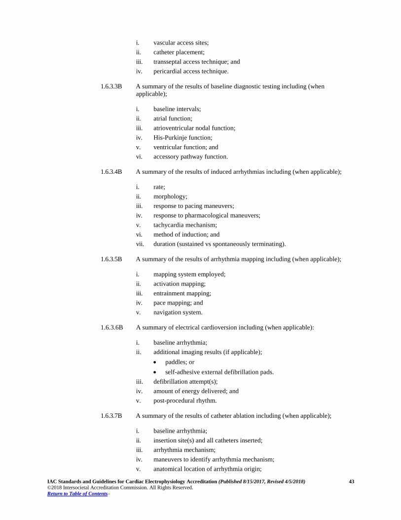
IAC Standards and Guidelines for Cardiac Electrophysiology Accreditation (Published 8/15/2017, Revised 4/5/2018) 43 ©2018 Intersocietal Accreditation Commission. All Rights Reserved. Return to Table of Contents»
i. vascular access sites; ii. catheter placement; iii. transseptal access technique; and iv. pericardial access technique.
1.6.3.3B A summary of the results of baseline diagnostic testing including (when
applicable);
i. baseline intervals; ii. atrial function; iii. atrioventricular nodal function; iv. His-Purkinje function; v. ventricular function; and vi. accessory pathway function.
1.6.3.4B A summary of the results of induced arrhythmias including (when applicable);
i. rate; ii. morphology; iii. response to pacing maneuvers; iv. response to pharmacological maneuvers; v. tachycardia mechanism; vi. method of induction; and vii. duration (sustained vs spontaneously terminating).
1.6.3.5B A summary of the results of arrhythmia mapping including (when applicable);
i. mapping system employed; ii. activation mapping; iii. entrainment mapping; iv. pace mapping; and v. navigation system.
1.6.3.6B A summary of electrical cardioversion including (when applicable): i. baseline arrhythmia; ii. additional imaging results (if applicable);
• paddles; or • self-adhesive external defibrillation pads.
iii. defibrillation attempt(s); iv. amount of energy delivered; and v. post-procedural rhythm.
1.6.3.7B A summary of the results of catheter ablation including (when applicable);
i. baseline arrhythmia; ii. insertion site(s) and all catheters inserted; iii. arrhythmia mechanism; iv. maneuvers to identify arrhythmia mechanism; v. anatomical location of arrhythmia origin;
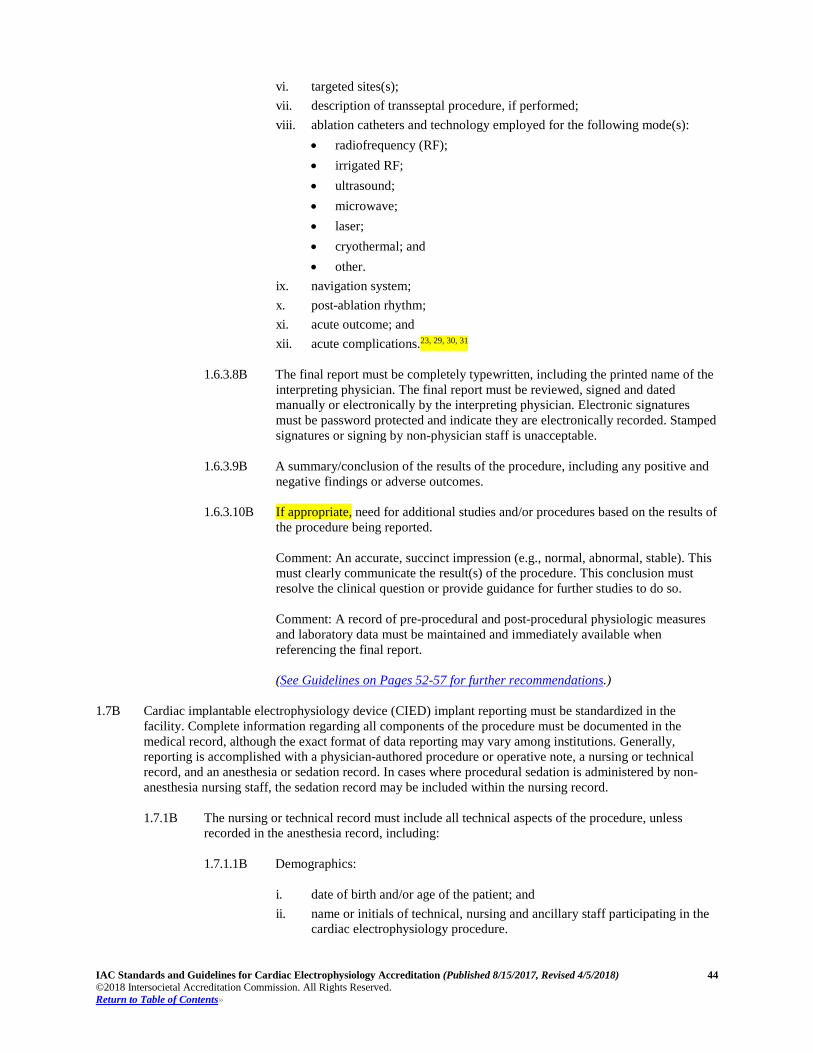
IAC Standards and Guidelines for Cardiac Electrophysiology Accreditation (Published 8/15/2017, Revised 4/5/2018) 44 ©2018 Intersocietal Accreditation Commission. All Rights Reserved. Return to Table of Contents»
vi. targeted sites(s); vii. description of transseptal procedure, if performed; viii. ablation catheters and technology employed for the following mode(s):
• radiofrequency (RF); • irrigated RF; • ultrasound; • microwave; • laser; • cryothermal; and • other.
ix. navigation system; x. post-ablation rhythm; xi. acute outcome; and xii. acute complications.23, 29, 30, 31
1.6.3.8B The final report must be completely typewritten, including the printed name of the
interpreting physician. The final report must be reviewed, signed and dated manually or electronically by the interpreting physician. Electronic signatures must be password protected and indicate they are electronically recorded. Stamped signatures or signing by non-physician staff is unacceptable.
1.6.3.9B A summary/conclusion of the results of the procedure, including any positive and
negative findings or adverse outcomes.
1.6.3.10B If appropriate, need for additional studies and/or procedures based on the results of the procedure being reported. Comment: An accurate, succinct impression (e.g., normal, abnormal, stable). This must clearly communicate the result(s) of the procedure. This conclusion must resolve the clinical question or provide guidance for further studies to do so. Comment: A record of pre-procedural and post-procedural physiologic measures and laboratory data must be maintained and immediately available when referencing the final report. (See Guidelines on Pages 52-57 for further recommendations.)
1.7B Cardiac implantable electrophysiology device (CIED) implant reporting must be standardized in the
facility. Complete information regarding all components of the procedure must be documented in the medical record, although the exact format of data reporting may vary among institutions. Generally, reporting is accomplished with a physician-authored procedure or operative note, a nursing or technical record, and an anesthesia or sedation record. In cases where procedural sedation is administered by non-anesthesia nursing staff, the sedation record may be included within the nursing record. 1.7.1B The nursing or technical record must include all technical aspects of the procedure, unless
recorded in the anesthesia record, including: 1.7.1.1B Demographics:
i. date of birth and/or age of the patient; and ii. name or initials of technical, nursing and ancillary staff participating in the
cardiac electrophysiology procedure.
IAC Standards and Guidelines for Cardiac Electrophysiology Accreditation (Published 8/15/2017, Revised 4/5/2018) 45 ©2018 Intersocietal Accreditation Commission. All Rights Reserved. Return to Table of Contents»
1.7.1.2B Baseline data: i. height; ii. weight; iii. gender; and iv. baseline blood pressure prior to the start of the procedure.
1.7.1.3B Procedural data:
i. blood pressure; ii. heart rate; iii. rhythm; iv. oxygenation; v. medications administered; vi. vascular access; vii. lead placement; viii. device placement; ix. induced arrhythmias; x. cardioversions; xi. fluoroscopic exposure, when applicable, list one or more of the following:
• fluoroscopy time; • radiation dose; and • dose-area product.
xii. contrast agent(s), if used, the following must be documented. • name of contrast(s); • volume(s) injected; and • other data, as required.
xiii. additional imaging, when applicable: • intracardiac echocardiography (ICE); • transthoracic echocardiography; • transesophageal echocardiography; • other imaging, as required; and • other data/information, as required.
1.7.1.4B Device documentation must include, but not limited to:
i. pulse generator manufacturer; model and serial number;
• Universal Device Identifier (UDI), when available; ii. pacing lead(s); iii. lead model and serial numbers; iv. Universal Device Identifier(s) (UDI), when available; and v. acute and chronic lead parameters (see Appendix B). Comment: Sidedness must be indicated for all lead positions and measurement sample sites for testing when referring to data described in this section (Anatomical description(s) for lead position and measurement sample sites may be used in cases of complex congenital heart disease).
IAC Standards and Guidelines for Cardiac Electrophysiology Accreditation (Published 8/15/2017, Revised 4/5/2018) 46 ©2018 Intersocietal Accreditation Commission. All Rights Reserved. Return to Table of Contents»
1.7.1.5B Post-procedural data: i. blood pressure; ii. heart rate; iii. rhythm; iv. level of consciousness; v. oxygenation; and vi. hemostasis.
1.7.2B The anesthesia record must include all aspects of the procedure relating to anesthesia or sedation, and the patient’s response to anesthesia or sedation. 1.7.2.1B Pre-procedural data:
i. height; ii. weight; iii. gender; iv. anesthesia risk assessment; and v. baseline blood pressure prior to the start of the procedure.
1.7.2.2B Procedural data:
i. blood pressure; ii. heart rate; iii. rhythm; iv. medications administered; v. level of anesthesia/sedation; vi. oxygenation; and vii. capnography measures, if applicable; viii. activated clotting time(s) (ACT), if applicable; ix. arterial blood gas, if applicable.
1.7.2.3B Post-procedural data:
i. blood pressure; ii. heart rate; iii. rhythm; iv. level of consciousness; and v. oxygenation.
1.7.3B Physicians reporting CIED implant procedures must accurately describe the key aspects of the
procedure. The report must include but may not be limited to:
1.7.3.1B Demographics: i. date of the study; ii. name and/or identifier of the facility; iii. name and/or identifier of the patient; iv. type of study; v. indication for the procedure; and vi. name of the performing physician(s):
IAC Standards and Guidelines for Cardiac Electrophysiology Accreditation (Published 8/15/2017, Revised 4/5/2018) 47 ©2018 Intersocietal Accreditation Commission. All Rights Reserved. Return to Table of Contents»
• primary operator; and • secondary operator (if applicable).
1.7.3.2B A summary of the technical aspects of the procedure including (when applicable):
i. incision site(s); ii. vascular access site(s); iii. additional introducer guide catheters or sheaths employed; iv. description of angiographic findings; v. lead position(s); vi. pocket location; and vii. wound closure.
1.7.3.3B A summary of the results of lead testing including (when applicable, if not
reported elsewhere): i. P/R wave amplitude; ii. pacing threshold(s) in pertinent lead configurations; iii. lead impedance(s); and iv. defibrillation threshold.
1.7.3.4B The final report must be completely typewritten, including the printed name of the
interpreting physician. The final report must be reviewed, signed and dated manually or electronically by the interpreting physician. Electronic signatures must be password protected and indicate they are electronically recorded. Stamped signatures or signing by non-physician staff is unacceptable.
1.7.3.5B A summary/conclusion of the results of the procedure, including any positive and negative findings or adverse outcomes.
1.7.3.6B Any need for additional studies and/or procedures based on the results of the
procedure being reported. Comment: An accurate, succinct impression (e.g., normal, abnormal, stable). This must clearly communicate the result(s) of the procedure. This conclusion must resolve the clinical question or provide guidance for further studies to do so. Comment: A record of pre-procedural and post-procedural physiologic measures and laboratory data must be maintained and immediately available when referencing the final report.
1.7.3.7B Any need for additional studies and/or procedures based on the results of the
procedure being reported. (See Guidelines on Pages 52-57 for further recommendations.)
1.8B Cardiac electrophysiology chronic lead extraction reporting must be standardized in the facility. Complete
information regarding all components of the procedure must be documented in the medical record, although the exact format of data reporting may vary among institutions. Generally, reporting is accomplished with a physician-authored procedure or operative note, a nursing or technical record, and an anesthesia or sedation record. In cases where procedural sedation is administered by non-anesthesia nursing staff, the sedation record may be included within the nursing record. 1.8.1B The nursing or technical record must include all technical aspects of the procedure as listed in
Standard 1.6.1.1B, unless recorded in the anesthesia record including:
IAC Standards and Guidelines for Cardiac Electrophysiology Accreditation (Published 8/15/2017, Revised 4/5/2018) 48 ©2018 Intersocietal Accreditation Commission. All Rights Reserved. Return to Table of Contents»
1.8.1.1B Demographics: i. date of birth and/or age of the patient; and ii. name or initials of technical, nursing and ancillary staff participating in the
cardiac electrophysiology procedure.
1.8.1.2B Baseline data: i. height; ii. weight; iii. gender; and iv. baseline blood pressure prior to the start of the procedure.
1.8.1.3B Procedural data, when applicable:
i. blood pressure measurement(s) to include invasive blood pressure
monitoring (e.g., arterial line); ii. heart rate; iii. rhythm; iv. oxygenation; v. activated clotting time(s) (ACT), if applicable; vi. arterial blood gas, if applicable; vii. medications administered; viii. vascular access; ix. induced arrhythmias; x. cardioversions; xi. sheath removal; xii. fluoroscopic exposure, when applicable, list one or more of the following:
• fluoroscopy time; • radiation dose; and • dose-area product.
xiii. contrast agent(s), if used, the following must be documented: • name of contrast(s); • volume(s) injected; and • other data, as required.
xiv. additional imaging, when applicable: • intracardiac echocardiography (ICE); • transthoracic echocardiography; • transesophageal echocardiography; and • other imaging, as required.
xv. other data/information, as required.
1.8.1.4B Device, lead and adapter information (connected and abandoned) must include, but not limited to: i. pulse generator manufacturer; model and serial number;
• Universal Device Identifier (UDI), when available ii. pacing lead(s);
• lead model and serial numbers
IAC Standards and Guidelines for Cardiac Electrophysiology Accreditation (Published 8/15/2017, Revised 4/5/2018) 49 ©2018 Intersocietal Accreditation Commission. All Rights Reserved. Return to Table of Contents»
iii. Universal Device Identifier(s) (UDI), when available: • access route; • insertion site(s)/lead placement(s); • other, as required;
iv. location and description of abandoned device, lead(s) and adapter(s); and v. disposition of explanted material (e.g., pathology, bacteriology, industry, etc.); vi. time to individual lead removal/extraction; vii. other data, as required. Comment: Sidedness must be indicated for all lead positions and measurement sample sites for testing when referring to data described in this section (Anatomical description(s) for lead position and measurement sample sites may be used in cases of complex congenital heart disease.).
1.8.1.5B Post-procedural data: i. blood pressure; ii. heart rate; iii. rhythm; iv. level of consciousness; v. oxygenation; and vi. hemostasis.
1.8.2B The anesthesia record must include all aspects of the procedure relating to anesthesia or
sedation, and the patient’s response to anesthesia or sedation: 1.8.2.1B Pre-procedural data:
i. height; ii. weight; iii. gender; iv. anesthesia risk assessment; and v. baseline blood pressure prior to the start of the procedure.
1.8.2.2B Procedural data:
i. blood pressure measurement(s) to include invasive blood pressure
monitoring (e.g., arterial line); ii. heart rate; iii. rhythm; iv. medications administered; v. level of anesthesia/sedation; vi. oxygenation; vii. capnography measures, if applicable; viii. activated clotting time(s) (ACT), if applicable; and ix. arterial blood gas, if applicable.
1.8.2.3B Post-procedural data:
i. blood pressure; ii. heart rate;
IAC Standards and Guidelines for Cardiac Electrophysiology Accreditation (Published 8/15/2017, Revised 4/5/2018) 50 ©2018 Intersocietal Accreditation Commission. All Rights Reserved. Return to Table of Contents»
iii. rhythm; iv. level of consciousness; and v. oxygenation.
1.8.3B Physicians reporting chronic lead extraction procedures must accurately describe the key
aspects of the procedure. The report must include but may not be limited to: 1.8.3.1B Demographics:
i. date of the study; ii. name and/or identifier of the facility; iii. name and/or identifier of the patient; iv. type of study; v. indication for the procedure; and vi. name of the performing physician(s): vii. primary operator; and viii. secondary operator (if applicable).
1.8.3.2B A summary of the technical aspects of the procedure including (when applicable):
i. incision site(s); ii. vascular access site(s); iii. additional introducer guide catheters or sheaths employed; iv. description of angiographic findings; v. lead position(s); vi. pocket location; vii. successful/partial or failed explant; viii. in the presence of infection, description of pocket findings and
documentation of cultures taken; and ix. wound closure.
1.8.3.3B A summary of the results of lead testing including (when applicable, if not
reported elsewhere): i. P/R wave amplitude; ii. pacing threshold(s) in pertinent lead configurations; iii. lead impedance(s); and iv. defibrillation threshold.
1.8.3.4B The final report must be completely typewritten, including the printed name of the
interpreting physician. The final report must be reviewed, signed and dated manually or electronically by the interpreting physician. Electronic signatures must be password protected and indicate they are electronically recorded. Stamped signatures or signing by non-physician staff is unacceptable.
1.8.3.5B A summary/conclusion of the results of the procedure, including any positive and negative findings or adverse outcomes.
1.8.3.6B Any need for additional studies and/or procedures based on the results of the
procedure being reported. Comment: An accurate, succinct impression (e.g., normal, abnormal, stable). This
IAC Standards and Guidelines for Cardiac Electrophysiology Accreditation (Published 8/15/2017, Revised 4/5/2018) 51 ©2018 Intersocietal Accreditation Commission. All Rights Reserved. Return to Table of Contents»
must clearly communicate the result(s) of the procedure. This conclusion must resolve the clinical question or provide guidance for further studies to do so. Comment: A record of pre-procedural and post-procedural physiologic measures and laboratory data must be maintained and immediately available when referencing the final report.
1.8.3.7B Any need for additional studies and/or procedures based on the results of the
procedure being reported. (See Guidelines on Pages 52-57 for further recommendations.)
STANDARD – Procedure Volumes 1.9B The procedure volume must be sufficient to maintain proficiency in procedure performance and
interpretation. 1.9.1B The facility must have specific privileging requirements for individual operators to perform
invasive cardiac electrophysiology procedures to include, but not limited to: cardiac electrophysiology testing and ablation, device implantation and chronic lead extraction.
(See Guidelines on Pages 52-57 for further recommendations.)
IAC Standards and Guidelines for Cardiac Electrophysiology Accreditation (Published 8/15/2017, Revised 4/5/2018) 52 ©2018 Intersocietal Accreditation Commission. All Rights Reserved. Return to Table of Contents»
Section 1B: Procedures and Protocols Guidelines
1.1B In most diagnostic and ablation cases, rhythm active drugs (including β-blockers and calcium-channel
blockers) are discontinued five half-lives before the procedure to allow the target arrhythmia to be induced, mapped, and ablated.
All physicians and staff are required to be familiar with identifying all potential procedural
complications and to understand their role in managing them. As many management strategies for arrhythmias require chronic and/or periprocedural anticoagulation,
careful evaluation, assessment, and planning are needed. 1.1.2B Because of the complexity of the EP procedures, patient safety and positive outcomes are critically
dependent on the skill levels of the staff. Additional staff is needed as the complexity of the case increases and more equipment is required.
Laboratory staffing recommendations include, but are not limited to:
• Staff physicians must have prerequisite training and appropriate credentialing reflecting expertise
in the management and treatment of cardiac arrhythmias. • It is desirable that anesthesia services be an integral part of clinical practice in the EP laboratory. • Advanced practice nurses (APNs) and physician assistants (PAs) should be used in areas where they
will have a maximum impact on patient care and where they can assume roles and responsibilities unique to their training and certification.
• At least one registered nurse should be present for every invasive procedure in the EP laboratory. • Industry representatives should function according to clear policies under the direction of the
laboratory manager, staff, or physician. • Additional laboratory staff should include, but is not limited to, registered nurses (RNs), EP
specialists/technologists, radiological technologists, and certified nurse practitioners (NPs) and Physician Assistants (PAs) as needed.
• Additional appropriately trained personnel should be provided to staff patient prep, recover and OR areas.
• Other key personnel that are important for the safe and efficient function of the laboratory include quality improvement (QI) staff, information technologists, biomedical engineers, scheduling coordinators, purchasing, inventory and supply personnel, and housekeeping.
1.2B In patients undergoing pacemaker or defibrillator lead extraction, or who require pericardial access for
epicardial ablation or left atrial ablation ligation, additional preparation may be required on a case-by-case basis, such as typing and crossmatching of blood products in select patients and immediate availability of thoracic surgical backup.
1.2.4B A complete description of the procedure, including the anticipated success rates and possible
complications, is best delivered in the outpatient setting before the EP procedure. Health care facilities should insist that clinicians administering or supervising the administration of
moderate sedation meet the requirements of the American Society of Anesthesiologists. 1.4.6B The decision for patient discharge takes into account procedural detail, patient age and health status,
potential for complications (such as blood loss), and the ability of the patient (or caregivers) to evaluate signs of concern.
1.4.3.1B and 1.6.3.7B Complication definitions include, but not limited to: 23-92,30,31
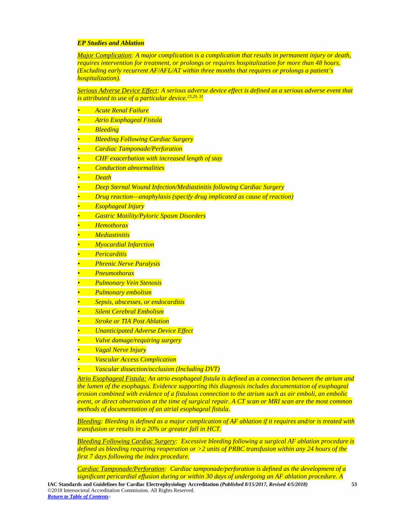
IAC Standards and Guidelines for Cardiac Electrophysiology Accreditation (Published 8/15/2017, Revised 4/5/2018) 53 ©2018 Intersocietal Accreditation Commission. All Rights Reserved. Return to Table of Contents»
EP Studies and Ablation
Major Complication: A major complication is a complication that results in permanent injury or death, requires intervention for treatment, or prolongs or requires hospitalization for more than 48 hours. (Excluding early recurrent AF/AFL/AT within three months that requires or prolongs a patient’s hospitalization).
Serious Adverse Device Effect: A serious adverse device effect is defined as a serious adverse event that is attributed to use of a particular device.23,29, 31
• Acute Renal Failure • Atrio Esophageal Fistula • Bleeding • Bleeding Following Cardiac Surgery • Cardiac Tamponade/Perforation • CHF exacerbation with increased length of stay • Conduction abnormalities • Death • Deep Sternal Wound Infection/Mediastinitis following Cardiac Surgery • Drug reaction—anaphylaxis (specify drug implicated as cause of reaction) • Esophageal Injury • Gastric Motility/Pyloric Spasm Disorders • Hemothorax • Mediastinitis • Myocardial Infarction • Pericarditis • Phrenic Nerve Paralysis • Pneumothorax • Pulmonary Vein Stenosis • Pulmonary embolism • Sepsis, abscesses, or endocarditis • Silent Cerebral Embolism • Stroke or TIA Post Ablation • Unanticipated Adverse Device Effect • Valve damage/requiring surgery • Vagal Nerve Injury • Vascular Access Complication • Vascular dissection/occlusion (Including DVT) Atrio Esophageal Fistula: An atrio esophageal fistula is defined as a connection between the atrium and the lumen of the esophagus. Evidence supporting this diagnosis includes documentation of esophageal erosion combined with evidence of a fistulous connection to the atrium such as air emboli, an embolic event, or direct observation at the time of surgical repair. A CT scan or MRI scan are the most common methods of documentation of an atrial esophageal fistula.
Bleeding: Bleeding is defined as a major complication of AF ablation if it requires and/or is treated with transfusion or results in a 20% or greater fall in HCT.
Bleeding Following Cardiac Surgery: Excessive bleeding following a surgical AF ablation procedure is defined as bleeding requiring reoperation or >2 units of PRBC transfusion within any 24 hours of the first 7 days following the index procedure.
Cardiac Tamponade/Perforation: Cardiac tamponade/perforation is defined as the development of a significant pericardial effusion during or within 30 days of undergoing an AF ablation procedure. A
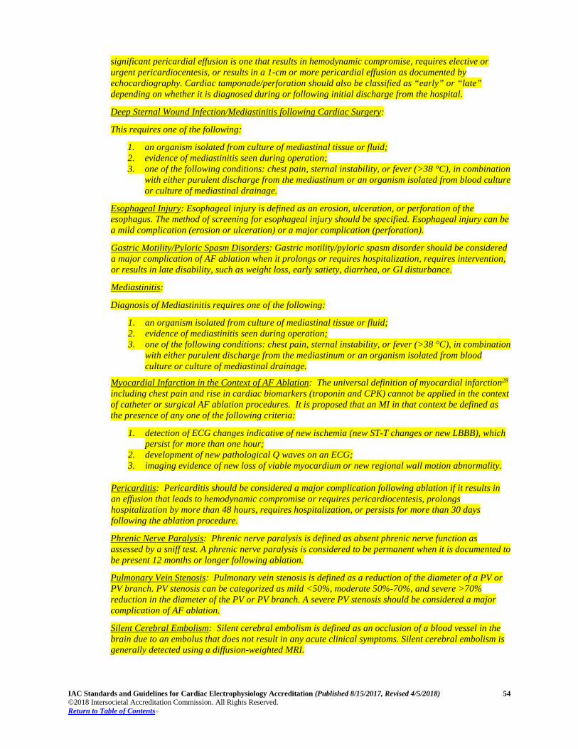
IAC Standards and Guidelines for Cardiac Electrophysiology Accreditation (Published 8/15/2017, Revised 4/5/2018) 54 ©2018 Intersocietal Accreditation Commission. All Rights Reserved. Return to Table of Contents»
significant pericardial effusion is one that results in hemodynamic compromise, requires elective or urgent pericardiocentesis, or results in a 1-cm or more pericardial effusion as documented by echocardiography. Cardiac tamponade/perforation should also be classified as “early” or “late” depending on whether it is diagnosed during or following initial discharge from the hospital.
Deep Sternal Wound Infection/Mediastinitis following Cardiac Surgery:
This requires one of the following:
1. an organism isolated from culture of mediastinal tissue or fluid; 2. evidence of mediastinitis seen during operation; 3. one of the following conditions: chest pain, sternal instability, or fever (>38 °C), in combination
with either purulent discharge from the mediastinum or an organism isolated from blood culture or culture of mediastinal drainage.
Esophageal Injury: Esophageal injury is defined as an erosion, ulceration, or perforation of the esophagus. The method of screening for esophageal injury should be specified. Esophageal injury can be a mild complication (erosion or ulceration) or a major complication (perforation).
Gastric Motility/Pyloric Spasm Disorders: Gastric motility/pyloric spasm disorder should be considered a major complication of AF ablation when it prolongs or requires hospitalization, requires intervention, or results in late disability, such as weight loss, early satiety, diarrhea, or GI disturbance.
Mediastinitis:
Diagnosis of Mediastinitis requires one of the following:
1. an organism isolated from culture of mediastinal tissue or fluid; 2. evidence of mediastinitis seen during operation; 3. one of the following conditions: chest pain, sternal instability, or fever (>38 °C), in combination
with either purulent discharge from the mediastinum or an organism isolated from blood culture or culture of mediastinal drainage.
Myocardial Infarction in the Context of AF Ablation: The universal definition of myocardial infarction28 including chest pain and rise in cardiac biomarkers (troponin and CPK) cannot be applied in the context of catheter or surgical AF ablation procedures. It is proposed that an MI in that context be defined as the presence of any one of the following criteria:
1. detection of ECG changes indicative of new ischemia (new ST-T changes or new LBBB), which persist for more than one hour;
2. development of new pathological Q waves on an ECG; 3. imaging evidence of new loss of viable myocardium or new regional wall motion abnormality.
Pericarditis: Pericarditis should be considered a major complication following ablation if it results in an effusion that leads to hemodynamic compromise or requires pericardiocentesis, prolongs hospitalization by more than 48 hours, requires hospitalization, or persists for more than 30 days following the ablation procedure.
Phrenic Nerve Paralysis: Phrenic nerve paralysis is defined as absent phrenic nerve function as assessed by a sniff test. A phrenic nerve paralysis is considered to be permanent when it is documented to be present 12 months or longer following ablation.
Pulmonary Vein Stenosis: Pulmonary vein stenosis is defined as a reduction of the diameter of a PV or PV branch. PV stenosis can be categorized as mild <50%, moderate 50%-70%, and severe >70% reduction in the diameter of the PV or PV branch. A severe PV stenosis should be considered a major complication of AF ablation.
Silent Cerebral Embolism: Silent cerebral embolism is defined as an occlusion of a blood vessel in the brain due to an embolus that does not result in any acute clinical symptoms. Silent cerebral embolism is generally detected using a diffusion-weighted MRI.
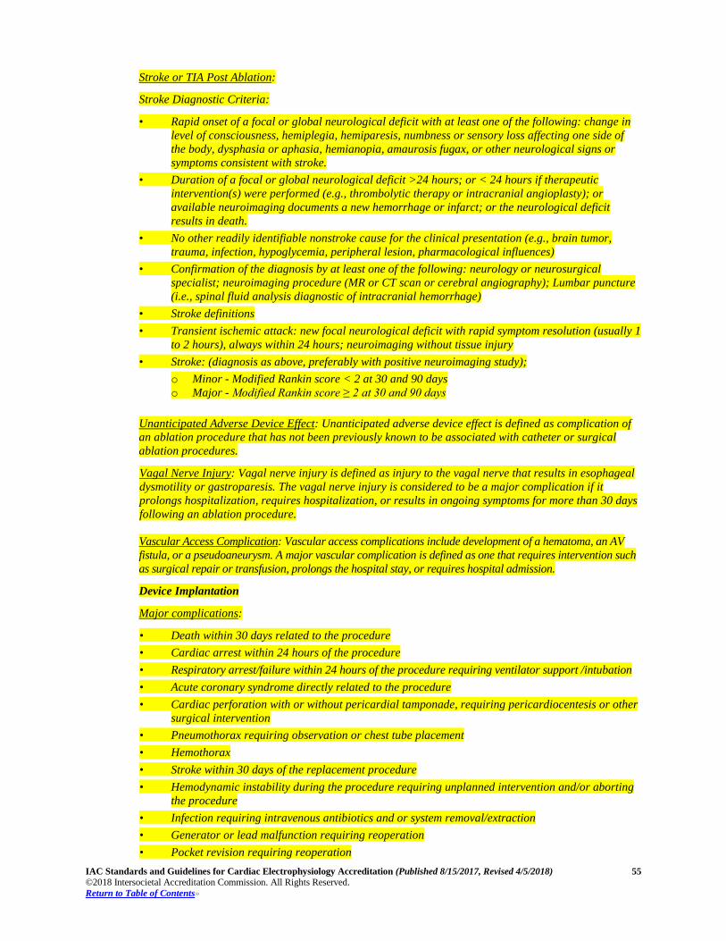
IAC Standards and Guidelines for Cardiac Electrophysiology Accreditation (Published 8/15/2017, Revised 4/5/2018) 55 ©2018 Intersocietal Accreditation Commission. All Rights Reserved. Return to Table of Contents»
Stroke or TIA Post Ablation:
Stroke Diagnostic Criteria:
• Rapid onset of a focal or global neurological deficit with at least one of the following: change in level of consciousness, hemiplegia, hemiparesis, numbness or sensory loss affecting one side of the body, dysphasia or aphasia, hemianopia, amaurosis fugax, or other neurological signs or symptoms consistent with stroke.
• Duration of a focal or global neurological deficit >24 hours; or < 24 hours if therapeutic intervention(s) were performed (e.g., thrombolytic therapy or intracranial angioplasty); or available neuroimaging documents a new hemorrhage or infarct; or the neurological deficit results in death.
• No other readily identifiable nonstroke cause for the clinical presentation (e.g., brain tumor, trauma, infection, hypoglycemia, peripheral lesion, pharmacological influences)
• Confirmation of the diagnosis by at least one of the following: neurology or neurosurgical specialist; neuroimaging procedure (MR or CT scan or cerebral angiography); Lumbar puncture (i.e., spinal fluid analysis diagnostic of intracranial hemorrhage)
• Stroke definitions • Transient ischemic attack: new focal neurological deficit with rapid symptom resolution (usually 1
to 2 hours), always within 24 hours; neuroimaging without tissue injury • Stroke: (diagnosis as above, preferably with positive neuroimaging study);
o Minor - Modified Rankin score < 2 at 30 and 90 days o Major - Modified Rankin score ≥ 2 at 30 and 90 days
Unanticipated Adverse Device Effect: Unanticipated adverse device effect is defined as complication of an ablation procedure that has not been previously known to be associated with catheter or surgical ablation procedures.
Vagal Nerve Injury: Vagal nerve injury is defined as injury to the vagal nerve that results in esophageal dysmotility or gastroparesis. The vagal nerve injury is considered to be a major complication if it prolongs hospitalization, requires hospitalization, or results in ongoing symptoms for more than 30 days following an ablation procedure. Vascular Access Complication: Vascular access complications include development of a hematoma, an AV fistula, or a pseudoaneurysm. A major vascular complication is defined as one that requires intervention such as surgical repair or transfusion, prolongs the hospital stay, or requires hospital admission.
Device Implantation
Major complications:
• Death within 30 days related to the procedure • Cardiac arrest within 24 hours of the procedure • Respiratory arrest/failure within 24 hours of the procedure requiring ventilator support /intubation • Acute coronary syndrome directly related to the procedure • Cardiac perforation with or without pericardial tamponade, requiring pericardiocentesis or other
surgical intervention • Pneumothorax requiring observation or chest tube placement • Hemothorax • Stroke within 30 days of the replacement procedure • Hemodynamic instability during the procedure requiring unplanned intervention and/or aborting
the procedure • Infection requiring intravenous antibiotics and or system removal/extraction • Generator or lead malfunction requiring reoperation • Pocket revision requiring reoperation
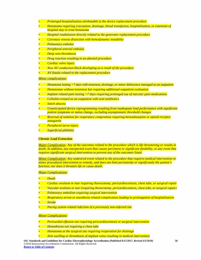
IAC Standards and Guidelines for Cardiac Electrophysiology Accreditation (Published 8/15/2017, Revised 4/5/2018) 56 ©2018 Intersocietal Accreditation Commission. All Rights Reserved. Return to Table of Contents»
• Prolonged hospitalization attributable to the device replacement procedure • Hematoma requiring evacuation, drainage, blood transfusion, hospitalization, or extension of
hospital stay to treat hematoma • Hospital readmission directly related to the generator replacement procedure • Coronary venous dissection with hemodynamic instability • Pulmonary embolus • Peripheral arterial embolus • Deep vein thrombosis • Drug reaction resulting in an aborted procedure • Cardiac valve injury • New AV conduction block developing as a result of the procedure • AV fistula related to the replacement procedure
Minor complications:
• Hematoma lasting >7 days with tenseness, drainage, or minor dehiscence managed as an outpatient • Hematomas without tenseness but requiring additional outpatient evaluation • Implant related pain lasting >7 days requiring prolonged use of narcotic pain medications • Cellulitis treated as an outpatient with oral antibiotics • Stitch abscess • Unanticipated device reprogramming resulting from inadequate lead performance with significant
patient symptoms or status change, excluding asymptomatic threshold changes • Reversal of sedation for respiratory compromise requiring benzodiazepine or opioid receptor
antagonist • Peripheral nerve injury • Superficial phlebitis
Chronic Lead Extraction
Major Complication: Any of the outcomes related to the procedure which is life threatening or results in death. In addition, any unexpected event that causes persistent or significant disability, or any event that requires significant surgical intervention to prevent any of the outcomes listed.
Minor Complication: Any undesired event related to the procedure that requires medical intervention or minor procedural intervention to remedy, and does not limit persistently or significantly the patient’s function, nor does it threaten life or cause death.
Major Complications:
• Death • Cardiac avulsion or tear requiring thoracotomy, pericardiocentesis, chest tube, or surgical repair • Vascular avulsion or tear (requiring thoracotomy, pericardiocentesis, chest tube, or surgical repair) • Pulmonary embolism requiring surgical intervention • Respiratory arrest or anesthesia related complication leading to prolongation of hospitalization • Stroke • Pacing system related infection of a previously non-infected site Minor Complications:
• Pericardial effusion not requiring pericardiocentesis or surgical intervention • Hemothorax not requiring a chest tube • Hematoma at the surgical site requiring reoperation for drainage • Arm swelling or thrombosis of implant veins resulting in medical intervention
IAC Standards and Guidelines for Cardiac Electrophysiology Accreditation (Published 8/15/2017, Revised 4/5/2018) 57 ©2018 Intersocietal Accreditation Commission. All Rights Reserved. Return to Table of Contents»
• Vascular repair near the implant site or venous entry site • Hemodynamically significant air embolism • Migrated lead fragment without sequelae • Blood transfusion related to blood loss during surgery • Pneumothorax requiring a chest tube • Pulmonary embolism not requiring surgical intervention • Distal embolization of lead fragment without clinical squeale
1.7.1.2B, 1.8.1.2B, 1.9.1.2B, 1.10.1.1B Adequate anticoagulation should be monitored with activated clotting
time (ACT) throughout the procedure.
Sedation records must include, but are not limited to the following information:
• type of sedation (e.g., moderate sedation vs. general anesthesia); • name of medication(s); • dose(s) and times(s) given; • route(s) of delivery; • staff administering medication; • other data, as required.
1.11B Procedure Volumes A facility should perform a minimum of 100 invasive cardiac electrophysiology and/or device procedures annually to include the following:
• Diagnostic electrophysiology testing and ablation: 50 procedures • Device Implantation: 50 procedures • Chronic Lead Extractions (if performed): 20 leads per operator10 Each member of the medical staff should interpret a minimum of 100 invasive cardiac electrophysiology procedures. Each member of the nursing and technical staff should assist in a minimum of 100 invasive cardiac electrophysiology procedures. The total volume of studies interpreted and performed by each staff member may be combined from sources other than the applicant facility. Lower volumes than those recommended here; however, should not dissuade a facility that is otherwise compliant with the IAC Cardiac Electrophysiology Standards from applying for accreditation.
Centers specializing in pediatric and adult congenital heart disease may need to perform a relatively large percentage of epicardial device implants to meet the challenges of patient size and anatomy. These cases may be counted in device implant quotas as long as the medical staff is directly involved with lead testing and patient care along with the surgeons. Similarly, operator experience for chronic lead extraction at many pediatric centers may be less than 20 cases per operator annually. It is recommended that pediatric and adult congenital lead extractions be performed at experienced centers.
IAC Standards and Guidelines for Cardiac Electrophysiology Accreditation (Published 8/15/2017, Revised 4/5/2018) 58 ©2018 Intersocietal Accreditation Commission. All Rights Reserved. Return to Table of Contents»
Part C: Quality Improvement
Section 1C: Quality Improvement
STANDARD – Quality Improvement (QI) Program 1.1C The facility must have a Quality Improvement (QI) Program and conduct internal quality assessment and
improvement at regular intervals that are appropriate for the facility’s stated purpose and include cardiac electrophysiology procedures. 1.1.1C The QI program must include the QI measures outlined below to include but may not be limited
to the evaluation and review of:
1.1.1.1C test appropriateness;
1.1.1.2C safety and procedural outcomes;
1.1.1.3C interpretive quality review;
1.1.1.4C report completeness and timeliness.
(See Guidelines on Page 59 for further recommendations.)
STANDARD – QI Oversight
1.2C The Medical Director, Nurse Manager, Technical Manager, staff and/or an appointed QI Committee must provide oversight to the QI program including, but not limited to: 1.2.1C performance of all medical, nursing, technical and ancillary staff;
1.2.2C assessment and evaluation of patient and personnel radiation dose; 1.2.3C adherence to National Patient Safety Goals must be documented;13 1.2.4C evidence of improvement activities or, if an assessment confirms acceptable quality of a
measure, the program must demonstrate improvement by selecting a new or an additional area for assessment;
1.2.5C pre-defined indicators of quality and pre-defined thresholds that indicate the need for corrective
action. Comparisons with external benchmarks are desirable; 1.2.6C review of procedural indications, safety and complications as well as standardized and
recognized clinical outcome measures;1,10,20 1.2.7C review of the reports of QI evaluations and any corrective actions taken to address any
deficiencies
IAC Standards and Guidelines for Cardiac Electrophysiology Accreditation (Published 8/15/2017, Revised 4/5/2018) 59 ©2018 Intersocietal Accreditation Commission. All Rights Reserved. Return to Table of Contents»
Section 1C: Quality Improvement Meetings Guidelines
1.1.1C A QI program should be in place to assess and improve the administrative quality of the facility’s
operation. Administrative areas that may be assessed include, but are not limited to:
• scheduling back logs; • patient wait times; • accuracy of patient information during scheduling; • completeness of documentation; • time from completion of procedure to signature and distribution of final report; • patient satisfaction and feedback; • referring physician satisfaction and feedback; and • patient education – on individual risk factors, smoking cessation, signs and symptoms of heart
arrhythmia, cardiovascular accident, stroke or myocardial infarction and calling 911, importance of follow-up after discharge, review of discharge medications including importance of adherence to antithrombotic therapy.
IAC Standards and Guidelines for Cardiac Electrophysiology Accreditation (Published 8/15/2017, Revised 4/5/2018) 60 ©2018 Intersocietal Accreditation Commission. All Rights Reserved. Return to Table of Contents»
Section 2C: Quality Improvement Measures
STANDARD – Test Appropriateness
2.1C As part of the ongoing QI Program, facilities must incorporate the measurement of the appropriateness of the procedure being performed based on criteria published and/or endorsed by professional medical organization(s).1,10 2.1.1C The facility must evaluate and document the appropriateness of the procedure performed and
categorize as:
2.1.1.1C appropriate / usually appropriate; 2.1.1.2C may be appropriate; and
2.1.1.3C rarely appropriate / usually not appropriate.
2.1.2C Appropriate indications must be measured for a minimum of four cases per cardiac
electrophysiology accreditation procedure type (testing and ablation, device implantation, chronic lead extraction) and be reviewed every six months.
(See Guidelines on Page 62 for further recommendations.)
STANDARD – Safety and Procedural Outcomes
2.2C The QI Program must include assessment of the safety of the procedures being performed. Must have a process for documentation of complications with the goal to decrease complications. 2.2.1C Areas that must be assessed include, but are not limited to:
2.2.1.1C all procedural complications including all serious adverse events;
2.2.1.2C patient and personnel safety must be evaluated to include, but not limited to:
i. accuracy of patient identification; ii. medication safety; iii. infection control measures; and iv. staff (occupational) and patient radiation exposure monitoring according to
state regulations and published guidelines where appropriate.11, 18, 19
2.2.1.3C documentation of adverse technical events such as equipment or device failure.
2.2.2C Participation in a national registry for all patients is strongly recommended.
2.2.3C Safety and procedural outcomes must be measured for a minimum of four cases per cardiac electrophysiology accreditation procedure type (testing and ablation, device implantation, chronic lead extraction) and be reviewed every six months.
2.2.4C Outcomes data, which must be consistent with national benchmarks when available, must be
used to improve processes and procedures (refer to Appendix C).
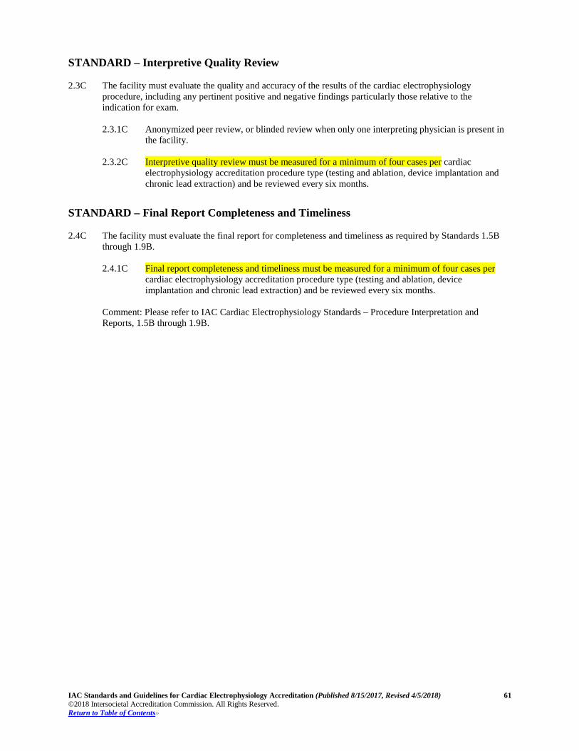
IAC Standards and Guidelines for Cardiac Electrophysiology Accreditation (Published 8/15/2017, Revised 4/5/2018) 61 ©2018 Intersocietal Accreditation Commission. All Rights Reserved. Return to Table of Contents»
STANDARD – Interpretive Quality Review
2.3C The facility must evaluate the quality and accuracy of the results of the cardiac electrophysiology procedure, including any pertinent positive and negative findings particularly those relative to the indication for exam. 2.3.1C Anonymized peer review, or blinded review when only one interpreting physician is present in
the facility.
2.3.2C Interpretive quality review must be measured for a minimum of four cases per cardiac electrophysiology accreditation procedure type (testing and ablation, device implantation and chronic lead extraction) and be reviewed every six months.
STANDARD – Final Report Completeness and Timeliness
2.4C The facility must evaluate the final report for completeness and timeliness as required by Standards 1.5B through 1.9B. 2.4.1C Final report completeness and timeliness must be measured for a minimum of four cases per
cardiac electrophysiology accreditation procedure type (testing and ablation, device implantation and chronic lead extraction) and be reviewed every six months.
Comment: Please refer to IAC Cardiac Electrophysiology Standards – Procedure Interpretation and Reports, 1.5B through 1.9B.
IAC Standards and Guidelines for Cardiac Electrophysiology Accreditation (Published 8/15/2017, Revised 4/5/2018) 62 ©2018 Intersocietal Accreditation Commission. All Rights Reserved. Return to Table of Contents»
Section 2C: Quality Improvement Measures Guidelines
2.1C There should be a mechanism for education of referring physicians to improve the appropriateness of
testing. A program for documentation and reporting should be developed and include:
• patterns of appropriate procedures performed; • baseline rate of appropriate procedures; • goals for improvement in the performance of appropriate procedures; and • measurement of improvement rate.
IAC Standards and Guidelines for Cardiac Electrophysiology Accreditation (Published 8/15/2017, Revised 4/5/2018) 63 ©2018 Intersocietal Accreditation Commission. All Rights Reserved. Return to Table of Contents»
Section 3C: Quality Improvement Meetings
STANDARD – QI Meetings
3.1C Quality Improvement (QI) meetings must be documented. 3.1.1C The facility must have a minimum of two QI meetings per year one of which is to review the
results of the QI analyses and any additional QI-related topics: 3.1.1.1C test appropriateness;
3.1.1.2C safety and procedural outcomes;
3.1.1.3C interpretive quality review;
3.1.1.4C report completeness and timeliness; and
3.1.1.5C other related topics.
3.1.2C All significant complications must be reviewed during these meetings.
3.1.2.1C Procedure outcomes, including success rates and complications, should be documented and recorded. Data acquired from the electrophysiology facility QI process should be used to benchmark the complication rates and outcomes of both individual practitioners and the overall electrophysiology facility.
3.1.2.2C Given the often poorly defined relationship between case volumes and outcomes, a more appropriate measure is to ensure that all major complications are reviewed by the QI committee and handled as described in the previous sections.
3.1.2.3C Complications and any identifiable root cause(s) and corrective action(s), must be
reviewed and documented in efforts to improve future outcomes. Complications should be tracked and recorded to allow for trend changes to be documented and addressed.
3.1.2.4C All relevant staff must participate in at least one meeting per year. All staff are
responsible for the content discussed during the QI meetings; therefore, every attempt should be made to either attend in person, via web conference or teleconference. If unable to attend one of the two biannual meetings, the staff member is required to review the meeting minutes and document their attendance with one of the following: Medical Director, Nurse Manager, Technical Manager and/or an appointed QI Committee member.
3.2C Morbidity and Mortality (M&M) conferences must be documented.
3.2.1C The Medical Director and medical staff must attend a minimum of one M&M conference per
quarter, related to cardiac electrophysiology.
IAC Standards and Guidelines for Cardiac Electrophysiology Accreditation (Published 8/15/2017, Revised 4/5/2018) 64 ©2018 Intersocietal Accreditation Commission. All Rights Reserved. Return to Table of Contents»
Section 4C: Quality Improvement Documentation/Record Retention
STANDARD – QI Documentation / Record Retention
4.1C The facility QI documentation must include, but is not be limited to: 4.1.1C the data for the QI measures;
4.1.2C minutes from the QI meetings; and 4.1.3C participant list (may include remote participation and/or review of minutes).
Comment: The QI documentation must be maintained and available to all appropriate personnel.

IAC Standards and Guidelines for Cardiac Electrophysiology Accreditation (Published 8/15/2017, Revised 4/5/2018) 65 ©2018 Intersocietal Accreditation Commission. All Rights Reserved. Return to Table of Contents»
Selected Bibliography
1. ACCF/HRS/AHA/ASE/HFSA/SCAI/SCCT/SCMR 2013 – Appropriate Use Criteria for Implantable Cardioverter Defibrillators and Cardiac Resynchronization Therapy: A Report of the American College of Cardiology Foundation Appropriate Use Criteria Task Force, Heart Rhythm Society, American Heart Association, American Society of Echocardiography, Heart Failure Society of America, Society for Cardiovascular Angiography and Interventions, Society of Cardiovascular Computed Tomography and Society for Cardiovascular Magnetic Resonance. Endorsed by the American Geriatrics Society. Russo, A., et al., J Amer Coll Cardiol, 2013; 61(12):1318-1368. content.onlinejacc.org/article.aspx?articleid=1659563
2. 2015 ACC/AHA/HRS Advanced Training Statement on Clinical Cardiac Electrophysiology (A Revision of the ACC/AHA 2006 Update of the Clinical Competence Statement on Invasive Electrophysiology Studies, Catheter Ablation and Cardioversion). Zipes, DP, Calkins, H, Daubert, JP, et al., Heart Rhythm, 2015 January: 13(1):e1-e35. content.onlinejacc.org/article.aspx?articleid=2442987
3. ACGME Program Requirements for Graduate Medical Education in Clinical Cardiac Electrophysiology (Internal Medicine). American Board of Internal Medicine: Accreditation Council for Graduate Medical Education. Effective July 1, 2017. www.acgme.org/Portals/0/PFAssets/ProgramRequirements/ 154_clinical_card_electrophys_07012017.pdf
4. Task Force 1: Training in Clinical Cardiology. Baughman, K., et al., J Am Coll Cardiol, 2008; 51(3):339-348. content.onlinejacc.org/article.aspx?articleid=1187211
5. American Board of Internal Medicine. Policies and Procedures for Certification. Published June 2015. www.abim.org/pdf/publications/Policies-and-Procedures-Certification.pdf
6. Task Force 6: Training in Specialized Electrophysiology, Cardiac Pacing, and Arrhythmia Management - Endorsed by the Heart Rhythm Society. Naccarelli, G., et al., J Am Coll Cardiol, 2008; 51(3):374-380. content.onlinejacc.org/article.aspx?articleid=1187235
7. Task Force 4: Recommendations for Training Guidelines in Pediatric Cardiac Electrophysiology - Endorsed by the Heart Rhythm Society. Vetter, V., et al., J Am Coll Cardiol, 2005; 46(7):1391-1395. content.onlinejacc.org/article.aspx?articleid=1136954
8. Recommendations for Advanced Fellowship Training in Clinical Pediatric and Congenital Electrophysiology: A Report from the Training and Credentialing Committee of the Pediatric and Congenital Electrophysiology Society. Walsh, E., et al., Heart Rhythm, 2013; 10(5):775-781. www.heartrhythmjournal.com/article/S1547-5271(13)00315-9/pdf
9. 2008 Heart Rhythm Society/Pediatric and Congenital Electrophysiology Society Clinical Competency Statement: Training Pathways for Implantation of Cardioverter-defibrillators (ICD) and Cardiac Resynchronization Therapy (CRT) Devices in Pediatric and Congenital Heart Patients. Saul, J., et al., Heart Rhythm, 2008; 5(6):1-8. www.hrsonline.org/Practice-Guidance/Clinical-Guidelines-Documents/2008-Heart-Rhythm-Society-Pediatric-and-Congenital-Electrophysiology-Society-Clinical-Competency-Statement
10. 2017 HRS Expert Consensus Statement on Cardiovascular Implantable Electronic Device Lead Management and Extraction Heart Rhythm. Kusumoto F., Schoenfeld M., Wilkoff B., et al., Heart Rhythm, 2017;14(12):e503-e551. www.hrsonline.org/Policy-Payment/Clinical-Guidelines-Documents/2017-HRS-Expert-Consensus-Statement-on-Cardiovascular-Implantable-Electronic-Device-Lead-Management-and-Extraction
11. FDA Code of Federal Regulations (CFR) Title 21 for system performance. 2014; (21)8. www.accessdata.fda.gov/scripts/cdrh/cfdocs/cfcfr/CFRSearch.cfm ?FR=1020.32
12. 2013 HRS Expert Consensus Statement on Electrophysiology (EP) Lab Standards: Process, Protocols, Equipment, Personnel and Safety. Haines, D., et al., Heart Rhythm, 2014 Aug; 11(8):10-51. www.heartrhythmjournal.com/article/S1547-5271(14)00334-8/fulltext
13. Quality Improvement Guidelines for Preventing Wrong Site, Wrong Procedure, and Wrong Person Errors: Application of the Joint Commission “Universal Protocol for Preventing Wrong Site, Wrong Procedure, Wrong Person Surgery” to the Practice of Interventional Radiology. Angle, J., et al., J Vasc Interv Radiol, 2008; 19:1145-1151. www.jvir.org/article/S1051-0443(08)00473-9/fulltext
14. USFDA, Recommendation for Healthcare Professionals on Preventing Surgical Fires, U.S. Food and Drug Administration. www.fda.gov/Drugs /DrugSafety/SafeUseInitiative/PreventingSurgicalFires/ ucm270636.htm
IAC Standards and Guidelines for Cardiac Electrophysiology Accreditation (Published 8/15/2017, Revised 4/5/2018) 66 ©2018 Intersocietal Accreditation Commission. All Rights Reserved. Return to Table of Contents»
15. The Joint Commission, Preventing Surgical Fires, 2003; Issue 29. www.jointcommission.org/assets/1/18/sea_29.pdf
16. Operating Room Fire Safety. Hart, S., et al, The Ochsner Journal, 2011; 11:37-42. www.ncbi.nlm.nih.gov/pmc/articles/PMC3096161/pdf/i1524-5012-11-1-37.pdf
17. Outline of Administrative Policies for Quality Assurance and Peer Review of Tissue Reactions Associated with Fluoroscopically-Guided Interventions. Balter, S., et al., NCRP Statement No. 11, December 31, 2014. ncrponline.org/wp-content/themes/ncrp/PDFs/Statement_11.pdf
18. Radiation Doses in Interventional Radiology Procedures: The RAD-IR Study - Part III: Dosimetric Performance of the Interventional Fluoroscopy Units. Balter, S., et al., J Vasc Interv Radiol, 2004; 15(9):919-926. www.jvir.org/article/S1051-0443(07)60496-5/fulltext
19. Quality Improvement Guidelines for Recording Patient Radiation Dose in the Medical Record. Miller, D., et al, J Vasc Interv Radiol, 2012; 23(1):11-18. www.jvir.org/article/S1051-0443(11)01256-5/fulltext
20. 2012 HRS/EHRA/ECAS Expert Consensus Statement on Catheter and Surgical Ablation of Atrial Fibrillation: Recommendations for Patient Selection, Procedural Techniques, Patient Management and Follow-up, Definitions, Endpoints, and Research Trial Design. Calkins, H., et al., Heart Rhythm, 2012 April; 9(4):632-696. www.heartrhythmjournal.com/article/S1547-5271(11)01501-3/fulltext
21. Guideline for Prevention of Surgical Site Infection. Mangram, A., et al., Infection Control and Hospital Epidemiology, 1999 April; 20(4):247-278. www.cdc.gov/hicpac/pdf/SSIguidelines.pdf
22. Incidence and Predictors of Major Complications from Contemporary Catheter Ablation to Treat Cardiac Arrhythmias. Bohnen M., Stevenson W., Tedrow B., et al., Heart Rhythm, 2011;8:1661-1666. www.heartrhythmjournal.com/article/S1547-5271(11)00604-7/fulltext
23. Updated Worldwide Survey on the Methods, Efficacy, and Safety of Catheter Ablation for Human Atrial Fibrillation. Cappato R., Calkins H., Chen S, et al., Circ Arrhythm Electrophysiol, 2010;3:32-38. circep.ahajournals.org/content/circae/3/1/32.full.pdf
24. Complication Rates Associated With Pacemaker or Implantable Cardioverter-Defibrillator Generator Replacements and Upgrade Procedures: Results from the REPLACE Registry. Poole J., Gleva M., Mela T., et al., Circulation, 2010;122(16):1553-561. circ.ahajournals.org/content/122/16/1553.abstract
25. Implantation-Related Complications of Implantable Cardioverter-Defibrillators and Cardiac Resynchronization Therapy Devices. Van Rees J., de Bie M., Thijssen J., et al., J Am Coll Cardiol, 2011;58(10):995-1000. content.onlinejacc.org/article.aspx?articleid=1206941
26. The Multicentre European Radiofrequency Survey (MERFS): Complications of Radiofrequency Catheter Ablation of Arrhythmias. Hindricks G., Eur Heart J, 1993;14(12):1644-1653. eurheartj.oxfordjournals.org/content/14/12/1644
27. Acute Complications of Permanent Pacemaker Implantation: Their Financial Implication and Relation to Volume and Operator Experience. Tobin K., et al., J Am Coll Cardiol, 2000;85(6):774-776. www.ajconline.org/article/S0002-9149(99)00861-9/abstract
28. Universal Definition of Myocardial Infarction. Kristian Thygesen, Joseph S. Alpert, Harvey D. White and on behalf of the Joint ESC/ACCF/AHA/WHF Task Force for the Redefinition of Myocardial Infarction, J Am Coll Cardiol, 2007 Nov 27;50(22):2173-95. onlinejacc.org/content/50/22/2173
29. 2012 HRS/EHRA/ECAS Expert Consensus Statement on Catheter and Surgical Ablation of Atrial Fibrillation: Recommendations for Patient Selection, Procedural Techniques, Patient Management and Follow-up, Definitions, Endpoints, and Research Trial Design: A report of the Heart Rhythm Society (HRS) Task Force on Catheter and Surgical Ablation of Atrial Fibrillation. Calkins H., Kuck K., Cappato R., et al. Heart Rhythm, 2012 Apr;9(4):632-696.e21. www.heartrhythmjournal.com/article/S1547-5271(11)01501-3/fulltext
30. 2017 AHA/ACC/HRS Guidelines for Management of Patients with Ventricular Arrhythmias and the Prevention of Sudden Cardiac Death: Executive Summary. Al-Khatib S., Stevenson W., Ackerman M., et al., J Am Coll Cardiol, 2017 10.1016/j.jacc.2017.10.054. www.onlinejacc.org/content/early/2017/10/19/j.jacc.2017.10.053
31. 2017 HRS/EHRA/ECAS/APHRS/SOLAECE Expert Consensus Statement on Catheter and Surgical Ablation of Atrial Fibrillation. Calkins H., Hindricks G., Cappato R., et al., Heart Rhythm, 2017 Oct;14(10) e275. www.hrsonline.org/Policy-Payment/Clinical-Guidelines-Documents/2017-HRS-EHRA-ECAS-APHRS-SOLAECE-Expert-Consensus-Statement-on-Catheter-and-Surgical-Ablation-of-Atrial-Fibrillation
IAC Standards and Guidelines for Cardiac Electrophysiology Accreditation (Published 8/15/2017, Revised 4/5/2018) 67 ©2018 Intersocietal Accreditation Commission. All Rights Reserved. Return to Table of Contents»
Appendix A Medical Staff Required Training and Experience
All medical staff member(s) must comply with national society training standards: The medical staff member(s) must meet one of the published national society training standards pertaining to cardiac arrhythmias and be credentialed by the health care facility to perform cardiac electrophysiology procedures. The currently acceptable national society training standards are:
i. 2015 ACC/AHA/HRS Advanced Training Statement on clinical cardiac electrophysiology (A Revision of the ACC/AHA 2006
Update of the Clinical Competence Statement on Invasive Electrophysiology Studies, Catheter Ablation and Cardioversion).2 ii. American College of Cardiology/American Heart Association 2006 Update of the Clinical Competence Statement on
Invasive Electrophysiology Studies, Catheter Ablation, and Cardio-version: A Report of the American College of Cardiology/American Heart Association/American College of Physicians Task Force on Clinical Competence and Training, developed in collaboration with the Heart Rhythm Society.2
iii. American Board of Internal Medicine: Accreditation Council for Graduate Medical Education. Program Requirements for Graduate Medical Education in Clinical Cardiac Electrophysiology (Internal Medicine).3
iv. Task Force 1: Training in Clinical Cardiology by the American College of Cardiology.4 v. American Board of Internal Medicine. Policies and Procedures for Certification.5 vi. Task Force 6: Training in Specialized Electrophysiology, Cardiac Pacing, and Arrhythmia Management, endorsed by the
Heart Rhythm Society.6 vii. ACCF/ASA/AAP Task Force 4: Recommendations for Training in Pediatric Cardiology Electrophysiology.7 viii. Heart Rhythm Society. Recommendations for Advanced Fellowship Training in Clinical Pediatric and Congenital
Electrophysiology. A Report from the Training and Credentialing Committee of the Pediatric and Congenital Electrophysiology Society.8
ix. Heart Rhythm Society/Pediatric and Congenital Electrophysiology Society Clinical Competency Statement: Training Pathways for Implantation of Cardioverter-Defibrillators and Cardiac Resynchronization Therapy Devices in Pediatric and Congenital Heart Patients.9
x. 2017 HRS Expert Consensus Statement on Cardiovascular Implantable Electronic Device Lead Management and Extraction31
xi. Other national society training standards may be considered appropriate subject to review and approval by the IAC Cardiac Electrophysiology Board of Directors.
Fluoroscopy: Equipment and Instrumentation
When fluoroscopy is required, equipment and instrumentation must include, but not limited to: 2.1.7.1A A fixed or portable, single or biplane angiography and/or fluoroscopy system that must meet the following specifications:
i. high quality, subtracted digital imaging; ii. road-mapping (recommended) with ability to refer back to an unsubtracted live image; iii. last image hold is desirable; iv. pulsed fluoroscopy is desirable; v. dose measurement capability and/or fluoro time; vi. Digital Imaging and Communications in Medicine (DICOM) compatible digital image storage with capability of
storing uncompressed images on portable format without loss of image resolution (as applicable); vii. ability to display and review prior relevant images during the procedure is desirable; viii. minimum detector diameter of 8 inches; ix. minimum spatial resolution of matrix of 1000 x 1000; x. minimum contrast resolution to see the 1.5 mm hole in a standard phantom (see Page 4, Section 4B (low
contrast performance) of Guidance Document Fluoro QA Guide posted on intersocietal.org/ep/seeking/sample_documents.htm.
xi. image monitor performance using the Society of Motion Picture and Television Engineers (SMPTE) pattern; and xii. for equipment installed before 2006 that does not display cumulative dose and or dose area product (DAP),
documentation of fluoroscopy time and the number of images per procedure is acceptable.
IAC Standards and Guidelines for Cardiac Electrophysiology Accreditation (Published 8/15/2017, Revised 4/5/2018) 68 ©2018 Intersocietal Accreditation Commission. All Rights Reserved. Return to Table of Contents»
Appendix B
Procedure Interpretation and Reports Requirements for device measures, testing and program setting data: 1.7.1.4B Device measures, testing and program setting data, if applicable must include, but not limited to:
i. baseline rate and rhythm;
ii. for permanent pacemaker;
a. for right ventricular lead:
• R-wave amplitude (millivolts)
• lead impedance (ohms);
• ventricular capturing threshold (volts); and
• no diaphragmatic stimulation at ___ (volts)
b. for right atrial lead:
iii. atrial P wave (millivolts);
a. atrial capturing threshold (volts);
• lead impedance (ohms); and
• no diaphragmatic stimulation at ___ (volts)
• for Implantable Cardiac Defibrillator (ICD): single or dual chamber:
• Pacing System Analyzer (PSA)-based measurements when measurable:
• P wave (millivolts);
• atrial pacing threshold (volts);
• atrial pacing pulse width (milliseconds);
• right ventricular R-wave (millivolts);
• right ventricular pacing threshold (volts);
• right ventricular pulse width (milliseconds);
• left ventricular R-wave (millivolts);
• left ventricular pacing threshold (volts);
• left ventricular pulse width (milliseconds);
• atrial lead impedance (ohms);
• right ventricular impedance (ohms); and
• left ventricular impedance (ohms)
b. Device measured data when measurable:
• P wave (millivolts);
• atrial pacing threshold (volts);
• atrial pacing pulse width (milliseconds);
• right ventricular R-wave (millivolts);
IAC Standards and Guidelines for Cardiac Electrophysiology Accreditation (Published 8/15/2017, Revised 4/5/2018) 69 ©2018 Intersocietal Accreditation Commission. All Rights Reserved. Return to Table of Contents»
• right ventricular pacing threshold (volts);
• right ventricular pulse width (milliseconds);
• left ventricular R-wave (millivolts);
• left ventricular pacing threshold (volts);
• left ventricular pulse width (milliseconds);
• atrial lead impedance (ohms);
• right ventricular impedance (ohms); and
• left ventricular impedance (ohms)
c. Defibrillation testing when performed:
• method of induction;
o detection and termination result (joule); and
o impedance (ohms)
• defibrillation threshold (joules);
d. Program settings:
• ventricular tachycardia detection range (beats per minute);
o first therapy cardioversion (joules); and
o second therapy cardioversion(s) (joules)
• ventricular fibrillation detection range (beats per minute);
o first therapy cardioversion (joules); and
o second therapy cardioversion(s) (joules)
• Bradycardia pacing mode;
o lower rate (pulses per minute); and
o upper rate (pulses per minute)
iv. for Cardiac Resynchronization Therapy Defibrillator (CRT-D):
a. Pacing System Analyzer (PSA)-based measurements:
• P wave (millivolts);
• atrial pacing threshold (volts);
• atrial pacing pulse width (milliseconds);
• right ventricular R-wave (millivolts);
• right ventricular pacing threshold (volts);
• right ventricular pulse width (milliseconds);
• left ventricular R-wave (millivolts);
• left ventricular pacing threshold (volts);
• left ventricular pulse width (milliseconds);
• atrial lead impedance (ohms);
• right ventricular impedance (ohms); and
• left ventricular impedance (ohms)
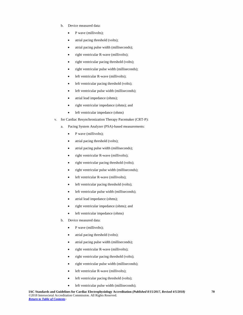
IAC Standards and Guidelines for Cardiac Electrophysiology Accreditation (Published 8/15/2017, Revised 4/5/2018) 70 ©2018 Intersocietal Accreditation Commission. All Rights Reserved. Return to Table of Contents»
b. Device measured data:
• P wave (millivolts);
• atrial pacing threshold (volts);
• atrial pacing pulse width (milliseconds);
• right ventricular R-wave (millivolts);
• right ventricular pacing threshold (volts);
• right ventricular pulse width (milliseconds);
• left ventricular R-wave (millivolts);
• left ventricular pacing threshold (volts);
• left ventricular pulse width (milliseconds);
• atrial lead impedance (ohms);
• right ventricular impedance (ohms); and
• left ventricular impedance (ohms)
v. for Cardiac Resynchronization Therapy Pacemaker (CRT-P):
a. Pacing System Analyzer (PSA)-based measurements:
• P wave (millivolts);
• atrial pacing threshold (volts);
• atrial pacing pulse width (milliseconds);
• right ventricular R-wave (millivolts);
• right ventricular pacing threshold (volts);
• right ventricular pulse width (milliseconds);
• left ventricular R-wave (millivolts);
• left ventricular pacing threshold (volts);
• left ventricular pulse width (milliseconds);
• atrial lead impedance (ohms);
• right ventricular impedance (ohms); and
• left ventricular impedance (ohms)
b. Device measured data:
• P wave (millivolts);
• atrial pacing threshold (volts);
• atrial pacing pulse width (milliseconds);
• right ventricular R-wave (millivolts);
• right ventricular pacing threshold (volts);
• right ventricular pulse width (milliseconds);
• left ventricular R-wave (millivolts);
• left ventricular pacing threshold (volts);
• left ventricular pulse width (milliseconds);
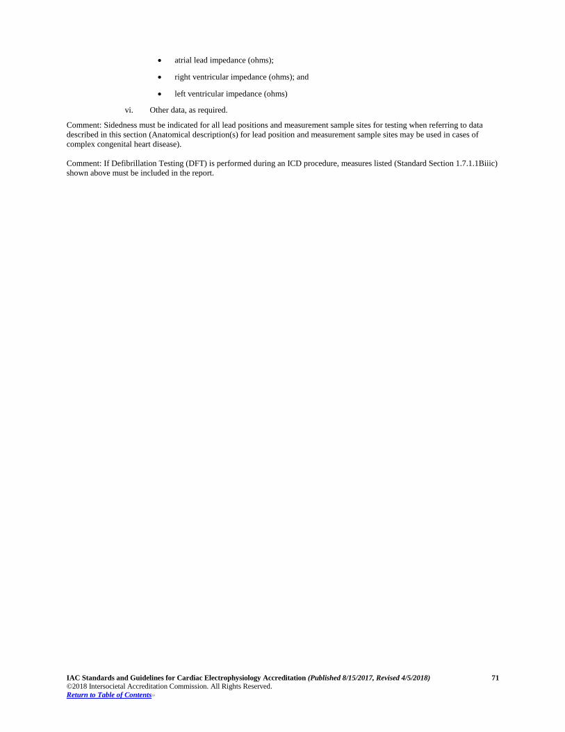
IAC Standards and Guidelines for Cardiac Electrophysiology Accreditation (Published 8/15/2017, Revised 4/5/2018) 71 ©2018 Intersocietal Accreditation Commission. All Rights Reserved. Return to Table of Contents»
• atrial lead impedance (ohms);
• right ventricular impedance (ohms); and
• left ventricular impedance (ohms)
vi. Other data, as required.
Comment: Sidedness must be indicated for all lead positions and measurement sample sites for testing when referring to data described in this section (Anatomical description(s) for lead position and measurement sample sites may be used in cases of complex congenital heart disease). Comment: If Defibrillation Testing (DFT) is performed during an ICD procedure, measures listed (Standard Section 1.7.1.1Biiic) shown above must be included in the report.
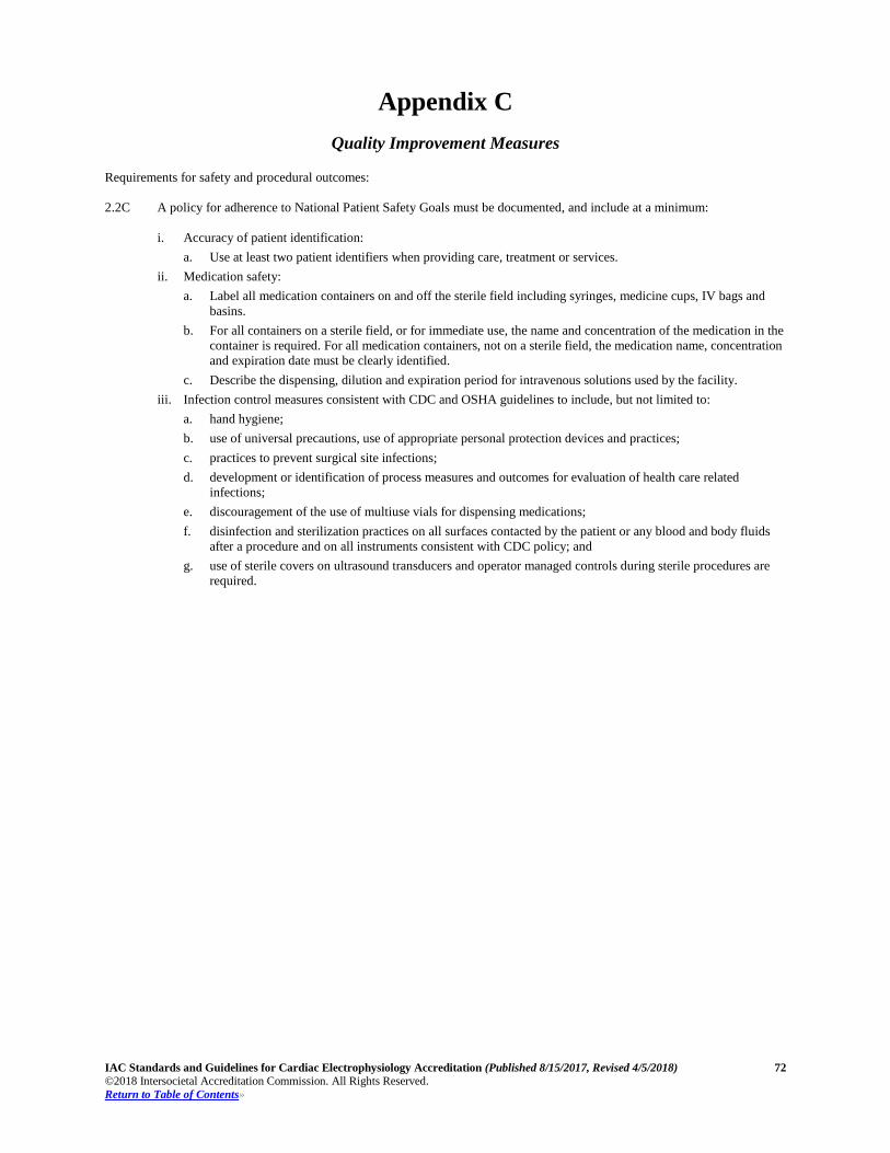
IAC Standards and Guidelines for Cardiac Electrophysiology Accreditation (Published 8/15/2017, Revised 4/5/2018) 72 ©2018 Intersocietal Accreditation Commission. All Rights Reserved. Return to Table of Contents»
Appendix C
Quality Improvement Measures Requirements for safety and procedural outcomes: 2.2C A policy for adherence to National Patient Safety Goals must be documented, and include at a minimum:
i. Accuracy of patient identification: a. Use at least two patient identifiers when providing care, treatment or services.
ii. Medication safety: a. Label all medication containers on and off the sterile field including syringes, medicine cups, IV bags and
basins. b. For all containers on a sterile field, or for immediate use, the name and concentration of the medication in the
container is required. For all medication containers, not on a sterile field, the medication name, concentration and expiration date must be clearly identified.
c. Describe the dispensing, dilution and expiration period for intravenous solutions used by the facility. iii. Infection control measures consistent with CDC and OSHA guidelines to include, but not limited to:
a. hand hygiene; b. use of universal precautions, use of appropriate personal protection devices and practices; c. practices to prevent surgical site infections; d. development or identification of process measures and outcomes for evaluation of health care related
infections; e. discouragement of the use of multiuse vials for dispensing medications; f. disinfection and sterilization practices on all surfaces contacted by the patient or any blood and body fluids
after a procedure and on all instruments consistent with CDC policy; and g. use of sterile covers on ultrasound transducers and operator managed controls during sterile procedures are
required.