Embed Size (px)
Citation preview
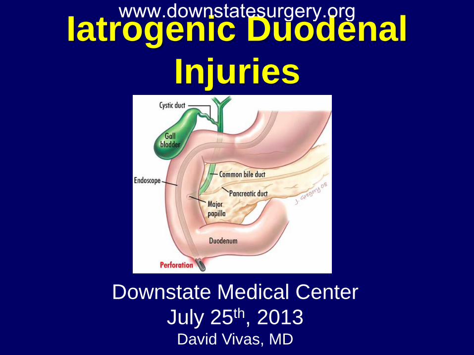
Iatrogenic Duodenal Injuries
Downstate Medical Center July 25th, 2013
David Vivas, MD
www.downstatesurgery.org
Case History
• 71 y/o female who was seen by her PMD c/o 2 h/o RUQ pain radiated to the back. Patient denied fevers, nausea or vomiting. Was found to have elevated LFT’s and was referred to the ED.
• Patient was admitted to our service for further work up. • PMH: Depression, Thalassemia, Reynaud’s
• PSH: Myomectomy
• NKDA
www.downstatesurgery.org
Case
• Patient in NAD, AAOx3
• Mild jaundice
• C-V: RRR, S1-S2
• Pulm: CTA b/l
• Abdomen: Non distended, soft, tender to palpation in
RUQ, neg Murphy’s sign. No masses.
Physical Exam
www.downstatesurgery.org
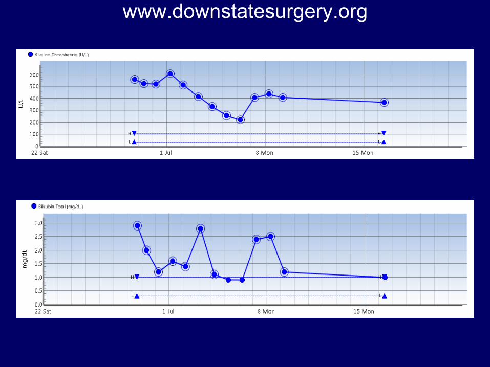
Case Labs: • CBC: 10.45>10.0/30.2<469
• BMP: 140/4.7/103/25/38/1.53<81
• LFT: 7.0/3.7/315/498/558/2.9 • Coags: 14.2/30.4/1.2
www.downstatesurgery.org
Case HD #1
• Patient had significant improvement in pain. No fevers, nausea or vomiting
• Started on clear liquid diet • Patient was assessed by GI
• MRCP • Trend LFT’s • Abx • Plan for possible ERCP
www.downstatesurgery.org
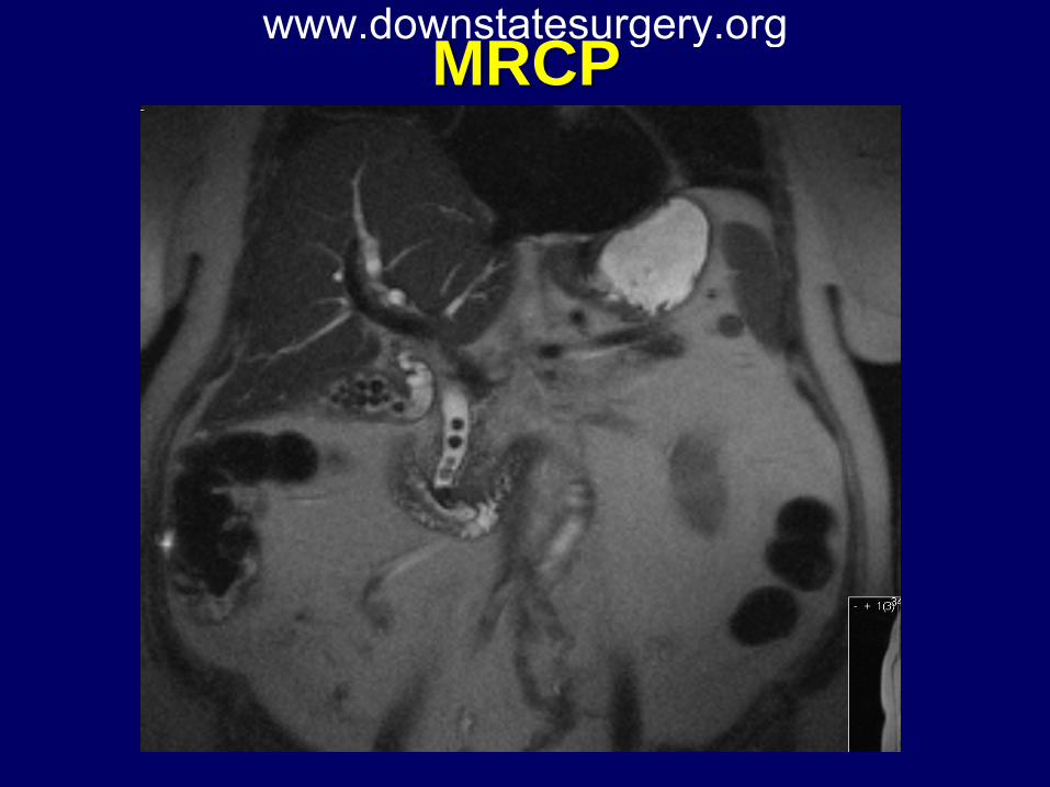
MRCP www.downstatesurgery.org
Case HD #2
• Patient denied abdominal pain
• Tolerating clear liquid diet
• Plan for ERCP next day
• LFT’s trending down
www.downstatesurgery.org
Case HD #3
• ERCP: • Unsuccessful • Incidental perforation of duodenum diverticulum
opposite to ampulla during difficult small bowel intubation
• 2 unsuccessful attempts for Hemoclip placement
www.downstatesurgery.org

Case www.downstatesurgery.org
Case
• Patient transferred to PACU in stable condition
• NGT in place
• Surgery notified of complication
• On initial assessment: • In NAD • AVSS • Abdomen soft, non tender
HD #3
www.downstatesurgery.org
Case
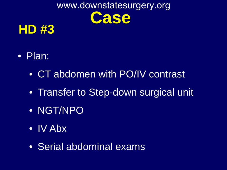
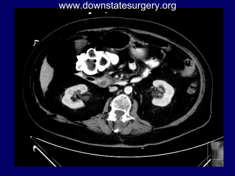
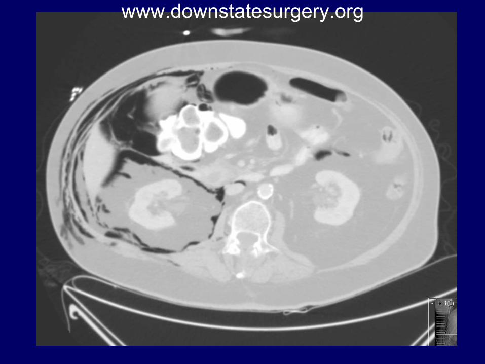
• Plan:
• CT abdomen with PO/IV contrast
• Transfer to Step-down surgical unit
• NGT/NPO
• IV Abx
• Serial abdominal exams
HD #3
www.downstatesurgery.org
www.downstatesurgery.org
www.downstatesurgery.org
Case
• Patient was transferred to Step Down unit
• Multiple assessments during the following
hours • Patient in NAD, denied abdominal pain • AVSS • Abdomen soft, non tender
HD #3
www.downstatesurgery.org
Case
• Approximately 9 hrs. post attempted ERCP: • Patient now complains of RUQ abdominal
pain • On physical exam:
• AVSS • Abdomen diffusely tender, particularly in
RUQ
• Decision was made to proceed with emergent surgical exploration
HD #3
www.downstatesurgery.org
Case
• Exploratory laparotomy • Gush of air • Large infiltration of air in the
retroperitoneum, colonic mesentery and small bowel
• Transverse colon intermittently adherent to duodenum without distinctive demarcation between the 2 structures
HD #3
www.downstatesurgery.org
Case
• Exploratory laparotomy • 5 mm perforation lateral aspect of 2nd portion of
duodenum with active leak of bile and air • GB appeared thickened and inflamed • Primary closure and omentoplasty • JP drain
HD #3
www.downstatesurgery.org
Case POD #1-4
• Patient NPO • NGT to LCS • Abx • Patient with no mayor complaints, afebrile,
VSS
www.downstatesurgery.org
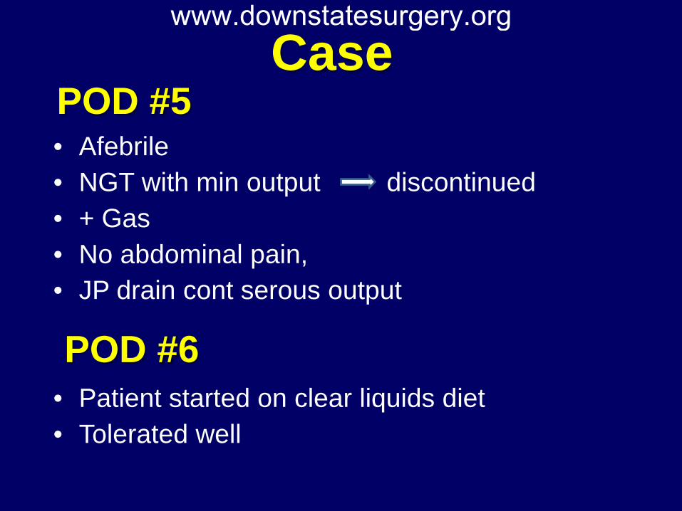
Case POD #5 • Afebrile • NGT with min output discontinued • + Gas • No abdominal pain, • JP drain cont serous output
• Patient started on clear liquids diet • Tolerated well
POD #6
www.downstatesurgery.org

Case POD #8
• Advanced to low fat diet • Tolerated
• Patient discharged home
POD #9
www.downstatesurgery.org
www.downstatesurgery.org

Questions?
www.downstatesurgery.org
Iatrogenic Duodenal Injuries
• ERCP was first introduced in 1968 by McCune et al and has evolved over the decades
• Currently, it is a valuable, widely used diagnostic
and therapeutic tool in hepato-biliary-pancreas diseases
• ERCP has a relatively high complication rate of nearly 10% and a mortality rate of 0.1 to 1%
www.downstatesurgery.org
Iatrogenic Duodenal Injuries
• Therapeutic aspects of ERCP are becoming more important
• Endoscopists take on increasingly more complex cases the risk of complication is increasing
• Pancreatitis, cholangitis, and hemorrhage are more
frequent ERCP complications
• ERCP-related perforation is one of the most feared, due to its potentially lethal nature
www.downstatesurgery.org
Iatrogenic Duodenal Injuries Classification
• Retroperitoneal duodenal perforation • The most common • Usually occur as a result of a sphincterotomy that
extends beyond the intramural portion of bile duct
• Perforation of the bile ducts • Usually occurs following dilation of strictures, forceful
cannulation, guidewire insertion, or stent migration
• Free bowel-wall perforation: • Rare, usually occurring in patients with a stricture or
anomalous anatomy, such as Billroth II gastrectomy
www.downstatesurgery.org
Iatrogenic Duodenal Injuries Classification
• Also reported, but rare following ERCP and sphincterotomy: • Gastric and esophageal perforations • Pneumomediastinum without evidence of
perforation • Intestinal perforation related to biliary stents
www.downstatesurgery.org
Incidence of Retroduodenal Perforation
• Retroduodenal perforation was reported in 0.5 to 2.1% of sphincterotomies in older large series
• More recently, the incidence of perforation has
appeared to decrease to less than 0.5%, probably because of improvement in experience and skill of the endoscopists
• Severe and fatal cases continue to occur
www.downstatesurgery.org
Risk Factors for Duodenal Perforation
• Risk factors for overall perforation: • Patient related
• Sphincter of Oddi dysfunction • Common bile duct dilation
• Procedure related
• Performance of a sphincterotomy • Longer duration of the procedure • Biliary stricture dilation
www.downstatesurgery.org
Risk Factors for Duodenal Perforation
• Risk factors for bowel wall perforation: • Stenosis in the upper GI tract or bile ducts • Abnormal GI anatomy (s/p gastrectomy, s/p
pancreaticoduodenectomy and situs inversus)
• Particular caution required with use of side-viewing scope in patients with Billroth II reconstruction
www.downstatesurgery.org
Risk Factors for Duodenal Perforation
• Risk factors for retroperitoneal perforation: • Precut and larger sphincterotomies
(particularly if cuts created outside the usual landmarks)
• Small caliber bile duct • Presence of periampullary diverticulum • Intramural injection of contrast
www.downstatesurgery.org
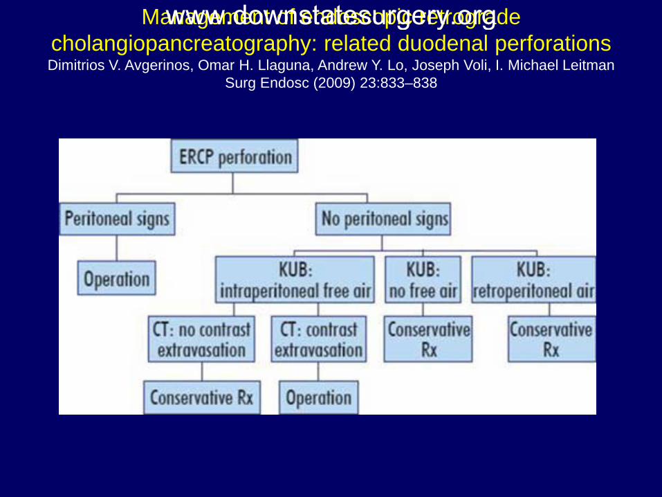
Clinical Manifestations and Diagnosis
• Perforation is rarely evident endoscopically • Free abdominal perforation:
• Almost always recognized immediately based upon clinical symptoms, physical signs and fluoro findings
• Retroduodenal perforation: • Usually determined by the presence of air or contrast
in the retroperitoneal space outside the confines of the bile ducts and duodenum during CT ordered for post ERCP pain
www.downstatesurgery.org

Clinical Manifestations and Diagnosis
• Patients with undetected leaks can present hours after the procedure with pain, fever and leukocytosis
• Other findings:
• Gas in the portal system • Pneumothorax • Pneumomediastinum • Pneumoretroperitoneum • Pneumoperitoneum • Subcutaneous emphysema
www.downstatesurgery.org
Diagnosis- Abdominal CT
• Should be obtained in patients suspected of having a perforation even if no evidence of retroperitoneal air on plain films • CT is the most sensitive means of detecting perforation
www.downstatesurgery.org
Diagnosis- Abdominal CT
• The clinical or radiographic amount of air: • Not always indicates the size of the perforation • Not always correlates with the severity of the complication
• The amount of air reflects the degree of manipulation after the perforation occurred
www.downstatesurgery.org
Retroperitoneal Air
• Typically associated with perforation
• However, may develop in clinically asymptomatic patients following sphincterotomy
• These patients may not require intervention
www.downstatesurgery.org
Retroperitoneal Air • Origin of retroperitoneal air in asymptomatic
patients: • Related to dissection through an injured or
intact bowel (similar to that described after colonoscopy)
• Sealed microperforations
• Presence of retroperitoneal air in the absence of symptoms should warrant careful observation but may not require intervention
www.downstatesurgery.org
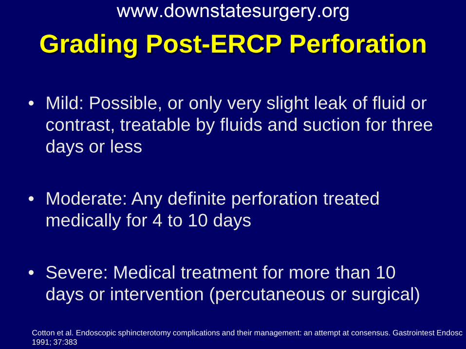
Grading Post-ERCP Perforation
• Mild: Possible, or only very slight leak of fluid or contrast, treatable by fluids and suction for three days or less
• Moderate: Any definite perforation treated
medically for 4 to 10 days • Severe: Medical treatment for more than 10
days or intervention (percutaneous or surgical)
Cotton et al. Endoscopic sphincterotomy complications and their management: an attempt at consensus. Gastrointest Endosc 1991; 37:383
www.downstatesurgery.org
Clasification Post-ERCP Perforation
• Coded in descending order of injury severity • Correlates with the mechanism of injury and
the anatomic location of damage • Used as a predictor for the need for surgical
intervention • Graded from Type I-IV
Miller et al. Perforations following endoscopic retrograde cholangiopancreatography: a single institution experience and surgical recommendations. Am J Surg. 2013 Aug;206(2):180-6
www.downstatesurgery.org
Clasification Post-ERCP Perforation
• Type I: • Lateral or the medial duodenal wall remote from the ampulla • Caused by acute angulation of the endoscope • Typically large with extensive contrast leakage • Requires immediate surgery
• Type II:
• Peri-Vaterian • Variable in their severity • Consequent to a precut sphincterotomy or occurring near a
periampullary diverticulum • Minimal or moderate contrast leakage • Some may be able to be managed conservatively
Miller et al. Perforations following endoscopic retrograde cholangiopancreatography: a single institution experience and surgical recommendations. Am J Surg. 2013 Aug;206(2):180-6
www.downstatesurgery.org
Clasification Post-ERCP Perforation
• Type III • Distal CBD • Secondary to wire manipulation or basket instrumentation during
stone retrieval • Often small and localized and may frequently be managed
conservatively
• Type IV • Reveal retroperitoneal air and result from insufflation during ERCP • Typically managed nonoperatively
Miller et al. Perforations following endoscopic retrograde cholangiopancreatography: a single institution experience and surgical recommendations. Am J Surg. 2013 Aug;206(2):180-6
www.downstatesurgery.org
Post-ERCP Perforation Management
• Free abdominal duodenal perforations usually require surgery
• Conservative approach to retroperitoneal perforation following sphincterotomy has been adopted
• Early surgical consultation and careful observation is mandatory
• Outcome poor in patients who do not receive prompt and
appropriate treatment
www.downstatesurgery.org
• Endoscopic Therapy: • Endoclips • Over the scope clips • Fibrin glue • Covered stents • Nasobiliary tube
• Percutaneous transhepatic drainage
Post-ERCP Perforation Management
www.downstatesurgery.org
• NPO, IV hydration, NG or nasoduodenal suction, IV antibiotics
• Consider percutaneous drainage as alternative
to surgical drainage of retroperitoneal collections • TPN if NPO status expected for more than one
week
Post-ERCP Perforation Management
www.downstatesurgery.org
Post-ERCP Perforation Management
• Surgery if: • Persistent biliary obstruction, cholangitis, and
if no improvement after brief nonoperative management
• Overall surgery is required in 20 to 40 percent of patients with perforation
• Type of surgical intervention depends upon
clinicopathological condition
www.downstatesurgery.org
Post-ERCP Perforation Management
• Simple Repair • Tube decompression
– Gastrostomy – Duodenostomy – Feeding Jejunostomy
• Serosal Patch • Duodenal Diverticulization • Pyloric exclusion
www.downstatesurgery.org
• Prognosis of patients with a perforation depends upon
• Rapidity of diagnosis • Clinical setting • Patient comorbidities
Post-ERCP Perforation Management
www.downstatesurgery.org
• Overall mortality has decreased from 16% percent in older reports to 8% in a review that considered major studies from the year 2000
• Lower mortality noted in recent years may reflect benefits related to a conservative team approach to the management of small retroperitoneal perforations
Post-ERCP Perforation Management
Machado NO. Management of duodenal perforation post-endoscopic retrograde cholangiopancreatography. When and whom to operate and what factors determine the outcome? A review article. JOP 2012; 13:18
www.downstatesurgery.org
• A retrospective review of ERCP-related perforations to the duodenum (April 1999 to February 2008)
• Incidence, Clinical outcomes
• Data included: ERCP indication, Clinical
presentation, Diagnostic methods, Time to diagnosis and treatment, Type of injury, Management
Management of endoscopic retrograde cholangiopancreatography: related duodenal perforations Dimitrios V. Avgerinos, Omar H. Llaguna, Andrew Y. Lo, Joseph Voli, I. Michael Leitman
Surg Endosc (2009) 23:833–838
www.downstatesurgery.org
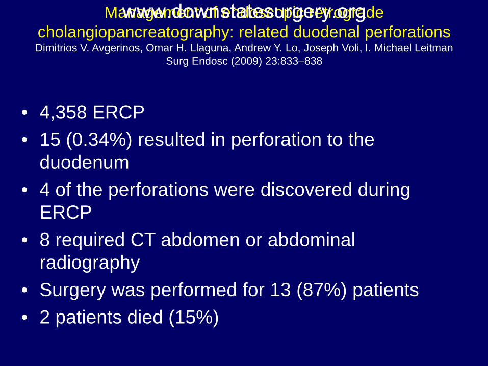
• 4,358 ERCP • 15 (0.34%) resulted in perforation to the
duodenum • 4 of the perforations were discovered during
ERCP • 8 required CT abdomen or abdominal
radiography • Surgery was performed for 13 (87%) patients • 2 patients died (15%)
Management of endoscopic retrograde cholangiopancreatography: related duodenal perforations Dimitrios V. Avgerinos, Omar H. Llaguna, Andrew Y. Lo, Joseph Voli, I. Michael Leitman
Surg Endosc (2009) 23:833–838
www.downstatesurgery.org
• One patient was managed conservatively with a successful outcome
• 9 patients underwent surgery within 24 h after the ERCP
• 1 patient underwent surgery after 24 h • The overall mortality rate was 20% (3 of 15
patients)
Management of endoscopic retrograde cholangiopancreatography: related duodenal perforations Dimitrios V. Avgerinos, Omar H. Llaguna, Andrew Y. Lo, Joseph Voli, I. Michael Leitman
Surg Endosc (2009) 23:833–838
www.downstatesurgery.org
• The interval between the perforation and the operation is of great significance
• The mortality rate increases dramatically with late surgical management (>24 hours)
• Clinical and radiographic features can be used to determine the surgical or conservative treatment of ERCP-related duodenal perforations
• Patient age and intraoperative findings can determined the final outcome and morbidity or mortality
Management of endoscopic retrograde cholangiopancreatography: related duodenal perforations Dimitrios V. Avgerinos, Omar H. Llaguna, Andrew Y. Lo, Joseph Voli, I. Michael Leitman
Surg Endosc (2009) 23:833–838
www.downstatesurgery.org
Management of endoscopic retrograde cholangiopancreatography: related duodenal perforations Dimitrios V. Avgerinos, Omar H. Llaguna, Andrew Y. Lo, Joseph Voli, I. Michael Leitman
Surg Endosc (2009) 23:833–838
www.downstatesurgery.org
Perforations following endoscopic retrograde cholangiopancreatography: a single institution experience
and surgical recommendations Rafi Miller, M.D. ,Andrew Zbar, M.D.. Yoram Klein, M.D., Victor Buyeviz, M.D., Ehud
Melzer, M.D., Bruce N. Mosenkis, M.D., Eli Mavor, M.D. The American Journal of Surgery (2013) 206, 180-186
• Records of patients undergoing ERCP 16-year period (1995-2011)
• Types of injuries, diagnosis, management, and patient outcome
• Injuries classified from I to IV • 1,638 ERCP • 27 perforations (1.6%) • Nearly 50% of the procedures were regarded as difficult
www.downstatesurgery.org
Perforations following endoscopic retrograde cholangiopancreatography: a single institution experience
and surgical recommendations Rafi Miller, M.D. ,Andrew Zbar, M.D.. Yoram Klein, M.D., Victor Buyeviz, M.D., Ehud
Melzer, M.D., Bruce N. Mosenkis, M.D., Eli Mavor, M.D. The American Journal of Surgery (2013) 206, 180-186
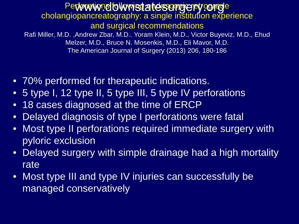
• 70% performed for therapeutic indications. • 5 type I, 12 type II, 5 type III, 5 type IV perforations • 18 cases diagnosed at the time of ERCP • Delayed diagnosis of type I perforations were fatal • Most type II perforations required immediate surgery with
pyloric exclusion • Delayed surgery with simple drainage had a high mortality
rate • Most type III and type IV injuries can successfully be
managed conservatively
www.downstatesurgery.org
Perforations following endoscopic retrograde cholangiopancreatography: a single institution experience
and surgical recommendations Rafi Miller, M.D. ,Andrew Zbar, M.D.. Yoram Klein, M.D., Victor Buyeviz, M.D., Ehud
Melzer, M.D., Bruce N. Mosenkis, M.D., Eli Mavor, M.D. The American Journal of Surgery (2013) 206, 180-186
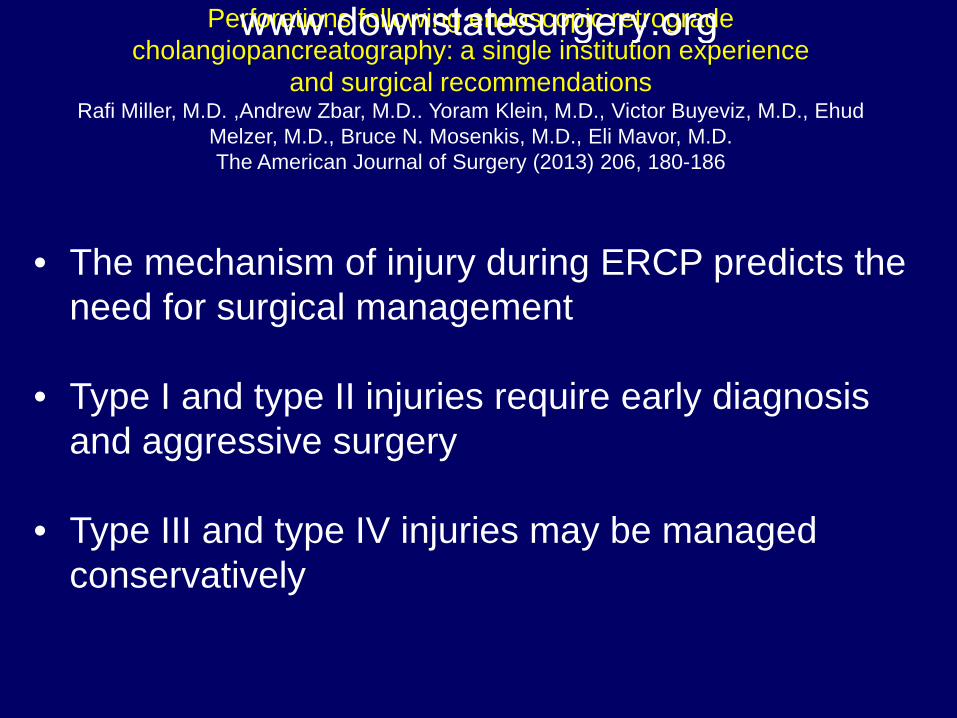
• The mechanism of injury during ERCP predicts the need for surgical management
• Type I and type II injuries require early diagnosis and aggressive surgery
• Type III and type IV injuries may be managed conservatively
www.downstatesurgery.org
Perforations following endoscopic retrograde cholangiopancreatography: a single institution experience
and surgical recommendations Rafi Miller, M.D. ,Andrew Zbar, M.D.. Yoram Klein, M.D., Victor Buyeviz, M.D., Ehud
Melzer, M.D., Bruce N. Mosenkis, M.D., Eli Mavor, M.D. The American Journal of Surgery (2013) 206, 180-186
www.downstatesurgery.org
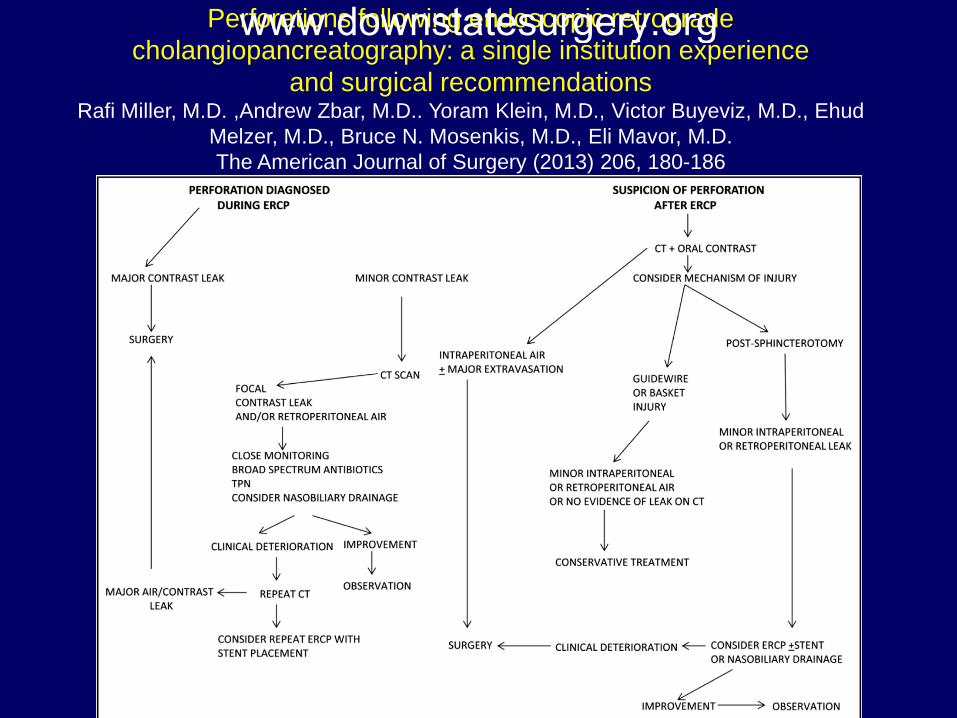
• Retroduodenal perforation has decreased from up to 2.1 percent of sphincterotomies to less than 0.5 percent in recent series
• Free abdominal perforation is almost always recognized immediately based upon clinical symptoms, physical signs, and fluoroscopic findings
Summary www.downstatesurgery.org
• Retroduodenal perforation is usually determined by the radiologic evidence of air or contrast in the retroperitoneal space
• Retroperitoneal air may also develop following
sphincterotomy in patients who are clinically asymptomatic. Such patients may not require intervention
Summary www.downstatesurgery.org
• Abdominal CT scan should be obtained in patients who are suspected of having a perforation. CT scan is the most sensitive means for detecting perforation
• Patients with free abdominal duodenal
perforation usually require surgery
Summary www.downstatesurgery.org

• By contrast, a conservative approach to small retroperitoneal perforation may be appropriate
• Early surgical consultation and careful
observation is mandatory since the outcome may be poor in patients who do not receive prompt and appropriate treatment
Summary www.downstatesurgery.org

1. Proposal of an endoscopic retrograde cholangiopancreatography-related perforation management
guideline based on perforation type. Kwon W, Jang J, Ryu J, Kim Y, Yoon Y, Kang M, Kim S. J Korean
Surg Soc 2012;83:218-226
2. Cotton PB, Lehman G, Vennes J, et al. Endoscopic sphincterotomy complications and their
management: an attempt at consensus. Gastrointest Endosc 1991; 37:383
3. Machado NO. Management of duodenal perforation post-endoscopic retrograde
cholangiopancreatography. When and whom to operate and what factors determine the outcome? A
review article. JOP 2012; 13:18
4. Miller R, Zbar A, Klein Y, Buyeviz V, Melzer E, Mosenkis BN, Mavor E. Perforations following
endoscopic retrograde cholangiopancreatography: a single institution experience and surgical
recommendations. Am J Surg. 2013 Aug;206(2):180-6
5. Loperfido S et al. Post-ERCP perforation. In: UpToDate, Basow, DS (Ed), UpToDate, Waltham, MA,
2013
6. Dimitrios V. Avgerinos, Omar H. Llaguna, Andrew Y. Lo, Joseph Voli, I. Michael Leitman. Management
of endoscopic retrograde cholangiopancreatography: related duodenal perforations. Surg Endosc
(2009) 23:833–838
References www.downstatesurgery.org