Embed Size (px)
Citation preview

Identification guide for cephalopod paralarvae from the Mediterranean Sea
ICES Cooperative Research ReportRapport des Recherches Collectives
No. 324 February 2015
ICES COOPERATIVE RESEARCH REPORT RAPPORT DES RECHERCHES COLLECTIVES
NO. 324
FEBRUARY 2015
Identification guide for cephalopod
paralarvae from the Mediterranean Sea
Authors
Núria Zaragoza, Antoni Quetglas, and Ana Moreno
International Council for the Exploration of the Sea
Conseil International pour l’Exploration de la Mer H. C. Andersens Boulevard 44–46
DK-1553 Copenhagen V
Denmark
Telephone (+45) 33 38 67 00
Telefax (+45) 33 93 42 15
www.ices.dk
Recommended format for purposes of citation:
Zaragoza, N., Quetglas, A. and Moreno, A. 2015. Identification guide for cephalopod
paralarvae from the Mediterranean Sea. ICES Cooperative Research Report No. 324. 91
pp.
Series Editor: Emory D. Anderson
The material in this report may be reused for non-commercial purposes using the rec-
ommended citation. ICES may only grant usage rights of information, data, images,
graphs, etc. of which it has ownership. For other third-party material cited in this re-
port, you must contact the original copyright holder for permission. For citation of da-
tasets or use of data to be included in other databases, please refer to the latest ICES
data policy on the ICES website. All extracts must be acknowledged. For other repro-
duction requests please contact the General Secretary.
This document is a report conducted under the auspices of the International Council
for the Exploration of the Sea and does not necessarily represent the view of the Coun-
cil.
ISBN 978-87-7482-156-4
ISSN 1017-6195
© 2015 International Council for the Exploration of the Sea
Contents
1 Introduction .................................................................................................................... 1
2 Checklist of species ....................................................................................................... 3
3 Identification key of early life stages of cephalopods ............................................ 7
4 Glossary of terms (from Sweeney et al., 1992) ......................................................... 9
5 Description and illustration of paralarvae .............................................................. 13
5.1 Family Sepiidae Leach, 1817 ............................................................................. 13
Sepia officinalis Linnaeus, 1758 ................................................................. 13
Sepia elegans Blainville, 1827..................................................................... 14
Sepia orbignyana Férussac, 1826 ................................................................ 14
5.2 Family Sepiolidae Leach, 1817 .......................................................................... 15
5.2.1 Subfamily Rossiinae Appellöf, 1898 .................................................... 15
Rossia macrosoma (Delle Chiaje, 1830) ..................................................... 15
Neorossia caroli (Joubin, 1902) ................................................................... 16
5.2.2 Subfamily Heteroteuthinae Appellöf, 1898 ........................................ 16
Heteroteuthis dispar (Rüppell, 1844) ......................................................... 16
Stoloteuthis leucoptera (A. E. Verrill, 1878) .............................................. 17
5.2.3 Subfamily Sepiolinae Appellöf, 1898 .................................................. 18
Rondeletiola minor (Naef, 1912) ................................................................. 18
Sepiola spp. Leach, 1817 ............................................................................ 19
Sepietta spp. Naef, 1912 ............................................................................. 19
5.3 Family Loliginidae Lesueur, 1821 ..................................................................... 19
Loligo vulgaris Lamarck, 1798 ................................................................... 21
Loligo forbesii Steenstrup, 1857 ................................................................. 23
Alloteuthis spp. Wülker, 1920 ................................................................... 23
5.4 Family Chtenopterygidae Grimpe, 1922 ......................................................... 23
Chtenopteryx sicula (Vérany, 1851) ........................................................... 23
5.5 Family Enoploteuthidae Pfeffer, 1900 .............................................................. 25
Abralia veranyi (Rüppell, 1844) ................................................................. 26
Abraliopsis morisii (Vérany, 1839) ............................................................. 27
5.6 Family Ancistrocheiridae Pfeffer, 1912 ............................................................ 29
Ancistrocheirus lesueurii (d'Orbigny, 1842) ............................................. 29
5.7 Family Octopoteuthidae Berry, 1912 ................................................................ 32
Octopoteuthis spp. Rüppell, 1844 ............................................................. 32

Taningia danae Joubin, 1931 ...................................................................... 34
5.8 Family Onychoteuthidae Gray, 1847................................................................ 35
Ancistroteuthis lichtensteinii (Férussac [in Férussac & d'Orbigny], 1835)
..................................................................................................................... 36
Onychoteuthis banksii (Leach, 1817) ......................................................... 37
5.9 Family Histioteuthidae Verril, 1881 ................................................................. 39
Histioteuthis reversa (Verrill, 1880) ........................................................... 41
Histioteuthis bonnellii (Férussac, 1835) ..................................................... 41
5.10 Family Brachioteuthidae Pfeffer, 1908 ............................................................. 42
Brachioteuthis riisei (Steenstrup, 1882) ..................................................... 42
5.11 Family Ommastrephidae Steenstrup, 1857 ..................................................... 44
Rhynchoteuthion type A .......................................................................... 45
Rhynchoteuthion type B ........................................................................... 46
Rhynchoteuthion type C .......................................................................... 47
Ommastrephes bartramii (Lesueur, 1821) ................................................. 48
5.12 Family Thysanoteuthidae Keferstein, 1866 ..................................................... 49
Thysanoteuthis rhombus Troschel, 1857 .................................................... 49
5.13 Family Chiroteuthidae Gray, 1849 ................................................................... 54
Chiroteuthis veranii (Férussac, 1835) ........................................................ 54
5.14 Family Cranchiidae Prosch, 1847 ...................................................................... 55
5.14.1 Subfamily Cranchiinae Prosch, 1849 .............................................. 56
Cranchia scabra Leach, 1817 ...................................................................... 56
5.14.2 Subfamily Taoniinae Pfeffer, 1912 .................................................. 58
Teuthowenia megalops (Prosch, 1849) ....................................................... 58
Galiteuthis armata Joubin, 1898 ................................................................. 59
5.15 Family Opisthoteuthidae Verril, 1896 .............................................................. 60
Opisthoteuthis sp. Verrill, 1883 ................................................................. 60
5.16 Family Octopodidae d’Orbigny, 1839 .............................................................. 61
5.16.1 Subfamily Octopodinae d’Orbigny, 1845 ...................................... 64
Octopus vulgaris Cuvier, 1797 ................................................................... 65
Octopus salutii Vérany, 1836 ..................................................................... 70
Callistoctopus macropus (Risso, 1826) ....................................................... 70
Macrotritopus defilippi (Vérany, 1851) ...................................................... 71
Scaeurgus unicirrhus (Delle Chiaje [in de Férussac & d’Orbigny], 1841)
..................................................................................................................... 73
Pteroctopus tetracirrhus (Delle Chiaje, 1830) ........................................... 74
5.16.2 Subfamily Eledoninae Grimpe, 1921 ............................................. 75
Eledone cirrhosa (Lamarck, 1798) .............................................................. 75
Eledone moschata (Lamarck, 1798) ............................................................ 76
5.16.3 Subfamily Bathypolypodinae Robson, 1929 ................................. 76
5.17 Family Tremoctopodidae (Brock, 1882) ........................................................... 76
Tremoctopus violaceus Delle Chiaje, 1830 ................................................. 76
5.18 Family Ocythoidea Gray, 1849 .......................................................................... 79
Ocythoe tuberculata Rafinesque, 1814 ...................................................... 79
5.19 Family Argonautidae Tryon, 1879 .................................................................... 80
Argonauta argo Linnaeus, 1758 ................................................................. 80
6 Acknowledgements ..................................................................................................... 84
7 References ..................................................................................................................... 85
8 Author contact information ....................................................................................... 91
Identification guide of cephalopod paralarvae from the Mediterranean Sea | 1
1 Introduction
Cephalopods are key components of marine trophic webs, where they constitute major
food resources for a large variety of predators including fish, other cephalopods, ma-
rine mammals, and seabirds (e.g. Clarke, 1996; Piatkowski et al., 2001; Cherel et al.,
2009). Cephalopods are, in turn, voracious predators of fish and crustaceans (Boyle and
Rodhouse, 2005). Octopuses, squid, and cuttlefish are also important living marine re-
sources, maintaining relevant fisheries around the world (FAO, 2012). Despite their
economic and ecological importance, the number of studies on these molluscs, until
relatively recently, has been small compared to other taxonomic groups such as fish,
crustaceans, or marine mammals (Piatkowski et al., 2001). The lack of knowledge is
even worse in the case of larval stages, which have been little studied worldwide and
represent, without doubt, a challenge for future studies on cephalopods. The difficul-
ties in sampling (low abundance and patchy distribution), the uncertainties of species
identification, and problems related to their maintenance in captivity are major limita-
tions to those studies (Vecchione, 1987; Boyle and Rodhouse, 2005). The taxonomy is
probably the biggest obstacle to overcome, since the identification of virtually all larval
and juvenile stages of cephalopods has been, and still is in many cases, confusing and
problematic (Sweeney et al., 1992).
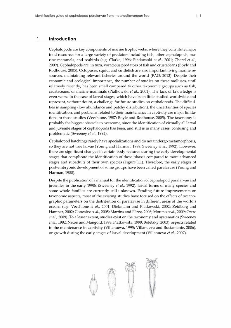
Cephalopod hatchings rarely have specializations and do not undergo metamorphosis,
so they are not true larvae (Young and Harman, 1988; Sweeney et al., 1992). However,
there are significant changes in certain body features during the early developmental
stages that complicate the identification of these phases compared to more advanced
stages and subadults of their own species (Figure 1.1). Therefore, the early stages of
post-embryonic development of some groups have been called paralarvae (Young and
Harman, 1988).
Despite the publication of a manual for the identification of cephalopod paralarvae and
juveniles in the early 1990s (Sweeney et al., 1992), larval forms of many species and
some whole families are currently still unknown. Pending future improvements on
taxonomic aspects, most of the existing studies have focused on the effects of oceano-
graphic parameters on the distribution of paralarvae in different areas of the world’s
oceans (e.g. Vecchione et al., 2001; Diekmann and Piatkowski, 2002; Zeidberg and
Hamner, 2002; González et al., 2005; Martins and Pérez, 2006; Moreno et al., 2009; Otero
et al., 2009). To a lesser extent, studies exist on the taxonomy and systematics (Sweeney
et al., 1992; Nixon and Mangold, 1998; Piatkowski, 1998; Boletzky, 2003), aspects related
to the maintenance in captivity (Villanueva, 1995; Villanueva and Bustamante, 2006),
or growth during the early stages of larval development (Villanueva et al., 2007).
2 | ICES Cooperative Research Report No. 324
Figure 1.1. Change in form during growth of an oceanic species of cephalopod (from Guerra,
1992).
The identification and analysis of cephalopod larval stages is interesting, not only from
a systematic point of view and for a comprehensive understanding of the biological
cycle of the species, but also for studies on population dynamics, especially to estimate
the recruitment of certain commercially important stocks (Guerra, 1992). In the Medi-
terranean, cephalopods are important resources for certain fisheries. The most im-
portant in terms of biomass is the common octopus (Octopus vulgaris), which may rep-
resent 20–40% of the bottom trawl landings (Quetglas et al., 1998). The seasonal fishery
for cuttlefish (Sepia officinalis) is vital for the maintenance of the small-scale fleet, since
a large number of vessels throughout the Mediterranean target this species. Finally,
although the importance of the common squid (Loligo vulgaris) for the commercial fleet
is limited in relative terms, it is one of the main targets for recreational fishers (Morales-
Nin et al., 2005). The remaining commercially sought species have negligible economic
importance owing to low catches or scarce flesh quality (Sartor et al., 1998).
Until now, a total of 67 cephalopod species has been reported in the Mediterranean Sea
(Bello, 2008), from which only 53 are represented by well established populations
(Bello, 2003). Despite having well-cataloged the Mediterranean teuthofauna from their
adult forms (e.g. Belcari and Sartor, 1993; Sánchez et al., 1998; Quetglas et al., 2000; Gon-
zález and Sánchez, 2002), studies on larval stages are very scarce (Roper, 1972, 1974;
Sánchez and Molí, 1985; Bello, 2004; Lefkaditou et al., 2005). As mentioned, such studies
are limited by a lack of taxonomic information for many species, which is essential for
the proper identification of specimens. The only available paralarva guide was pro-
duced more than 20 years ago (Sweeney et al., 1992), but valuable taxonomic infor-
mation has been published since then. To assist with the identification of cephalopods
in plankton samples collected in the Balearic Sea (western Mediterranean), we com-
piled the currently available information on paralarvae and early life stages of the
cephalopod species inhabiting our study area. This CRR is the result of such a compi-
lation, which also incorporates new, unpublished pictures from our own material. We
hope this work will also assist scientists and students interested in identifying cepha-
lopod paralarvae.
Identification guide of cephalopod paralarvae from the Mediterranean Sea | 3
2 Checklist of species
Taxonomic list of the cephalopods recorded in the Mediterranean.
Class CEPHALOPODA Cuvier, 1795
Subclass COLEOIDEA Bather, 1888
Order SEPIIDA Zittel, 1895
Family SEPIIDAE Leach, 1817
Genus Sepia Linnaeus, 1758
Sepia officinalis Linnaeus, 1758
Sepia elegans Blainville, 1827
Sepia orbignyana Férussac, 1826
Order SEPIOLIDA Fioroni, 1981
Family SEPIOLIDAE Leach, 1817
Subfamily ROSSINAE Appellöf, 1898
Genus Rossia Owen, 1834
Rossia macrosoma (Delle Chiaje, 1830)
Genus Neorossia von Boletzky, 1971
Neorossia caroli (Joubin, 1902)
Subfamily HETEROTEUTHINAE Appellöf, 1898
Genus Heteroteuthis Gray, 1849
Heteroteuthis dispar (Rüppell, 1844)
Genus Stoloteuthis Verrill, 1881
Stoloteuthis leucoptera (A. E. Verrill, 1878)
Subfamily SEPIOLINAE Appellöf, 1898
Genus Rondeletiola Naef, 1921
Rondeletiola minor (Naef, 1912)
Genus Sepiola Leach, 1817
Sepiola rondeletii Leach, 1817
Sepiola robusta Naef, 1912
Sepiola ligulata Naef, 1912
Sepiola intermedia Naef, 1912
Sepiola affinis Naef, 1912
Genus Sepietta Naef, 1912
Sepietta oweniana (d’Orbigny, 1841)
Sepietta obscura Naef, 1916
Sepietta neglecta Naef, 1916
Order TEUTHIDA Naef, 1916
Suborder MYOPSIDA d’Orbigny, 1845
Family LOLIGINIDAE Lesueur, 1821

4 | ICES Cooperative Research Report No. 324
Genus Loligo Lamarck, 1798
Loligo vulgaris Lamarck, 1798
Loligo forbesii Steenstrup, 1857
Genus Alloteuthis Wülker, 1920
Alloteuthis media (Linnaeus, 1758)
Alloteuthis subulata (Lamarck, 1798)
Suborder OEGOPSIDA d’Orbigny, 1845
Family CHTENOPTERYGIDAE Grimpe, 1922
Genus Chtenopteryx Appellöf, 1890
Chtenopteryx sicula (Vérany, 1851)
Family ENOPLOTEUTHIDAE Pfeffer, 1900
Genus Abralia Gray, 1849
Abralia veranyi (Rüppell, 1844)
Abraliopsis morisii (Vérany, 1839)
Family ANCISTROCHEIRIDAE Pfeffer, 1912
Genus Ancistrocheirus Gray, 1849
Ancistrocheirus lesueurii (d'Orbigny, 1842)
Family OCTOPOTEUTHIDAE Berry, 1912
Genus Octopoteuthis Rüppell, 1844
Octopoteuthis sicula Rüppell, 1844
Genus Taningia Joubin, 1931
Taningia danae Joubin, 1931
Family ONYCHOTEUTHIDAE Gray, 1847
Genus Onychoteuthis Lichtenstein, 1818
Onychoteuthis banksii (Leach, 1817)
Genus Ancistroteuthis Gray, 1849
Ancistroteuthis lichtensteinii (Férussac [in Férussac &
d’Orbigny], 1835)
Family HISTIOTEUTHIDAE Verril, 1881
Genus Histioteuthis d’Orbigny, 1841
Histioteuthis reversa (Verril, 1880)
Histioteuthis bonnellii (Férussac, 1835)
Family BRACHIOTEUTHIDAE Pfeffer, 1908
Genus Brachioteuthis Verrill, 1881
Brachioteuthis riisei (Steenstrup, 1882)
Family OMMASTREPHIDAE Steenstrup, 1857
Subfamily ILLICINAE Posselt, 1890
Identification guide of cephalopod paralarvae from the Mediterranean Sea | 5
Genus Illex Steenstrup, 1880
Illex coindetii (Vérany, 1839)
Genus Todaropsis Girard, 1890
Todaropsis eblanae (Ball, 1841)
Subfamily TODARODINAE Adam, 1960
Genus Todarodes Steenstrup, 1880
Todarodes sagittatus (Lamarck, 1798)
Subfamily OMMASTREPHINAE Steenstrup, 1857
Genus Ommastrephes d’Orbigny, 1835
Ommastrephes bartramii (Lesueur, 1821)
Family THYSANOTEUTHIDAE Keferstein, 1866
Genus Thysanoteuthis Troschel, 1857
Thysanoteuthis rhombus Troschel, 1857
Family CHIROTEUTHIDAE Gray, 1849
Genus Chiroteuthis d’Orbigny, 1841
Chiroteuthis veranii (Férussac, 1835)
Family CRANCHIIDAE Prosch, 1847
Subfamily CRANCHIINAE Prosch, 1849
Genus Cranchia Leach, 1817
Cranchia scabra Leach, 1817
Subfamily TAONIINAE Pfeffer, 1912
Genus Teuthowenia Chun, 1910
Teuthowenia megalops (Prosch, 1849)
Genus Galiteuthis Joubin, 1898
Galiteuthis armata Joubin, 1898
Order OCTOPODA Leach, 1818
Suborder CIRRATA Grimpe, 1916
Family OPISTHOTEUTHIDAE Verril, 1896
Genus Opisthoteuthis Verrill, 1883
Opisthoteuthis calypso Villanueva, Collins, Sánchez & Voss,
2002
Suborder INCIRRITA Grimpe, 1916
Family OCTOPODIDAE d’Orbigny, 1839
Subfamily OCTOPODINAE d’Orbigny, 1845
Genus Octopus Cuvier, 1798
Octopus salutii Vérany, 1836
6 | ICES Cooperative Research Report No. 324
Octopus vulgaris Cuvier, 1797
Genus Callistoctopus Iw. Taki, 1964
Callistoctopus macropus (Risso, 1826)
Genus Macrotritropus Grimpe, 1922
Macrotritropus defilippi (Vérany, 1851)
Genus Scaeurgus Troschel, 1857
Scaeurgus unicirrhus (Delle Chiaje [in de Férussac & d’Orbi-
gny], 1841)
Genus Pteroctopus P. Fischer, 1882
Pteroctopus tetracirrhus (Delle Chiaje, 1830)
Subfamily ELEDONINAE Grimpe, 1921
Genus Eledone Leach, 1817
Eledone cirrhosa (Lamarck, 1798)
Eledone moschata (Lamarck, 1798)
Subfamily BATHYPOLYPODINAE Robson, 1929
Genus Bathypolypus Grimpe, 1921
Bathypolypus sponsalis (P. Fischer & H. Fischer, 1892)
Family TREMOCTOPODIDAE Brock, 1882
Genus Tremoctopus delle Chiaje, 1830
Tremoctopus violaceus Delle Chiaje, 1830
Family OCYTHOIDAE Gray, 1849
Genus Ocythoe Rafinesque, 1814
Ocythoe tuberculata Rafinesque, 1814
Family ARGONAUTIDAE Tryon, 1879
Genus Argonauta Linnaeus, 1758
Argonauta argo Linnaeus, 1758
Identification guide of cephalopod paralarvae from the Mediterranean Sea | 7
3 Identification key of early life stages of cephalopods
1. • Mantle without fins; arm crown without tentacles................. Order Octopoda 16
• Mantle with fins or fin rudiments; one pair of tentacles or a trunk-like structure
(proboscis) exists .............................................................................................................. 2
2. • Mantle with lateral fins................................................................................................ 3
• Mantle with subterminal or terminal fins, sometimes dorsally attached
........................................................................................................... Order Teuthoidea 5
3. • Arm crown with eight subequal arms with suckers only (cirri develop in later
juvenile stage). Suborder Cirrata .................................................... Opisthoteuthidae
• Arm crown with 6–8 arms and two tentacles between arm III and IV; all append-
ages with stalked suckers ...................................................................................... 4
4. • Fins long and narrow (not paddle-shaped); extend laterally from near posterior
end to near anterior margin of mantle, but never united posteriorly ....... Sepiidae
• Fins paddle or ear-shaped, each fin at least as wide as long (antero-posteriorly)
............................................................................................................................ Sepiolidae
5. • Eye covered by a transparent membrane (cornea), tentacular clubs with clubs
with four rows of suckers, no external photophores. Suborder Myopsida
.......................................................................................................................... Loliginidae
• Eye without cornea, thus, in contact with seawater, many species with external
photophores. Suborder Oegopsida ............................................................................... 6
6. • Tentacles fused into trunk like structure (proboscis) ................ Ommastrephidae
• Pair of tentacles; no proboscis .................................................................................... 7
7. • Head with long neck .................................................................................................... 8
• Head without long neck .............................................................................................. 9
8. • Neck with dorsal hump; arm crown not stalked ....................... Brachioteuthidae
• Neck multiple chambered and without dorsal hump; long tail with secondary fin
(often missing); arm crown stalked ............................................... Chiroteuthidae
9. • Funnel locking cartilage and mantle fused in nuchal region ............ Cranchiidae
• Funnel locking cartilage and mantle not fused; mantle always free in nuchal re-
gion ................................................................................................................................. . 10
10. • Transverse T-shape funnel locking-cartilage; mantle densely covered with small
chromatophores, even visible in preserved specimens
.............................................................................................................. Thysanoteuthidae
• Funnel locking-cartilage of other shape (straight, round or subtriangular) ….. 11
11. • Fins with muscular ribs; tentacular club in small paralarvae
.............................................................................................................. Chtenopterygidae
• Fins without ribs ......................................................................................................... 12
12. • Mantle sharply pointed posteriorly; funnel locking-cartilage straight; head often
withdrawn into mantle up to eye lenses; in juvenile stages arm pair IV rudimentary
............................................................................................... Onychoteuthidae
8 | ICES Cooperative Research Report No. 324
• Features other than above ......................................................................................... 13
13. • Eyes stalked, tubular, anterolaterally directed; tentacles present only in early
stage, generally lost in older stage; tentacular club with two rows of suckers
............................................................................................................… Octopoteuthidae
• Eyes not stalked, tubular, or anterolaterally directed; tentacular club with 4–8
rows of suckers .......................................................………….................….……….… 14
14. • Integumental light organs around margin of eye lid; in large juvenile to adult
individuals, left larger than the right one .....................................… Histioteuthidae
• Integumental light organs absent around eye lid; in large juvenile to adult indi-
viduals, eyes of equal size …......................................……………………................. 15
15. • Light organs on mantle, arms and/or intestine, but no on eyes
............................................................................................................... Ancistrocheiridae
• Light organs on mantle, arms and/or intestine and eyes, well defined even in
early juvenile stages, but no light organs on viscera
................................................................................................................. Enoploteuthidae
16. • Mantle muscular, arms of equal length or only slightly enlarged ...................... 17
• Mantle muscular, arm pairs I, or I and IV greatly enlarged ................................ 18
17. • Specialized funnel locking cartilage present (groove with a small knob below),
conspicuous even in hatchlings; in juvenile females arm pair I slightly enlarged;
dwarfed males with hectocotylus enveloped in a small sac
...................................................................................................................... Argonautidae
• No specialized funnel locking cartilage (roughly a small “bump”), mantle lock-
ing apparatus absent; arms not modified in juvenile males; arms equal in length
and generally short and compact ............................................................ Octopodidae
18. • Arm pairs I and IV greatly enlarged; in early juvenile stages, not enclosed in
brachial membrane; funnel elongated ........................................................Ocythoidae
• Arm pair I greatly enlarged and robust, arm pair III reduced; in hatchlings, head
and arms enveloped by brachial membrane ....................... Tremoctopodidae
Identification guide of cephalopod paralarvae from the Mediterranean Sea | 9
4 Glossary of terms (from Sweeney et al., 1992)
• Antitragus: Small knob-like cartilaginous projection from the posterior wall of the
funnel locking-cartilage in some families (e.g. Chiroteuthidae). See tragus.
• Arms: Eight circumoral appendages in adults of coleoid cephalopods. (One pair of
modified appendages called "tentacles" lies between the ventral and ventrolateral arms
in the "decapodous" Sepioidea and Teuthoidea).
• Arm crown: Inclusive term encompassing all circumoral appendages (arms, tenta-
cles). See circumoral appendages.
• Arm-crown stalk: Elongation of the head between the eyes and the arm crown. Com-
mon in many "larval" and juvenile squids (e.g. Brachioteuthidae, Chiroteuthidae,
Cranchiidae). Sometimes referred to as armcrown pillar.
• Band: Unbroken transverse line or series of chromatophores; may be simple or com-
plex.
• Bar: Short transverse line of chromatophores that represents broken or interrupted
bands.
• Buccal connectives: Muscular rods that connect the supports of the buccal membrane
to the bases of the arms.
• Buccal lappet: Small, subtriangular flap formed by the tip of the buccal membrane
support and the adjoining buccal membrane; may bear suckers.
• Buccal membrane: Thin web of tissue that encircles the mouth, reinforced by 6–8
buccal supports.
• Bullet-shape: Refers to posteriorly blunt, rounded, rather broad body (mantle) form
common in "larval" cephalopods.
• Calamus: Conical papilla or projection on the hectocotylus of octopods at the distal
terminus of the sperm groove, distal to the last sucker and proximal to the ligula. See
ligula.
• Carpal cluster (Carpal pad): Usually distinct group of suckers and knobs on the car-
pus of the tentacular club.
• Carpal suckers: Small suckers on the carpus of the club that usually adhere to knobs
on the opposite carpus during the locking of the clubs.
• Carpus: Proximal zone of suckers and/or knobs on the tentacular club.
• Cartilage (-inous): Solid concentration of connective tissue-derived material occur-
ring in funnnel-mantle locking apparatus, nuchal attachment, integumental
"scales",cranium, etc.
• Chitinous: Generalized term for some hard structures in cephalopods that may con-
tain chitin.
• Chromatophore: Organs consisting of pigment-filled sacs with associated muscles
and nerves that provide much of the background color, color patterns, and pattern
changes in cephalopods.
• Chromatophore fields: Suites of chromatophores that produce species-specific pat-
terns in discrete regions of the body, namely arm, arm base, head, eye, mantle, viscera,
and funnel.
10 | ICES Cooperative Research Report No. 324
• Circumoral appendages: Eight arms (squid, cuttlefish, and octopuses) and (squid
and cuttlefish) or the very numerous tentacles (Nautilus) that protrude from the head
and encircle the mouth of cephalopods.
• Cirri: Arm – elongate, fleshy tendrils along the lateral edges of the oral surface of the
arms, especially in cirrate octopods. Body – fleshy protuberances of skin that can be
erected as papillae, usually dorsal to the eyes.
• Club: See tentacular club.
• Complex band or stripe: Single irregular or multiple series of chromatophores form-
ing a thick but distinct line.
• Cone, conus: Spoon-like or cup-like conical posterior terminus of the gladius or cut-
tlebone; homologous to the phragmacone of fossil teuthoids.
• Cuttlebone: Calcareous, oblong, supporting plate in the dorsal part of the mantle of
cuttlefish.
• Dactylus: Distal, terminal section of the tentacular club, often characterized by suck-
ers of reduced size.
• Fins: Muscular flaps that arise along the lateral or dorsolateral surface of the mantle
of sepioids, teuthoids, vampyromorphs, and cirrate octopods; used for locomotion,
steering, and stabilization.
• Fin lobe: Portion of each fin that protrudes anteriorly from the anterior point of at-
tachment of the fin to the mantle.
• Funnel: Ventral, subconical tube through which water is expelled from the mantle
cavity during locomotion and respiration (reproductive and waste products; ink also
passes through the funnel). (Archaic term: siphon).
• Funnel locking-cartilage: Cartilaginous pad that contains variously shaped grooves,
pits, pockets, or depressions on each ventrolateral side of the posterior part of the fun-
nel that joins with the mantle component to lock the funnel and mantle together during
locomotion. See mantle locking-cartilage.
• Funnel organ: Glandular structure on the inside of the funnel, generally a single W-
shape form in octopods and a dorsal inverted V-shape component with opposed ven-
tral oblong components in decapods.
• Funnel valve: Semilunar muscular flap, a one-way valve, on the inner, dorsal surface
near the distal opening of the funnel.
• Gill lamellae: Leaf-like convoluted individual components of the gill through which
gas exchange occurs.
• Gills: In decapods and octopods (other than cirromorphs), the gills are not flattened;
the inner and outer demibranchs are attached to a narrow central axis and typically are
arranged vertically in two diverging rows or are oriented parallel to the gill axis (la-
mellae perpendicular to axis). In sepioids, the gills have free lamellae (not attached at
tip) and have no branchial canal.
• Gladius: Feather or rod-shape chitinous supporting structure in the dorsal midline
of teuthoids and nonsepiid sepioids; the homolog of the shell of ancestral forms. For-
merly termed pen.
Identification guide of cephalopod paralarvae from the Mediterranean Sea | 11
• Hectocotylus: One (or more) arm(s) of male cephalopods modified for transferring
spermatophores to the female; modifications may involve suckers, sucker stalks, pro-
tective membranes, trabeculae, and arm shape. Not all species have a hectocotylus. See
calamus, ligula.
• Hooks: Chitinous, claw-like structures ontogenetically derived from the suckers on
the arms and/or clubs of some oegopsids.
• Ink sac: Organ that produces and stores the ink of cephalopods; it generally lies along
the intestine (sometimes imbedded in the digestive gland) and empties via a duct into
the rectum.
• Koelliker organs: Minute, bristle-like structures that cover the body of planktonic
octopod larvae.
• Lanceola: Expanded portion of the gladius vane.
• Light organ: Simple or complex structure that produces bioluminescence by intrinsic
(self-generated) or extrinsic (bacterial) means. Also termed photophore.
• Ligula: Spatulate to spoon-shape, terminal structure of the hectocotylus of octopods,
which includes the calamus proximally (basally) and usually a series of transverse
ridges and grooves on the oral surface. See calamus, hectocotylus.
• Mantle: Fleshy (muscular) tubular or sac-like body of cephalopods; contraction pro-
vides propulsion through jet-like expulsion of water as well as respiration; contains the
viscera.
• Mantle length (ML): In decapods, measured dorsally from anterior most point of
mantle to posterior apex of mantle or tip of united fins, whichever is longest. In octo-
pods, measured dorsally from midpoint between eyes to posterior end of mantle. For
exceptions, see Introduction.
• Mantle locking-cartilage: Cartilaginous ridge, knob, or swelling on each side of the
ventrolateral, internal surface of the mantle that locks into a corresponding funnel car-
tilage during locomotion. See funnel locking-cartilage.
• Manus: Central portion of club between the dactylus distally and the carpus proxi-
mally.
• Nuchal folds: Series of longitudinal folds or pleats of skin on the nuchal region.
• Nuchal region: Dorsolateral area around the posterior part of the head, normally
covered by the anterior mantle wall.
• Pedicel: Cylindrical stalk that supports a sucker in sepioids and teuthoids.
• Photophore: Organ of greater or lesser complexity that produces and distributes bi-
oluminescence, either intrinsically through biochemical reaction or extrinsically
through luminescent bacteria. See light organ.
• Protective membrane: Thin fold of integument along the lateral angles of the oral
surface of the arms and clubs lateral to the suckers, usually supported by muscular
rods called trabeculae. See trabeculae.
• Rachis: Thickened central axis that usually extends the entire length of the gladius.
Free rachis is the portion that does not support vanes. See gladius, vane.
• Radula: Chitinous, ribbon-like band in the mouth of cephalopods containing numer-
ous transverse rows of teeth.
12 | ICES Cooperative Research Report No. 324
• Rostrum: See spine.
• Sepion: See cuttlebone.
• Simple band or stripe: Single unbroken series of chromatophores forming a straight
line.
• Spine: Sharp, spike-like extension on the posterior tip of the gladius or cuttlebone (=
rostrum).
• Spot: Regular color marking, typically circular, of fixed diameter that may occur an-
ywhere on the body, may be either darker or lighter than the background color of the
cephalopod. Dark spots consist of either single large chromatophores or clusters of
small chromatophores, and light spots are defined by concentrations of leucophores in
the skin.
• Suckers: Muscular, suction-cup structure on the arms and tentacles (rarely on the
buccal membrane) of cephalopods; some are stalked, placed on muscular rods that
contract (squid and cuttlefish); some are sessile, embedded without stalks on the oral
surface of the arms (octopuses); are usually counted either in longitudinal or in trans-
verse (oblique) rows.
• Sucker ring: Chitinous, often serrated or denticulate ring that encircles the opening
of suckers of squid and cuttlefish.
• Tail: Posterior extension generally of the gladius and mantle epithelium, frequently
elongate. Fins may extend posteriorly along the tail, and the tail may be swollen by the
inclusion of vacuolated tissue.
• Tentacles: Elongate, fourth circumoral appendages of cuttlefish and squid used to
capture prey; divided into a proximal stalk and a distal club; clubs generally expanded
with arrangement of suckers (or hooks); stalks commonly devoid of suckers. Tentacles
can retract into pockets on the head of cuttlefish, or merely contract, as in squid.
• Tentacular club: Terminal portion of a tentacle; armed with suckers (or suckers
and/or hooks), used for capturing prey.
• Trabeculae: Muscular rods that support the protective membranes on the arms and
clubs of cephalopods; occasionally membranes are reduced and/or trabeculae are elon-
gated, so they extend beyond the edge of the membrane, papilla-like.
• Tragus: Small, cartilaginous, knob-like projection from the inner wall of the funnel
locking-cartilage in some families (e.g. Chiroteuthidae, Mastigoteuthidae). See antitra-
gus.
• Vane: Thin lateral expansion of the gladius that arises from the rachis. See rachis.
• Visceral chromatophores: Large tegumental chromatophores located deep in the
mantle region in the integument (skin) covering the dorsal surface of the visceral mass.
• Water pores: Small orifices at the base of the web of some pelagic octopuses, e.g.
Tremoctopus.
• Web: Thin, muscular fold of skin of greater or lesser extent that extends between the
arms of many octopuses and a few squid, giving an umbrella-like appearance when
the arms are spread (e.g. Cirroteuthidae, Histioteuthidae).
Identification guide of cephalopod paralarvae from the Mediterranean Sea | 13
5 Description and illustration of paralarvae
5.1 Family Sepiidae Leach, 1817
Family characters: Cuttlebone (shell or sepion) internal, usually calcareous, porous,
and finely laminate; mantle broad, robust, oval to circular in outline, and slightly flat-
tened dorso-ventrally (Figure 5.1); fins narrow, lateral, and occupy almost entirely the
mantle length; posterior fin lobes free, not connected at midline; arms with 2–4 rows of
suckers and tentacular clubs with 4–8 rows or more longitudinal rows of suckers; re-
tractile tentacles into pockets on ventro-lateral sides of the head; funnel locking-appa-
ratus curved to angular, not straight.
Remarks: Three species of this family inhabit the Mediterranean (Sepia officinalis, Sepia
elegans, and Sepia orbignyana). Hatchlings of all three species are not found in plankton
samplings because they have benthonic habits. There are not paralarvae forms, since
hatchlings are like miniature adults.
References: Sweeney et al. (1992).
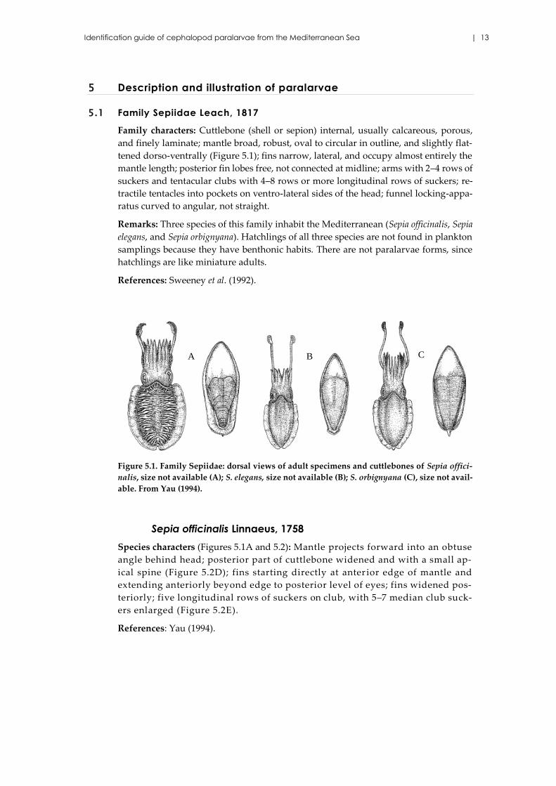
Figure 5.1. Family Sepiidae: dorsal views of adult specimens and cuttlebones of Sepia offici-
nalis, size not available (A); S. elegans, size not available (B); S. orbignyana (C), size not avail-
able. From Yau (1994).
Sepia officinalis Linnaeus, 1758
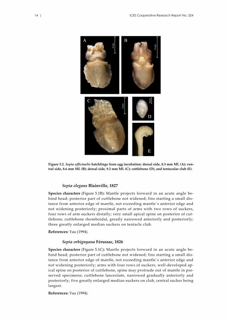
Species characters (Figures 5.1A and 5.2): Mantle projects forward into an obtuse
angle behind head; posterior part of cuttlebone widened and with a small ap-
ical spine (Figure 5.2D); fins starting directly at anter ior edge of mantle and
extending anteriorly beyond edge to posterior level of eyes; fins widened pos-
teriorly; five longitudinal rows of suckers on club, with 5–7 median club suck-
ers enlarged (Figure 5.2E).
References: Yau (1994).
A B C
14 | ICES Cooperative Research Report No. 324
Figure 5.2. Sepia officinalis hatchlings from egg incubation: dorsal side, 8.3 mm ML (A); ven-
tral side, 8.6 mm ML (B); dorsal side, 9.2 mm ML (C); cuttlebone (D), and tentacular club (E).
Sepia elegans Blainville, 1827
Species characters (Figure 5.1B): Mantle projects forward in an acute angle be-
hind head; posterior part of cuttlebone not widened; fins starting a small dis-
tance from anterior edge of mantle, not exceeding mantle´s anterior edge and
not widening posteriorly; proximal parts of arms with two rows of suckers,
four rows of arm suckers distally; very small apical spine on posterior of cut-
tlebone; cuttlebone rhomboidal, greatly narrowed anteriorly and posteriorly;
three greatly enlarged median suckers on tentacle club.
References: Yau (1994).
Sepia orbignyana Férussac, 1826
Species characters (Figure 5.1C): Mantle projects forward in an acute angle be-
hind head; posterior part of cuttlebone not widened; fins starting a small dis-
tance from anterior edge of mantle, not exceeding mantle´s anterior edge and
not widening posteriorly; arms with four rows of suckers, well-developed ap-
ical spine on posterior of cuttlebone, spine may protrude out of mantle in pre-
served specimens; cuttlebone lanceolate, narrowed gradually anteriorly and
posteriorly; five greatly enlarged median suckers on club, central sucker being
largest.
References: Yau (1994).
Identification guide of cephalopod paralarvae from the Mediterranean Sea | 15
5.2 Family Sepiolidae Leach, 1817
Family characters: Mantle short, broad, sac-like, rounded posteriorly; fins large, round,
separated; funnel locking-cartilage simple, straight; shell absent or reduced to a chitin-
ous gladius; eye covered by transparent skin.
Remarks: The characters used here for most sepiolid paralarvae descriptions are those
of juveniles or adults.
References: Sweeney et al. (1992); Nesis (1999).
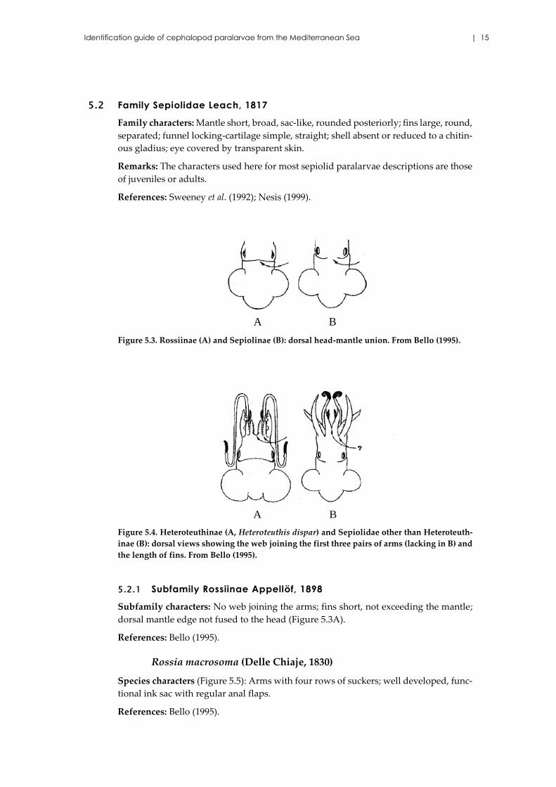
Figure 5.3. Rossiinae (A) and Sepiolinae (B): dorsal head-mantle union. From Bello (1995).
Figure 5.4. Heteroteuthinae (A, Heteroteuthis dispar) and Sepiolidae other than Heteroteuth-
inae (B): dorsal views showing the web joining the first three pairs of arms (lacking in B) and
the length of fins. From Bello (1995).
5.2.1 Subfamily Rossiinae Appellöf, 1898
Subfamily characters: No web joining the arms; fins short, not exceeding the mantle;
dorsal mantle edge not fused to the head (Figure 5.3A).
References: Bello (1995).
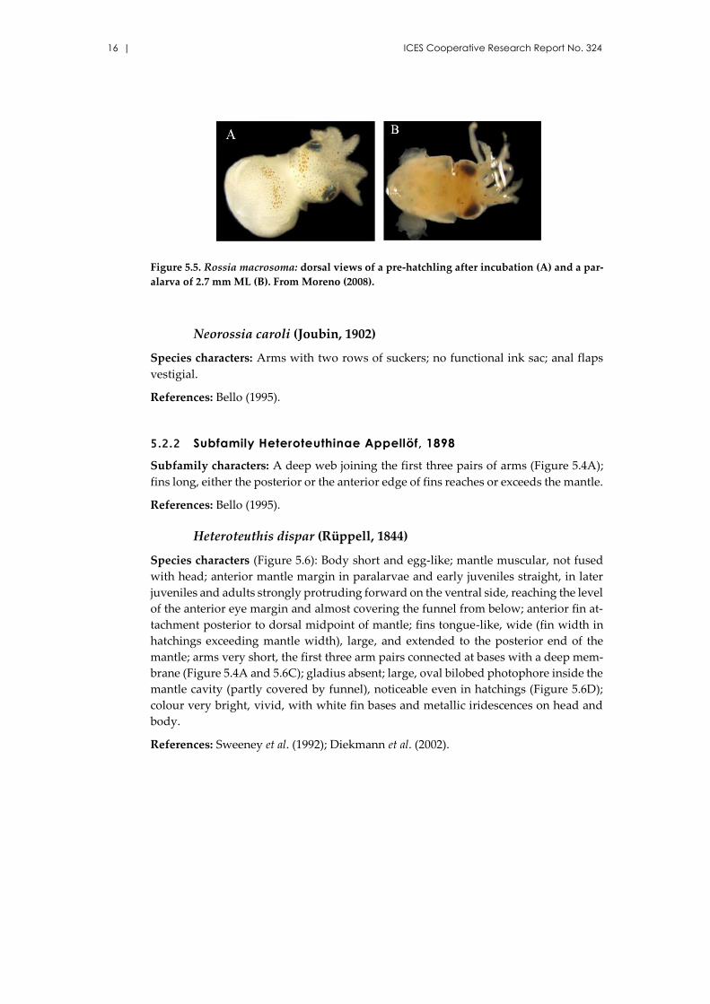
Rossia macrosoma (Delle Chiaje, 1830)
Species characters (Figure 5.5): Arms with four rows of suckers; well developed, func-
tional ink sac with regular anal flaps.
References: Bello (1995).
A B
A B
16 | ICES Cooperative Research Report No. 324
Figure 5.5. Rossia macrosoma: dorsal views of a pre-hatchling after incubation (A) and a par-
alarva of 2.7 mm ML (B). From Moreno (2008).
Neorossia caroli (Joubin, 1902)
Species characters: Arms with two rows of suckers; no functional ink sac; anal flaps
vestigial.
References: Bello (1995).
5.2.2 Subfamily Heteroteuthinae Appellöf, 1898
Subfamily characters: A deep web joining the first three pairs of arms (Figure 5.4A);
fins long, either the posterior or the anterior edge of fins reaches or exceeds the mantle.
References: Bello (1995).
Heteroteuthis dispar (Rüppell, 1844)
Species characters (Figure 5.6): Body short and egg-like; mantle muscular, not fused
with head; anterior mantle margin in paralarvae and early juveniles straight, in later
juveniles and adults strongly protruding forward on the ventral side, reaching the level
of the anterior eye margin and almost covering the funnel from below; anterior fin at-
tachment posterior to dorsal midpoint of mantle; fins tongue-like, wide (fin width in
hatchings exceeding mantle width), large, and extended to the posterior end of the
mantle; arms very short, the first three arm pairs connected at bases with a deep mem-
brane (Figure 5.4A and 5.6C); gladius absent; large, oval bilobed photophore inside the
mantle cavity (partly covered by funnel), noticeable even in hatchings (Figure 5.6D);
colour very bright, vivid, with white fin bases and metallic iridescences on head and
body.
References: Sweeney et al. (1992); Diekmann et al. (2002).
Identification guide of cephalopod paralarvae from the Mediterranean Sea | 17
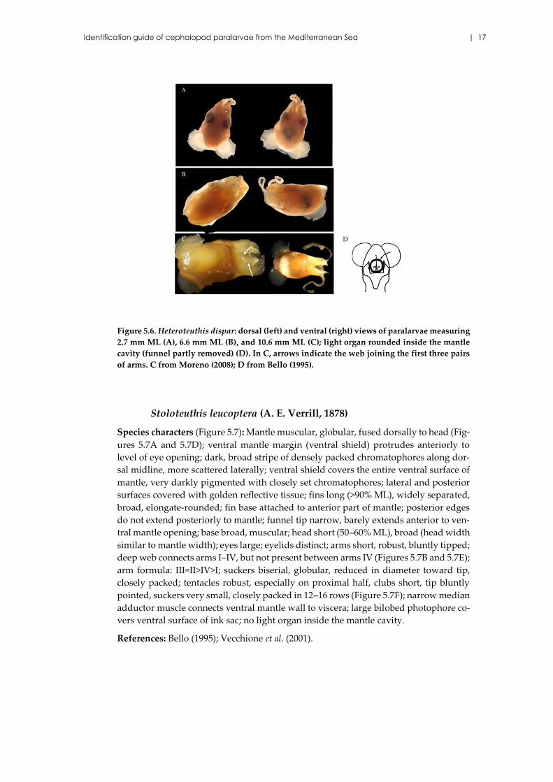
Figure 5.6. Heteroteuthis dispar: dorsal (left) and ventral (right) views of paralarvae measuring
2.7 mm ML (A), 6.6 mm ML (B), and 10.6 mm ML (C); light organ rounded inside the mantle
cavity (funnel partly removed) (D). In C, arrows indicate the web joining the first three pairs
of arms. C from Moreno (2008); D from Bello (1995).
Stoloteuthis leucoptera (A. E. Verrill, 1878)
Species characters (Figure 5.7): Mantle muscular, globular, fused dorsally to head (Fig-
ures 5.7A and 5.7D); ventral mantle margin (ventral shield) protrudes anteriorly to
level of eye opening; dark, broad stripe of densely packed chromatophores along dor-
sal midline, more scattered laterally; ventral shield covers the entire ventral surface of
mantle, very darkly pigmented with closely set chromatophores; lateral and posterior
surfaces covered with golden reflective tissue; fins long (>90% ML), widely separated,
broad, elongate-rounded; fin base attached to anterior part of mantle; posterior edges
do not extend posteriorly to mantle; funnel tip narrow, barely extends anterior to ven-
tral mantle opening; base broad, muscular; head short (50–60% ML), broad (head width
similar to mantle width); eyes large; eyelids distinct; arms short, robust, bluntly tipped;
deep web connects arms I–IV, but not present between arms IV (Figures 5.7B and 5.7E);
arm formula: III=II>IV>I; suckers biserial, globular, reduced in diameter toward tip,
closely packed; tentacles robust, especially on proximal half, clubs short, tip bluntly
pointed, suckers very small, closely packed in 12–16 rows (Figure 5.7F); narrow median
adductor muscle connects ventral mantle wall to viscera; large bilobed photophore co-
vers ventral surface of ink sac; no light organ inside the mantle cavity.
References: Bello (1995); Vecchione et al. (2001).
18 | ICES Cooperative Research Report No. 324
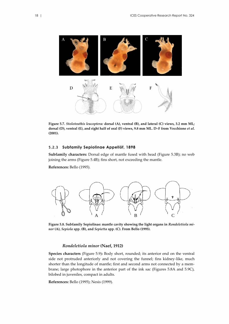
Figure 5.7. Stoloteuthis leucoptera: dorsal (A), ventral (B), and lateral (C) views, 3.2 mm ML;
dorsal (D), ventral (E), and right half of oral (F) views, 9.8 mm ML. D–F from Vecchione et al.
(2001).
5.2.3 Subfamily Sepiolinae Appellöf, 1898
Subfamily characters: Dorsal edge of mantle fused with head (Figure 5.3B); no web
joining the arms (Figure 5.4B); fins short, not exceeding the mantle.
References: Bello (1995).
Figure 5.8. Subfamily Sepiolinae: mantle cavity showing the light organs in Rondeletiola mi-
nor (A), Sepiola spp. (B), and Sepietta spp. (C). From Bello (1995).
Rondeletiola minor (Naef, 1912)
Species characters (Figure 5.9): Body short, rounded; its anterior end on the ventral
side not protruded anteriorly and not covering the funnel; fins kidney-like, much
shorter than the longitude of mantle; first and second arms not connected by a mem-
brane; large photophore in the anterior part of the ink sac (Figures 5.8A and 5.9C),
bilobed in juveniles, compact in adults.
References: Bello (1995); Nesis (1999).
A CB
Identification guide of cephalopod paralarvae from the Mediterranean Sea | 19
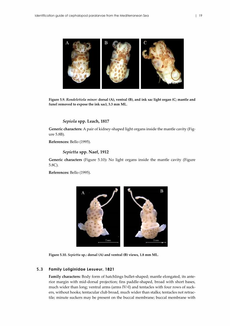
Figure 5.9. Rondeletiola minor: dorsal (A), ventral (B), and ink sac light organ (C; mantle and
funel removed to expose the ink sac), 3.3 mm ML.
Sepiola spp. Leach, 1817
Generic characters: A pair of kidney-shaped light organs inside the mantle cavity (Fig-
ure 5.8B).
References: Bello (1995).
Sepietta spp. Naef, 1912
Generic characters (Figure 5.10): No light organs inside the mantle cavity (Figure
5.8C).
References: Bello (1995).
Figure 5.10. Sepietta sp.: dorsal (A) and ventral (B) views, 1.8 mm ML.
5.3 Family Loliginidae Lesueur, 1821
Family characters: Body form of hatchlings bullet-shaped; mantle elongated, its ante-
rior margin with mid-dorsal projection; fins paddle-shaped, broad with short bases,
much wider than long; ventral arms (arms IV>I) and tentacles with four rows of suck-
ers, without hooks; tentacular club broad, much wider than stalks; tentacles not retrac-
tile; minute suckers may be present on the buccal membrane; buccal membrane with
20 | ICES Cooperative Research Report No. 324
eight lappets, connectives to arms IV attached ventrally; funnel cartilage straight, elon-
gated; gladius feather-like, with short free rachis; in some species, two (rarely one)
photophores on ink sac; head squarish; eyes covered by transparent corneal membrane
with only a minute pore at its anterior end; the number of chromatophores decreases
from the ventral to dorsal side.
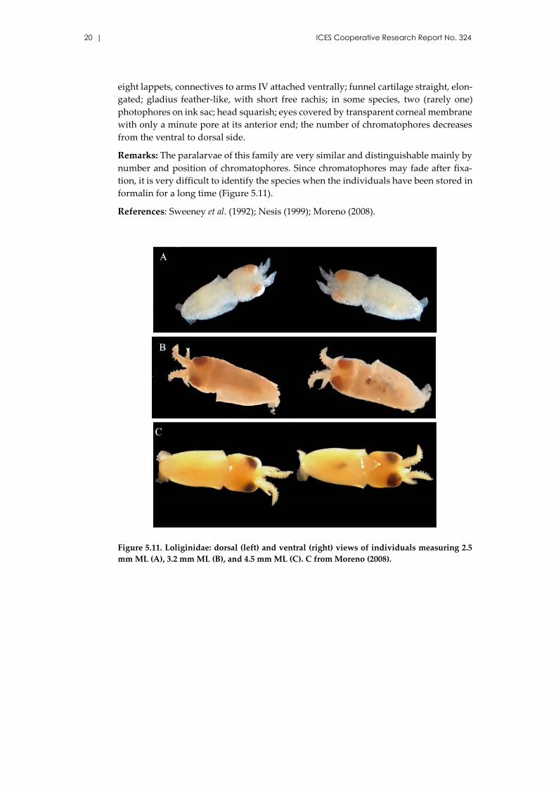
Remarks: The paralarvae of this family are very similar and distinguishable mainly by
number and position of chromatophores. Since chromatophores may fade after fixa-
tion, it is very difficult to identify the species when the individuals have been stored in
formalin for a long time (Figure 5.11).
References: Sweeney et al. (1992); Nesis (1999); Moreno (2008).
Figure 5.11. Loliginidae: dorsal (left) and ventral (right) views of individuals measuring 2.5
mm ML (A), 3.2 mm ML (B), and 4.5 mm ML (C). C from Moreno (2008).
Identification guide of cephalopod paralarvae from the Mediterranean Sea | 21
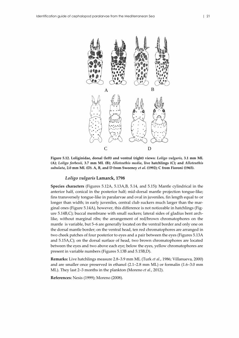
Figure 5.12. Loliginidae, dorsal (left) and ventral (right) views: Loligo vulgaris, 3.1 mm ML
(A); Loligo forbesii, 3.7 mm ML (B); Alloteuthis media, live hatchlings (C); and Alloteuthis
subulata, 2.0 mm ML (D). A, B, and D from Sweeney et al. (1992); C from Fioroni (1965).
Loligo vulgaris Lamarck, 1798
Species characters (Figures 5.12A, 5.13A,B, 5.14, and 5.15): Mantle cylindrical in the
anterior half, conical in the posterior half; mid-dorsal mantle projection tongue-like;
fins transversely tongue-like in paralarvae and oval in juveniles, fin length equal to or
longer than width; in early juveniles, central club suckers much larger than the mar-
ginal ones (Figure 5.14A), however, this difference is not noticeable in hatchlings (Fig-
ure 5.14B,C); buccal membrane with small suckers; lateral sides of gladius bent arch-
like, without marginal ribs; the arrangement of red/brown chromatophores on the
mantle is variable, but 5–6 are generally located on the ventral border and only one on
the dorsal mantle border; on the ventral head, ten red chromatophores are arranged in
two cheek patches of four posterior to eyes and a pair between the eyes (Figures 5.13A
and 5.15A,C); on the dorsal surface of head, two brown chromatophores are located
between the eyes and two above each eye; below the eyes, yellow chromatophores are
present in variable numbers (Figures 5.13B and 5.15B,D).
Remarks: Live hatchlings measure 2.8–3.9 mm ML (Turk et al., 1986; Villanueva, 2000)
and are smaller once preserved in ethanol (2.1–2.8 mm ML) or formalin (1.6–3.0 mm
ML). They last 2–3 months in the plankton (Moreno et al., 2012).
References: Nesis (1999); Moreno (2008).
D C
BA
22 | ICES Cooperative Research Report No. 324
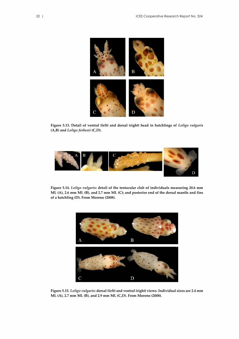
Figure 5.13. Detail of ventral (left) and dorsal (right) head in hatchlings of Loligo vulgaris
(A,B) and Loligo forbesii (C,D).
Figure 5.14. Loligo vulgaris: detail of the tentacular club of individuals measuring 20.6 mm
ML (A), 2.6 mm ML (B), and 2.7 mm ML (C); and posterior end of the dorsal mantle and fins
of a hatchling (D). From Moreno (2008).
Figure 5.15. Loligo vulgaris: dorsal (left) and ventral (right) views. Individual sizes are 2.4 mm
ML (A), 2.7 mm ML (B), and 2.9 mm ML (C,D). From Moreno (2008).
Identification guide of cephalopod paralarvae from the Mediterranean Sea | 23
Loligo forbesii Steenstrup, 1857
Species characters (Figure 5.12B and 5.13C,D): Body form bullet-shaped with well-de-
veloped, paddle-shape terminal fins; mantle with few large dorsal chromatophores
and numerous ventral chromatophores; the arrangement of chromatophores in hatch-
lings is similar to L. vulgaris, but in higher numbers in each body part; e.g. on the ven-
tral surface of head, three red chromatophores are located on each arm IV (Figure
5.13C).
Remarks: Hatchling sizes range from 3.5–4.1 mm ML (mean 3.7 mm ML).
References: Sweeney et al. (1992); Moreno (2008); Yau (1994).
Alloteuthis spp. Wülker, 1920
Generic characters (Figure 5.12C,D): Juveniles have bullet-shaped body; paddle-
shaped terminal fins that form a simple point at the tip at this stage, which develops
into a tail in the subadult stage; in early juveniles, the median rows of suckers of the
tentacle club are three–four fold larger than marginal suckers; however, such a differ-
ence in sucker size is not noticeable in hatchlings.
Remarks: Juvenile stages are nearly indistinguishable from Loligo spp., but hatchlings
show a different chromatophore arrangement with a greater number of yellow chro-
matophores than L. forbesii. Two species of this genus (Alloteuthis media and A. subulata)
inhabit the Mediterranean (Bello, 2008). Hatchlings measure 2.0–2.8 mm ML and last
15–30 d in the plankton.
References: Yau (1994); Hastie et al. (2013).
5.4 Family Chtenopterygidae Grimpe, 1922
Since this family is monotypic, family/species characters are described for the only cur-
rently known species.
Chtenopteryx sicula (Vérany, 1851)
Species characters (Figures 5.16 and 5.17): mantle short, wide, slightly depressed
dorso-ventrally, rounded behind; head short and half-retractable (up the midline of
eye); eyes small, widely separated; funnel straight, large; fins separate dorsally, fringe
mantle laterally; hatchlings with transversely elongate fins, result of first trabeculae;
fins clearly with muscular trabeculae (at 3.5 mm ML); fins length increase with size,
but much shorter than mantle, consisting of a series of flexible muscular ribs joined by
thin transparent membrane; arms short, arms IV longer and wider than others; arms
suckers small, in two rows in proximal, 4–6 rows in distal part of arms I-III, 1–2 zig-
zag rows on arms IV; tentacles short and robust with broad, oval clubs with suckers
forming a distinct circular pad (at <4 mm ML), and the sucker surface directed towards
the front (already visible in hatching ~1 mm ML; Figure 5.16A–C); pointed tip develops
and becomes dactylus (at >4 mm ML), equal length of manus (at 6 mm ML); buccal
membrane with seven lappets, with 12–15 minute suckers in two rows on lappets, con-
nectives to arms IV attached ventrally; gladius with long rachis and wide vane, without
end conus; a single chromatophore occurs on the aboral surface of the club (at ≥2 mm
ML); photophores on eye-ball (one elongated) and ink sac (large, round, resembling a
fried egg on a black frying pan); ink sac photophore appearing in late paralarvae.
24 | ICES Cooperative Research Report No. 324
Individuals ca. 2.0 mm ML (Figure 5.16D,E): Mantle large, muscular, broad, tapers to
a point posteriorly; mantle much longer than head and arms combined; head dorso-
ventrally compressed; eyes prominent, tubular, subspherical; slightly dorso-ventrally
elongate; funnel large, robust, muscular, extends to level of mid-eye; fins minute, ter-
minal flaps; arms IV longest; arms II and III subequal, very short, stubby at bases; sud-
den attenuate papilla-like tips; 2–3 suckers; arms I are minute papillae, just developing;
clubs terminal, broad, round, nearly equal in diameter to eyes, with about 25 suckers
in round cluster of 5–6 suckers across; digestive gland subspherical; ink sac well-de-
veloped (Figure 5.16F), spherical, with concentration of bronze reflective tissue on ven-
tral surface (precursor to photophore).
Individuals ca. 3.5 mm ML (Figure 5.17A-E): Head very short; eyes tubular, directed
antero-laterally, slightly elongate dorso-ventrally; funnel very large, robust; extends
anterior to base of arms; fins very small, with short bases; fins extend well posterior to
mantle tip, about 10 (at 3.2 mm ML) to 12 (at >3.5 mm ML) short muscular supports
(ribs) extend from muscular bases, connected by thin, easily torn membrane, giving
comb-like appearance to fins; arms short with attenuate tips and few very small suck-
ers; tentacle stalks, longer than arms; clubs terminal, expanded, round, with about 20
suckers and a papilla-like dactylus; digestive gland globular, slightly elongate dorso-
ventrally; photophore anlage a round, reflective, bronze patch at ventral tip, ringed
with black.
Remarks: The species is easily identifiable at all developmental stages by its distinctive
clubs and the typical ribbed fins made of muscular supports joined by a thin mem-
brane.
References: Sweeney et al. (1992); Nesis (1999); Diekmann et al. (2002); Haimovici et al.
(2002); Moreno (2008).
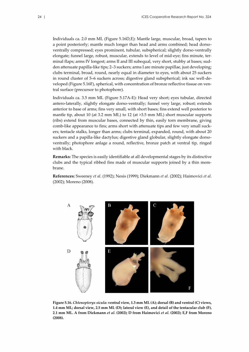
Figure 5.16. Chtenopteryx sicula: ventral view, 1.3 mm ML (A); dorsal (B) and ventral (C) views,
1.4 mm ML; dorsal view, 2.5 mm ML (D); lateral view (E), and detail of the tentacular club (F),
2.1 mm ML. A from Diekmann et al. (2002); D from Haimovici et al. (2002); E,F from Moreno
(2008).
Identification guide of cephalopod paralarvae from the Mediterranean Sea | 25
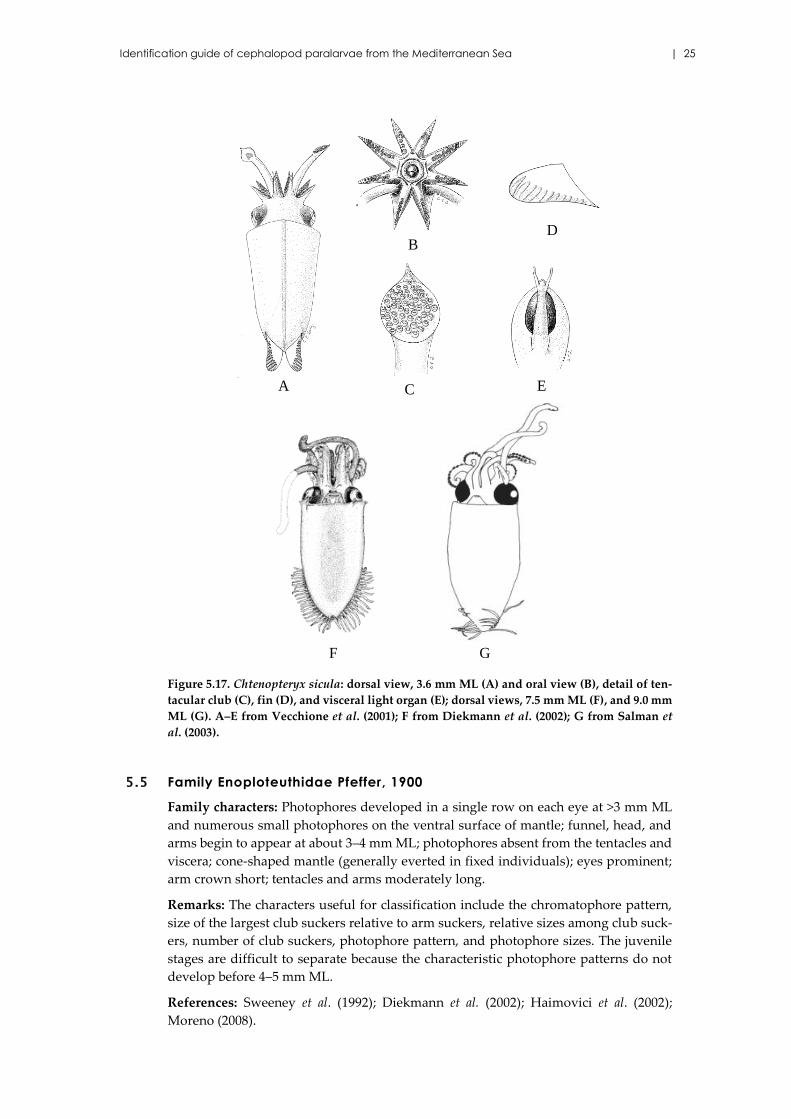
Figure 5.17. Chtenopteryx sicula: dorsal view, 3.6 mm ML (A) and oral view (B), detail of ten-
tacular club (C), fin (D), and visceral light organ (E); dorsal views, 7.5 mm ML (F), and 9.0 mm
ML (G). A–E from Vecchione et al. (2001); F from Diekmann et al. (2002); G from Salman et
al. (2003).
5.5 Family Enoploteuthidae Pfeffer, 1900
Family characters: Photophores developed in a single row on each eye at >3 mm ML
and numerous small photophores on the ventral surface of mantle; funnel, head, and
arms begin to appear at about 3–4 mm ML; photophores absent from the tentacles and
viscera; cone-shaped mantle (generally everted in fixed individuals); eyes prominent;
arm crown short; tentacles and arms moderately long.
Remarks: The characters useful for classification include the chromatophore pattern,
size of the largest club suckers relative to arm suckers, relative sizes among club suck-
ers, number of club suckers, photophore pattern, and photophore sizes. The juvenile
stages are difficult to separate because the characteristic photophore patterns do not
develop before 4–5 mm ML.
References: Sweeney et al. (1992); Diekmann et al. (2002); Haimovici et al. (2002);
Moreno (2008).
GF
D
C E
B
A
26 | ICES Cooperative Research Report No. 324
Abralia veranyi (Rüppell, 1844)
Species characters (Figures 5.18 and 5.19): Arms I–III long, but never as long or longer
than the ML, and attenuate, with about 14–20 suckers at <4.5 mm ML; chromatophores
on aboral surface of arms I–III; arms IV much less developed, with 6–16 suckers at <4.5
mm ML, no hook; arm formula II>I=III>IV; tentacular stalks long and robust with a row
of large aboral chromatophores; three photophores forming a single row on the ventral
surface of eyes occurring at >3.0 mm ML, anterior largest, posterior intermediate, cen-
tral smallest (Figures 5.18D,E); in juveniles, posterior photophore different from the
others; no trace of light organs in positions two or four; absence of light organs on the
arm tips (key characteristic to distinguish between Abralia and Abraliopsis).
Individuals ca. 3.0 mm ML: Club region undifferentiated, minute suckers along distal
one fourth of tentacle; a few small, integumentary photophores, evenly distributed
over ventral and ventro-lateral surface of head, mantle, and funnel, most in association
with small chromatophores; fins very small, terminal flaps; meet posteriorly.
Individuals ca. 4.5 mm ML: Carpus with 4–5 suckers, four rows of suckers on manus
with 6–8 median suckers enlarged and no hook development; head narrower than
mantle opening; seven large dark chromatophores on dorsal and lateral surface of
head; small photophores in longitudinal rows on ventral and lateral surface of head;
one row extends into arms IV; funnel strongly developed, extends to the level of pos-
terior edge of eye; six small photophores on ventral surface of funnel; mantle elongated
and muscular, with broad opening that tapers to blunt posterior end; many small chro-
matophores evenly distributed over ventral and ventro-lateral surface of mantle; large
chromatophores in bands around mantle, corresponding to photophores on ventral
and lateral surfaces; very large chromatophore on each postero-lateral end of mantle
ventral, to posterior part of fins; fins terminal, muscular, short, triangular with rounded
angles and meet at posterior end of mantle.
Remarks: Late juveniles and adults are characterized by one series of hooks and two
series of suckers on the manus club. Hooks are not yet formed in paralarvae.
References: Vecchione et al. (2001); Haimovici et al. (2002); Moreno (2008).
Identification guide of cephalopod paralarvae from the Mediterranean Sea | 27
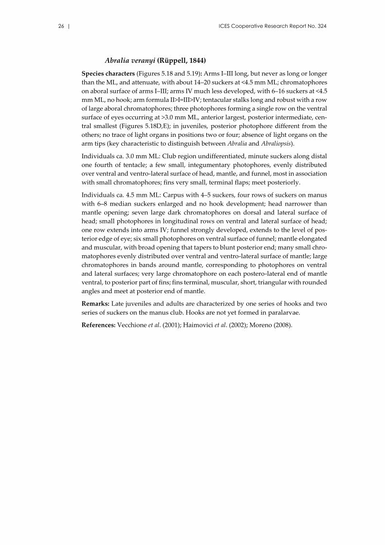
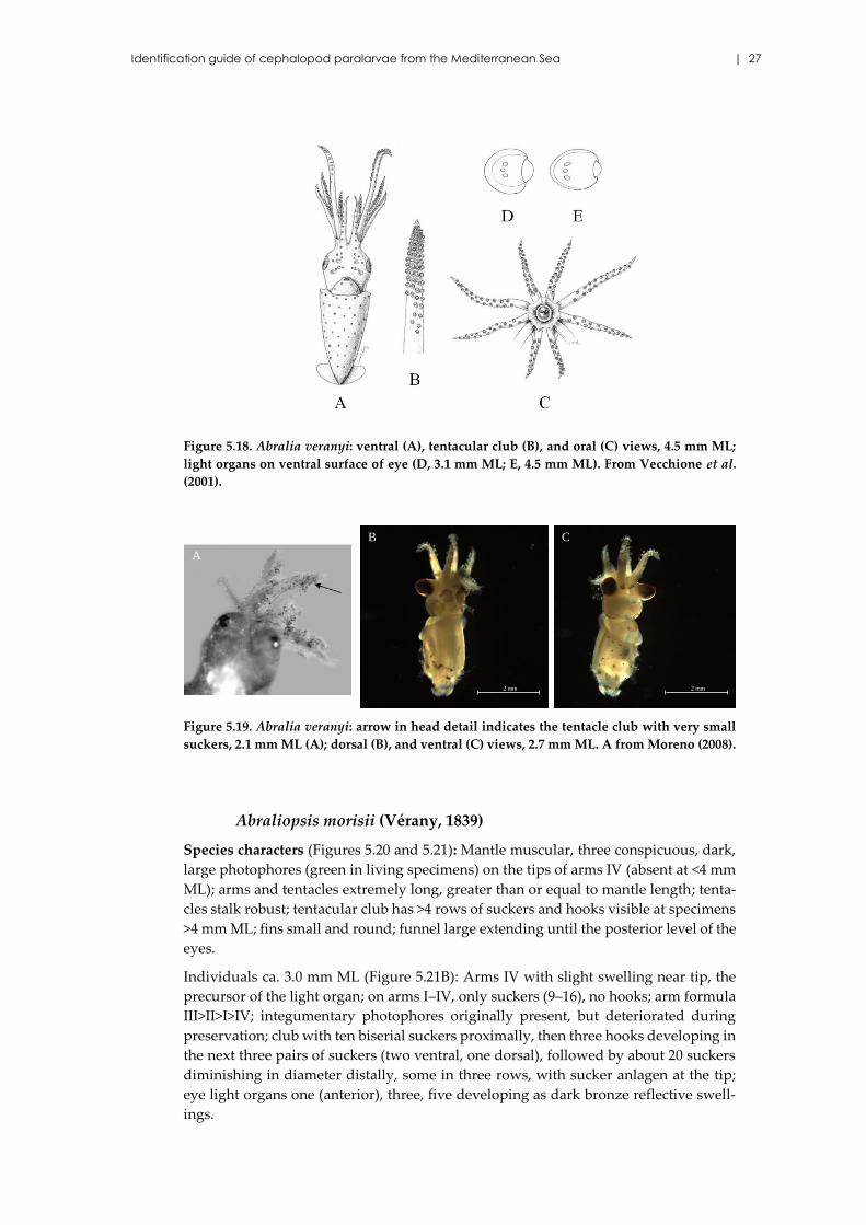
Figure 5.18. Abralia veranyi: ventral (A), tentacular club (B), and oral (C) views, 4.5 mm ML;
light organs on ventral surface of eye (D, 3.1 mm ML; E, 4.5 mm ML). From Vecchione et al.
(2001).
Figure 5.19. Abralia veranyi: arrow in head detail indicates the tentacle club with very small
suckers, 2.1 mm ML (A); dorsal (B), and ventral (C) views, 2.7 mm ML. A from Moreno (2008).
Abraliopsis morisii (Vérany, 1839)
Species characters (Figures 5.20 and 5.21): Mantle muscular, three conspicuous, dark,
large photophores (green in living specimens) on the tips of arms IV (absent at <4 mm
ML); arms and tentacles extremely long, greater than or equal to mantle length; tenta-
cles stalk robust; tentacular club has >4 rows of suckers and hooks visible at specimens
>4 mm ML; fins small and round; funnel large extending until the posterior level of the
eyes.
Individuals ca. 3.0 mm ML (Figure 5.21B): Arms IV with slight swelling near tip, the
precursor of the light organ; on arms I–IV, only suckers (9–16), no hooks; arm formula
III>II>I>IV; integumentary photophores originally present, but deteriorated during
preservation; club with ten biserial suckers proximally, then three hooks developing in
the next three pairs of suckers (two ventral, one dorsal), followed by about 20 suckers
diminishing in diameter distally, some in three rows, with sucker anlagen at the tip;
eye light organs one (anterior), three, five developing as dark bronze reflective swell-
ings.
A
2 mm
CB
2 mm
28 | ICES Cooperative Research Report No. 324
Individuals ca. 5.0 mm ML: Arms IV with two small terminal light organs and median
row of photophores on proximal half, but hooks absent; four hooks present on arms I,
seven hooks on arms II, nine hooks on arms III; photophores absent on arms I–III; three
rows of photophores on head, row on ventral midline most conspicuous; eye photo-
phores one (anterior), three, and five developed, two and four anlagen present.
Individuals ca. 13.0 mm ML (Figure 5.20C): Arms IV with three terminal light organs,
two rows of photophores that extend to one half of arm length, and 16 hooks; arms I
with 13 hooks, II with 17 hooks and no photophores; arm formula IV>III>II>I; tentacu-
lar club with four large hooks in ventral row and five small hooks in dorsal row (Figure
5.20D), carpus with eight suckers, dactylus with 3–4 rows of small suckers; aboral keel
of club developing; two kinds of photophores (large, spherical, dark; and small, spher-
ical, translucent) in about nine indistinct rows on ventral surface of head; ventral pe-
riphery of eyes with five photophores in single row; one and five largest, two and four
small, three intermediate; 13 small integumentary photophores around eyelids; photo-
phores on mantle and funnel similar to those on head, kinds in indistinct rows; dense
ventrally, sparse dorsally on mantle; ventral midline devoid of photophores.
Remarks: In juveniles and adults, five round photophores of similar structure develop
on the ventral side of each eye; they form a typical row with the posterior and anterior
photophore enlarged; the manus of the club presents two series of hooks in specimens
>9 mm ML.
References: Sweeney et al. (1992); Diekmann et al. (2002); Vecchione et al (2001); Moreno
(2008).
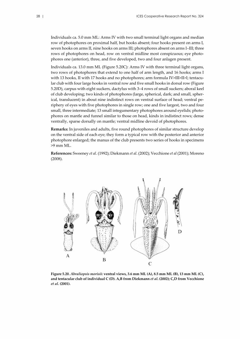
Figure 5.20. Abraliopsis morisii: ventral views, 3.6 mm ML (A), 8.5 mm ML (B), 13 mm ML (C),
and tentacular club of individual C (D). A,B from Diekmann et al. (2002); C,D from Vecchione
et al. (2001).
Identification guide of cephalopod paralarvae from the Mediterranean Sea | 29
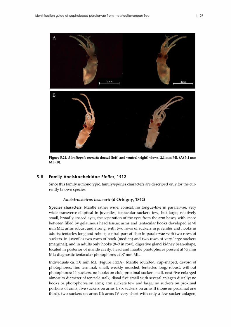
Figure 5.21. Abraliopsis morisii: dorsal (left) and ventral (right) views, 2.1 mm ML (A) 3.1 mm
ML (B).
5.6 Family Ancistrocheiridae Pfeffer, 1912
Since this family is monotypic, family/species characters are described only for the cur-
rently known species.
Ancistrocheirus lesueurii (d'Orbigny, 1842)
Species characters: Mantle rather wide, conical; fin tongue-like in paralarvae, very
wide transverse-elliptical in juveniles; tentacular suckers few, but large; relatively
small, broadly spaced eyes, the separation of the eyes from the arm bases, with space
between filled by gelatinous head tissue; arms and tentacular hooks developed at >8
mm ML; arms robust and strong, with two rows of suckers in juveniles and hooks in
adults; tentacles long and robust, central part of club in paralarvae with two rows of
suckers, in juveniles two rows of hook (median) and two rows of very large suckers
(marginal), and in adults only hooks (8–9 in row); digestive gland kidney bean-shape,
located in posterior of mantle cavity; head and mantle photophores present at >5 mm
ML; diagnostic tentacular photophores at >7 mm ML.
Individuals ca. 3.0 mm ML (Figure 5.22A): Mantle rounded, cup-shaped, devoid of
photophores; fins terminal, small, weakly muscled; tentacles long, robust, without
photophores; 11 suckers, no hooks on club, proximal sucker small, next five enlarged
almost to diameter of tentacle stalk, distal five small with several anlagen distally; no
hooks or photophores on arms; arm suckers few and large; no suckers on proximal
portions of arms; five suckers on arms I, six suckers on arms II (none on proximal one
third), two suckers on arms III; arms IV very short with only a few sucker anlagen;
30 | ICES Cooperative Research Report No. 324
photophores absent on head; eyes and buccal assemblage stalked with gelatinous ma-
terial filling spaces between stalks.
Individuals ca. 4.0 mm ML (Figures 5.22B and 5.23A): Mantle without obvious photo-
phores; fins small, rounded; tentacles long, robust, without photophores; 15 suckers on
club plus several distal anlagen, no hooks; all suckers large except proximal one and
distal two; arm formula II>I=III>>IV; arm suckers large, few in number, none on prox-
imal sections of arms; eight suckers on arms I, 12 on arms II, nine on arms III, none on
arms IV; head lacks detectable photophores; eyes and buccal assemblage stalked with
gelatinous material between stalks.
Individuals ca. 5.5 mm ML (Figures 5.22C and 5.23B–G): Mantle short, broad, bluntly
rounded posteriorly, muscular; 12 photophores on ventral surface: four along anterior
margin, four pairs that form two zig-zag rows posteriorly to the tip; fins small, elon-
gate, semilunar, posterior; funnel tubular, base broad; head broad; two rows of five
photophores on ventral surface of head in an arc from posterolateral corner to base of
arms IV; arms long, robust, attenuate; arm formula III>II>I>IV (Figure 5.23F); number
of suckers on arms: 15 (I), 16 (II), 22 (III), eight (IV); no hooks; suckers relatively large,
on long stalks; tentacles long, robust to the attenuate tip; suckers on club begin with
one small proximal sucker, set apart from the rest; manal suckers relatively large, bise-
rial proximally, enlarge gradually to maximum diameter in third–sixth pairs, then di-
minish to tip; lateral suckers larger than medial suckers; about 27 suckers in total, no
hooks (Figure 5.23G); six small spherical photophores embedded along the tentacular
stalk.
Individuals ca. 8.0 mm ML: Mantle muscular, broadest anteriorly, tapers evenly to
bluntly rounded posterior tip; 18 small, spheroidal photophores on ventral surface of
mantle in distinct pattern of transverse rows, anterior to posterior: four, two, two, four,
two, two, two; the posterior-most photophores form at the very tip as elevated knobs
(Figure 5.23H); fins rounded, triangular, terminal; funnel large, base broad, tube ex-
tends to posterior level of eyes; head large, wider than mantle; eyes prominent, no oc-
ular photophores; at least five photophores in arc on each side of ventral surface of
head; arms very long, robust, attenuate; arms I–III subequal, longer than IV; armature
on arms I: one proximal sucker, five hooks, 13+ distal suckers; arms II: 8–9 hooks, 14+
suckers; arms III: 11 hooks, 16+ suckers; arms IV: none hooks, about 20 suckers; tenta-
cles long, robust to tip; club with four pairs biserial carpal suckers; four transverse rows
with two medial hooks and two large marginal suckers each (i.e. eight hooks) on ma-
nus, 8–10 diminishing biserial suckers on dactylus; low aboral keel extends from level
of first manal row to tip of dactylus; tentacular stalk with nine small embedded sphe-
roidal photophores.
References: Sweeney et al. (1992); Nesis (1999); Vecchione et al. (2001); Moreno (2008).
Identification guide of cephalopod paralarvae from the Mediterranean Sea | 31
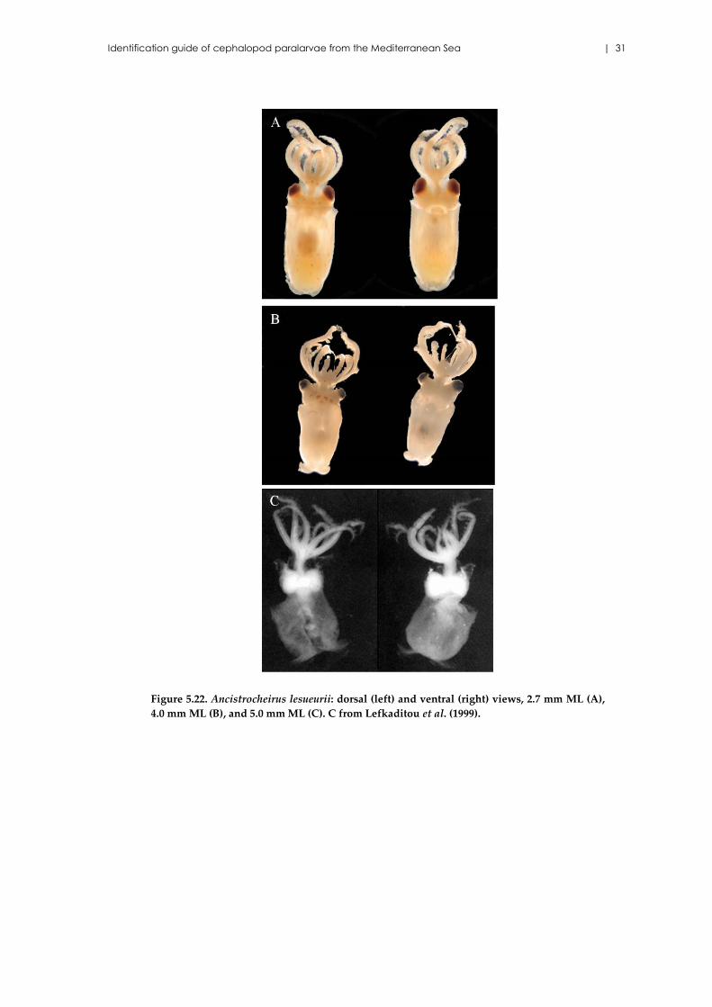
Figure 5.22. Ancistrocheirus lesueurii: dorsal (left) and ventral (right) views, 2.7 mm ML (A),
4.0 mm ML (B), and 5.0 mm ML (C). C from Lefkaditou et al. (1999).
32 | ICES Cooperative Research Report No. 324
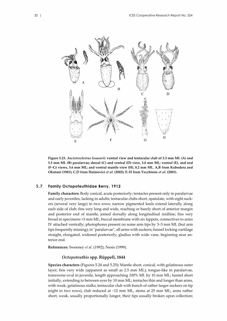
Figure 5.23. Ancistrocheirus lesueurii: ventral view and tentacular club of 3.3 mm ML (A) and
5.5 mm ML (B) paralarvae; dorsal (C) and ventral (D) view, 5.0 mm ML; ventral (E), and oral
(F–G) views, 5.6 mm ML; and ventral mantle view (H), 8.2 mm ML. A,B from Kubodera and
Okutani (1981); C,D from Haimovici et al. (2002); E–H from Vecchione et al. (2001).
5.7 Family Octopoteuthidae Berry, 1912
Family characters: Body conical, acute posteriorly; tentacles present only in paralarvae
and early juveniles, lacking in adults; tentacular clubs short, spatulate, with eight suck-
ers (several very large) in two rows; narrow pigmented keels extend laterally along
each side of club; fins very long and wide, reaching or barely short of anterior margin
and posterior end of mantle, joined dorsally along longitudinal midline; fins very
broad in specimens >3 mm ML; buccal membrane with six lappets, connectives to arms
IV attached ventrally; photophores present on some arm tips by 3–5 mm ML (but arm
tips frequently missing); in "paralarvae", all arms with suckers; funnel locking-cartilage
straight, elongated, widened posteriorly; gladius with wide vane, beginning near an-
terior end.
References: Sweeney et al. (1992); Nesis (1999).
Octopoteuthis spp. Rüppell, 1844
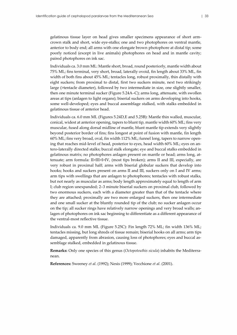
Species characters (Figures 5.24 and 5.25): Mantle short, conical, with gelatinous outer
layer; fins very wide (apparent as small as 2.5 mm ML), tongue-like in paralarvae,
transverse-oval in juvenile, length approaching 100% ML by 10 mm ML; funnel short
initially, extending to between eyes by 10 mm ML; tentacles thin and longer than arms,
with weak, gelatinous stalks; tentacular club with bunch of rather larger suckers on tip
(eight in two rows); club reduced at ~12 mm ML, stems at 25 mm ML; arms rather
short, weak, usually proportionally longer, their tips usually broken upon collection;
Identification guide of cephalopod paralarvae from the Mediterranean Sea | 33
gelatinous tissue layer on head gives smaller specimens appearance of short arm-
crown stalk and short, wide eye-stalks; one and two photophores on ventral mantle,
anterior to body end; all arms with one elongate brown photophore at distal tip; some
poorly noticed (except in live animals) photophores on head and in mantle cavity;
paired photophores on ink sac.
Individuals ca. 3.0 mm ML: Mantle short, broad, round posteriorly, mantle width about
75% ML; fins terminal, very short, broad, laterally ovoid, fin length about 33% ML, fin
width of both fins about 45% ML; tentacles long, robust proximally, thin distally with
eight suckers; from proximal to distal, first two suckers minute, next two strikingly
large (>tentacle diameter), followed by two intermediate in size, one slightly smaller,
then one minute terminal sucker (Figure 5.24A–C); arms long, attenuate, with swollen
areas at tips (anlagen to light organs); biserial suckers on arms developing into hooks,
some well-developed; eyes and buccal assemblage stalked, with stalks embedded in
gelatinous tissue of anterior head.
Individuals ca. 6.0 mm ML (Figures 5.24D,E and 5.25B): Mantle thin walled, muscular,
conical, widest at anterior opening, tapers to blunt tip, mantle width 60% ML; fins very
muscular, fused along dorsal midline of mantle, blunt mantle tip extends very slightly
beyond posterior border of fins; fins longest at point of fusion with mantle, fin length
60% ML; fins very broad, oval, fin width 112% ML; funnel long, tapers to narrow open-
ing that reaches mid-level of head, posterior to eyes, head width 60% ML; eyes on an-
tero-laterally directed stalks; buccal stalk elongate; eye and buccal stalks embedded in
gelatinous matrix; no photophores anlagen present on mantle or head; arms long, at-
tenuate; arm formula: II>III>I>IV, (most tips broken); arms II and III, especially, are
very robust in proximal half; arms with biserial globular suckers that develop into
hooks; hooks and suckers present on arms II and III, suckers only on I and IV arms;
arm tips with swellings that are anlagen to photophores; tentacles with robust stalks,
but not nearly as muscular as arms; body length approximately equal to length of arm
I; club region unexpanded; 2–3 minute biserial suckers on proximal club, followed by
two enormous suckers, each with a diameter greater than that of the tentacle where
they are attached; proximally are two more enlarged suckers, then one intermediate
and one small sucker at the bluntly rounded tip of the club; no sucker anlagen occur
on the tip; all sucker rings have relatively narrow openings and very broad walls; an-
lagen of photophores on ink sac beginning to differentiate as a different appearance of
the ventral-most reflective tissue.
Individuals ca. 9.0 mm ML (Figure 5.25C): Fin length 72% ML; fin width 136% ML;
tentacles missing, but long shreds of tissue remain; biserial hooks on all arms; arm tips
damaged, apparently from abrasion, causing loss of photophores; eyes and buccal as-
semblage stalked, embedded in gelatinous tissue.
Remarks: Only one species of this genus (Octopoteuthis sicula) inhabits the Mediterra-
nean.
References: Sweeney et al. (1992); Nesis (1999); Vecchione et al. (2001).
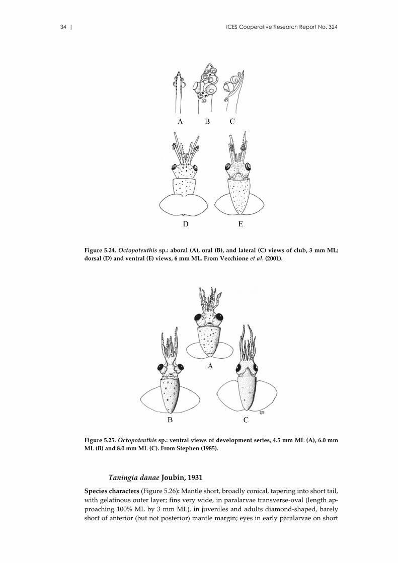
34 | ICES Cooperative Research Report No. 324
Figure 5.24. Octopoteuthis sp.: aboral (A), oral (B), and lateral (C) views of club, 3 mm ML;
dorsal (D) and ventral (E) views, 6 mm ML. From Vecchione et al. (2001).
Figure 5.25. Octopoteuthis sp.: ventral views of development series, 4.5 mm ML (A), 6.0 mm
ML (B) and 8.0 mm ML (C). From Stephen (1985).
Taningia danae Joubin, 1931
Species characters (Figure 5.26): Mantle short, broadly conical, tapering into short tail,
with gelatinous outer layer; fins very wide, in paralarvae transverse-oval (length ap-
proaching 100% ML by 3 mm ML), in juveniles and adults diamond-shaped, barely
short of anterior (but not posterior) mantle margin; eyes in early paralarvae on short
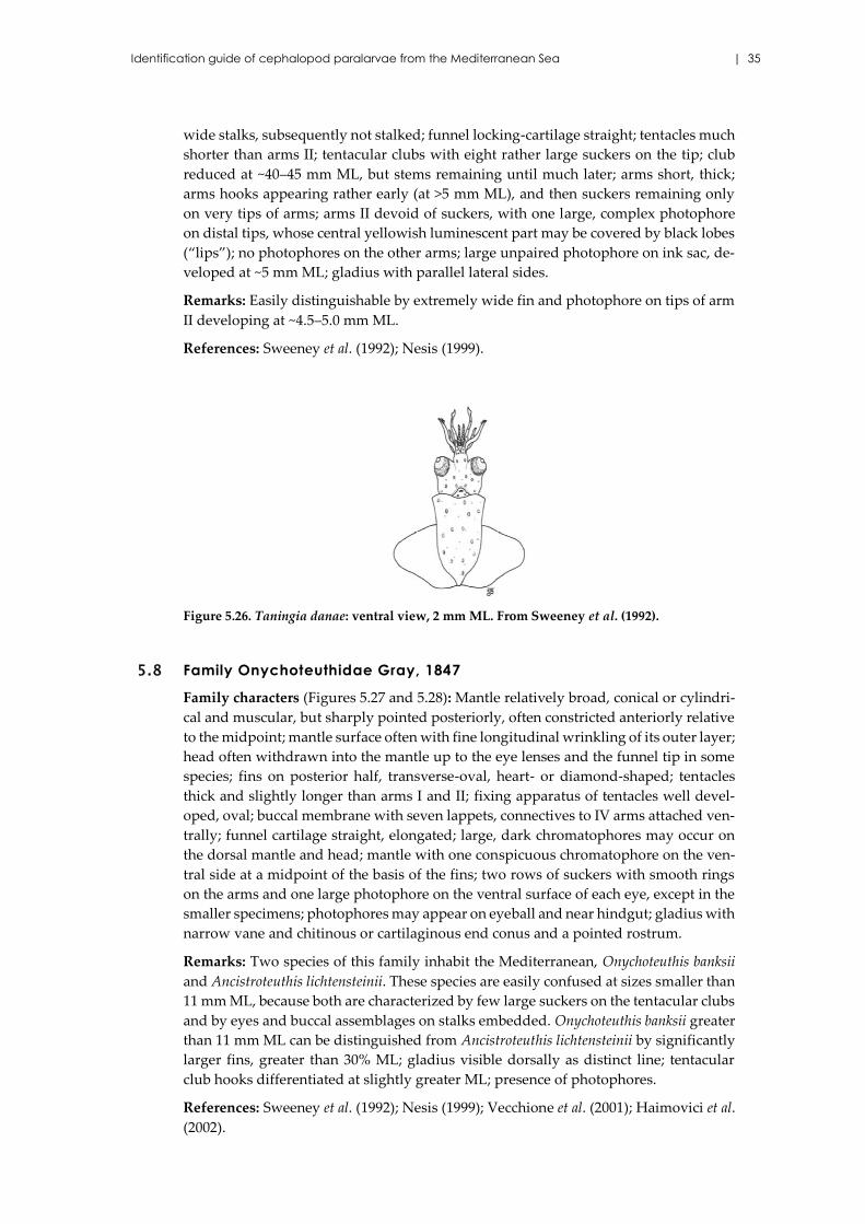
Identification guide of cephalopod paralarvae from the Mediterranean Sea | 35
wide stalks, subsequently not stalked; funnel locking-cartilage straight; tentacles much
shorter than arms II; tentacular clubs with eight rather large suckers on the tip; club
reduced at ~40–45 mm ML, but stems remaining until much later; arms short, thick;
arms hooks appearing rather early (at >5 mm ML), and then suckers remaining only
on very tips of arms; arms II devoid of suckers, with one large, complex photophore
on distal tips, whose central yellowish luminescent part may be covered by black lobes
(“lips”); no photophores on the other arms; large unpaired photophore on ink sac, de-
veloped at ~5 mm ML; gladius with parallel lateral sides.
Remarks: Easily distinguishable by extremely wide fin and photophore on tips of arm
II developing at ~4.5–5.0 mm ML.
References: Sweeney et al. (1992); Nesis (1999).
Figure 5.26. Taningia danae: ventral view, 2 mm ML. From Sweeney et al. (1992).
5.8 Family Onychoteuthidae Gray, 1847
Family characters (Figures 5.27 and 5.28): Mantle relatively broad, conical or cylindri-
cal and muscular, but sharply pointed posteriorly, often constricted anteriorly relative
to the midpoint; mantle surface often with fine longitudinal wrinkling of its outer layer;
head often withdrawn into the mantle up to the eye lenses and the funnel tip in some
species; fins on posterior half, transverse-oval, heart- or diamond-shaped; tentacles
thick and slightly longer than arms I and II; fixing apparatus of tentacles well devel-
oped, oval; buccal membrane with seven lappets, connectives to IV arms attached ven-
trally; funnel cartilage straight, elongated; large, dark chromatophores may occur on
the dorsal mantle and head; mantle with one conspicuous chromatophore on the ven-
tral side at a midpoint of the basis of the fins; two rows of suckers with smooth rings
on the arms and one large photophore on the ventral surface of each eye, except in the
smaller specimens; photophores may appear on eyeball and near hindgut; gladius with
narrow vane and chitinous or cartilaginous end conus and a pointed rostrum.
Remarks: Two species of this family inhabit the Mediterranean, Onychoteuthis banksii
and Ancistroteuthis lichtensteinii. These species are easily confused at sizes smaller than
11 mm ML, because both are characterized by few large suckers on the tentacular clubs
and by eyes and buccal assemblages on stalks embedded. Onychoteuthis banksii greater
than 11 mm ML can be distinguished from Ancistroteuthis lichtensteinii by significantly
larger fins, greater than 30% ML; gladius visible dorsally as distinct line; tentacular
club hooks differentiated at slightly greater ML; presence of photophores.
References: Sweeney et al. (1992); Nesis (1999); Vecchione et al. (2001); Haimovici et al.
(2002).
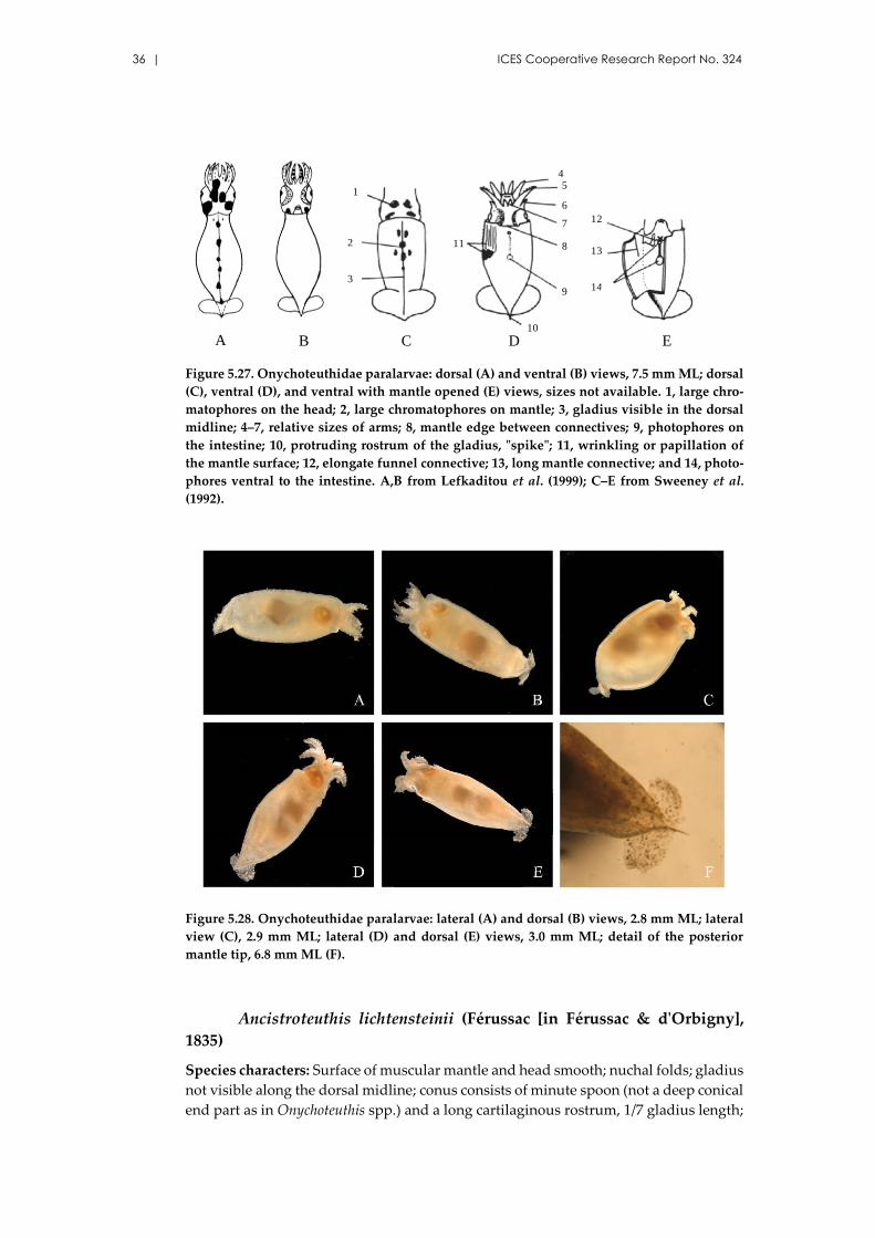
36 | ICES Cooperative Research Report No. 324
Figure 5.27. Onychoteuthidae paralarvae: dorsal (A) and ventral (B) views, 7.5 mm ML; dorsal
(C), ventral (D), and ventral with mantle opened (E) views, sizes not available. 1, large chro-
matophores on the head; 2, large chromatophores on mantle; 3, gladius visible in the dorsal
midline; 4–7, relative sizes of arms; 8, mantle edge between connectives; 9, photophores on
the intestine; 10, protruding rostrum of the gladius, "spike"; 11, wrinkling or papillation of
the mantle surface; 12, elongate funnel connective; 13, long mantle connective; and 14, photo-
phores ventral to the intestine. A,B from Lefkaditou et al. (1999); C–E from Sweeney et al.
(1992).
Figure 5.28. Onychoteuthidae paralarvae: lateral (A) and dorsal (B) views, 2.8 mm ML; lateral
view (C), 2.9 mm ML; lateral (D) and dorsal (E) views, 3.0 mm ML; detail of the posterior
mantle tip, 6.8 mm ML (F).
Ancistroteuthis lichtensteinii (Férussac [in Férussac & d'Orbigny],
1835)
Species characters: Surface of muscular mantle and head smooth; nuchal folds; gladius
not visible along the dorsal midline; conus consists of minute spoon (not a deep conical
end part as in Onychoteuthis spp.) and a long cartilaginous rostrum, 1/7 gladius length;
A B
1
2
3
C
45
6
7
8
9
10
D
12
13
14
E
11
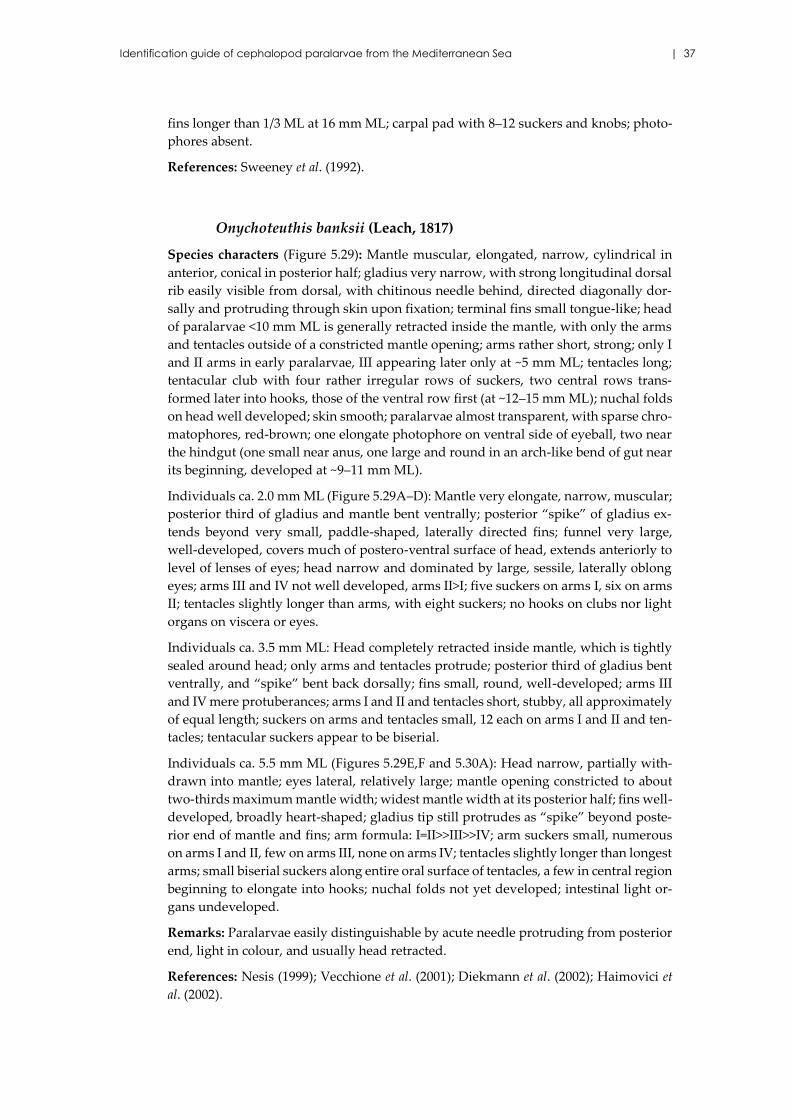
Identification guide of cephalopod paralarvae from the Mediterranean Sea | 37
fins longer than 1/3 ML at 16 mm ML; carpal pad with 8–12 suckers and knobs; photo-
phores absent.
References: Sweeney et al. (1992).
Onychoteuthis banksii (Leach, 1817)
Species characters (Figure 5.29): Mantle muscular, elongated, narrow, cylindrical in
anterior, conical in posterior half; gladius very narrow, with strong longitudinal dorsal
rib easily visible from dorsal, with chitinous needle behind, directed diagonally dor-
sally and protruding through skin upon fixation; terminal fins small tongue-like; head
of paralarvae <10 mm ML is generally retracted inside the mantle, with only the arms
and tentacles outside of a constricted mantle opening; arms rather short, strong; only I
and II arms in early paralarvae, III appearing later only at ~5 mm ML; tentacles long;
tentacular club with four rather irregular rows of suckers, two central rows trans-
formed later into hooks, those of the ventral row first (at ~12–15 mm ML); nuchal folds
on head well developed; skin smooth; paralarvae almost transparent, with sparse chro-
matophores, red-brown; one elongate photophore on ventral side of eyeball, two near
the hindgut (one small near anus, one large and round in an arch-like bend of gut near
its beginning, developed at ~9–11 mm ML).
Individuals ca. 2.0 mm ML (Figure 5.29A–D): Mantle very elongate, narrow, muscular;
posterior third of gladius and mantle bent ventrally; posterior “spike” of gladius ex-
tends beyond very small, paddle-shaped, laterally directed fins; funnel very large,
well-developed, covers much of postero-ventral surface of head, extends anteriorly to
level of lenses of eyes; head narrow and dominated by large, sessile, laterally oblong
eyes; arms III and IV not well developed, arms II>I; five suckers on arms I, six on arms
II; tentacles slightly longer than arms, with eight suckers; no hooks on clubs nor light
organs on viscera or eyes.
Individuals ca. 3.5 mm ML: Head completely retracted inside mantle, which is tightly
sealed around head; only arms and tentacles protrude; posterior third of gladius bent
ventrally, and “spike” bent back dorsally; fins small, round, well-developed; arms III
and IV mere protuberances; arms I and II and tentacles short, stubby, all approximately
of equal length; suckers on arms and tentacles small, 12 each on arms I and II and ten-
tacles; tentacular suckers appear to be biserial.
Individuals ca. 5.5 mm ML (Figures 5.29E,F and 5.30A): Head narrow, partially with-
drawn into mantle; eyes lateral, relatively large; mantle opening constricted to about
two-thirds maximum mantle width; widest mantle width at its posterior half; fins well-
developed, broadly heart-shaped; gladius tip still protrudes as “spike” beyond poste-
rior end of mantle and fins; arm formula: I=II>>III>>IV; arm suckers small, numerous
on arms I and II, few on arms III, none on arms IV; tentacles slightly longer than longest
arms; small biserial suckers along entire oral surface of tentacles, a few in central region
beginning to elongate into hooks; nuchal folds not yet developed; intestinal light or-
gans undeveloped.
Remarks: Paralarvae easily distinguishable by acute needle protruding from posterior
end, light in colour, and usually head retracted.
References: Nesis (1999); Vecchione et al. (2001); Diekmann et al. (2002); Haimovici et
al. (2002).
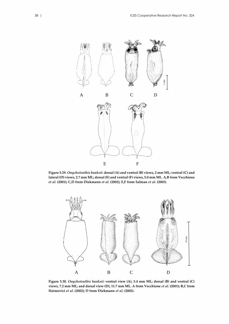
38 | ICES Cooperative Research Report No. 324
Figure 5.29. Onychoteuthis banksii: dorsal (A) and ventral (B) views, 2 mm ML; ventral (C) and
lateral (D) views, 2.7 mm ML; dorsal (E) and ventral (F) views, 5.0 mm ML. A,B from Vecchione
et al. (2001); C,D from Diekmann et al. (2002); E,F from Salman et al. (2003).
Figure 5.30. Onychoteuthis banksii: ventral view (A), 5.4 mm ML; dorsal (B) and ventral (C)
views, 7.2 mm ML; and dorsal view (D), 11.7 mm ML. A from Vecchione et al. (2001); B,C from
Haimovici et al. (2002); D from Diekmann et al. (2002).
C DA B
E F 1
mm
A CB D
10 m
m
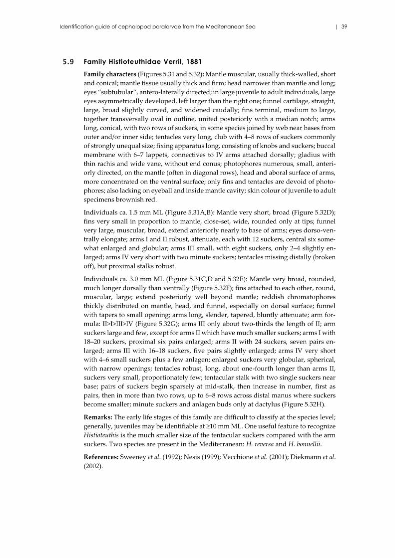
Identification guide of cephalopod paralarvae from the Mediterranean Sea | 39
5.9 Family Histioteuthidae Verril, 1881
Family characters (Figures 5.31 and 5.32): Mantle muscular, usually thick-walled, short
and conical; mantle tissue usually thick and firm; head narrower than mantle and long;
eyes “subtubular”, antero-laterally directed; in large juvenile to adult individuals, large
eyes asymmetrically developed, left larger than the right one; funnel cartilage, straight,
large, broad slightly curved, and widened caudally; fins terminal, medium to large,
together transversally oval in outline, united posteriorly with a median notch; arms
long, conical, with two rows of suckers, in some species joined by web near bases from
outer and/or inner side; tentacles very long, club with 4–8 rows of suckers commonly
of strongly unequal size; fixing apparatus long, consisting of knobs and suckers; buccal
membrane with 6–7 lappets, connectives to IV arms attached dorsally; gladius with
thin rachis and wide vane, without end conus; photophores numerous, small, anteri-
orly directed, on the mantle (often in diagonal rows), head and aboral surface of arms,
more concentrated on the ventral surface; only fins and tentacles are devoid of photo-
phores; also lacking on eyeball and inside mantle cavity; skin colour of juvenile to adult
specimens brownish red.
Individuals ca. 1.5 mm ML (Figure 5.31A,B): Mantle very short, broad (Figure 5.32D);
fins very small in proportion to mantle, close-set, wide, rounded only at tips; funnel
very large, muscular, broad, extend anteriorly nearly to base of arms; eyes dorso-ven-
trally elongate; arms I and II robust, attenuate, each with 12 suckers, central six some-
what enlarged and globular; arms III small, with eight suckers, only 2–4 slightly en-
larged; arms IV very short with two minute suckers; tentacles missing distally (broken
off), but proximal stalks robust.
Individuals ca. 3.0 mm ML (Figure 5.31C,D and 5.32E): Mantle very broad, rounded,
much longer dorsally than ventrally (Figure 5.32F); fins attached to each other, round,
muscular, large; extend posteriorly well beyond mantle; reddish chromatophores
thickly distributed on mantle, head, and funnel, especially on dorsal surface; funnel
with tapers to small opening; arms long, slender, tapered, bluntly attenuate; arm for-
mula: II>I>III>IV (Figure 5.32G); arms III only about two-thirds the length of II; arm
suckers large and few, except for arms II which have much smaller suckers; arms I with
18–20 suckers, proximal six pairs enlarged; arms II with 24 suckers, seven pairs en-
larged; arms III with 16–18 suckers, five pairs slightly enlarged; arms IV very short
with 4–6 small suckers plus a few anlagen; enlarged suckers very globular, spherical,
with narrow openings; tentacles robust, long, about one-fourth longer than arms II,
suckers very small, proportionately few; tentacular stalk with two single suckers near
base; pairs of suckers begin sparsely at mid-stalk, then increase in number, first as
pairs, then in more than two rows, up to 6–8 rows across distal manus where suckers
become smaller; minute suckers and anlagen buds only at dactylus (Figure 5.32H).
Remarks: The early life stages of this family are difficult to classify at the species level;
generally, juveniles may be identifiable at ≥10 mm ML. One useful feature to recognize
Histioteuthis is the much smaller size of the tentacular suckers compared with the arm
suckers. Two species are present in the Mediterranean: H. reversa and H. bonnellii.
References: Sweeney et al. (1992); Nesis (1999); Vecchione et al. (2001); Diekmann et al.
(2002).
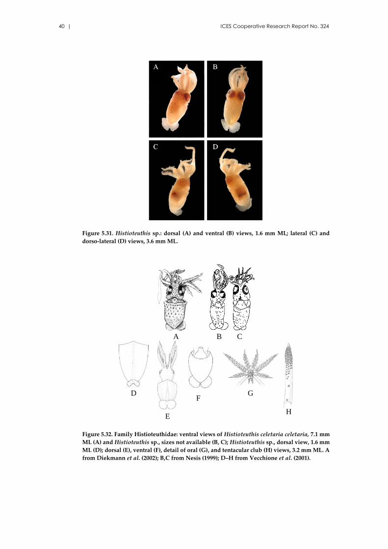
40 | ICES Cooperative Research Report No. 324
Figure 5.31. Histioteuthis sp.: dorsal (A) and ventral (B) views, 1.6 mm ML; lateral (C) and
dorso-lateral (D) views, 3.6 mm ML.
Figure 5.32. Family Histioteuthidae: ventral views of Histioteuthis celetaria celetaria, 7.1 mm
ML (A) and Histioteuthis sp., sizes not available (B, C); Histioteuthis sp., dorsal view, 1.6 mm
ML (D); dorsal (E), ventral (F), detail of oral (G), and tentacular club (H) views, 3.2 mm ML. A
from Diekmann et al. (2002); B,C from Nesis (1999); D–H from Vecchione et al. (2001).
B CA
D
E
F
H
G
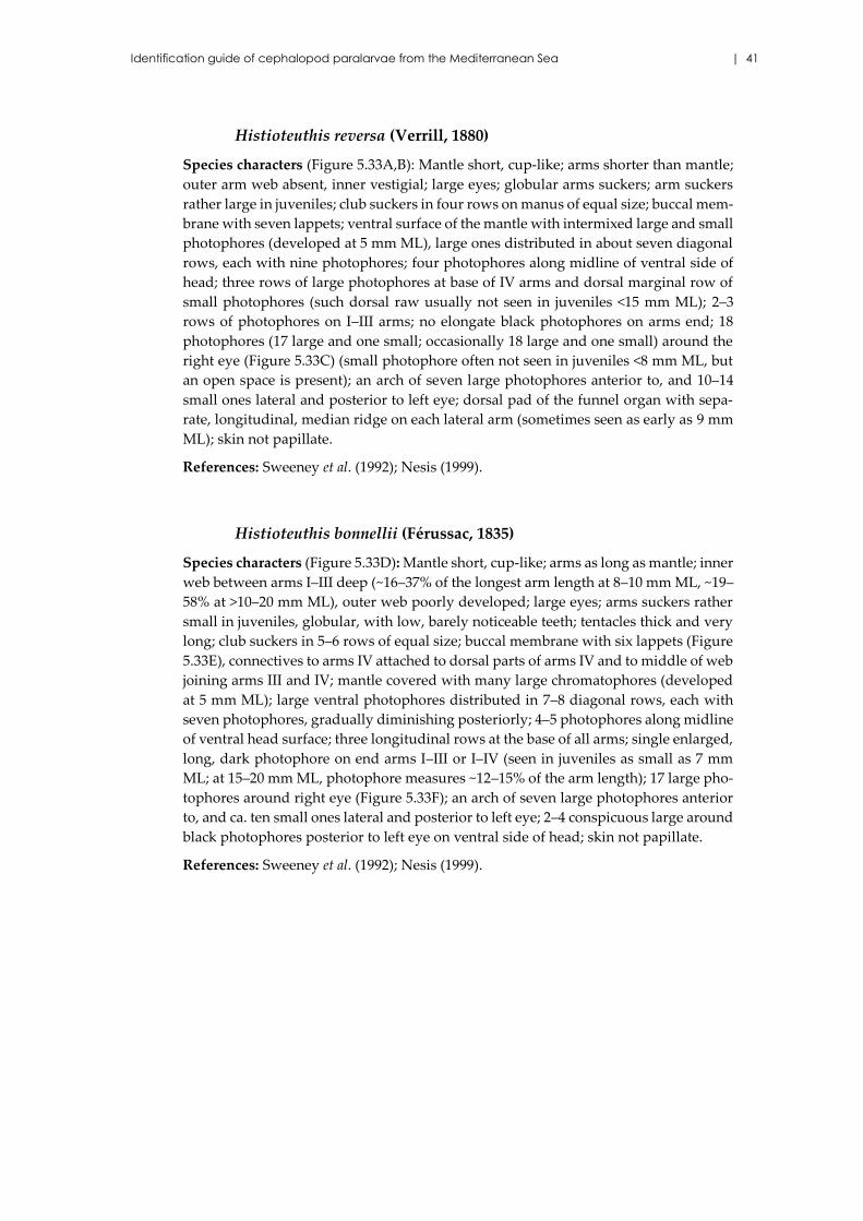
Identification guide of cephalopod paralarvae from the Mediterranean Sea | 41
Histioteuthis reversa (Verrill, 1880)
Species characters (Figure 5.33A,B): Mantle short, cup-like; arms shorter than mantle;
outer arm web absent, inner vestigial; large eyes; globular arms suckers; arm suckers
rather large in juveniles; club suckers in four rows on manus of equal size; buccal mem-
brane with seven lappets; ventral surface of the mantle with intermixed large and small
photophores (developed at 5 mm ML), large ones distributed in about seven diagonal
rows, each with nine photophores; four photophores along midline of ventral side of
head; three rows of large photophores at base of IV arms and dorsal marginal row of
small photophores (such dorsal raw usually not seen in juveniles <15 mm ML); 2–3
rows of photophores on I–III arms; no elongate black photophores on arms end; 18
photophores (17 large and one small; occasionally 18 large and one small) around the
right eye (Figure 5.33C) (small photophore often not seen in juveniles <8 mm ML, but
an open space is present); an arch of seven large photophores anterior to, and 10–14
small ones lateral and posterior to left eye; dorsal pad of the funnel organ with sepa-
rate, longitudinal, median ridge on each lateral arm (sometimes seen as early as 9 mm
ML); skin not papillate.
References: Sweeney et al. (1992); Nesis (1999).
Histioteuthis bonnellii (Férussac, 1835)
Species characters (Figure 5.33D): Mantle short, cup-like; arms as long as mantle; inner
web between arms I–III deep (~16–37% of the longest arm length at 8–10 mm ML, ~19–
58% at >10–20 mm ML), outer web poorly developed; large eyes; arms suckers rather
small in juveniles, globular, with low, barely noticeable teeth; tentacles thick and very
long; club suckers in 5–6 rows of equal size; buccal membrane with six lappets (Figure
5.33E), connectives to arms IV attached to dorsal parts of arms IV and to middle of web
joining arms III and IV; mantle covered with many large chromatophores (developed
at 5 mm ML); large ventral photophores distributed in 7–8 diagonal rows, each with
seven photophores, gradually diminishing posteriorly; 4–5 photophores along midline
of ventral head surface; three longitudinal rows at the base of all arms; single enlarged,
long, dark photophore on end arms I–III or I–IV (seen in juveniles as small as 7 mm
ML; at 15–20 mm ML, photophore measures ~12–15% of the arm length); 17 large pho-
tophores around right eye (Figure 5.33F); an arch of seven large photophores anterior
to, and ca. ten small ones lateral and posterior to left eye; 2–4 conspicuous large around
black photophores posterior to left eye on ventral side of head; skin not papillate.
References: Sweeney et al. (1992); Nesis (1999).
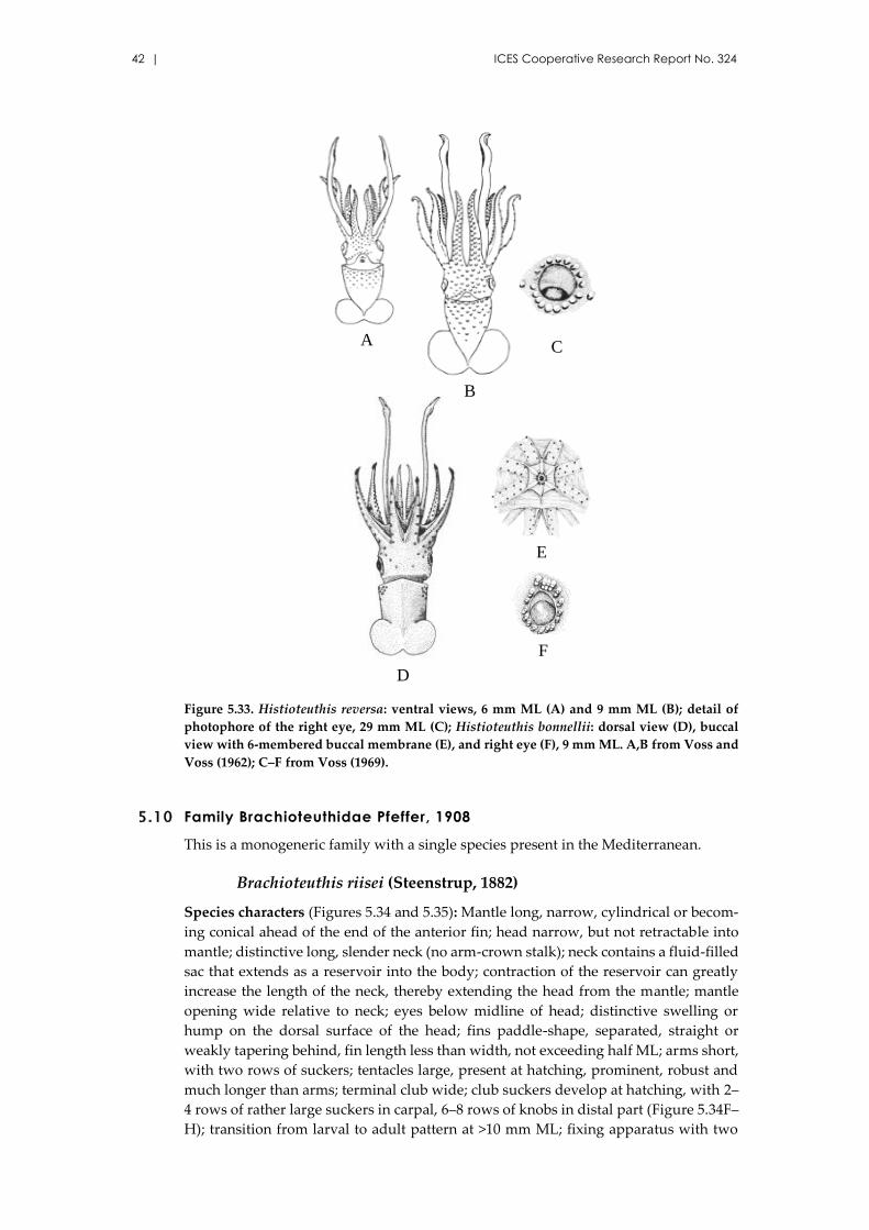
42 | ICES Cooperative Research Report No. 324
Figure 5.33. Histioteuthis reversa: ventral views, 6 mm ML (A) and 9 mm ML (B); detail of
photophore of the right eye, 29 mm ML (C); Histioteuthis bonnellii: dorsal view (D), buccal
view with 6-membered buccal membrane (E), and right eye (F), 9 mm ML. A,B from Voss and
Voss (1962); C–F from Voss (1969).
5.10 Family Brachioteuthidae Pfeffer, 1908
This is a monogeneric family with a single species present in the Mediterranean.
Brachioteuthis riisei (Steenstrup, 1882)
Species characters (Figures 5.34 and 5.35): Mantle long, narrow, cylindrical or becom-
ing conical ahead of the end of the anterior fin; head narrow, but not retractable into
mantle; distinctive long, slender neck (no arm-crown stalk); neck contains a fluid-filled
sac that extends as a reservoir into the body; contraction of the reservoir can greatly
increase the length of the neck, thereby extending the head from the mantle; mantle
opening wide relative to neck; eyes below midline of head; distinctive swelling or
hump on the dorsal surface of the head; fins paddle-shape, separated, straight or
weakly tapering behind, fin length less than width, not exceeding half ML; arms short,
with two rows of suckers; tentacles large, present at hatching, prominent, robust and
much longer than arms; terminal club wide; club suckers develop at hatching, with 2–
4 rows of rather large suckers in carpal, 6–8 rows of knobs in distal part (Figure 5.34F–
H); transition from larval to adult pattern at >10 mm ML; fixing apparatus with two
D
A
B
F
E
C
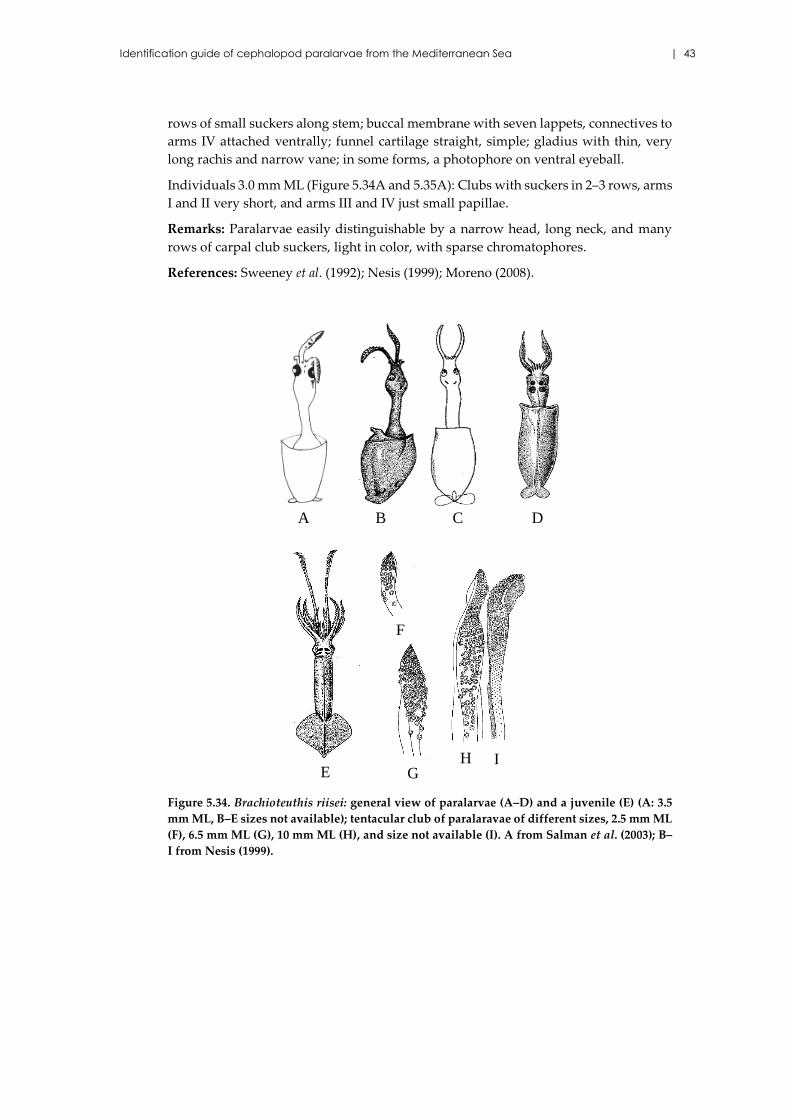
Identification guide of cephalopod paralarvae from the Mediterranean Sea | 43
rows of small suckers along stem; buccal membrane with seven lappets, connectives to
arms IV attached ventrally; funnel cartilage straight, simple; gladius with thin, very
long rachis and narrow vane; in some forms, a photophore on ventral eyeball.
Individuals 3.0 mm ML (Figure 5.34A and 5.35A): Clubs with suckers in 2–3 rows, arms
I and II very short, and arms III and IV just small papillae.
Remarks: Paralarvae easily distinguishable by a narrow head, long neck, and many
rows of carpal club suckers, light in color, with sparse chromatophores.
References: Sweeney et al. (1992); Nesis (1999); Moreno (2008).
Figure 5.34. Brachioteuthis riisei: general view of paralarvae (A–D) and a juvenile (E) (A: 3.5
mm ML, B–E sizes not available); tentacular club of paralaravae of different sizes, 2.5 mm ML
(F), 6.5 mm ML (G), 10 mm ML (H), and size not available (I). A from Salman et al. (2003); B–
I from Nesis (1999).
B C D
E
F
IHG
A
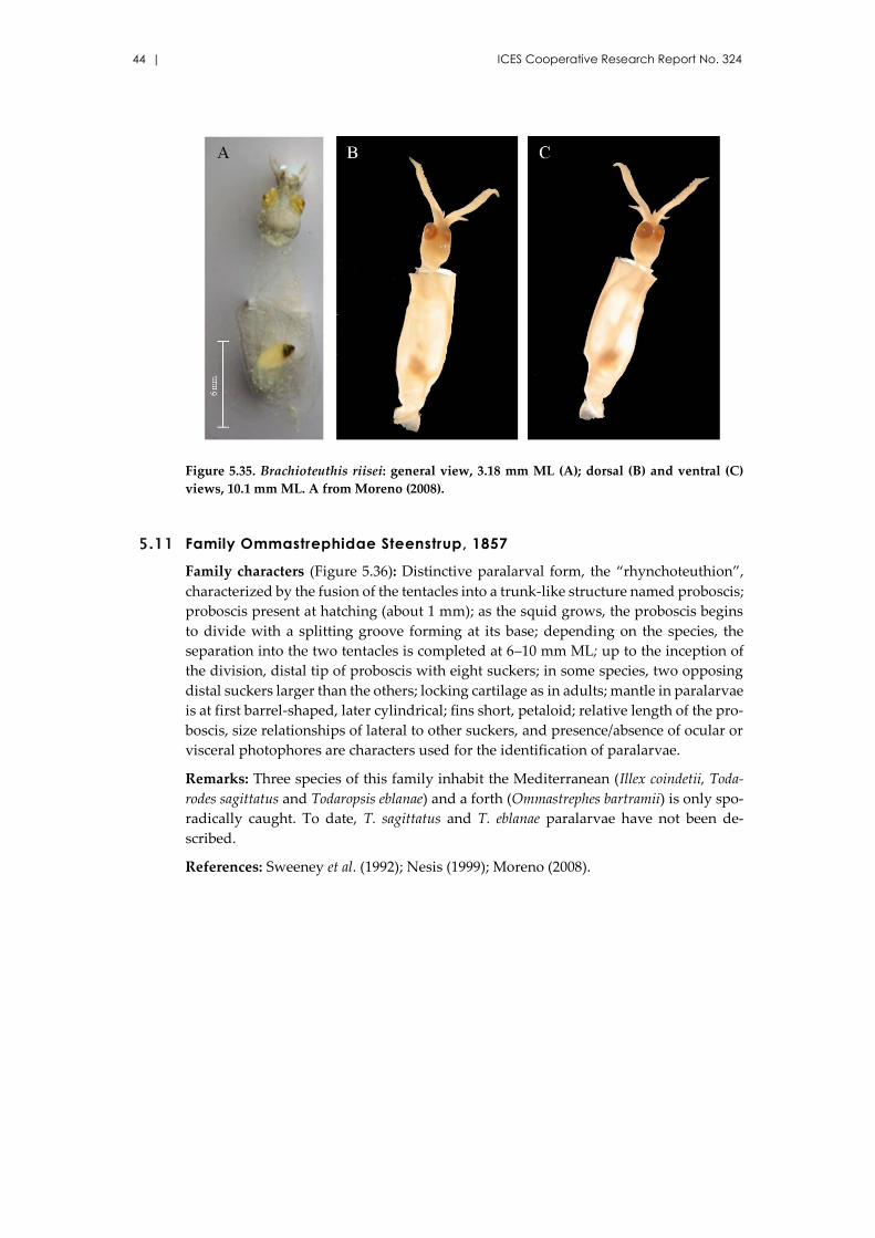
44 | ICES Cooperative Research Report No. 324
Figure 5.35. Brachioteuthis riisei: general view, 3.18 mm ML (A); dorsal (B) and ventral (C)
views, 10.1 mm ML. A from Moreno (2008).
5.11 Family Ommastrephidae Steenstrup, 1857
Family characters (Figure 5.36): Distinctive paralarval form, the “rhynchoteuthion”,
characterized by the fusion of the tentacles into a trunk-like structure named proboscis;
proboscis present at hatching (about 1 mm); as the squid grows, the proboscis begins
to divide with a splitting groove forming at its base; depending on the species, the
separation into the two tentacles is completed at 6–10 mm ML; up to the inception of
the division, distal tip of proboscis with eight suckers; in some species, two opposing
distal suckers larger than the others; locking cartilage as in adults; mantle in paralarvae
is at first barrel-shaped, later cylindrical; fins short, petaloid; relative length of the pro-
boscis, size relationships of lateral to other suckers, and presence/absence of ocular or
visceral photophores are characters used for the identification of paralarvae.
Remarks: Three species of this family inhabit the Mediterranean (Illex coindetii, Toda-
rodes sagittatus and Todaropsis eblanae) and a forth (Ommastrephes bartramii) is only spo-
radically caught. To date, T. sagittatus and T. eblanae paralarvae have not been de-
scribed.
References: Sweeney et al. (1992); Nesis (1999); Moreno (2008).
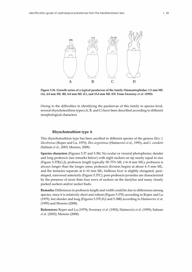
Identification guide of cephalopod paralarvae from the Mediterranean Sea | 45
Figure 5.36. Growth series of a typical paralarvae of the family Ommastrephidae: 1.5 mm ML
(A), 4.0 mm ML (B), 8.0 mm ML (C), and 15.0 mm ML (D). From Sweeney et al. (1992).
Owing to the difficulties in identifying the paralarvae of this family to species level,
several rhynchoteuthion types (A, B, and C) have been described according to different
morphological characters.
Rhynchoteuthion type A
This rhynchoteuthion type has been ascribed to different species of the genera Illex: I.
illecebrosus (Roper and Lu, 1979), Illex argentinus (Haimovici et al., 1995), and I. coindetii
(Salman et al., 2003; Moreno, 2008).
Species characters (Figures 5.37 and 5.38): No ocular or visceral photophores; slender
and long proboscis (see remarks below) with eight suckers on tip nearly equal in size
(Figure 5.37B,G,J); proboscis length typically 50−75% ML (<4–8 mm ML); proboscis is
always longer than the longer arms; proboscis division begins at about 4–5 mm ML,
and the tentacles separate at 6–10 mm ML; bulbous liver is slightly elongated, pear-
shaped, narrowed anteriorly (Figure 5.37C); post-proboscis juveniles are characterized
by the presence of more than four rows of suckers on the dactylus and many closely
packed suckers and/or sucker buds.
Remarks: Differences in proboscis length and width could be due to differences among
species, since it is relatively short and robust (Figure 5.37E) according to Roper and Lu
(1979), but slender and long (Figures 5.37F,H,I and 5.38B) according to Haimovici et al.
(1995) and Moreno (2008).
References: Roper and Lu (1979); Sweeney et al. (1992); Haimovici et al. (1995); Salman
et al. (2003); Moreno (2008).
B DA C
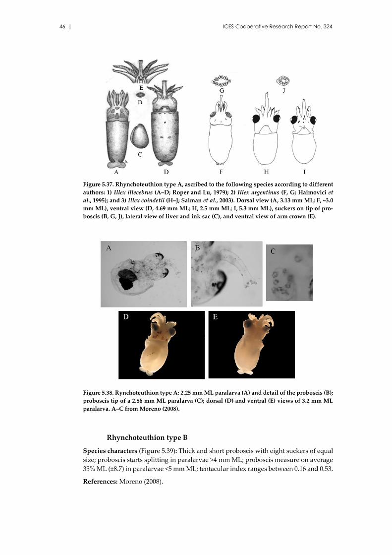
46 | ICES Cooperative Research Report No. 324
Figure 5.37. Rhynchoteuthion type A, ascribed to the following species according to different
authors: 1) Illex illecebrus (A–D; Roper and Lu, 1979); 2) Illex argentinus (F, G; Haimovici et
al., 1995); and 3) Illex coindetii (H–J; Salman et al., 2003). Dorsal view (A, 3.13 mm ML; F, ~3.0
mm ML), ventral view (D, 4.69 mm ML; H, 2.5 mm ML; I, 5.3 mm ML), suckers on tip of pro-
boscis (B, G, J), lateral view of liver and ink sac (C), and ventral view of arm crown (E).
Figure 5.38. Rynchoteuthion type A: 2.25 mm ML paralarva (A) and detail of the proboscis (B);
proboscis tip of a 2.86 mm ML paralarva (C); dorsal (D) and ventral (E) views of 3.2 mm ML
paralarva. A–C from Moreno (2008).
Rhynchoteuthion type B
Species characters (Figure 5.39): Thick and short proboscis with eight suckers of equal
size; proboscis starts splitting in paralarvae >4 mm ML; proboscis measure on average
35% ML (±8.7) in paralarvae <5 mm ML; tentacular index ranges between 0.16 and 0.53.
References: Moreno (2008).
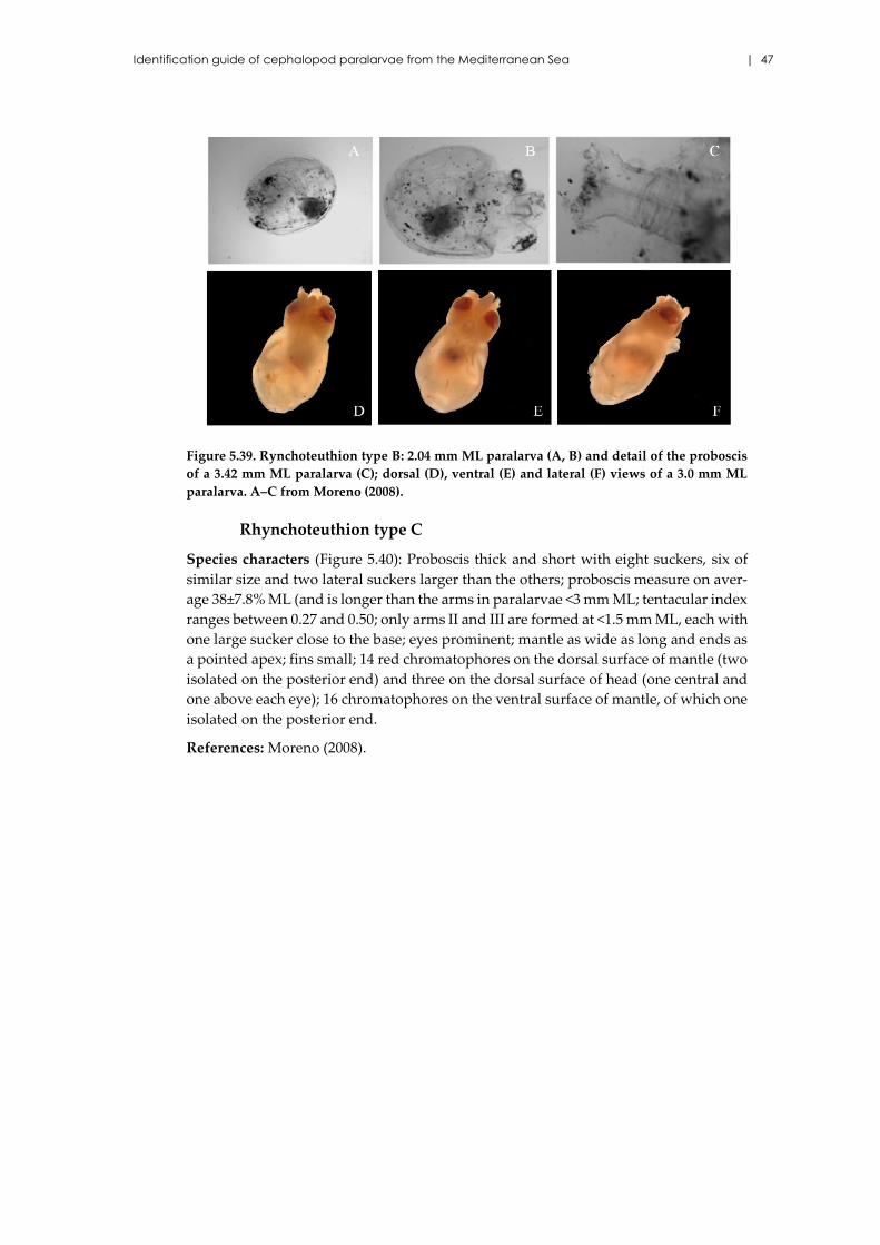
Identification guide of cephalopod paralarvae from the Mediterranean Sea | 47
Figure 5.39. Rynchoteuthion type B: 2.04 mm ML paralarva (A, B) and detail of the proboscis
of a 3.42 mm ML paralarva (C); dorsal (D), ventral (E) and lateral (F) views of a 3.0 mm ML
paralarva. A–C from Moreno (2008).
Rhynchoteuthion type C
Species characters (Figure 5.40): Proboscis thick and short with eight suckers, six of
similar size and two lateral suckers larger than the others; proboscis measure on aver-
age 38±7.8% ML (and is longer than the arms in paralarvae <3 mm ML; tentacular index
ranges between 0.27 and 0.50; only arms II and III are formed at <1.5 mm ML, each with
one large sucker close to the base; eyes prominent; mantle as wide as long and ends as
a pointed apex; fins small; 14 red chromatophores on the dorsal surface of mantle (two
isolated on the posterior end) and three on the dorsal surface of head (one central and
one above each eye); 16 chromatophores on the ventral surface of mantle, of which one
isolated on the posterior end.
References: Moreno (2008).
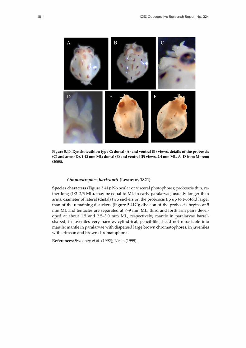
48 | ICES Cooperative Research Report No. 324
Figure 5.40. Rynchoteuthion type C: dorsal (A) and ventral (B) views, details of the proboscis
(C) and arms (D), 1.43 mm ML; dorsal (E) and ventral (F) views, 2.4 mm ML. A–D from Moreno
(2008).
Ommastrephes bartramii (Lesueur, 1821)
Species characters (Figure 5.41): No ocular or visceral photophores; proboscis thin, ra-
ther long (1/2–2/3 ML), may be equal to ML in early paralarvae, usually longer than
arms; diameter of lateral (distal) two suckers on the proboscis tip up to twofold larger
than of the remaining 6 suckers (Figure 5.41C); division of the proboscis begins at 5
mm ML and tentacles are separated at 7–9 mm ML; third and forth arm pairs devel-
oped at about 1.5 and 2.5–3.0 mm ML, respectively; mantle in paralarvae barrel-
shaped, in juveniles very narrow, cylindrical, pencil-like; head not retractable into
mantle; mantle in paralarvae with dispersed large brown chromatophores, in juveniles
with crimson and brown chromatophores.
References: Sweeney et al. (1992); Nesis (1999).
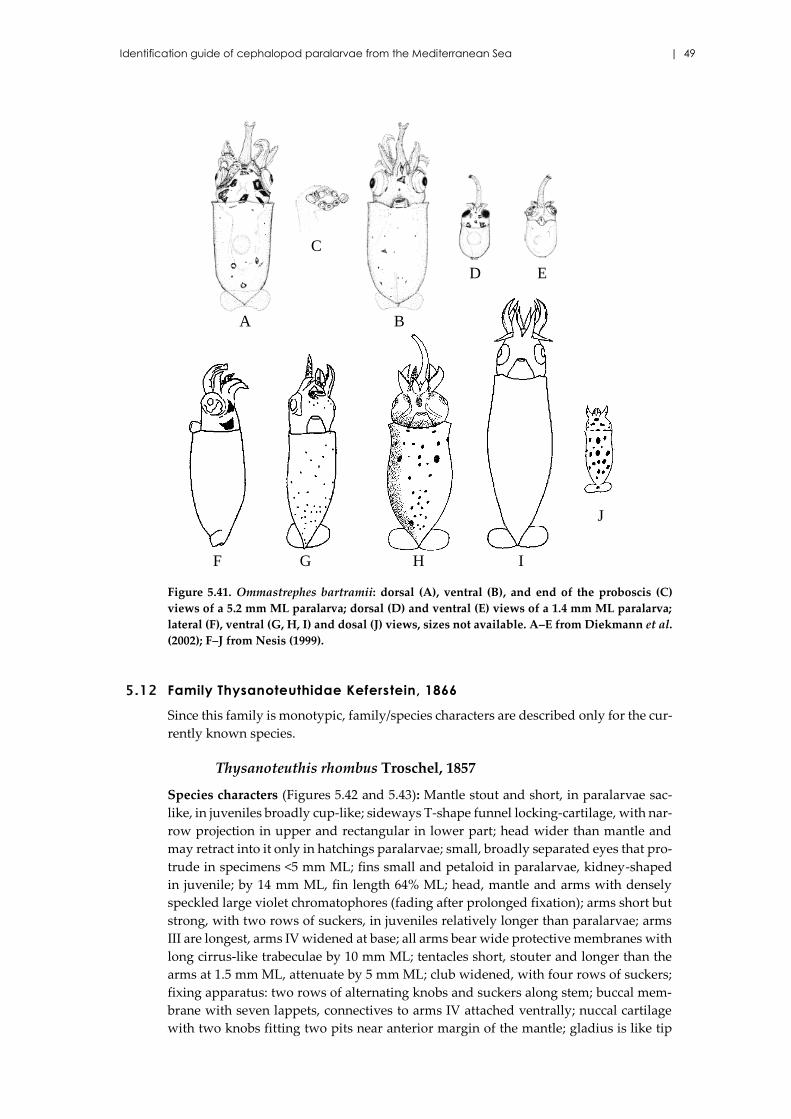
Identification guide of cephalopod paralarvae from the Mediterranean Sea | 49
Figure 5.41. Ommastrephes bartramii: dorsal (A), ventral (B), and end of the proboscis (C)
views of a 5.2 mm ML paralarva; dorsal (D) and ventral (E) views of a 1.4 mm ML paralarva;
lateral (F), ventral (G, H, I) and dosal (J) views, sizes not available. A–E from Diekmann et al.
(2002); F–J from Nesis (1999).
5.12 Family Thysanoteuthidae Keferstein, 1866
Since this family is monotypic, family/species characters are described only for the cur-
rently known species.
Thysanoteuthis rhombus Troschel, 1857
Species characters (Figures 5.42 and 5.43): Mantle stout and short, in paralarvae sac-
like, in juveniles broadly cup-like; sideways T-shape funnel locking-cartilage, with nar-
row projection in upper and rectangular in lower part; head wider than mantle and
may retract into it only in hatchings paralarvae; small, broadly separated eyes that pro-
trude in specimens <5 mm ML; fins small and petaloid in paralarvae, kidney-shaped
in juvenile; by 14 mm ML, fin length 64% ML; head, mantle and arms with densely
speckled large violet chromatophores (fading after prolonged fixation); arms short but
strong, with two rows of suckers, in juveniles relatively longer than paralarvae; arms
III are longest, arms IV widened at base; all arms bear wide protective membranes with
long cirrus-like trabeculae by 10 mm ML; tentacles short, stouter and longer than the
arms at 1.5 mm ML, attenuate by 5 mm ML; club widened, with four rows of suckers;
fixing apparatus: two rows of alternating knobs and suckers along stem; buccal mem-
brane with seven lappets, connectives to arms IV attached ventrally; nuccal cartilage
with two knobs fitting two pits near anterior margin of the mantle; gladius is like tip
A
D E
C
F G H I
J
B
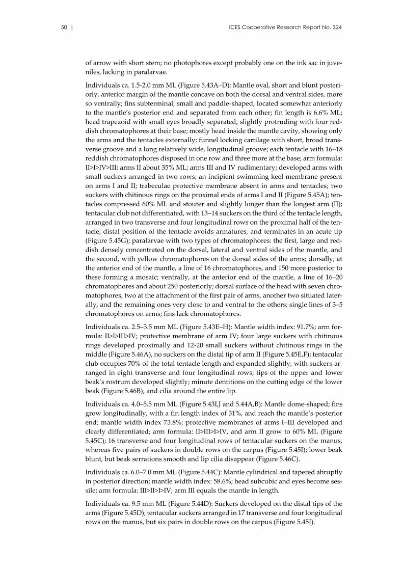
50 | ICES Cooperative Research Report No. 324
of arrow with short stem; no photophores except probably one on the ink sac in juve-
niles, lacking in paralarvae.
Individuals ca. 1.5-2.0 mm ML (Figure 5.43A–D): Mantle oval, short and blunt posteri-
orly, anterior margin of the mantle concave on both the dorsal and ventral sides, more
so ventrally; fins subterminal, small and paddle-shaped, located somewhat anteriorly
to the mantle’s posterior end and separated from each other; fin length is 6.6% ML;
head trapezoid with small eyes broadly separated, slightly protruding with four red-
dish chromatophores at their base; mostly head inside the mantle cavity, showing only
the arms and the tentacles externally; funnel locking cartilage with short, broad trans-
verse groove and a long relatively wide, longitudinal groove; each tentacle with 16–18
reddish chromatophores disposed in one row and three more at the base; arm formula:
II>I>IV>III; arms II about 35% ML; arms III and IV rudimentary; developed arms with
small suckers arranged in two rows; an incipient swimming keel membrane present
on arms I and II; trabeculae protective membrane absent in arms and tentacles; two
suckers with chitinous rings on the proximal ends of arms I and II (Figure 5.45A); ten-
tacles compressed 60% ML and stouter and slightly longer than the longest arm (II);
tentacular club not differentiated, with 13–14 suckers on the third of the tentacle length,
arranged in two transverse and four longitudinal rows on the proximal half of the ten-
tacle; distal position of the tentacle avoids armatures, and terminates in an acute tip
(Figure 5.45G); paralarvae with two types of chromatophores: the first, large and red-
dish densely concentrated on the dorsal, lateral and ventral sides of the mantle, and
the second, with yellow chromatophores on the dorsal sides of the arms; dorsally, at
the anterior end of the mantle, a line of 16 chromatophores, and 150 more posterior to
these forming a mosaic; ventrally, at the anterior end of the mantle, a line of 16–20
chromatophores and about 250 posteriorly; dorsal surface of the head with seven chro-
matophores, two at the attachment of the first pair of arms, another two situated later-
ally, and the remaining ones very close to and ventral to the others; single lines of 3–5
chromatophores on arms; fins lack chromatophores.
Individuals ca. 2.5–3.5 mm ML (Figure 5.43E–H): Mantle width index: 91.7%; arm for-
mula: II>I>III>IV; protective membrane of arm IV; four large suckers with chitinous
rings developed proximally and 12-20 small suckers without chitinous rings in the
middle (Figure 5.46A), no suckers on the distal tip of arm II (Figure 5.45E,F); tentacular
club occupies 70% of the total tentacle length and expanded slightly, with suckers ar-
ranged in eight transverse and four longitudinal rows; tips of the upper and lower
beak’s rostrum developed slightly; minute dentitions on the cutting edge of the lower
beak (Figure 5.46B), and cilia around the entire lip.
Individuals ca. 4.0–5.5 mm ML (Figure 5.43I,J and 5.44A,B): Mantle dome-shaped; fins
grow longitudinally, with a fin length index of 31%, and reach the mantle’s posterior
end; mantle width index 73.8%; protective membranes of arms I–III developed and
clearly differentiated; arm formula: II>III>I>IV, and arm II grow to 60% ML (Figure
5.45C); 16 transverse and four longitudinal rows of tentacular suckers on the manus,
whereas five pairs of suckers in double rows on the carpus (Figure 5.45I); lower beak
blunt, but beak serrations smooth and lip cilia disappear (Figure 5.46C).
Individuals ca. 6.0–7.0 mm ML (Figure 5.44C): Mantle cylindrical and tapered abruptly
in posterior direction; mantle width index: 58.6%; head subcubic and eyes become ses-
sile; arm formula: III>II>I>IV; arm III equals the mantle in length.
Individuals ca. 9.5 mm ML (Figure 5.44D): Suckers developed on the distal tips of the
arms (Figure 5.45D); tentacular suckers arranged in 17 transverse and four longitudinal
rows on the manus, but six pairs in double rows on the carpus (Figure 5.45J).
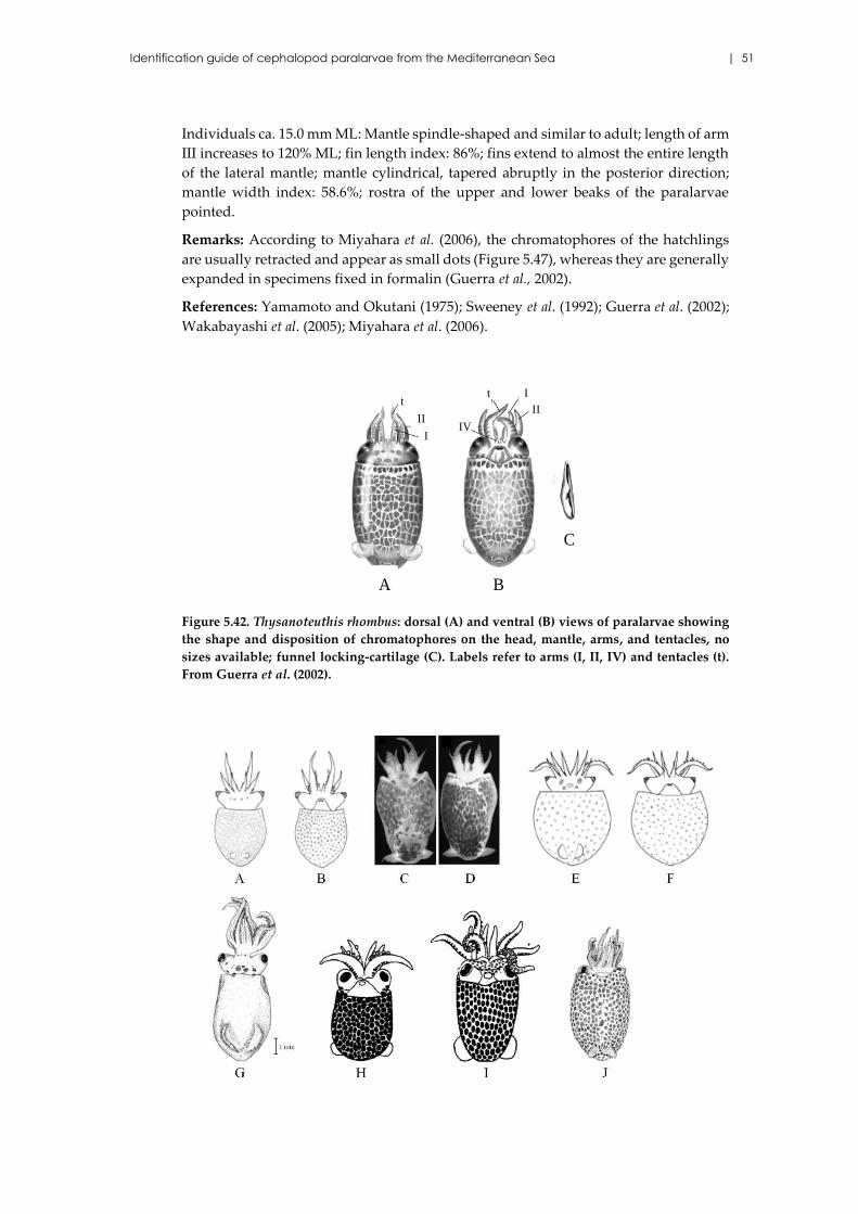
Identification guide of cephalopod paralarvae from the Mediterranean Sea | 51
Individuals ca. 15.0 mm ML: Mantle spindle-shaped and similar to adult; length of arm
III increases to 120% ML; fin length index: 86%; fins extend to almost the entire length
of the lateral mantle; mantle cylindrical, tapered abruptly in the posterior direction;
mantle width index: 58.6%; rostra of the upper and lower beaks of the paralarvae
pointed.
Remarks: According to Miyahara et al. (2006), the chromatophores of the hatchlings
are usually retracted and appear as small dots (Figure 5.47), whereas they are generally
expanded in specimens fixed in formalin (Guerra et al., 2002).
References: Yamamoto and Okutani (1975); Sweeney et al. (1992); Guerra et al. (2002);
Wakabayashi et al. (2005); Miyahara et al. (2006).
Figure 5.42. Thysanoteuthis rhombus: dorsal (A) and ventral (B) views of paralarvae showing
the shape and disposition of chromatophores on the head, mantle, arms, and tentacles, no
sizes available; funnel locking-cartilage (C). Labels refer to arms (I, II, IV) and tentacles (t).
From Guerra et al. (2002).
t I
II
IV
BA
t
II
I
C
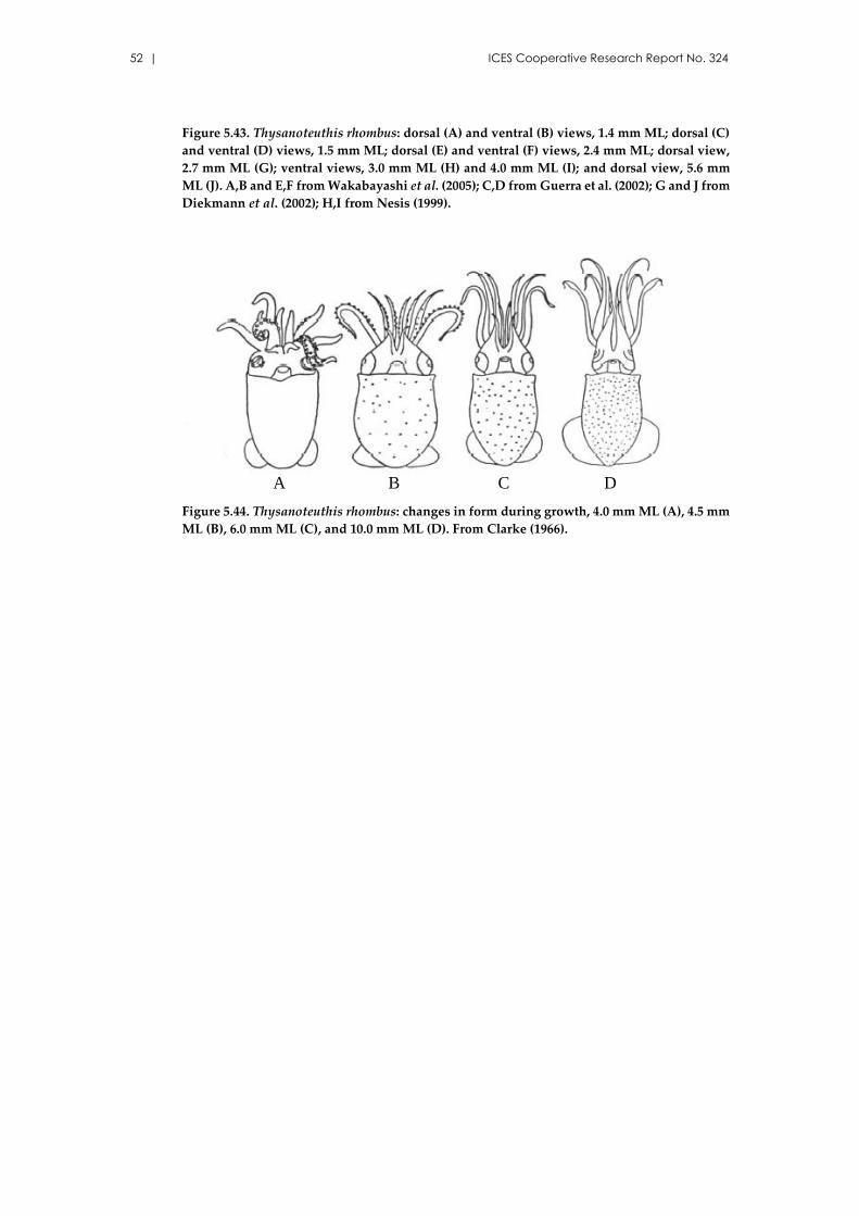
52 | ICES Cooperative Research Report No. 324
Figure 5.43. Thysanoteuthis rhombus: dorsal (A) and ventral (B) views, 1.4 mm ML; dorsal (C)
and ventral (D) views, 1.5 mm ML; dorsal (E) and ventral (F) views, 2.4 mm ML; dorsal view,
2.7 mm ML (G); ventral views, 3.0 mm ML (H) and 4.0 mm ML (I); and dorsal view, 5.6 mm
ML (J). A,B and E,F from Wakabayashi et al. (2005); C,D from Guerra et al. (2002); G and J from
Diekmann et al. (2002); H,I from Nesis (1999).
Figure 5.44. Thysanoteuthis rhombus: changes in form during growth, 4.0 mm ML (A), 4.5 mm
ML (B), 6.0 mm ML (C), and 10.0 mm ML (D). From Clarke (1966).
A DCB
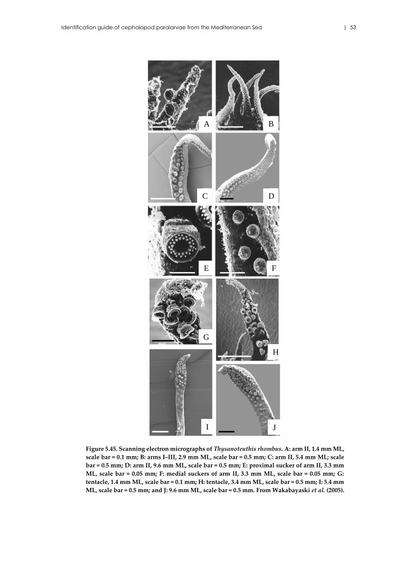
Identification guide of cephalopod paralarvae from the Mediterranean Sea | 53
Figure 5.45. Scanning electron micrographs of Thysanoteuthis rhombus. A: arm II, 1.4 mm ML,
scale bar = 0.1 mm; B: arms I–III, 2.9 mm ML, scale bar = 0.5 mm; C: arm II, 5.4 mm ML; scale
bar = 0.5 mm; D: arm II, 9.6 mm ML, scale bar = 0.5 mm; E: proximal sucker of arm II, 3.3 mm
ML, scale bar = 0.05 mm; F: medial suckers of arm II, 3.3 mm ML, scale bar = 0.05 mm; G:
tentacle, 1.4 mm ML, scale bar = 0.1 mm; H: tentacle, 3.4 mm ML, scale bar = 0.5 mm; I: 5.4 mm
ML, scale bar = 0.5 mm; and J: 9.6 mm ML, scale bar = 0.5 mm. From Wakabayaski et al. (2005).
G
H
I J
A B
C D
E F
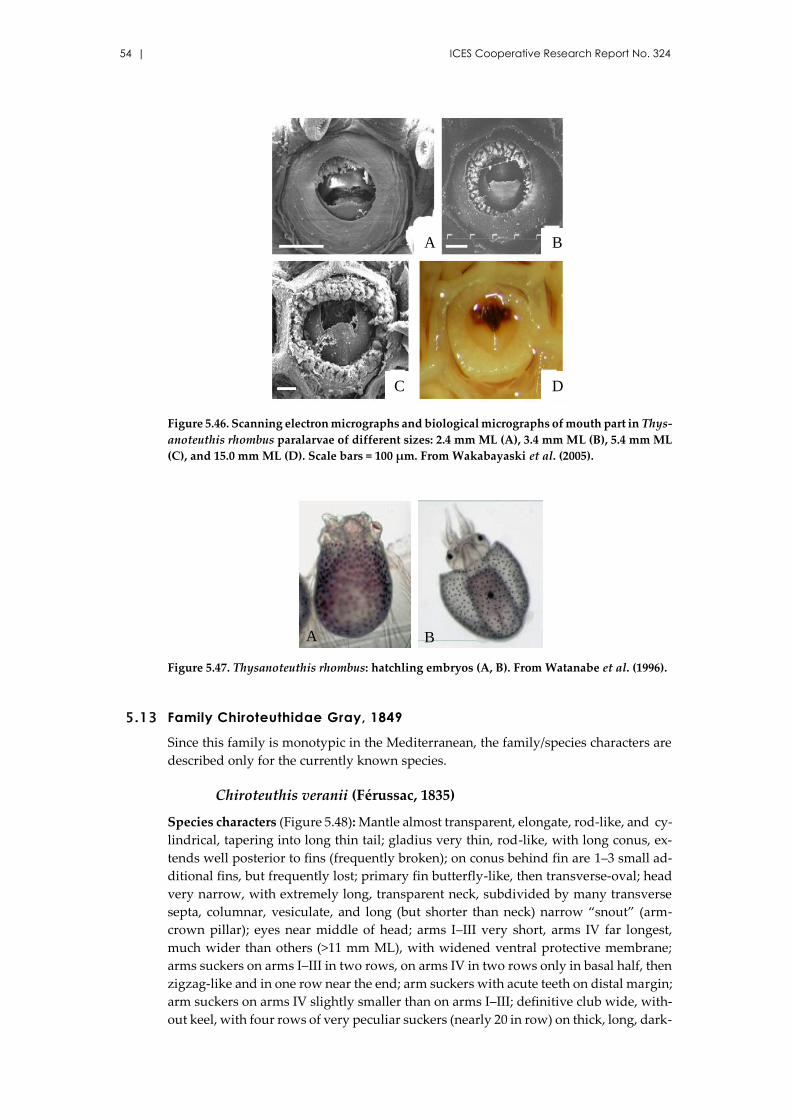
54 | ICES Cooperative Research Report No. 324
Figure 5.46. Scanning electron micrographs and biological micrographs of mouth part in Thys-
anoteuthis rhombus paralarvae of different sizes: 2.4 mm ML (A), 3.4 mm ML (B), 5.4 mm ML
(C), and 15.0 mm ML (D). Scale bars = 100 µm. From Wakabayaski et al. (2005).
Figure 5.47. Thysanoteuthis rhombus: hatchling embryos (A, B). From Watanabe et al. (1996).
5.13 Family Chiroteuthidae Gray, 1849
Since this family is monotypic in the Mediterranean, the family/species characters are
described only for the currently known species.
Chiroteuthis veranii (Férussac, 1835)
Species characters (Figure 5.48): Mantle almost transparent, elongate, rod-like, and cy-
lindrical, tapering into long thin tail; gladius very thin, rod-like, with long conus, ex-
tends well posterior to fins (frequently broken); on conus behind fin are 1–3 small ad-
ditional fins, but frequently lost; primary fin butterfly-like, then transverse-oval; head
very narrow, with extremely long, transparent neck, subdivided by many transverse
septa, columnar, vesiculate, and long (but shorter than neck) narrow “snout” (arm-
crown pillar); eyes near middle of head; arms I–III very short, arms IV far longest,
much wider than others (>11 mm ML), with widened ventral protective membrane;
arms suckers on arms I–III in two rows, on arms IV in two rows only in basal half, then
zigzag-like and in one row near the end; arm suckers with acute teeth on distal margin;
arm suckers on arms IV slightly smaller than on arms I–III; definitive club wide, with-
out keel, with four rows of very peculiar suckers (nearly 20 in row) on thick, long, dark-
A B
C D
A B
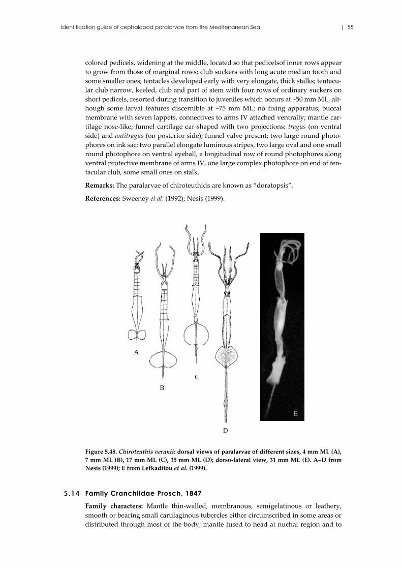
Identification guide of cephalopod paralarvae from the Mediterranean Sea | 55
colored pedicels, widening at the middle, located so that pedicelsof inner rows appear
to grow from those of marginal rows; club suckers with long acute median tooth and
some smaller ones; tentacles developed early with very elongate, thick stalks; tentacu-
lar club narrow, keeled, club and part of stem with four rows of ordinary suckers on
short pedicels, resorted during transition to juveniles which occurs at ~50 mm ML, alt-
hough some larval features discernible at ~75 mm ML; no fixing apparatus; buccal
membrane with seven lappets, connectives to arms IV attached ventrally; mantle car-
tilage nose-like; funnel cartilage ear-shaped with two projections: tragus (on ventral
side) and antitragus (on posterior side); funnel valve present; two large round photo-
phores on ink sac; two parallel elongate luminous stripes, two large oval and one small
round photophore on ventral eyeball, a longitudinal row of round photophores along
ventral protective membrane of arms IV, one large complex photophore on end of ten-
tacular club, some small ones on stalk.
Remarks: The paralarvae of chiroteuthids are known as “doratopsis”.
References: Sweeney et al. (1992); Nesis (1999).
Figure 5.48. Chiroteuthis veranii: dorsal views of paralarvae of different sizes, 4 mm ML (A),
7 mm ML (B), 17 mm ML (C), 35 mm ML (D); dorso-lateral view, 31 mm ML (E). A–D from
Nesis (1999); E from Lefkaditou et al. (1999).
5.14 Family Cranchiidae Prosch, 1847
Family characters: Mantle thin-walled, membranous, semigelatinous or leathery,
smooth or bearing small cartilaginous tubercles either circumscribed in some areas or
distributed through most of the body; mantle fused to head at nuchal region and to
A
B
C
D
E
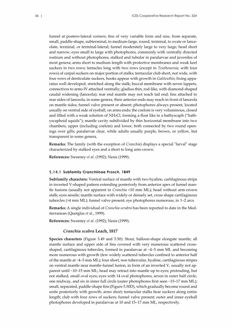
56 | ICES Cooperative Research Report No. 324
funnel at postero-lateral corners; fins of very variable form and size, from separate,
small, paddle-shape, subterminal, to medium-large, round, terminal, to ovate or lance-
olate, terminal, or terminal-lateral; funnel moderately large to very large; head short
and narrow; eyes small to large with photophores, commonly with ventrally directed
rostrum and without photophores, stalked and tubular in paralarvae and juveniles of
most genera; arms short to medium length with protective membranes and weak keel
suckers in two rows; tentacles long with two rows (except in Teuthowenia, with four
rows) of carpal suckers on major portion of stalks; tentacular club short, not wide, with
four rows of denticulate suckers, hooks appear with growth in Galiteuthis; fixing appa-
ratus well developed, stretched along the stalk; buccal membrane with seven lappets,
connectives to arms IV attached ventrally; gladius thin, rod-like, with diamond-shaped
caudal widening (lanceola); rear end mantle may not reach tail end; fins attached to
rear sides of lanceola, in some genera, their anterior ends may reach in front of lanceola
on mantle sides; funnel valve present or absent; photophores always present, located
usually on ventral side of eyeball, on arms ends; the coelom is very voluminous, closed
and filled with a weak solution of NH4Cl, forming a float like in a bathyscaph (“bath-
yscaphoid squids”); mantle cavity subdivided by thin horizontal membrane into two
chambers, upper (including coelom) and lower, both connected by two round open-
ings over gills; paralarvae clear, while adults usually purple, brown, or yellow, but
transparent in some genera.
Remarks: The family (with the exception of Cranchia) displays a special "larval" stage
characterized by stalked eyes and a short to long arm-crown.
References: Sweeney et al. (1992); Nesis (1999).
5.14.1 Subfamily Cranchiinae Prosch, 1849
Subfamily characters: Ventral surface of mantle with two hyaline, cartilaginous strips
in inverted V-shaped pattern extending posteriorly from anterior apex of funnel man-
tle fusions (usually not apparent in Cranchia <10 mm ML); head without arm-crown
stalk; eyes sessile; mantle surface with widely or densely set, cross shape cartilaginous
tubercles (>4 mm ML); funnel valve present; eye photophores numerous, in 1–2 arcs.
Remarks: A single individual of Cranchia scrabra has been reported to date in the Med-
iterranean (Quetglas et al., 1999).
References: Sweeney et al. (1992); Nesis (1999).
Cranchia scabra Leach, 1817
Species characters (Figure 5.49 and 5.50): Stout, balloon-shape elongate mantle; all
mantle surface and upper side of fins covered with very numerous scattered cross-
shaped, cartilaginous tubercles, formed in paralarvae at ~4–5 mm ML and becoming
more numerous with growth (few widely scattered tubercles confined to anterior half
of the mantle at ~4–5 mm ML); four short, non tubercular, hyaline, cartilaginous stripes
on ventral mantle near mantle-funnel fusion, in form of an inverted V, usually not ap-
parent until ~10–15 mm ML; head may retract into mantle up to eyes; protruding, but
not stalked, small oval eyes; eyes with 14 oval photophores, seven in outer half circle,
one midway, and six in inner full circle (outer photophores first seen ~15–17 mm ML);
small, separated, paddle-shape fins (Figure 5.50D), which gradually become round and
unite posteriorly with growth; arms short; tentacular stalks bear suckers along entire
length; club with four rows of suckers; funnel valve present; outer and inner eyeball
photophores developed in paralarvae at 10 and 15–17 mm ML, respectively.
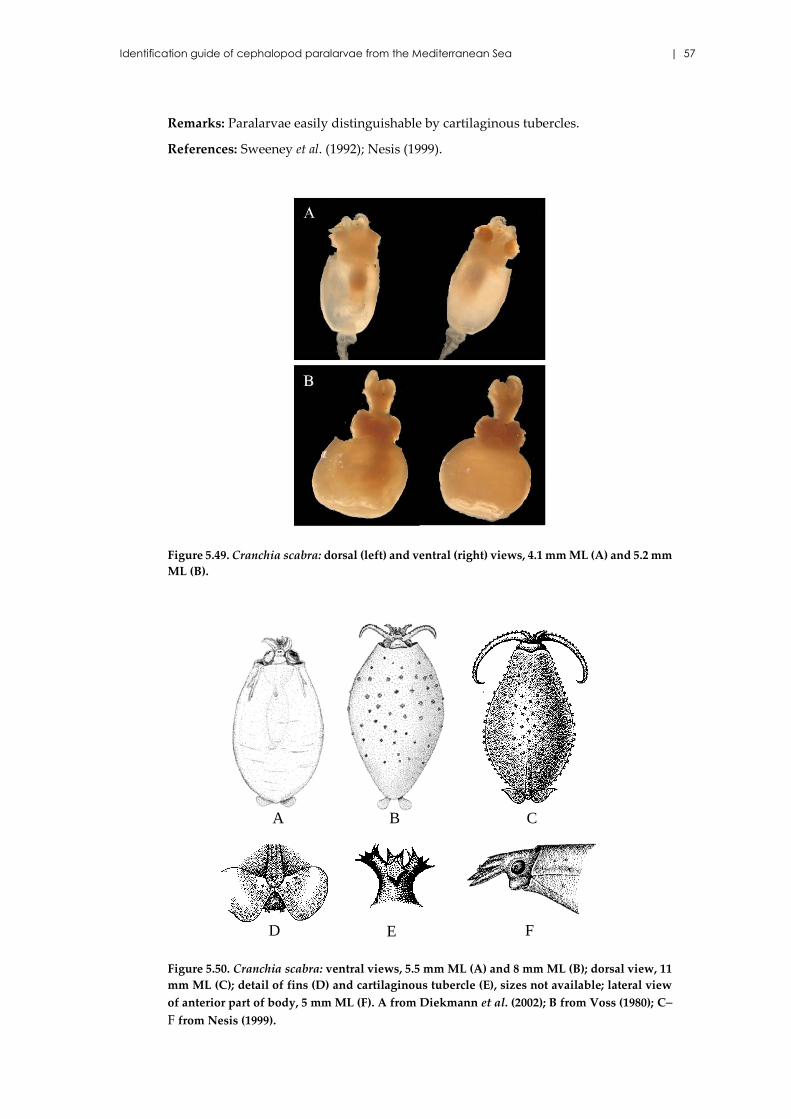
Identification guide of cephalopod paralarvae from the Mediterranean Sea | 57
Remarks: Paralarvae easily distinguishable by cartilaginous tubercles.
References: Sweeney et al. (1992); Nesis (1999).
Figure 5.49. Cranchia scabra: dorsal (left) and ventral (right) views, 4.1 mm ML (A) and 5.2 mm
ML (B).
Figure 5.50. Cranchia scabra: ventral views, 5.5 mm ML (A) and 8 mm ML (B); dorsal view, 11
mm ML (C); detail of fins (D) and cartilaginous tubercle (E), sizes not available; lateral view
of anterior part of body, 5 mm ML (F). A from Diekmann et al. (2002); B from Voss (1980); C–
F from Nesis (1999).
BA C
FD E
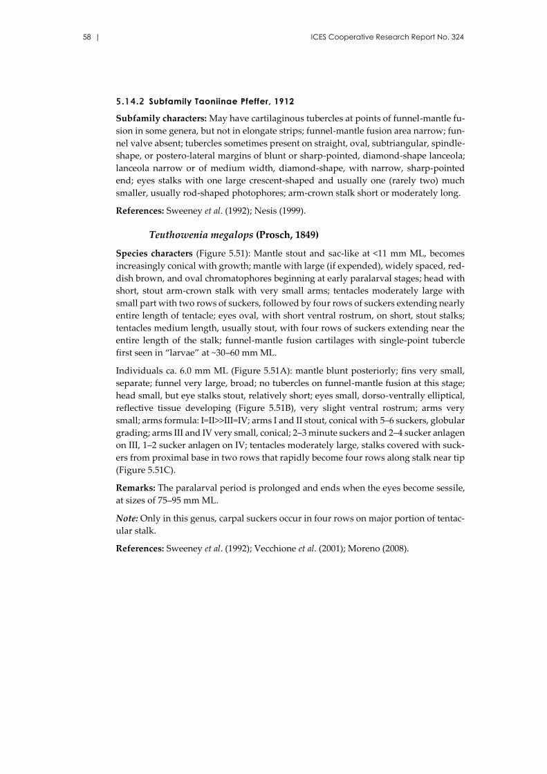
58 | ICES Cooperative Research Report No. 324
5.14.2 Subfamily Taoniinae Pfeffer, 1912
Subfamily characters: May have cartilaginous tubercles at points of funnel-mantle fu-
sion in some genera, but not in elongate strips; funnel-mantle fusion area narrow; fun-
nel valve absent; tubercles sometimes present on straight, oval, subtriangular, spindle-
shape, or postero-lateral margins of blunt or sharp-pointed, diamond-shape lanceola;
lanceola narrow or of medium width, diamond-shape, with narrow, sharp-pointed
end; eyes stalks with one large crescent-shaped and usually one (rarely two) much
smaller, usually rod-shaped photophores; arm-crown stalk short or moderately long.
References: Sweeney et al. (1992); Nesis (1999).
Teuthowenia megalops (Prosch, 1849)
Species characters (Figure 5.51): Mantle stout and sac-like at <11 mm ML, becomes
increasingly conical with growth; mantle with large (if expended), widely spaced, red-
dish brown, and oval chromatophores beginning at early paralarval stages; head with
short, stout arm-crown stalk with very small arms; tentacles moderately large with
small part with two rows of suckers, followed by four rows of suckers extending nearly
entire length of tentacle; eyes oval, with short ventral rostrum, on short, stout stalks;
tentacles medium length, usually stout, with four rows of suckers extending near the
entire length of the stalk; funnel-mantle fusion cartilages with single-point tubercle
first seen in “larvae” at ~30–60 mm ML.
Individuals ca. 6.0 mm ML (Figure 5.51A): mantle blunt posteriorly; fins very small,
separate; funnel very large, broad; no tubercles on funnel-mantle fusion at this stage;
head small, but eye stalks stout, relatively short; eyes small, dorso-ventrally elliptical,
reflective tissue developing (Figure 5.51B), very slight ventral rostrum; arms very
small; arms formula: I=II>>III=IV; arms I and II stout, conical with 5–6 suckers, globular
grading; arms III and IV very small, conical; 2–3 minute suckers and 2–4 sucker anlagen
on III, 1–2 sucker anlagen on IV; tentacles moderately large, stalks covered with suck-
ers from proximal base in two rows that rapidly become four rows along stalk near tip
(Figure 5.51C).
Remarks: The paralarval period is prolonged and ends when the eyes become sessile,
at sizes of 75–95 mm ML.
Note: Only in this genus, carpal suckers occur in four rows on major portion of tentac-
ular stalk.
References: Sweeney et al. (1992); Vecchione et al. (2001); Moreno (2008).
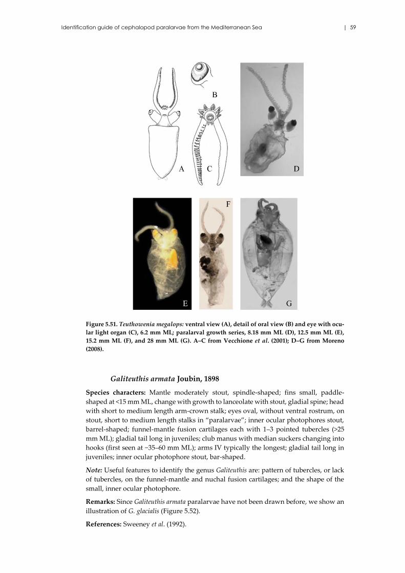
Identification guide of cephalopod paralarvae from the Mediterranean Sea | 59
Figure 5.51. Teuthowenia megalops: ventral view (A), detail of oral view (B) and eye with ocu-
lar light organ (C), 6.2 mm ML; paralarval growth series, 8.18 mm ML (D), 12.5 mm ML (E),
15.2 mm ML (F), and 28 mm ML (G). A–C from Vecchione et al. (2001); D–G from Moreno
(2008).
Galiteuthis armata Joubin, 1898
Species characters: Mantle moderately stout, spindle-shaped; fins small, paddle-
shaped at <15 mm ML, change with growth to lanceolate with stout, gladial spine; head
with short to medium length arm-crown stalk; eyes oval, without ventral rostrum, on
stout, short to medium length stalks in “paralarvae”; inner ocular photophores stout,
barrel-shaped; funnel-mantle fusion cartilages each with 1–3 pointed tubercles (>25
mm ML); gladial tail long in juveniles; club manus with median suckers changing into
hooks (first seen at ~35–60 mm ML); arms IV typically the longest; gladial tail long in
juveniles; inner ocular photophore stout, bar-shaped.
Note: Useful features to identify the genus Galiteuthis are: pattern of tubercles, or lack
of tubercles, on the funnel-mantle and nuchal fusion cartilages; and the shape of the
small, inner ocular photophore.
Remarks: Since Galiteuthis armata paralarvae have not been drawn before, we show an
illustration of G. glacialis (Figure 5.52).
References: Sweeney et al. (1992).
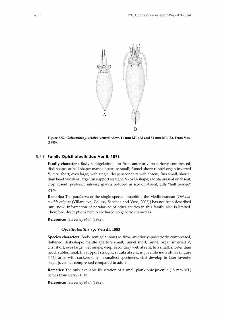
60 | ICES Cooperative Research Report No. 324
Figure 5.52. Galiteuthis glacialis: ventral view, 11 mm ML (A) and 54 mm ML (B). From Voss
(1980).
5.15 Family Opisthoteuthidae Verril, 1896
Family characters: Body semigelatinous to firm, anteriorly–posteriorly compressed,
disk-shape, or bell-shape; mantle aperture small; funnel short; funnel organ inverted
V; cirri short; eyes large; web single, deep; secondary web absent; fins small, shorter
than head width or large; fin support straight, V- or U-shape; radula present or absent;
crop absent; posterior salivary glands reduced in size or absent; gills “half orange”
type.
Remarks: The paralarva of the single species inhabiting the Mediterranean [Opistho-
teuthis calypso (Villanueva, Collins, Sánchez and Voss, 2002)] has not been described
until now. Information of paralarvae of other species in this family also is limited.
Therefore, descriptions herein are based on generic characters.
References: Sweeney et al. (1992).
Opisthoteuthis sp. Verrill, 1883
Species characters: Body semigelatinous to firm, anteriorly–posteriorly compressed,
flattened, disk-shape; mantle aperture small; funnel short; funnel organ inverted V;
cirri short; eyes large; web single, deep; secondary web absent; fins small, shorter than
head, subterminal; fin support straight; radula absent; in juvenile individuals (Figure
5.53), arms with suckers only in smallest specimens, cirri develop in later juvenile
stage; juveniles compressed compared to adults.
Remarks: The only available illustration of a small planktonic juvenile (15 mm ML)
comes from Berry (1912).
References: Sweeney et al. (1992).
A
B
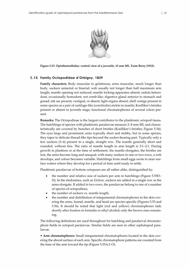
Identification guide of cephalopod paralarvae from the Mediterranean Sea | 61
Figure 5.53. Opisthoteuthidae: ventral view of a juvenile, 15 mm ML. From Berry (1912).
5.16 Family Octopodidae d’Orbigny, 1839
Family characters: Body muscular to gelatinous; arms muscular, much longer than
body, suckers uniserial or biserial; web usually not longer than half maximum arm
length; mantle opening not reduced; mantle locking-apparatus absent; radula hetero-
dont, occasionally homodont, not comb-like; digestive gland anterior to stomach and
gonad; ink sac present, vestigial, or absent; light organs absent; shell vestige present in
some species as a pair of cartilage-like (conchiolin) stylets in mantle; Koelliker's bristles
present or absent in juvenile stage; functional chromatophores of several colors pre-
sent.
Remarks: The Octopodinae is the largest contributor to the planktonic octopod fauna.
The hatchlings of species with planktonic paralarvae measure 2–8 mm ML and charac-
teristically are covered by bunches of short bristles (Koelliker’s bristles; Figure 5.54).
The eyes large and prominent; arms typically short and stubby, but in some species,
they taper to delicate thread-like tips beyond the sucker-bearing part. Typically only a
few suckers (3–4) present in a single, straight row. The mantle generally short and
rounded, without fins. The ratio of mantle length to arm length is 3:1–2:1. During
growth in plankton or at the time of settlement, the mantle elongates, the bristles are
lost, the arms become long and unequal; with many suckers in one or two rows, a web
develops, and colour becomes variable. Hatchlings from small eggs swim to near-sur-
face waters where they develop for a period of time until ready to settle.
Planktonic paralarvae of bottom octopuses are all rather alike, distinguished by:
the number and relative size of suckers per arm in hatchlings (Figure 5.55D–
H). In the eledonines, such as Eledone, suckers are added in a single row as the
arms elongate. If added in two rows, the paralarvae belong to one of a number
of species of octopodines;
the number of suckers vs. mantle length;
the number and distribution of integumental chromatophores in the skin cov-
ering the arms, funnel, mantle, and head are species-specific (Figures 5.55 and
5.56). It should be noted that light (red and yellow) chromatophores fade
shortly after fixation in formalin or ethyl alcohol; only the brown ones remain-
ing.
The following definitions are used throughout for hatchling and paralarval chromato-
phore fields in octopod paralarvae. Similar fields are seen in other cephalopod para-
larvae.
• Arm chromatophores: Small integumental chromatophores located in the skin cov-
ering the aboral surface of each arm. Specific chromatophore patterns are counted from
the base of the arm toward the tip (Figure 5.55A,I–O).
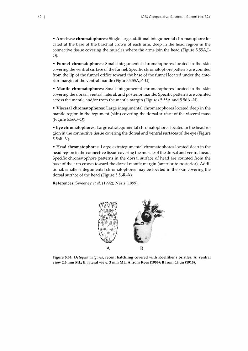
62 | ICES Cooperative Research Report No. 324
• Arm-base chromatophores: Single large additonal integumental chromatophore lo-
cated at the base of the brachial crown of each arm, deep in the head region in the
connective tissue covering the muscles where the arms join the head (Figure 5.55A,I–
O).
• Funnel chromatophores: Small integumental chromatophores located in the skin
covering the ventral surface of the funnel. Specific chromatophore patterns are counted
from the lip of the funnel orifice toward the base of the funnel located under the ante-
rior margin of the ventral mantle (Figure 5.55A,P–U).
• Mantle chromatophores: Small integumental chromatophores located in the skin
covering the dorsal, ventral, lateral, and posterior mantle. Specific patterns are counted
across the mantle and/or from the mantle margin (Figures 5.55A and 5.56A–N).
• Visceral chromatophores: Large integumental chromatophores located deep in the
mantle region in the tegument (skin) covering the dorsal surface of the visceral mass
(Figure 5.56O–Q).
• Eye chromatophores: Large extrategumental chromatophores located in the head re-
gion in the connective tissue covering the dorsal and ventral surfaces of the eye (Figure
5.56R–V).
• Head chromatophores: Large extrategumental chromatophores located deep in the
head region in the connective tissue covering the muscle of the dorsal and ventral head.
Specific chromatophore patterns in the dorsal surface of head are counted from the
base of the arm crown toward the dorsal mantle margin (anterior to posterior). Addi-
tional, smaller integumental chromatophores may be located in the skin covering the
dorsal surface of the head (Figure 5.56R–X).
References: Sweeney et al. (1992); Nesis (1999).
Figure 5.54. Octopus vulgaris, recent hatchling covered with Koelliker’s bristles: A, ventral
view 2.6 mm ML; B, lateral view, 3 mm ML. A from Rees (1953); B from Chun (1915).
A B
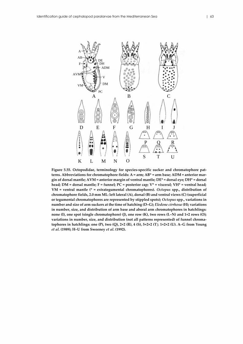
Identification guide of cephalopod paralarvae from the Mediterranean Sea | 63
Figure 5.55. Octopodidae, terminology for species-specific sucker and chromatophore pat-
terns. Abbreviations for chromatophore fields: A = arm; AB* = arm base; ADM = anterior mar-
gin of dorsal mantle; AVM = anterior margin of ventral mantle; DE* = dorsal eye; DH* = dorsal
head; DM = dorsal mantle; F = funnel; PC = posterior cap; V* = visceral; VH* = ventral head;
VM = ventral mantle (* = extrategumental chromatophores). Octopus spp., distribution of
chromatophore fields, 2.0 mm ML: left lateral (A), dorsal (B) and ventral views (C) (superficial
or tegumental chromatophores are represented by stippled spots); Octopus spp., variations in
number and size of arm suckers at the time of hatching (D–G); Eledone cirrhosa (H); variations
in number, size, and distribution of arm base and aboral arm chromatophores in hatchlings:
none (I), one spot (single chromatophore) (J), one row (K), two rows (L–N) and 1+2 rows (O);
variations in number, size, and distribution (not all patterns represented) of funnel chroma-
tophores in hatchlings: one (P), two (Q), 2+2 (R), 4 (S), 5+2+2 (T); 1+2+2 (U). A–G from Young
et al. (1989); H–U from Sweeney et al. (1992).
A B C
D E F G H I J
K O
RQP
M NLT US
DEDH
ADM
DM
V
PC
A
AB
AVM
VM
F
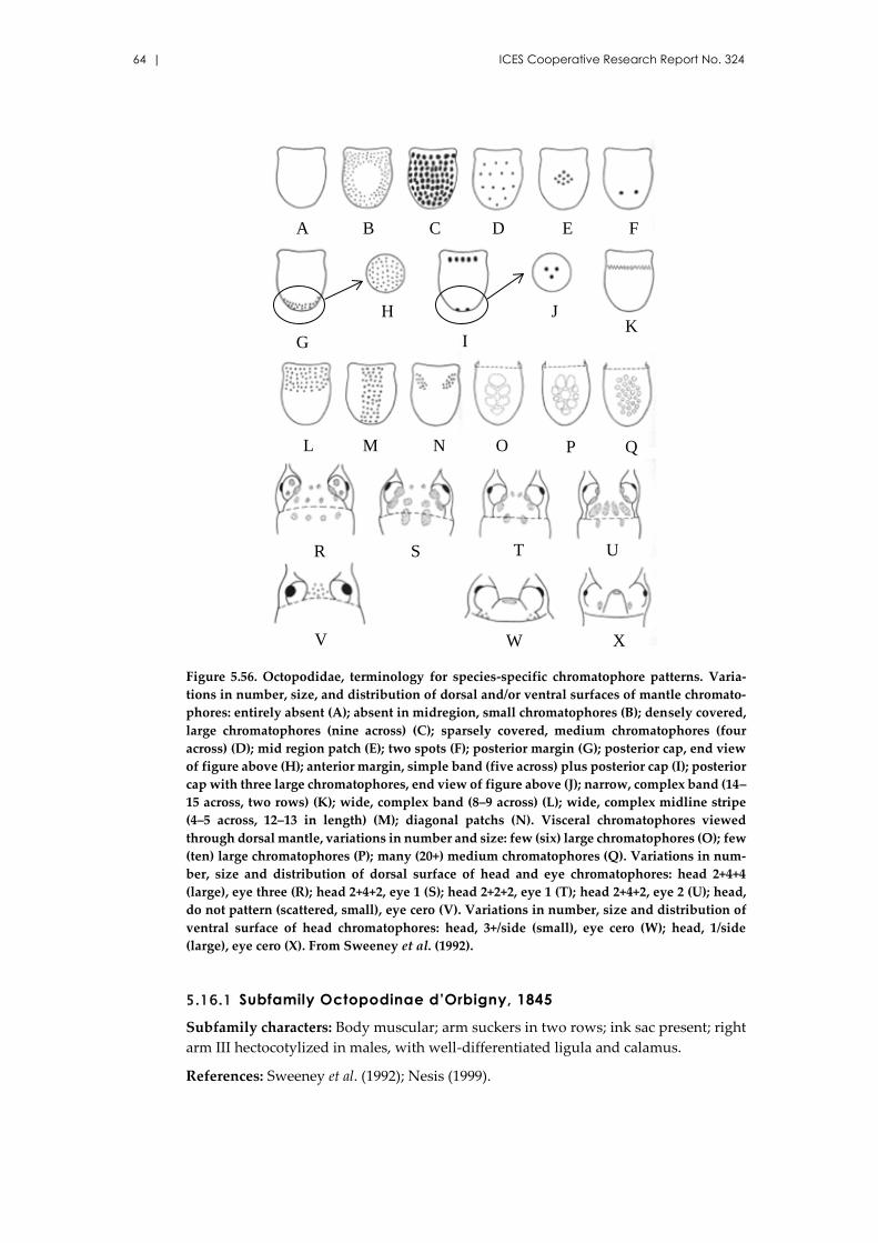
64 | ICES Cooperative Research Report No. 324
Figure 5.56. Octopodidae, terminology for species-specific chromatophore patterns. Varia-
tions in number, size, and distribution of dorsal and/or ventral surfaces of mantle chromato-
phores: entirely absent (A); absent in midregion, small chromatophores (B); densely covered,
large chromatophores (nine across) (C); sparsely covered, medium chromatophores (four
across) (D); mid region patch (E); two spots (F); posterior margin (G); posterior cap, end view
of figure above (H); anterior margin, simple band (five across) plus posterior cap (I); posterior
cap with three large chromatophores, end view of figure above (J); narrow, complex band (14–
15 across, two rows) (K); wide, complex band (8–9 across) (L); wide, complex midline stripe
(4–5 across, 12–13 in length) (M); diagonal patchs (N). Visceral chromatophores viewed
through dorsal mantle, variations in number and size: few (six) large chromatophores (O); few
(ten) large chromatophores (P); many (20+) medium chromatophores (Q). Variations in num-
ber, size and distribution of dorsal surface of head and eye chromatophores: head 2+4+4
(large), eye three (R); head 2+4+2, eye 1 (S); head 2+2+2, eye 1 (T); head 2+4+2, eye 2 (U); head,
do not pattern (scattered, small), eye cero (V). Variations in number, size and distribution of
ventral surface of head chromatophores: head, 3+/side (small), eye cero (W); head, 1/side
(large), eye cero (X). From Sweeney et al. (1992).
5.16.1 Subfamily Octopodinae d’Orbigny, 1845
Subfamily characters: Body muscular; arm suckers in two rows; ink sac present; right
arm III hectocotylized in males, with well-differentiated ligula and calamus.
References: Sweeney et al. (1992); Nesis (1999).
A B C D E F
G I
H
NML
K
R S
V
J
UT
W X
QPO
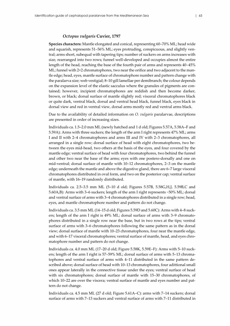
Identification guide of cephalopod paralarvae from the Mediterranean Sea | 65
Octopus vulgaris Cuvier, 1797
Species characters: Mantle elongated and conical, representing 60–70% ML; head wide
and squarish, represents 51–56% ML; eyes protruding, conspicuous, and slightly ven-
tral; arms short, subequal with tapering tips; number of suckers on arms increases with
size, rearranged into two rows; funnel well-developed and occupies almost the entire
length of the head, reaching the base of the fourth pair of arms and represents 40–45%
ML; funnel with 2+2 chromatophores, two near the orifice and two adjacent to the man-
tle edge; head, eyes, mantle surface of chromatophore number and pattern change with
the paralarva size; web vestigial; 8–10 gill lamellae per demibranch; the colour depends
on the expansion level of the elastic sacculus where the granules of pigments are con-
tained; however, incipient chromatophores are reddish and then become darker,
brown, or black; dorsal surface of mantle slightly red; visceral chromatophores black
or quite dark, ventral black, dorsal and ventral head black, funnel black, eyes black in
dorsal view and red in ventral view, dorsal arms mostly red and ventral arms black.
Due to the availability of detailed information on O. vulgaris paralarvae, descriptions
are presented in order of increasing sizes.
Individuals ca. 1.5–2.0 mm ML (newly hatched and 1 d old; Figures 5.57A, 5.58A–F and
5.59A): Arms with three suckers; the length of the arm I right represents 47% ML; arms
I and II with 2–4 chromatophores and arms III and IV with 2–3 chromatophores, all
arranged in a single row; dorsal surface of head with eight chromatophores, two be-
tween the eyes mid-head, two others at the basis of the eyes, and four covered by the
mantle edge; ventral surface of head with four chromatophores, two behind the funnel
and other two near the base of the arms; eyes with one postero-dorsally and one on
mid-ventral; dorsal surface of mantle with 10–12 chromatophores, 2–3 on the mantle
edge; underneath the mantle and above the digestive gland, there are 6–7 large visceral
chromatophores distributed in oval form, and two on the posterior cap; ventral surface
of mantle, with 16–19 randomly distributed.
Individuals ca. 2.5–3.5 mm ML (5–10 d old; Figures 5.57B, 5.58G,H,J, 5.59B,C and
5.60A,B): Arms with 3–6 suckers; length of the arm I right represents ~50% ML; dorsal
and ventral surface of arms with 3–4 chromatophores distributed in a single row; head,
eyes, and mantle chromatophore number and pattern do not change.
Individuals ca. 3.5 mm ML (14–15 d old; Figures 5.59D and 5.60C): Arms with 4–8 suck-
ers; length of the arm I right is 49% ML; dorsal surface of arms with 3–9 chromato-
phores distributed in a single row near the base, but in two rows at the tips; ventral
surface of arms with 3–6 chromatophores following the same pattern as in the dorsal
view; dorsal surface of mantle with 10–23 chromatophores, four near the mantle edge,
and with 6–17 visceral chromatophores; ventral surface of mantle, head, and eyes chro-
matophore number and pattern do not change.
Individuals ca. 4.0 mm ML (17–20 d old; Figure 5.58K, 5.59E–F): Arms with 5–10 suck-
ers; length of the arm I right is 57–59% ML; dorsal surface of arms with 5–13 chroma-
tophores and ventral surface of arms with 4–11 distributed in the same pattern de-
scribed above; dorsal surface of head with 10–13 chromatophores, four adittional small
ones appear laterally in the connective tissue under the eyes; ventral surface of head
with six chromatophores; dorsal surface of mantle with 15–30 chromatophores, of
which 10–22 are over the viscera; ventral surface of mantle and eyes number and pat-
tern do not change.
Individuals ca. 4.5 mm ML (27 d old; Figure 5.61A–C): arms with 7–14 suckers; dorsal
surface of arms with 7–13 suckers and ventral surface of arms with 7–11 distributed in
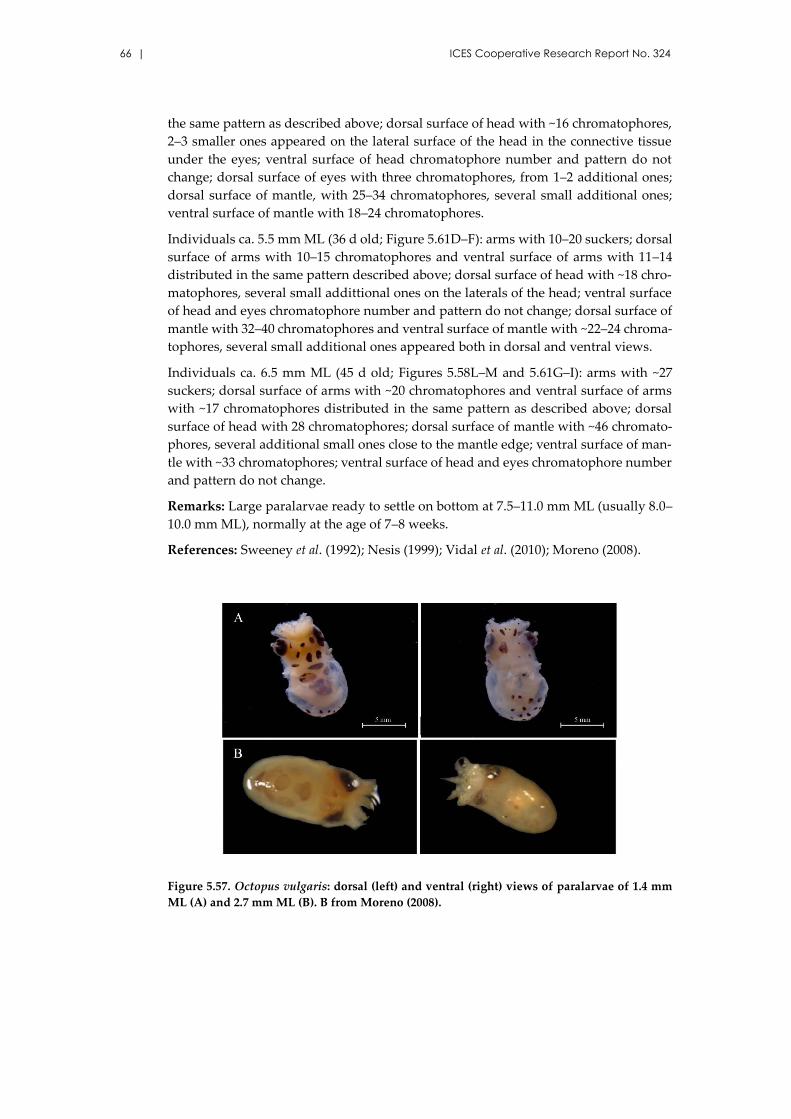
66 | ICES Cooperative Research Report No. 324
the same pattern as described above; dorsal surface of head with ~16 chromatophores,
2–3 smaller ones appeared on the lateral surface of the head in the connective tissue
under the eyes; ventral surface of head chromatophore number and pattern do not
change; dorsal surface of eyes with three chromatophores, from 1–2 additional ones;
dorsal surface of mantle, with 25–34 chromatophores, several small additional ones;
ventral surface of mantle with 18–24 chromatophores.
Individuals ca. 5.5 mm ML (36 d old; Figure 5.61D–F): arms with 10–20 suckers; dorsal
surface of arms with 10–15 chromatophores and ventral surface of arms with 11–14
distributed in the same pattern described above; dorsal surface of head with ~18 chro-
matophores, several small addittional ones on the laterals of the head; ventral surface
of head and eyes chromatophore number and pattern do not change; dorsal surface of
mantle with 32–40 chromatophores and ventral surface of mantle with ~22–24 chroma-
tophores, several small additional ones appeared both in dorsal and ventral views.
Individuals ca. 6.5 mm ML (45 d old; Figures 5.58L–M and 5.61G–I): arms with ~27
suckers; dorsal surface of arms with ~20 chromatophores and ventral surface of arms
with ~17 chromatophores distributed in the same pattern as described above; dorsal
surface of head with 28 chromatophores; dorsal surface of mantle with ~46 chromato-
phores, several additional small ones close to the mantle edge; ventral surface of man-
tle with ~33 chromatophores; ventral surface of head and eyes chromatophore number
and pattern do not change.
Remarks: Large paralarvae ready to settle on bottom at 7.5–11.0 mm ML (usually 8.0–
10.0 mm ML), normally at the age of 7–8 weeks.
References: Sweeney et al. (1992); Nesis (1999); Vidal et al. (2010); Moreno (2008).
Figure 5.57. Octopus vulgaris: dorsal (left) and ventral (right) views of paralarvae of 1.4 mm
ML (A) and 2.7 mm ML (B). B from Moreno (2008).
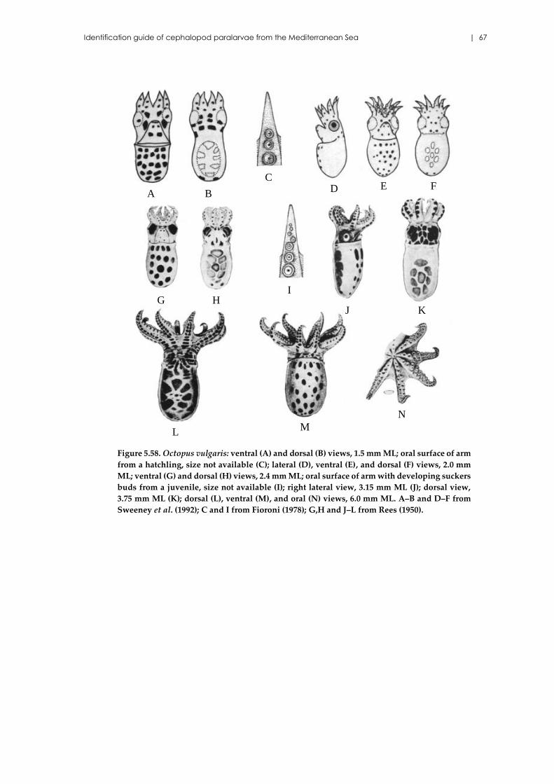
Identification guide of cephalopod paralarvae from the Mediterranean Sea | 67
Figure 5.58. Octopus vulgaris: ventral (A) and dorsal (B) views, 1.5 mm ML; oral surface of arm
from a hatchling, size not available (C); lateral (D), ventral (E), and dorsal (F) views, 2.0 mm
ML; ventral (G) and dorsal (H) views, 2.4 mm ML; oral surface of arm with developing suckers
buds from a juvenile, size not available (I); right lateral view, 3.15 mm ML (J); dorsal view,
3.75 mm ML (K); dorsal (L), ventral (M), and oral (N) views, 6.0 mm ML. A–B and D–F from
Sweeney et al. (1992); C and I from Fioroni (1978); G,H and J–L from Rees (1950).
J
B
C
IHG
D FE
N
ML
K
A
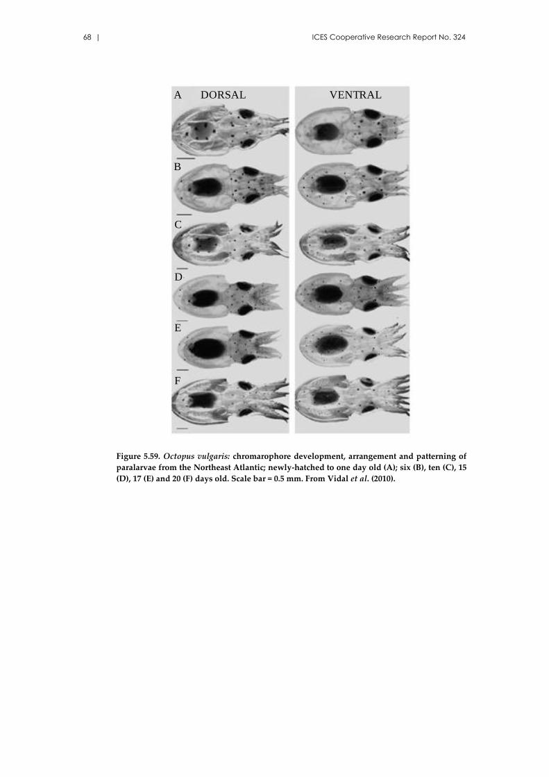
68 | ICES Cooperative Research Report No. 324
Figure 5.59. Octopus vulgaris: chromarophore development, arrangement and patterning of
paralarvae from the Northeast Atlantic; newly-hatched to one day old (A); six (B), ten (C), 15
(D), 17 (E) and 20 (F) days old. Scale bar = 0.5 mm. From Vidal et al. (2010).
A
B
C
D
E
F
DORSAL VENTRAL
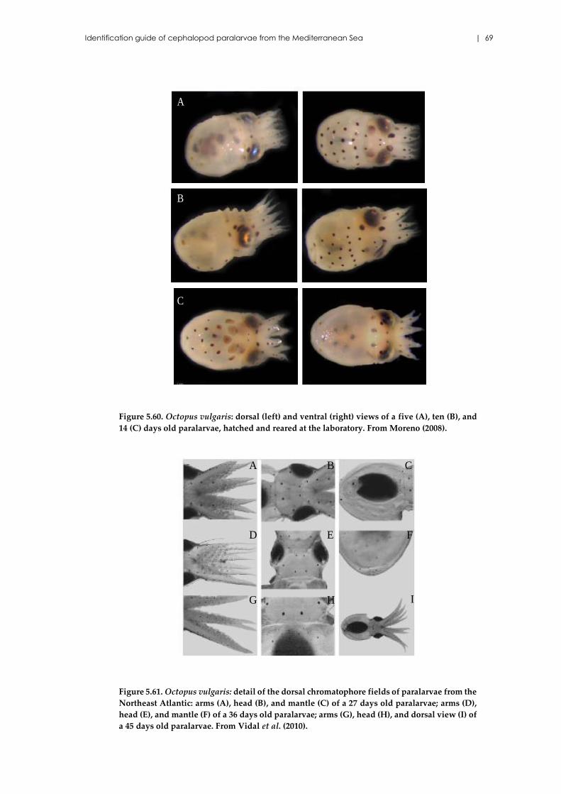
Identification guide of cephalopod paralarvae from the Mediterranean Sea | 69
Figure 5.60. Octopus vulgaris: dorsal (left) and ventral (right) views of a five (A), ten (B), and
14 (C) days old paralarvae, hatched and reared at the laboratory. From Moreno (2008).
Figure 5.61. Octopus vulgaris: detail of the dorsal chromatophore fields of paralarvae from the
Northeast Atlantic: arms (A), head (B), and mantle (C) of a 27 days old paralarvae; arms (D),
head (E), and mantle (F) of a 36 days old paralarvae; arms (G), head (H), and dorsal view (I) of
a 45 days old paralarvae. From Vidal et al. (2010).
A
B
C
A B C
D
IG H
E F
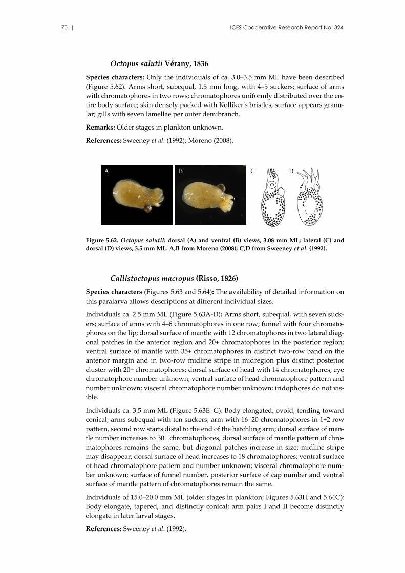
70 | ICES Cooperative Research Report No. 324
Octopus salutii Vérany, 1836
Species characters: Only the individuals of ca. 3.0–3.5 mm ML have been described
(Figure 5.62). Arms short, subequal, 1.5 mm long, with 4–5 suckers; surface of arms
with chromatophores in two rows; chromatophores uniformly distributed over the en-
tire body surface; skin densely packed with Kolliker's bristles, surface appears granu-
lar; gills with seven lamellae per outer demibranch.
Remarks: Older stages in plankton unknown.
References: Sweeney et al. (1992); Moreno (2008).
Figure 5.62. Octopus salutii: dorsal (A) and ventral (B) views, 3.08 mm ML; lateral (C) and
dorsal (D) views, 3.5 mm ML. A,B from Moreno (2008); C,D from Sweeney et al. (1992).
Callistoctopus macropus (Risso, 1826)
Species characters (Figures 5.63 and 5.64): The availability of detailed information on
this paralarva allows descriptions at different individual sizes.
Individuals ca. 2.5 mm ML (Figure 5.63A-D): Arms short, subequal, with seven suck-
ers; surface of arms with 4–6 chromatophores in one row; funnel with four chromato-
phores on the lip; dorsal surface of mantle with 12 chromatophores in two lateral diag-
onal patches in the anterior region and 20+ chromatophores in the posterior region;
ventral surface of mantle with 35+ chromatophores in distinct two-row band on the
anterior margin and in two-row midline stripe in midregion plus distinct posterior
cluster with 20+ chromatophores; dorsal surface of head with 14 chromatophores; eye
chromatophore number unknown; ventral surface of head chromatophore pattern and
number unknown; visceral chromatophore number unknown; iridophores do not vis-
ible.
Individuals ca. 3.5 mm ML (Figure 5.63E–G): Body elongated, ovoid, tending toward
conical; arms subequal with ten suckers; arm with 16–20 chromatophores in 1+2 row
pattern, second row starts distal to the end of the hatchling arm; dorsal surface of man-
tle number increases to 30+ chromatophores, dorsal surface of mantle pattern of chro-
matophores remains the same, but diagonal patches increase in size; midline stripe
may disappear; dorsal surface of head increases to 18 chromatophores; ventral surface
of head chromatophore pattern and number unknown; visceral chromatophore num-
ber unknown; surface of funnel number, posterior surface of cap number and ventral
surface of mantle pattern of chromatophores remain the same.
Individuals of 15.0–20.0 mm ML (older stages in plankton; Figures 5.63H and 5.64C):
Body elongate, tapered, and distinctly conical; arm pairs I and II become distinctly
elongate in later larval stages.
References: Sweeney et al. (1992).
B
A B DC
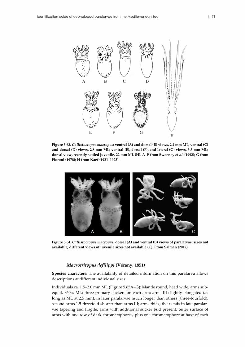
Identification guide of cephalopod paralarvae from the Mediterranean Sea | 71
Figure 5.63. Callistoctopus macropus: ventral (A) and dorsal (B) views, 2.4 mm ML; ventral (C)
and dorsal (D) views, 2.8 mm ML; ventral (E), dorsal (F), and lateral (G) views, 3.3 mm ML;
dorsal view, recently settled juvenile, 22 mm ML (H). A–F from Sweeney et al. (1992); G from
Fioroni (1978); H from Naef (1921–1923).
Figure 5.64. Callistoctopus macropus: dorsal (A) and ventral (B) views of paralarvae, sizes not
available; different views of juvenile sizes not available (C). From Salman (2012).
Macrotritopus defilippi (Vérany, 1851)
Species characters: The availability of detailed information on this paralarva allows
descriptions at different individual sizes.
Individuals ca. 1.5–2.0 mm ML (Figure 5.65A–G): Mantle round, head wide; arms sub-
equal, ~50% ML; three primary suckers on each arm; arms III slightly elongated (as
long as ML at 2.5 mm), in later paralarvae much longer than others (three-fourfold);
second arms 1.5-threefold shorter than arms III; arms thick, their ends in late paralar-
vae tapering and fragile; arms with additional sucker bud present; outer surface of
arms with one row of dark chromatophores, plus one chromatophore at base of each
A B
GFE
DC
H
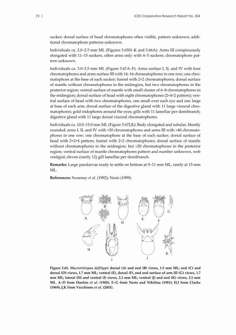
72 | ICES Cooperative Research Report No. 324
sucker; dorsal surface of head chromatophores often visible, pattern unknown; addi-
tional chromatophore patterns unknown.
Individuals ca. 2.0–2.5 mm ML (Figures 5.65H–K and 5.66A): Arms III conspicuously
elongated with 11–15 suckers, other arms only with 4–5 suckers; chromatophore pat-
tern unknown.
Individuals ca. 3.0–3.5 mm ML (Figure 5.67A–F): Arms surface I, II, and IV with four
chromatophores and arms surface III with 14–16 chromatophores in one row; one chro-
matophore at the base of each sucker; funnel with 2+2 chromatophores; dorsal surface
of mantle without chromatophores in the midregion, but two chromatophores in the
posterior region; ventral surface of mantle with small cluster of 6–8 chromatophores in
the midregion; dorsal surface of head with eight chromatophores (2+4+2 pattern); ven-
tral surface of head with two chromatophores, one small over each eye and one large
at base of each arm; dorsal surface of the digestive gland with 11 large visceral chro-
matophores; gold iridophores around the eyes; gills with 11 lamellae per demibranch;
digestive gland with 11 large dorsal visceral chromatophores.
Individuals ca. 10.0–15.0 mm ML (Figure 5.67J,K): Body elongated and tubular, bluntly
rounded; arms I, II, and IV with >20 chromatophores and arms III with >40 chromato-
phores in one row; one chromatophore at the base of each sucker; dorsal surface of
head with 2+2+4 pattern; funnel with 2+2 chromatophores; dorsal surface of mantle
without chromatophores in the midregion, but >20 chromatophores in the posterior
region; ventral surface of mantle chromatophores pattern and number unknown, web
vestigial; eleven (rarely 12) gill lamellae per demibranch.
Remarks: Large paralarvae ready to settle on bottom at 9–11 mm ML, rarely at 15 mm
ML.
References: Sweeney et al. (1992); Nesis (1999).
Figure 5.65. Macrotritopus defilippi: dorsal (A) and oral (B) views, 1.5 mm ML; oral (C) and
dorsal (D) views, 1.7 mm ML; ventral (E), dorsal (F), and oral surface of arm III (G) views, 1.7
mm ML; lateral (H) and ventral (I) views, 2.2 mm ML; ventral (J) and oral (K) views, 2.5 mm
ML. A–D from Hanlon et al. (1985), E–G from Nesis and Nikitina (1981); H,I from Clarke
(1969); J,K from Vecchione et al. (2001).
G
K
A
B
CD
EF
G
H
IJ
K
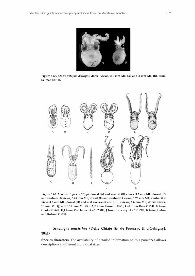
Identification guide of cephalopod paralarvae from the Mediterranean Sea | 73
Figure 5.66. Macrotritopus defilippi: dorsal views, 2.1 mm ML (A) and 3 mm ML (B). From
Salman (2012).
Figure 5.67. Macrotritopus defilippi: dorsal (A) and ventral (B) views, 3.2 mm ML; dorsal (C)
and ventral (D) views, 3.45 mm ML; dorsal (E) and ventral (F) views, 3.75 mm ML; ventral (G)
view, 4.5 mm ML; dorsal (H) and oral surface of arm III (I) views, 6.6 mm ML; dorsal views,
10 mm ML (J) and 13.2 mm ML (K). A,B from Fioroni (1965); C–F from Rees (1954); G from
Clarke (1969); H,I from Vecchione et al. (2001); J from Sweeney et al. (1992); K from Joubin
and Robson (1929).
Scaeurgus unicirrhus (Delle Chiaje [in de Férussac & d’Orbigny],
1841)
Species characters: The availability of detailed information on this paralarva allows
descriptions at different individual sizes.
A B
GH I
A B C D
KJ
E F
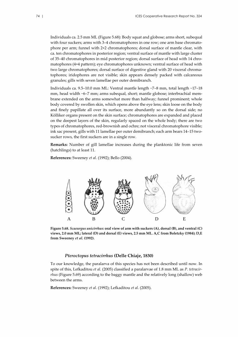
74 | ICES Cooperative Research Report No. 324
Individuals ca. 2.5 mm ML (Figure 5.68): Body squat and globose; arms short, subequal
with four suckers; arms with 3–4 chromatophores in one row; one arm base chromato-
phore per arm; funnel with 2+2 chromatophores; dorsal surface of mantle clear, with
ca. ten chromatophores in posterior region; ventral surface of mantle with large cluster
of 35–40 chromatophores in mid posterior region; dorsal surface of head with 14 chro-
matophores (4+4 pattern); eye chromatophores unknown; ventral surface of head with
two large chromatophores; dorsal surface of digestive gland with 20 visceral chroma-
tophores; iridophores are not visible; skin appears densely packed with calcareous
granules; gills with seven lamellae per outer demibranch.
Individuals ca. 9.5–10.0 mm ML: Ventral mantle length ~7–8 mm, total length ~17–18
mm, head width ~6–7 mm; arms subequal, short; mantle globose; interbrachial mem-
brane extended on the arms somewhat more than halfway; funnel prominent; whole
body covered by swollen skin, which opens above the eye lens; skin loose on the body
and finely papillate all over its surface, more abundantly so on the dorsal side; no
Kölliker organs present on the skin surface; chromatophores are expanded and placed
on the deepest layers of the skin, regularly spaced on the whole body; there are two
types of chromatophores, red-brownish and ochre; not visceral chromatophore visible;
ink sac present, gills with 11 lamellae per outer demibranch; each arm bears 14–15 two-
sucker rows, the first suckers are in a single row.
Remarks: Number of gill lamellae increases during the planktonic life from seven
(hatchlings) to at least 11.
References: Sweeney et al. (1992); Bello (2004).
Figure 5.68. Scaeurgus unicirrhus: oral view of arm with suckers (A), dorsal (B), and ventral (C)
views, 2.0 mm ML; lateral (D) and dorsal (E) views, 2.5 mm ML. A,C from Boletzky (1984); D,E
from Sweeney et al. (1992).
Pteroctopus tetracirrhus (Delle Chiaje, 1830)
To our knowledge, the paralarva of this species has not been described until now. In
spite of this, Lefkaditou et al. (2005) classified a paralarvae of 1.8 mm ML as P. tetracir-
rhus (Figure 5.69) according to the baggy mantle and the relatively long (shallow) web
between the arms.
References: Sweeney et al. (1992); Lefkaditou et al. (2005).
A DCB E
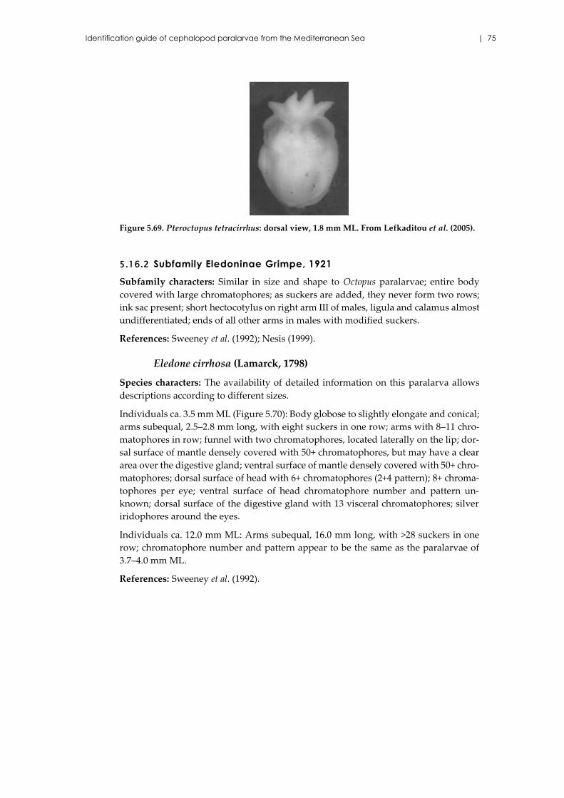
Identification guide of cephalopod paralarvae from the Mediterranean Sea | 75
Figure 5.69. Pteroctopus tetracirrhus: dorsal view, 1.8 mm ML. From Lefkaditou et al. (2005).
5.16.2 Subfamily Eledoninae Grimpe, 1921
Subfamily characters: Similar in size and shape to Octopus paralarvae; entire body
covered with large chromatophores; as suckers are added, they never form two rows;
ink sac present; short hectocotylus on right arm III of males, ligula and calamus almost
undifferentiated; ends of all other arms in males with modified suckers.
References: Sweeney et al. (1992); Nesis (1999).
Eledone cirrhosa (Lamarck, 1798)
Species characters: The availability of detailed information on this paralarva allows
descriptions according to different sizes.
Individuals ca. 3.5 mm ML (Figure 5.70): Body globose to slightly elongate and conical;
arms subequal, 2.5–2.8 mm long, with eight suckers in one row; arms with 8–11 chro-
matophores in row; funnel with two chromatophores, located laterally on the lip; dor-
sal surface of mantle densely covered with 50+ chromatophores, but may have a clear
area over the digestive gland; ventral surface of mantle densely covered with 50+ chro-
matophores; dorsal surface of head with 6+ chromatophores (2+4 pattern); 8+ chroma-
tophores per eye; ventral surface of head chromatophore number and pattern un-
known; dorsal surface of the digestive gland with 13 visceral chromatophores; silver
iridophores around the eyes.
Individuals ca. 12.0 mm ML: Arms subequal, 16.0 mm long, with >28 suckers in one
row; chromatophore number and pattern appear to be the same as the paralarvae of
3.7–4.0 mm ML.
References: Sweeney et al. (1992).
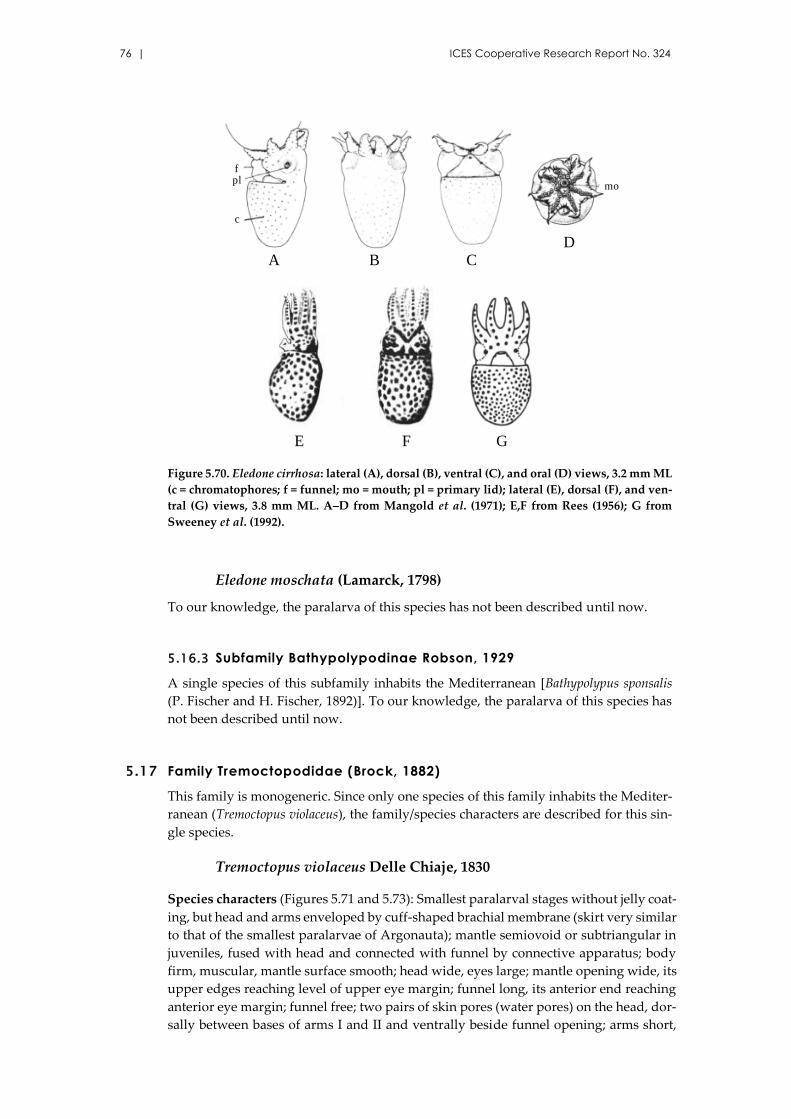
76 | ICES Cooperative Research Report No. 324
Figure 5.70. Eledone cirrhosa: lateral (A), dorsal (B), ventral (C), and oral (D) views, 3.2 mm ML
(c = chromatophores; f = funnel; mo = mouth; pl = primary lid); lateral (E), dorsal (F), and ven-
tral (G) views, 3.8 mm ML. A–D from Mangold et al. (1971); E,F from Rees (1956); G from
Sweeney et al. (1992).
Eledone moschata (Lamarck, 1798)
To our knowledge, the paralarva of this species has not been described until now.
5.16.3 Subfamily Bathypolypodinae Robson, 1929
A single species of this subfamily inhabits the Mediterranean [Bathypolypus sponsalis
(P. Fischer and H. Fischer, 1892)]. To our knowledge, the paralarva of this species has
not been described until now.
5.17 Family Tremoctopodidae (Brock, 1882)
This family is monogeneric. Since only one species of this family inhabits the Mediter-
ranean (Tremoctopus violaceus), the family/species characters are described for this sin-
gle species.
Tremoctopus violaceus Delle Chiaje, 1830
Species characters (Figures 5.71 and 5.73): Smallest paralarval stages without jelly coat-
ing, but head and arms enveloped by cuff-shaped brachial membrane (skirt very similar
to that of the smallest paralarvae of Argonauta); mantle semiovoid or subtriangular in
juveniles, fused with head and connected with funnel by connective apparatus; body
firm, muscular, mantle surface smooth; head wide, eyes large; mantle opening wide, its
upper edges reaching level of upper eye margin; funnel long, its anterior end reaching
anterior eye margin; funnel free; two pairs of skin pores (water pores) on the head, dor-
sally between bases of arms I and II and ventrally beside funnel opening; arms short,
A
E
CBD
F G
c
plf
mo
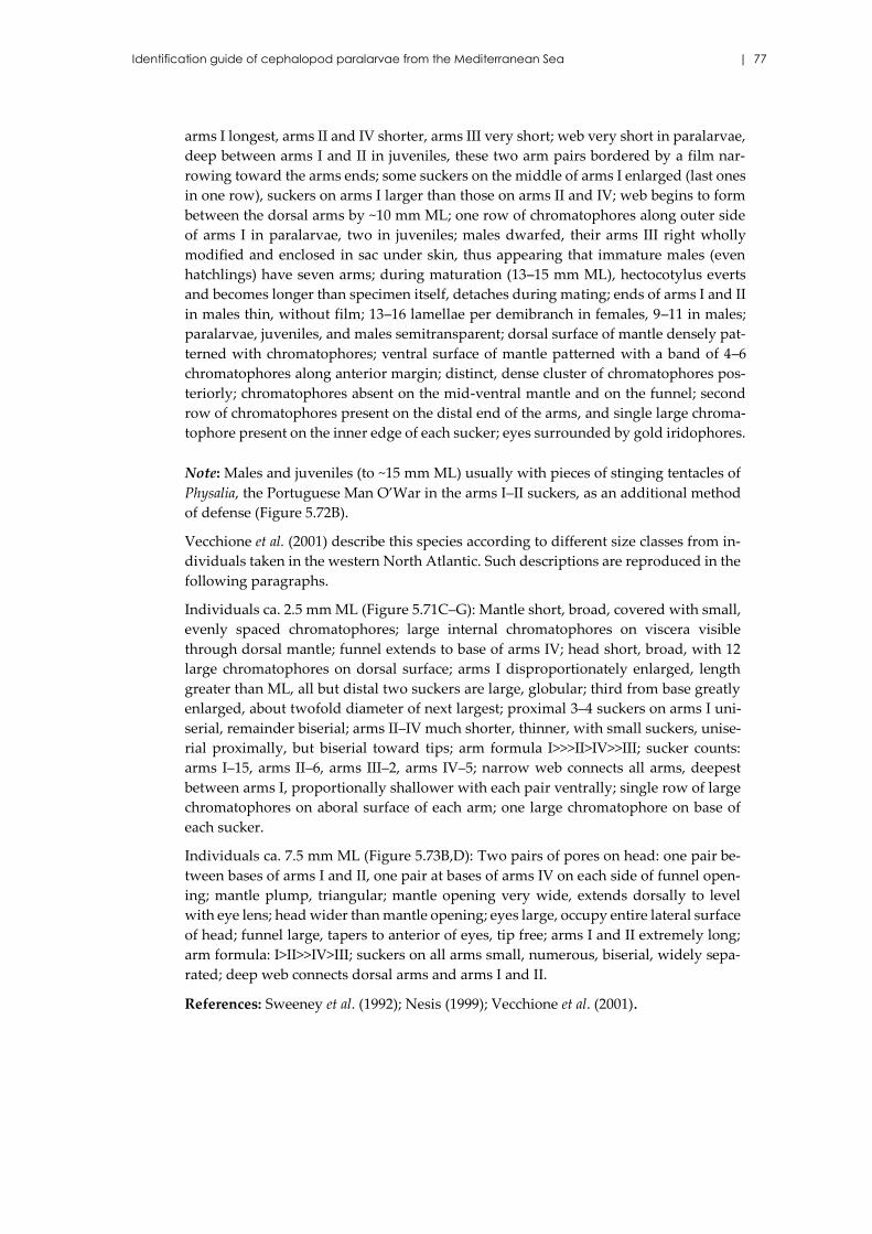
Identification guide of cephalopod paralarvae from the Mediterranean Sea | 77
arms I longest, arms II and IV shorter, arms III very short; web very short in paralarvae,
deep between arms I and II in juveniles, these two arm pairs bordered by a film nar-
rowing toward the arms ends; some suckers on the middle of arms I enlarged (last ones
in one row), suckers on arms I larger than those on arms II and IV; web begins to form
between the dorsal arms by ~10 mm ML; one row of chromatophores along outer side
of arms I in paralarvae, two in juveniles; males dwarfed, their arms III right wholly
modified and enclosed in sac under skin, thus appearing that immature males (even
hatchlings) have seven arms; during maturation (13–15 mm ML), hectocotylus everts
and becomes longer than specimen itself, detaches during mating; ends of arms I and II
in males thin, without film; 13–16 lamellae per demibranch in females, 9–11 in males;
paralarvae, juveniles, and males semitransparent; dorsal surface of mantle densely pat-
terned with chromatophores; ventral surface of mantle patterned with a band of 4–6
chromatophores along anterior margin; distinct, dense cluster of chromatophores pos-
teriorly; chromatophores absent on the mid-ventral mantle and on the funnel; second
row of chromatophores present on the distal end of the arms, and single large chroma-
tophore present on the inner edge of each sucker; eyes surrounded by gold iridophores.
Note: Males and juveniles (to ~15 mm ML) usually with pieces of stinging tentacles of
Physalia, the Portuguese Man O’War in the arms I–II suckers, as an additional method
of defense (Figure 5.72B).
Vecchione et al. (2001) describe this species according to different size classes from in-
dividuals taken in the western North Atlantic. Such descriptions are reproduced in the
following paragraphs.
Individuals ca. 2.5 mm ML (Figure 5.71C–G): Mantle short, broad, covered with small,
evenly spaced chromatophores; large internal chromatophores on viscera visible
through dorsal mantle; funnel extends to base of arms IV; head short, broad, with 12
large chromatophores on dorsal surface; arms I disproportionately enlarged, length
greater than ML, all but distal two suckers are large, globular; third from base greatly
enlarged, about twofold diameter of next largest; proximal 3–4 suckers on arms I uni-
serial, remainder biserial; arms II–IV much shorter, thinner, with small suckers, unise-
rial proximally, but biserial toward tips; arm formula I>>>II>IV>>III; sucker counts:
arms I–15, arms II–6, arms III–2, arms IV–5; narrow web connects all arms, deepest
between arms I, proportionally shallower with each pair ventrally; single row of large
chromatophores on aboral surface of each arm; one large chromatophore on base of
each sucker.
Individuals ca. 7.5 mm ML (Figure 5.73B,D): Two pairs of pores on head: one pair be-
tween bases of arms I and II, one pair at bases of arms IV on each side of funnel open-
ing; mantle plump, triangular; mantle opening very wide, extends dorsally to level
with eye lens; head wider than mantle opening; eyes large, occupy entire lateral surface
of head; funnel large, tapers to anterior of eyes, tip free; arms I and II extremely long;
arm formula: I>II>>IV>III; suckers on all arms small, numerous, biserial, widely sepa-
rated; deep web connects dorsal arms and arms I and II.
References: Sweeney et al. (1992); Nesis (1999); Vecchione et al. (2001).
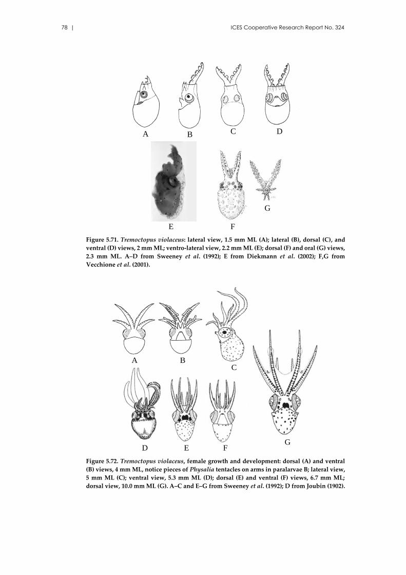
78 | ICES Cooperative Research Report No. 324
Figure 5.71. Tremoctopus violaceus: lateral view, 1.5 mm ML (A); lateral (B), dorsal (C), and
ventral (D) views, 2 mm ML; ventro-lateral view, 2.2 mm ML (E); dorsal (F) and oral (G) views,
2.3 mm ML. A–D from Sweeney et al. (1992); E from Diekmann et al. (2002); F,G from
Vecchione et al. (2001).
Figure 5.72. Tremoctopus violaceus, female growth and development: dorsal (A) and ventral
(B) views, 4 mm ML, notice pieces of Physalia tentacles on arms in paralarvae B; lateral view,
5 mm ML (C); ventral view, 5.3 mm ML (D); dorsal (E) and ventral (F) views, 6.7 mm ML;
dorsal view, 10.0 mm ML (G). A–C and E–G from Sweeney et al. (1992); D from Joubin (1902).
F
G
A B C D
E
BC
E FG
D
A
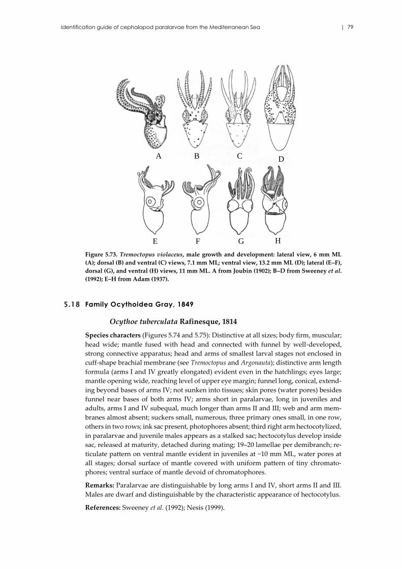
Identification guide of cephalopod paralarvae from the Mediterranean Sea | 79
Figure 5.73. Tremoctopus violaceus, male growth and development: lateral view, 6 mm ML
(A); dorsal (B) and ventral (C) views, 7.1 mm ML; ventral view, 13.2 mm ML (D); lateral (E–F),
dorsal (G), and ventral (H) views, 11 mm ML. A from Joubin (1902); B–D from Sweeney et al.
(1992); E–H from Adam (1937).
5.18 Family Ocythoidea Gray, 1849
Ocythoe tuberculata Rafinesque, 1814
Species characters (Figures 5.74 and 5.75): Distinctive at all sizes; body firm, muscular;
head wide; mantle fused with head and connected with funnel by well-developed,
strong connective apparatus; head and arms of smallest larval stages not enclosed in
cuff-shape brachial membrane (see Tremoctopus and Argonauta); distinctive arm length
formula (arms I and IV greatly elongated) evident even in the hatchlings; eyes large;
mantle opening wide, reaching level of upper eye margin; funnel long, conical, extend-
ing beyond bases of arms IV; not sunken into tissues; skin pores (water pores) besides
funnel near bases of both arms IV; arms short in paralarvae, long in juveniles and
adults, arms I and IV subequal, much longer than arms II and III; web and arm mem-
branes almost absent; suckers small, numerous, three primary ones small, in one row,
others in two rows; ink sac present, photophores absent; third right arm hectocotylized,
in paralarvae and juvenile males appears as a stalked sac; hectocotylus develop inside
sac, released at maturity, detached during mating; 19–20 lamellae per demibranch; re-
ticulate pattern on ventral mantle evident in juveniles at ~10 mm ML, water pores at
all stages; dorsal surface of mantle covered with uniform pattern of tiny chromato-
phores; ventral surface of mantle devoid of chromatophores.
Remarks: Paralarvae are distinguishable by long arms I and IV, short arms II and III.
Males are dwarf and distinguishable by the characteristic appearance of hectocotylus.
References: Sweeney et al. (1992); Nesis (1999).
A B C D
E HGF
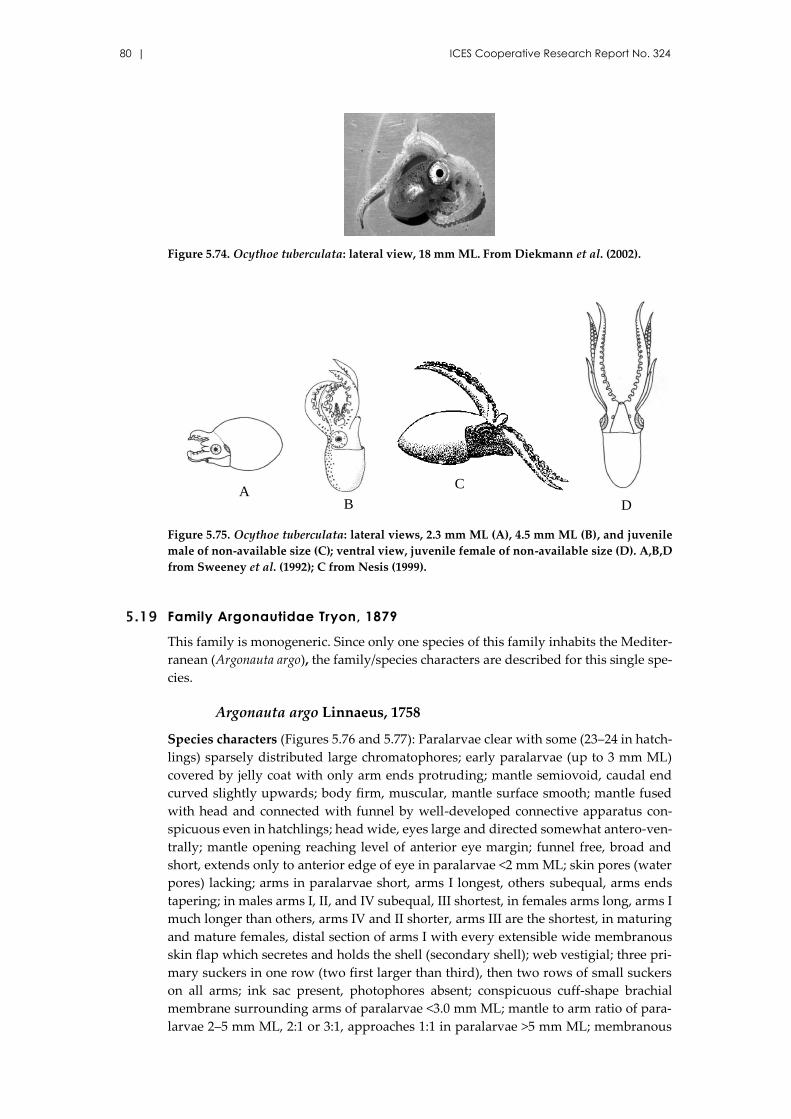
80 | ICES Cooperative Research Report No. 324
Figure 5.74. Ocythoe tuberculata: lateral view, 18 mm ML. From Diekmann et al. (2002).
Figure 5.75. Ocythoe tuberculata: lateral views, 2.3 mm ML (A), 4.5 mm ML (B), and juvenile
male of non-available size (C); ventral view, juvenile female of non-available size (D). A,B,D
from Sweeney et al. (1992); C from Nesis (1999).
5.19 Family Argonautidae Tryon, 1879
This family is monogeneric. Since only one species of this family inhabits the Mediter-
ranean (Argonauta argo), the family/species characters are described for this single spe-
cies.
Argonauta argo Linnaeus, 1758
Species characters (Figures 5.76 and 5.77): Paralarvae clear with some (23–24 in hatch-
lings) sparsely distributed large chromatophores; early paralarvae (up to 3 mm ML)
covered by jelly coat with only arm ends protruding; mantle semiovoid, caudal end
curved slightly upwards; body firm, muscular, mantle surface smooth; mantle fused
with head and connected with funnel by well-developed connective apparatus con-
spicuous even in hatchlings; head wide, eyes large and directed somewhat antero-ven-
trally; mantle opening reaching level of anterior eye margin; funnel free, broad and
short, extends only to anterior edge of eye in paralarvae <2 mm ML; skin pores (water
pores) lacking; arms in paralarvae short, arms I longest, others subequal, arms ends
tapering; in males arms I, II, and IV subequal, III shortest, in females arms long, arms I
much longer than others, arms IV and II shorter, arms III are the shortest, in maturing
and mature females, distal section of arms I with every extensible wide membranous
skin flap which secretes and holds the shell (secondary shell); web vestigial; three pri-
mary suckers in one row (two first larger than third), then two rows of small suckers
on all arms; ink sac present, photophores absent; conspicuous cuff-shape brachial
membrane surrounding arms of paralarvae <3.0 mm ML; mantle to arm ratio of para-
larvae 2–5 mm ML, 2:1 or 3:1, approaches 1:1 in paralarvae >5 mm ML; membranous
AC
DB
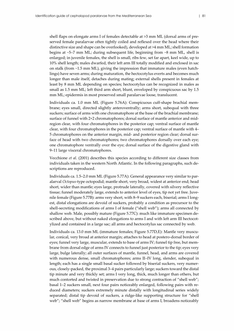
Identification guide of cephalopod paralarvae from the Mediterranean Sea | 81
shell flaps on elongate arms I of females detectable at >3 mm ML (dorsal arms of pre-
served female paralarvae often tightly coiled and reflexed over the head where their
distinctive size and shape can be overlooked), developed at >4 mm ML; shell formation
begins at ~5–7 mm ML; during subsequent life, beginning from ~8 mm ML, shell is
enlarged; in juvenile females, the shell is small, ribs few, set far apart, keel wide, up to
10% shell length; males dwarfed, their left arm III totally modified and enclosed in sac
on stalk (from ~1.5 mm ML), giving the impression that immature males (even hatch-
lings) have seven arms; during maturation, the hectocotylus everts and becomes much
longer than male itself, detaches during mating; external shells present in females at
least by 8 mm ML depending on species; hectocotylus can be recognized in males as
small as 1.5 mm ML; left third arm short, blunt, enveloped by conspicuous sac by 1.5
mm ML; epidermis in most preserved small paralarvae loose, translucent.
Individuals ca. 1.0 mm ML (Figure 5.76A): Conspicuous cuff-shape brachial mem-
brane; eyes small, directed slightly anteroventrally; arms short, subequal with three
suckers; surface of arms with one chromatophore at the base of the brachial membrane;
surface of funnel with 2+2 chromatophores; dorsal surface of mantle anterior and mid-
region clear, with four chromatophores in the posterior cap; ventral surface of mantle
clear, with four chromatophores in the posterior cap; ventral surface of mantle with 4–
5 chromatophores on the anterior margin, mid- and posterior region clear; dorsal sur-
face of head with two chromatophores; two chromatophores dorsally over each eye;
one chromatophore ventrally over the eye; dorsal surface of the digestive gland with
9–11 large visceral chromatophores.
Vecchione et al. (2001) describes this species according to different size classes from
individuals taken in the western North Atlantic. In the following paragraphs, such de-
scriptions are reproduced.
Individuals ca. 1.5–2.0 mm ML (Figure 5.77A): General appearance very similar to par-
alarval Octopus-type octopodid; mantle short, very broad, widest at anterior end; head
short, wider than mantle; eyes large, protrude laterally, covered with silvery reflective
tissue; funnel moderately large, extends to anterior level of eyes, tip not yet free. Juve-
nile female (Figure 5.77B): arms very short, with 8–9 suckers each, biserial; arms I long-
est, distal elongations are devoid of suckers, probably a condition as precursor to the
shell-secreting modifications of arms I of female (“shell web”); arms all connected by
shallow web. Male, possibly mature (Figure 5.77C): much like immature specimen de-
scribed above, but without naked elongations to arms I and with left arm III hectocot-
ylized and contained in a large sac; all arms and hectocotylus sac connected by web.
Individuals ca. 13.0 mm ML (immature females; Figure 5.77D,E): Mantle very muscu-
lar, conical, very broad at anterior margin; attaches to head at postero-dorsal border of
eyes; funnel very large, muscular, extends to base of arms IV; funnel tip free, but mem-
brane from dorsal edge of arms IV connects to funnel just posterior to the tip; eyes very
large, bulge laterally; all outer surfaces of mantle, funnel, head, and arms are covered
with numerous dense, small chromatophores; arms II–IV long, slender, subequal in
length; each has a single small basal sucker followed by biserial suckers, very numer-
ous, closely-packed, the proximal 3–4 pairs particularly large; suckers toward the distal
tip minute and very thickly set; arms I very long, thick, much longer than others, but
much contorted and twisted in preservation due to strong contraction of “shell web”;
basal 1–2 suckers small, next four pairs noticeably enlarged, following pairs with re-
duced diameters; suckers extremely minute distally with longitudinal series widely
separated; distal tip devoid of suckers, a ridge-like supporting structure for “shell
web”; “shell web” begins as narrow membrane at base of arms I, broadens noticeably
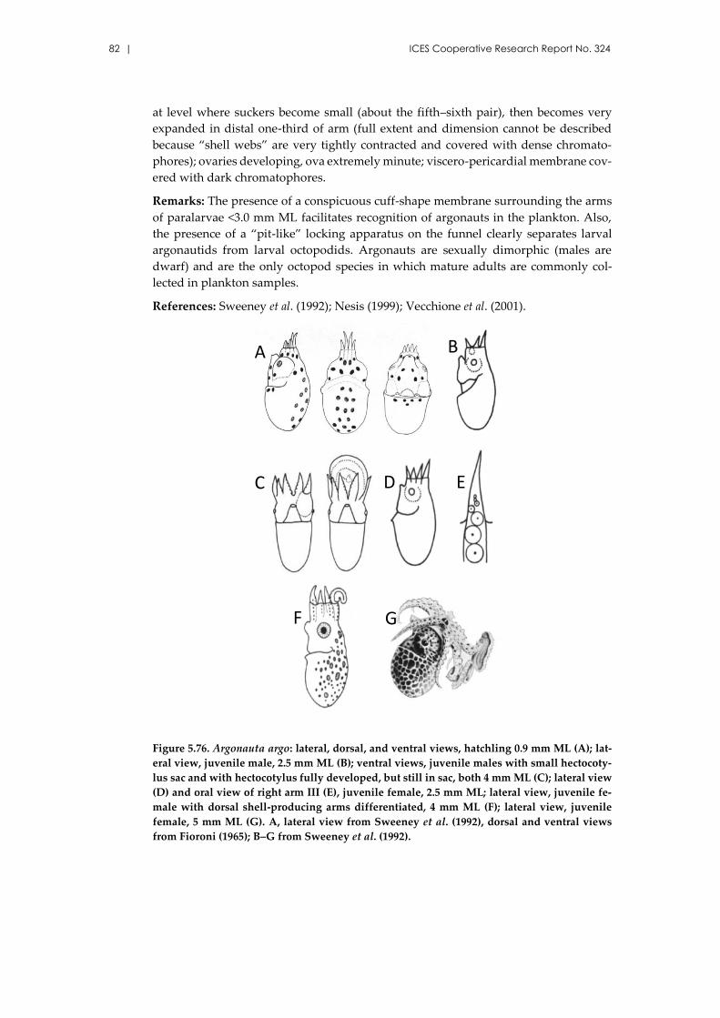
82 | ICES Cooperative Research Report No. 324
at level where suckers become small (about the fifth–sixth pair), then becomes very
expanded in distal one-third of arm (full extent and dimension cannot be described
because “shell webs” are very tightly contracted and covered with dense chromato-
phores); ovaries developing, ova extremely minute; viscero-pericardial membrane cov-
ered with dark chromatophores.
Remarks: The presence of a conspicuous cuff-shape membrane surrounding the arms
of paralarvae <3.0 mm ML facilitates recognition of argonauts in the plankton. Also,
the presence of a “pit-like” locking apparatus on the funnel clearly separates larval
argonautids from larval octopodids. Argonauts are sexually dimorphic (males are
dwarf) and are the only octopod species in which mature adults are commonly col-
lected in plankton samples.
References: Sweeney et al. (1992); Nesis (1999); Vecchione et al. (2001).
Figure 5.76. Argonauta argo: lateral, dorsal, and ventral views, hatchling 0.9 mm ML (A); lat-
eral view, juvenile male, 2.5 mm ML (B); ventral views, juvenile males with small hectocoty-
lus sac and with hectocotylus fully developed, but still in sac, both 4 mm ML (C); lateral view
(D) and oral view of right arm III (E), juvenile female, 2.5 mm ML; lateral view, juvenile fe-
male with dorsal shell-producing arms differentiated, 4 mm ML (F); lateral view, juvenile
female, 5 mm ML (G). A, lateral view from Sweeney et al. (1992), dorsal and ventral views
from Fioroni (1965); B–G from Sweeney et al. (1992).
A B
C D E
F G
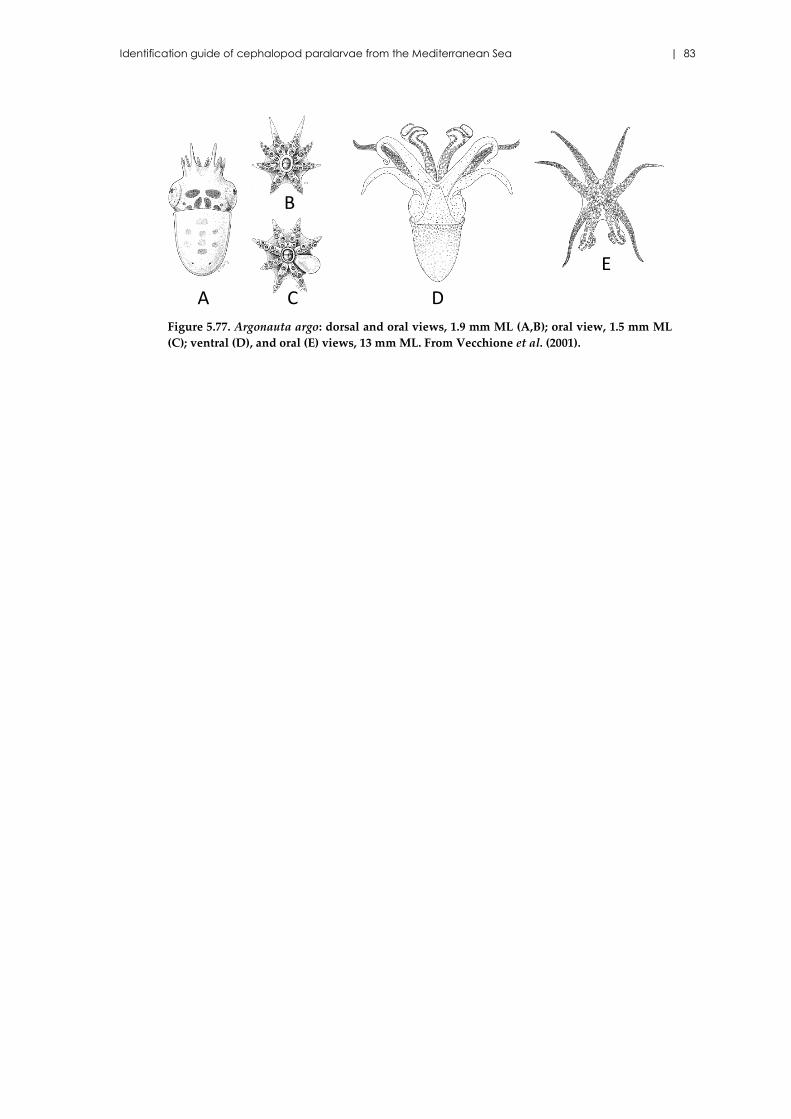
Identification guide of cephalopod paralarvae from the Mediterranean Sea | 83
Figure 5.77. Argonauta argo: dorsal and oral views, 1.9 mm ML (A,B); oral view, 1.5 mm ML
(C); ventral (D), and oral (E) views, 13 mm ML. From Vecchione et al. (2001).
A
B
C D
E

84 | ICES Cooperative Research Report No. 324
6 Acknowledgements
This work was carried out in the framework of the grant AAEE049/09 cofunded by the
Conselleria d’Innovació, Interior i Justícia del Govern de les Illes Balears and the Euro-
pean Regional Development Fund (ERDF). The original pictures of paralarvae were
taken using material from two competitive projects of the Spanish Government I+D+i
National Plan: BALEARES project (CTM 2009-07944 MAR) and the TUNIBAL project
(REN 2003-01176).
Identification guide of cephalopod paralarvae from the Mediterranean Sea | 85
7 References
Adam, W. 1937. Resultats scientifiques des croisières du Navire-école Belge "Mercator,"
VI: Cephalopoda. Memories du Musée Royal d’Historie Naturelle de Belgique,
2(9): 43–82.
Belcari, P., and Sartor, P. 1993. Bottom trawling teuthofauna of the northern Tyrrhenian
Sea. Scientia Marina, 57(2–3): 145–152.
Bello, G. 1995. A key for identification of the Mediterranean sepiolids (Mollusca: Ceph-
alopoda). Bulletin de l’Institut Océanographique de Monaco, 16: 41–55.
Bello, G. 2003. The biogeography of Mediterranean cephalopods. Biogeographia, 33:
209–226.
Bello, G. 2004. Firts record of paralarvae of Scaergus unicirrhus (Cephalopoda: Octo-
podidae). Journal of Plankton Research, 26(12): 1555–1558.
Bello, G. 2008. Cephalopoda. Biologia Marina Mediterranea, 15 (Suppl.): 318–322.
Berry, S. S. 1912. A review of the cephalopods of Western North America. Bulletin of
the Bureau of Fisheries, 30: 269–336.
Boletzky, S. von. 1984. The embryonic development of the octopus Scaeurgus unicirrhus
(Mollusca, Cephalopoda), additional data and discussion. Vie et Milieu, 34(2/3):
87–93.
Boletzky, S. von. 2003. Biology of early life stages in cephalopod Molluscs. Advances
in Marine Biology, 44: 143–203.
Boyle, P. R., and Rodhouse, P. 2005. Cephalopods: Ecology and Fisheries. Blackwell
Science, Oxford.
Cherel, Y., Ridoux, V., Spitz, J., and Richard, P. 2009. Stable isotopes document the
trophic structure of a deep-sea cephalopod assemblage including giant octopod
and giant squid. Biology Letters, 5: 364–367.
Chun, C. 1915. Die Cephalopoden, II: Myopsida, Octopoda. Wissenschaftliche
Ergebnisse der Deulschen Tiefsee-Expedition "Valdivia" 1898–1899, 18: 311–552.
Clarke, M. R. 1966. A review of the systematics and ecology of oceanic squids. Ad-
vances in Marine Biology, 4: 91–300.
Clarke, M. R. 1969. Cephalopoda collected on the Sond cruise. Journal of the Marine
Biological Association of the United Kingdom, 49: 961–976.
Clarke, M. R. 1996. The role of cephalopods in the world’s oceans: general conclusion
and the future. Philosophical Transaction of the Royal Society B: Biological Sci-
ences, 351(1343): 1105–1112.
Diekmann, R., Piatkowski, U., and Schneider, M. 2002. Early life and juvenile cephalo-
pods around seamounts of the subtropical eastern North Atlantic: illustrations
and a key for their identification. Berichte aus dem Institut für Meereskunde an
der Christian-Aabrechts-Universität Kiel, 326: 1–42.
Diekmann, R and Piatkowski, U. 2002. Early life stages of cephalopods in the Sargasso
Sea: distribution and diversity relative ti hydrographic conditions. Marine Biol-
ogy, 141: 123–130.
FAO. 2012. The State of World Fisheries and Aquaculture. Rome. 209 pp.
86 | ICES Cooperative Research Report No. 324
Fioroni, P. 1965. Die embryonale Musterentwicklung bei einigen mediterraneen
Tintenfischarten. Vie et Milieu, 16: 655–756.
Fioroni, P. 1978. Cephalopoda, Tintenfische. In Morphogenese der Tiere; Lieferung 2,
G5-I. Ed. by F. Seidel. Gustv Fischer Verlag, Stuttgart. 181 pp.
Guerra, A. 1992. Mollusca, Cephalopopoda. In Fauna Ibérica, vol. 1. Ed. by M. A. Ra-
mos, et al. Museo Nacional de Ciencias Naturales. Consejo Superior de Investi-
gaciones Científicas, Madrid. 327 pp.
Guerra, A., González, A. F., Rocha, F. J., Sagarminaga, R., and Cañadas, A. 2002. Plank-
tonic egg masses of the diamond-shaped squid Thysanoteuthis rhombus in the
eastern Atlantic and the Mediterranean Sea. Journal of Plankton Research, 24(4):
333–338.
González, A. F., Otero, J., Guerra, A., Prego, R., Rocha, F. J., and Dale, A. W. 2005. Dis-
tribution of common octopus and common squid paralarvae in a wind-driven
upwelling area (Ria of Vigo, northwestern Spain). Journal of Plankton Research,
27: 271–277.
González, M., and Sánchez, P. 2002. Cephalopod assemblages caught by trawling along
the Iberian Peninsula Mediterranean coast. Scientia Marina, 66(suppl. 2): 199–
208.
Haimovici, M., Vidal, E. A. G., and Perez, J. A. A. 1995. Larvae of Illex argentinus from
five surveys on the continental shelf of southern Brazil. ICES Marine Science
Symposia, 199: 414–424.
Haimovici, M., Piatkowski, U., and Aguilar dos Santos, R. 2002. Cephalopod paralar-
vae around tropical seamounts and oceanic islands off north-eastern coast of
Brazil. Bulletin of Marine Science, 71(1): 313–330.
Hanlon, R. T., Forsythe, J. W., and Boletzky, S. von. 1985. Field and laboratory behavior
of "Macrotritopus larvae" reared to Octopus defilippi Verany, 1851 (Mollusca:
Cephalopoda). Vie et Milieu, 35(3/4): 237–242.
Hastie, L. C., Allcock, A. L., Jereb, P., Lefkaditou, E., Moreno, A., Oesterwina, D., and
Pierce, G. J. 2013. Chapter IV: Alloteuthis subulata, European Common Squid. In
Advances in Squid Biology, Ecology and Fisheries. Part I–Mysid Squids, pp. 52–
100. Ed. by R. Rosa, R. O’Dor, and G. J. Pierce. Nova Biomedical, New York. 349
pp.
Joubin, L. 1902. Notice sur les travaux scientifiques. Francis Simon, Rennes, France. 134
pp.
Joubin, L., and Robson, G. C. 1929. On a new species of Macrotritopus obtained by Dr.
J. Schmidt's "Dana" expedition, with remarks on the Genus. Proceedings of the
Zoological Society of London, 1: 89–94.
Kubodera, T., and Okutani, T. 1981. The systematics and identification of larval ceph-
alopods from the northern North Pacific. Research Institute of the North Pacific
Fisheries, Hokkaido University, Special volume, 131–159.
Lefkaditou, E., Papaconstantinou, C., and Anastasopoulou, K. 1999. Juvenile cephalo-
pods collected in the midwater macroplankton over a trench in the Aegean Sea
(northeastern Mediterranean). Israel Journal of Zoology, 45: 395–405.
Identification guide of cephalopod paralarvae from the Mediterranean Sea | 87
Lefkaditou, E., Siapatis, A., and Somarakis, S. 2005. Juvenile planktonic cephalopods
sampled off the coasts of central Greece (Eastern Mediterranean) during winter.
Phuket Marine Biological Centre Research Bulletin, 66: 259–265.
Mangold, K., Boletzky, S. von, and Frosch, D. 1971. Reproductive biology and embry-
onic development of Eledone cirrosa (Cephalopoda, Octopoda). Marine Biology,
8: 109–117.
Martins, R. S., and Pérez, J. A. A. 2006. Occurrence of loliginid paralarvae around Santa
Catarina Island, Southern Brazil. Pan-American Journal of Aquatic Sciences, 1:
24–27.
Miyahara, K., Fukui, K., Nagahama, T., and Ohatani, T. 2006. First record of planktonic
egg masses of the diamond squid, Thysanoteuthis rhombus Troschel, in the Sea of
Japan. Plankton and Benthos Research, 1(1): 59–63.
Morales-Nin, B., Moranta, J., García, C., Tugores, M. P., Grau, A. M., Riera, F., and
Cerdà, M. 2005. The recreational fishery off Majorca Island (western Mediterra-
nean): some implications for coastal resource management. ICES Journal of Ma-
rine Science, 62: 727–739.
Moreno, A. 2008. Geographic variation in cephalopod life history traits. Dissertação
apresentada para accesso à categoria de Investigador Auxiliar. Unidade de Re-
cursos Marinhos e Sustentabilidade L-IPIMAR. 181 pp.
Moreno, A., Santos, A., Piatkowski, U., Santos, A. M. P., and Cabral, H. 2009. Distribu-
tion of cephalopod paralarvae in relation to the regional oceanography of the
western Iberia. Journal of Plankton Research, 31: 73–91.
Moreno, A., Pierce, G. J., Azevedo, M., Pereira, J., and Santos, A. M. P. 2012. The effect
of temperature on growth of early life stages of the common squid Loligo vulgaris.
Journal of Marine Biological Association of the United Kingdom, 92:, 1619-1628.
Naef, A. (1921–1923). Die Cephalopoden (Systematik). In Fauna e Flora del Golfo di
Napoli. Monograph 35, Pubblicazioni della Stazione Zoologica di Napoli. R.
Friedländer and Sohn, Berlin. 863 pp.
Nesis, K. N. 1999. Cephalopoda. In South Atlantic zooplankton, vol. 1, pp. 707–795. Ed.
by D. Boltovskoy. Backhuys Publishers, Leiden.
Nesis, K. N., and Nikitina, L. V. 1981. Macrotritopus, a planktonic larva of the benthic
octopus Octopus defilippi: identification and distribution. Zool Zhurnal, 60(6):
835–847.
Nixon, M., and Mangold, K. 1998. The early life of Sepia officinalis, and the contrast with
that of Octopus vulgaris (Cephalopoda). Journal of Zoology, 245: 407–421.
Otero, J., Alvarez-Salgado, X. A., González, A. F., Gilcoto, M., and Guerra, A. 2009.
High-frequency coastal upwelling events influence Octopus vulgaris larval dy-
namics on the NW Iberian shelf. Marine Ecology Progress Series, 386: 123–132.
Piatkowski, U. 1998. Modern target sampling techniques provide new insights into the
biology of early life stages of pelagic cephalopods. Biologia Marina Mediterra-
nea, 5: 260–272.
Piatkowski, U., Pierce, G. J., and Morais da Cunha, M. 2001. Impact of cephalopods in
the food chain and their interaction with the environment: preface. Fisheries Re-
search, 52: 5–
88 | ICES Cooperative Research Report No. 324
Quetglas, A., Alemany, F., Carbonell, A., Merella, P., and Sánchez, P. 1998. Some as-
pects of the biology of Todarodes sagittatus (Cephalopoda: Ommastrephidae)
from the Balearic Sea (Western Mediterranean). Scientia Marina, 62: 73–82.
Quetglas, A., Alemany, F., Carbonell, A., and Sánchez, P. 1999. First record of Cranchia
scabra Leach, 1817 (Cephalopoda: Cranchiidae) in the Mediterranean Sea. Bol-
letino Malacologico, 35(1–4): 1–2.
Quetglas, A., Carbonell, A., and Sánchez, P. 2000. Demersal continental shelf and up-
per slope cephalopod assemblages from de Balearic Sea (North-Western Medi-
terranean). Biological aspects of some deep-sea species. Estuarine, Coastal and
Shelf Science, 50: 739–749.
Rees, W. J. 1950. The distribution of Octopus vulgaris Lamarck in British waters. Journal
of the Marine Biological Association of the United Kingdom, 29: 361–378.
Rees, W. J. 1953. The Octopus larvae of the "Thor." Proceedings of the Malacological
Society of London, 29(6): 215–218.
Rees, W. J. 1954. The Macrotritopus problem. Bulletin of the British Museum (Natural
History), 2(4): 67–100.
Rees, W. J. 1956. Notes on the European species of Eledone, with special reference to
eggs and larvae. Bulletin of the British Museum (Natural History), 3(6): 283–293.
Roper, C. F. E. 1972. Ecology and vertical distribution of Mediterranean pelagic ceph-
alopods. Mediterranean biological studies. Washington: Smithsonian Institu-
tion. Final report, 5: 282–346.
Roper, C. F. E. 1974. Vertical and seasonal distribution of pelagic cephalopods in the
Mediterranean Sea: preliminary report. Bulletin of the American Malacological
Union, 39: 27–30.
Roper, C. F. E., and Lu, C. C. 1979. Rynchoteuthion larvae of ommastrephid squids of
the western north Atlantic, with the first decription of the larvae and juveniles
of Illex illecebrosus. Proceedings of the Biological Society of Washington, 91: 1039–
1059
Salman, A. 2012. Two new records paralarva in the Eastern Mediterranean (Cephalo-
pods: Mollusca). Journal of the Black Sea/Mediterranean Environment, 18(2):
197–207.
Salman, A., Katagan, T., and Benli, H. A. 2003. Vertical distribution and abundance of
juvenile cephalopods in the Aegean Sea. Scientia Marina, 67(2): 167–176.
Sánchez, P., Belcari, P., and Sartor, P. 1998. Composition and spatial distribution of
cephalopods in two north-western Mediterranean areas. South African Journal
of Marine Science, 20: 17–24.
Sánchez, P., and Molí, B. 1985. An annotated list of cephalopod larvae collected off the
Mediterranean coast of Spain, 1976-1981. Vie et Milieu, 35(3/4): 171–173.
Sartor, P., Belcari, P., Carbonell, A., Gonzalez, M., Quetglas, A., and Sánchez, P. 1998.
The importance of cephalopods to trawl fisheries in the western Mediterranean.
South African Journal of Marine Science, 20: 67–72.
Stephen, S. J. 1985. The distribution of larvae of the genus Octopoteuthis Rupell, 1844
(Cephalopoda, Teuthoidea). Vie et Milieu, 35(3/4): 175–179.
Identification guide of cephalopod paralarvae from the Mediterranean Sea | 89
Sweeney, M. J., Roper, C. F. E., Mangold, K. M., Clarke, M. R., and Boletzky, S. von.
(Eds). 1992. “Larval” and juvenile cephalopods: a manual for their identification.
Smithsonian Contributions to Zoology, 513: 1–282.
Turk, P. E., Hanlon, R. T., Bradford, L. A, and Yang, W.T. 1986. Aspects of feeding,
growth and survival of the European squid Loligo vulgaris Lamarck 1799, reared
through the early growth stages. Vie et Milieu, 36: 9–13.
Vecchione, M. 1987. Juvenile ecology. In Cephalopod Life Cycles, Vol. II, Comparative
Reviews, pp. 61–84. Ed. by P. R. Boyle. Academic Press, London.
Vecchione, M., Roper, C. F. E., and Sweeney, M. J. 2001. Distribution, relative abun-
dance and developmental morphology of larval cephalopods in the western
North Atlantic Ocean. NOAA Technical Reports NMFS 152. 54 pp.
Vidal, E. A. G., Fuentes, L., and da Silva, L. B. 2010. Defining Octopus vulgaris popula-
tions: A comparative study of the morphology and chromatophore pattern of
paralarvae from Northeastern and Southwestern Atlantic. Fisheries Research,
106: 199–208.
Villanueva, R. 1992. Deep-sea cephalopods of the north-western Mediterranean: indi-
cations of up-slope ontogenetic migration in two bathybenthic species. Journal
of Zoology, 227: 267–276.
Villanueva, R. 1995. Experimental rearing and growth of planktonic Octopus vulgaris
from hatching to settlement. Canadian Journal of Fisheries and Aquatic Sciences,
52: 2639–2650.
Villanueva, R. 2000. Effect of temperature on statolith growth of the European squid
Loligo vulgaris during early life. Marine Biology, 136: 449–460.
Villanueva, R., and Bustamante, P. 2006. Composition in essential and non-essential
elements of early stages of cephalopods and dietary effects on the elemental pro-
files of Octopus vulgaris paralarvae. Aquaculture, 261: 225–240.
Villanueva, R., Collins, M. A., Sánchez, P., and Voss, N. A. 2002. Systematics, distribu-
tion and biology of the cirrate octopods of the genus Opisthoteuthis (Mollusca,
Cephalopoda) in the Atlantic Ocean, with description of two new species. Bul-
letin of Marine Science, 71(2): 933–985.
Villanueva, R., Moltschaniwskyj, N. A., and Bozzano, A. 2007. Abiotic influences on
embryo growth: statoliths as experimental tools in the squid early life history.
Reviews in Fish Biology and Fisheries, 17: 101–110.
Voss, N. A. 1969. A monograph of the Cephalopoda of the North Atlantic. The Family
Histioteuthidae. Bulletin of Marine Science, 19: 713–867.
Voss, N. A. 1980. A generic revision of the Cranchiidae (Cephalopoda; Oegopsida).
Bulletin of Marine Science, 30: 365–412.
Voss, N. A., and Voss, G. L. 1962. Two new species of squids of the Genus Calliteuthis
from the Western Atlantic with a redescription of Calliteuthis reversa Verrill. Bul-
letin of Marine Science of the Gulf and Caribbean, 12(2): 169–200.
Wakabayashi, T., Tsuchiya K., and Segawa, S. 2005. Morphological changes with
growth in the paralarvae of the diamondback squid Thysanoteuthis rhombus Tro-
schel, 1857. Phuket Marine Biological Center Research Bulletin, 66: 167–174.
90 | ICES Cooperative Research Report No. 324
Watanabe, K., Sakurai, Y., Segawa, S., and Okutani, T. 1996. Development of the om-
mastrephid squid Todarodes pacificus, from fertilized egg to rhynchoteuthion par-
alarva. American Malacological Bulletin, 13: 73–88.
Yamamoto, K., and Okutani, T. 1975. Studies on early life history of Decapodan Mol-
lusca, V: Systematics and distribution of epipelagic larvae of Decapod cephalo-
pods in the southwestern waters of Japan during the summer of 1970. Bulletin
of the Tokai Regional Fisheries Research Laboratory, 83: 45–96.
Yau, C. 1994. The ecology and ontogeny of cephalopod juveniles in Scottish waters.
PhD thesis, University of Aderdeen, Scotland.
Young, R. E., and Harman, R. F. 1988. “Larva”, “paralarva” and “subadult” in cepha-
lopod terminology. Malacologia, 29: 201–207.
Young, R. E., Harman, R. F., and Hochberg, F. G. 1989. Octopodid paralarvae from
Hawaiian waters. Veliger 32(2): 152–165.
Zeidberg, L. D., and Hamner, W. M. 2002. Distribution of squid paralarvae, Loligo opal-
escens (Cephalopoda: Myopsida), in the Southern California Bight in the three
years following the 1997–1998 El Niño. Marine Biology, 141: 111–122.

Identification guide of cephalopod paralarvae from the Mediterranean Sea | 91
8 Author contact information
Núria Zaragoza
Instituto Español de Oceanografía (IEO)
Centre Oceanogràfic de les Balears
Moll de Ponent, s/n
07015-Palma de Mallorca, Spain
Antoni Quetglas
Instituto Español de Oceanografía (IEO)
Centre Oceanogràfic de les Balears
Moll de Ponent, s/n
07015-Palma de Mallorca, Spain
Ana Moreno
Instituto Português do Mar e da Atmosfera (IPMA)
Departamento do Mar
Avenida de Brasília, 1449-006
Lisboa, Portugal