Embed Size (px)
Citation preview
IDEXX VetAutoreadHematology Analyzer
Operator’s Guide
™

Proprietary Rights Notice
Information in this document is subject to change without notice. Companies, names and data used in examples are fictitious unless otherwise noted. No part of this document may be reproduced or transmitted in any form or by any means, electronic, mechanical or otherwise, for any purpose, without the express written permission of IDEXX Laboratories. IDEXX may have patents or pending patent applications, trademarks, copyrights or other intellectual or industrial property rights covering this document or subject matter in this document. The furnishing of this document does not give a license to these property rights except as expressly provided in any written license agreement from IDEXX Laboratories.
© 2008 IDEXX Laboratories, Inc. All rights reserved. • 06-02822-05
IDEXX VetLab, VetTest, VetLyte, IDEXX VetCentrifuge, VetTube and VetCom are trademarks or registered trademarks of IDEXX Laboratories, Inc. in the United States and/or other countries. VetAutoread and E-Z Prep are trademarks of QBC Diagnostics, Inc.
One IDEXX DriveWestbrook, Maine 04092 USA
idexx.com

v
Contents
Introduction .................................................................................................................................... 1Welcome ..............................................................................................................................................1Contact Us ...........................................................................................................................................1Overview ...............................................................................................................................................2Components ........................................................................................................................................2Other Required Materials .....................................................................................................................3
Setup Procedures .......................................................................................................................... 4Setting Up the IDEXX VetAutoread™ Hematology Analyzer ................................................................4
Setting the Language.....................................................................................................................6Setting up VetTest Communication ................................................................................................7Viewing the Software Version .........................................................................................................7Selecting the Printer Type ..............................................................................................................7Setting the Printout Units (non-VetTest analyzer connection only) ................................................8Setting the Printout Format ............................................................................................................8Setting Letterhead Information (non-VetTest analyzer connection only) .......................................9Setting the Date and Time (non-VetTest analyzer connection only) ..............................................9
IDEXX VetAutoread Analyzer Precautions ...........................................................................................9Setting Up the IDEXX VetCentrifuge ..................................................................................................10
Connecting the Power Supply .....................................................................................................10Disconnecting the Power Supply .................................................................................................10Spinning a Sample .......................................................................................................................10General Operation Notes and Precautions .................................................................................11
Setting Up the IDEXX Fibrinogen Precipitator ...................................................................................12
Sample Collection and Preparation ............................................................................................ 14Preparing a Canine, Feline or Equine Sample ...................................................................................14Preparing a Bovine Sample ...............................................................................................................16
Running Tests on the IDEXX VetAutoread™ Analyzer ................................................................ 19Before You Begin Testing ...................................................................................................................19Calibration Check ...............................................................................................................................19Test Procedure ...................................................................................................................................20Flashes and Flags ..............................................................................................................................20Dashes ...............................................................................................................................................21
Interpreting Test Results .............................................................................................................. 22Principles of the IDEXX VetAutoread Hematology Analyzer ..............................................................22Reading Test Results .........................................................................................................................23
Messages and Symbols ..............................................................................................................24Technical Notes ............................................................................................................................24
Buffy Coat Profile Graph ....................................................................................................................25
vi
IDEXX VetAutoread Hematology Analyzer Operator’s Guide
Understanding the Buffy Coat Profile Graph .............................................................................. 26Normal Sample ..................................................................................................................................26Reticulocytes ......................................................................................................................................27Nucleated Red Blood Cells ................................................................................................................28Eosinophils .........................................................................................................................................29Aggregated Platelets .........................................................................................................................29Clotted Samples ................................................................................................................................30Fibrinogen ..........................................................................................................................................30
Reference Intervals ...................................................................................................................... 31
Technical Notes ............................................................................................................................ 32Startup Service Alerts .........................................................................................................................32Test Alerts ...........................................................................................................................................32Sample Alerts .....................................................................................................................................33
Buffy Coat Profile Reference Graphs .......................................................................................... 37Normal Dog—Eosinophils Identified .................................................................................................37Dog—Elevated Granulocytes and Lymphocytes/Monocytes ...........................................................38Normal Cat .........................................................................................................................................39Anemic Cat .........................................................................................................................................40Normal Horse .....................................................................................................................................41Horse—Elevated Granulocytes and Lymphocytes/Monocytes .........................................................42Dog—Regenerative Anemia ..............................................................................................................43Dog—Regenerative Anemia with Reticulocytes and nRBCs ...........................................................44Dog—Possible Lymphopenia or Inadequate Staining ......................................................................45Cat—Clumped Platelets ....................................................................................................................46Dog—Uneven Layering .....................................................................................................................47Cat—Uneven Layering with Clotting ..................................................................................................48Dog—Additional Platelets ..................................................................................................................49
Specifications ............................................................................................................................... 50
Appendix A: IDEXX VetLab® Installation Guide .......................................................................... 51Connecting the VetTest®, VetLyte® and IDEXX VetAutoread™ Analyzers to the
IDEXX VetLab® Station .................................................................................................................51
1
Introduction
WelcomeCongratulations on becoming a new owner of the IDEXX VetAutoread™ Hematology Analyzer. We want you to gain the most from your investment, and are committed to serving your needs. If you have any problems or have any suggestions on how we can improve our service to you, please give us a call.
In most cases, an IDEXX representative will install your analyzer and train your staff on its proper use. However, we recommend that you carefully read the sections on Sample Collection and Preparation, and Reading Test Results.
It is especially important that you learn to read and use the buffy coat profile graph as part of your analysis. It is produced through the unique staining methods of the analyzer and provides a virtual “picture” of the blood, much like a blood film. And like human fingerprints, they are unique to each animal.
You can expect to hear from us, too, as we strive to make your IDEXX VetAutoread Hematology Analyzer help you practice better medicine. We’ll send updates by mail with full instructions that can be stored with this manual.
Contact Us
IDEXX Representative:
Telephone/Voice Mail:
United States/CanadaToll-free Technical Support ................................................1-800-248-2483
Toll-free Fax .......................................................................1-800-248-3010
www.idexx.com
EuropeToll-free Technical Support ............................................. 00800-1234-3399
Toll-free Fax .................................................................... 00800-1234-3333
www.idexx.nl
AustraliaToll-free Technical Support ................................................... 1800-655-978
Toll-free Fax .......................................................................... 1800-634-409
www.idexx.com.au
JapanToll-free Technical Support ..............................................81-0120-71-4921
Toll-free Fax .....................................................................81-0422-71-4922
www.idexx.co.jp
2
IDEXX VetAutoread Hematology Analyzer Operator’s Guide
OverviewIDEXX has used scientific methods to develop the IDEXX VetAutoread Hematology Analyzer as a valuable blood analysis tool to help you practice better medicine. In little more than 10 minutes, within the control of your own practice and technicians, the analyzer offers quantification of twelve important hematological parameters, including special notes to indicate whether or not an anemia is regenerative. Fast analysis is especially important in studying blood, as its composition begins to change immediately upon drawing of the sample.
The importance of cell morphologyGood laboratory methodology calls for a stained blood film to complete analyses. A blood film provides the necessary morphology to fully investigate the nature of the condition. The IDEXX VetAutoread Hematology Analyzer will provide informational notes on the printout whenever a blood film may be helpful to complete your blood examination. However, it is good practice to briefly examine blood films from all cases.
Attention to sample qualityThe IDEXX VetAutoread Hematology Analyzer uses special notes and flags (the # symbol) to indicate if the sample’s quality may affect the accuracy of the results. On such samples, we recommend that you examine the graph for abnormalities, and in some cases, we suggest that you redraw and retest to obtain the best results.
Additional flaggingOn extremely sick animals, the analyzer may flag results. The flags simply indicate that you should examine the graph and investigate the blood film for complete understanding.
Finally, a clinical diagnosis is a conclusion based on science that necessitates the full integration of a detailed medical history and a careful physical examination. The interpretations offered by the IDEXX VetAutoread Hematology Analyzer printout are only suggestions based upon a limited examination of a portion of the patient’s hematologic status. The statements have value only to a clinician who is able to use them as part of the complete diagnostic process.
ComponentsYour IDEXX VetAutoread Hematology Analyzer will be installed by an IDEXX representative. Choose a work area that is stable and flat for the analyzer and samples. Be sure to keep blood samples away from direct sunlight and other heat sources.
Verify the following components and accessories in each carton:
IDEXX VetAutoread Hematology Analyzer Carton• IDEXX VetAutoread Hematology Analyzer
• Power pack
• Power cord
• IDEXX E-Z Prep™ pipettor
• Workstation
• Accessory pack
– Calibration rod
– Tweezers
– Screwdriver
3
IDEXX VetCentrifuge™ Carton• IDEXX VetCentrifuge
• Power pack
• Power cords
• Accessory pack
– Rotor wrench
– Rotor removal wire
Starter Kit Carton• Software cartridge
• IDEXX VetTubes™ (one tray)
• Surge protector
• Buffy coat poster
• Connector pack
• IDEXX VetAutoread Hematology Analyzer Operator’s Guide
Other Required MaterialsYou will need to purchase:
• One or more of the following:
– New syringes and needles
– Vacuum collection devices with tube holder and appropriately sized needles
– Butterfly needles
• Blood collection tubes containing tri-potassium (K3) EDTA or disodium EDTA
We recommend liquid tri-potassium (K3) EDTA as it tends to mix better and more thoroughly with the sample. Make sure the blood-to-EDTA ratio is appropriate.
• IDEXX VetTubes
Be sure the tubes have not exceeded their labeled expiration date. Tubes can be used for 30 days after first opening a vial. Keep the vial stoppered tightly when not in use. Be sure to record the date you open the vial to monitor expiration.
• Lint-free laboratory wipes
Facial tissue and paper towels are not recommended.
Introduction
Setup Procedures
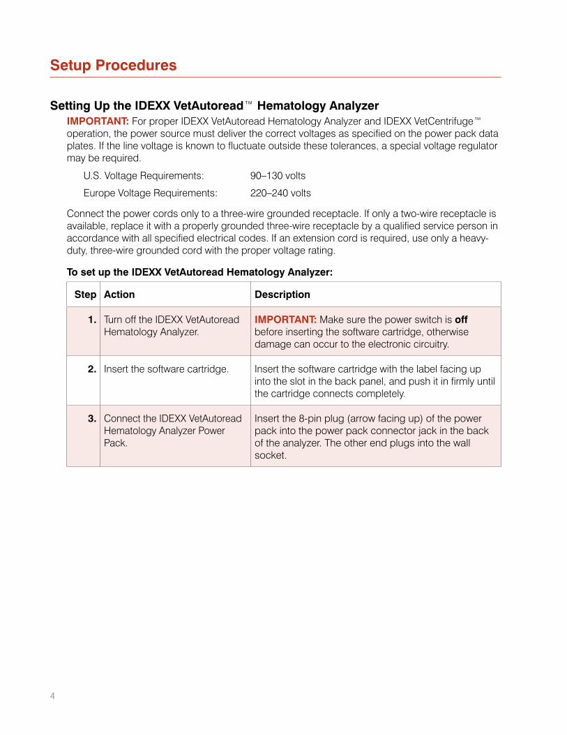
Setting Up the IDEXX VetAutoread™ Hematology AnalyzerIMPORTANT: For proper IDEXX VetAutoread Hematology Analyzer and IDEXX VetCentrifuge™ operation, the power source must deliver the correct voltages as specified on the power pack data plates. If the line voltage is known to fluctuate outside these tolerances, a special voltage regulator may be required.
U.S. Voltage Requirements: 90–130 volts
Europe Voltage Requirements: 220–240 volts
Connect the power cords only to a three-wire grounded receptacle. If only a two-wire receptacle is available, replace it with a properly grounded three-wire receptacle by a qualified service person in accordance with all specified electrical codes. If an extension cord is required, use only a heavy-duty, three-wire grounded cord with the proper voltage rating.
To set up the IDEXX VetAutoread Hematology Analyzer:
Step Action Description
1. Turn off the IDEXX VetAutoread Hematology Analyzer.
IMPORTANT: Make sure the power switch is off before inserting the software cartridge, otherwise damage can occur to the electronic circuitry.
2. Insert the software cartridge. Insert the software cartridge with the label facing up into the slot in the back panel, and push it in firmly until the cartridge connects completely.
3. Connect the IDEXX VetAutoread Hematology Analyzer Power Pack.
Insert the 8-pin plug (arrow facing up) of the power pack into the power pack connector jack in the back of the analyzer. The other end plugs into the wall socket.
4
5
Step Action Description
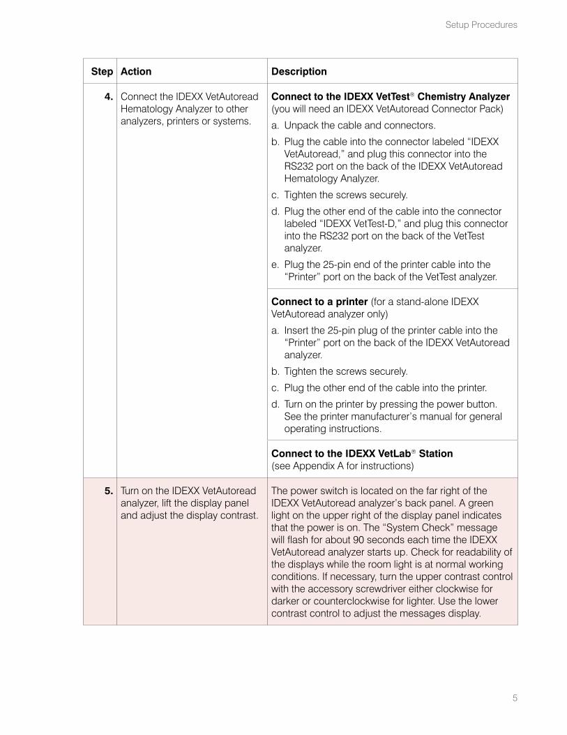
4. Connect the IDEXX VetAutoread Hematology Analyzer to other analyzers, printers or systems.
Connect to the IDEXX VetTest® Chemistry Analyzer (you will need an IDEXX VetAutoread Connector Pack)
a. Unpack the cable and connectors.
b. Plug the cable into the connector labeled “IDEXX VetAutoread,” and plug this connector into the RS232 port on the back of the IDEXX VetAutoread Hematology Analyzer.
c. Tighten the screws securely.
d. Plug the other end of the cable into the connector labeled “IDEXX VetTest-D,” and plug this connector into the RS232 port on the back of the VetTest analyzer.
e. Plug the 25-pin end of the printer cable into the “Printer” port on the back of the VetTest analyzer.
Connect to a printer (for a stand-alone IDEXX VetAutoread analyzer only)
a. Insert the 25-pin plug of the printer cable into the “Printer” port on the back of the IDEXX VetAutoread analyzer.
b. Tighten the screws securely.
c. Plug the other end of the cable into the printer.
d. Turn on the printer by pressing the power button. See the printer manufacturer’s manual for general operating instructions.
Connect to the IDEXX VetLab® Station (see Appendix A for instructions)
5. Turn on the IDEXX VetAutoread analyzer, lift the display panel and adjust the display contrast.
The power switch is located on the far right of the IDEXX VetAutoread analyzer’s back panel. A green light on the upper right of the display panel indicates that the power is on. The “System Check” message will flash for about 90 seconds each time the IDEXX VetAutoread analyzer starts up. Check for readability of the displays while the room light is at normal working conditions. If necessary, turn the upper contrast control with the accessory screwdriver either clockwise for darker or counterclockwise for lighter. Use the lower contrast control to adjust the messages display.
Setup Procedures
6
IDEXX VetAutoread Hematology Analyzer Operator’s Guide
Step Action Description
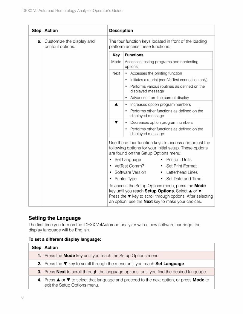
6. Customize the display and printout options.
The four function keys located in front of the loading platform access these functions:
Key Functions
Mode Accesses testing programs and nontesting options
Next • Accesses the printing function
• Initiates a reprint (non-VetTest connection only)
• Performs various routines as defined on the displayed message
• Advances from the current display
s • Increases option program numbers
• Performs other functions as defined on the displayed message
t • Decreases option program numbers
• Performs other functions as defined on the displayed message
Use these four function keys to access and adjust the following options for your initial setup. These options are found on the Setup Options menu:• Set Language • Printout Units • VetTest Comm? • Set Print Format • Software Version • Letterhead Lines • Printer Type • Set Date and Time
To access the Setup Options menu, press the Mode key until you reach Setup Options. Select s or t. Press the t key to scroll through options. After selecting an option, use the Next key to make your choices.
Setting the LanguageThe first time you turn on the IDEXX VetAutoread analyzer with a new software cartridge, the display language will be English.
To set a different display language:
Step Action
1. Press the Mode key until you reach the Setup Options menu.
2. Press the t key to scroll through the menu until you reach Set Language.
3. Press Next to scroll through the language options, until you find the desired language.
4. Press s or t to select that language and proceed to the next option, or press Mode to exit the Setup Options menu.
7
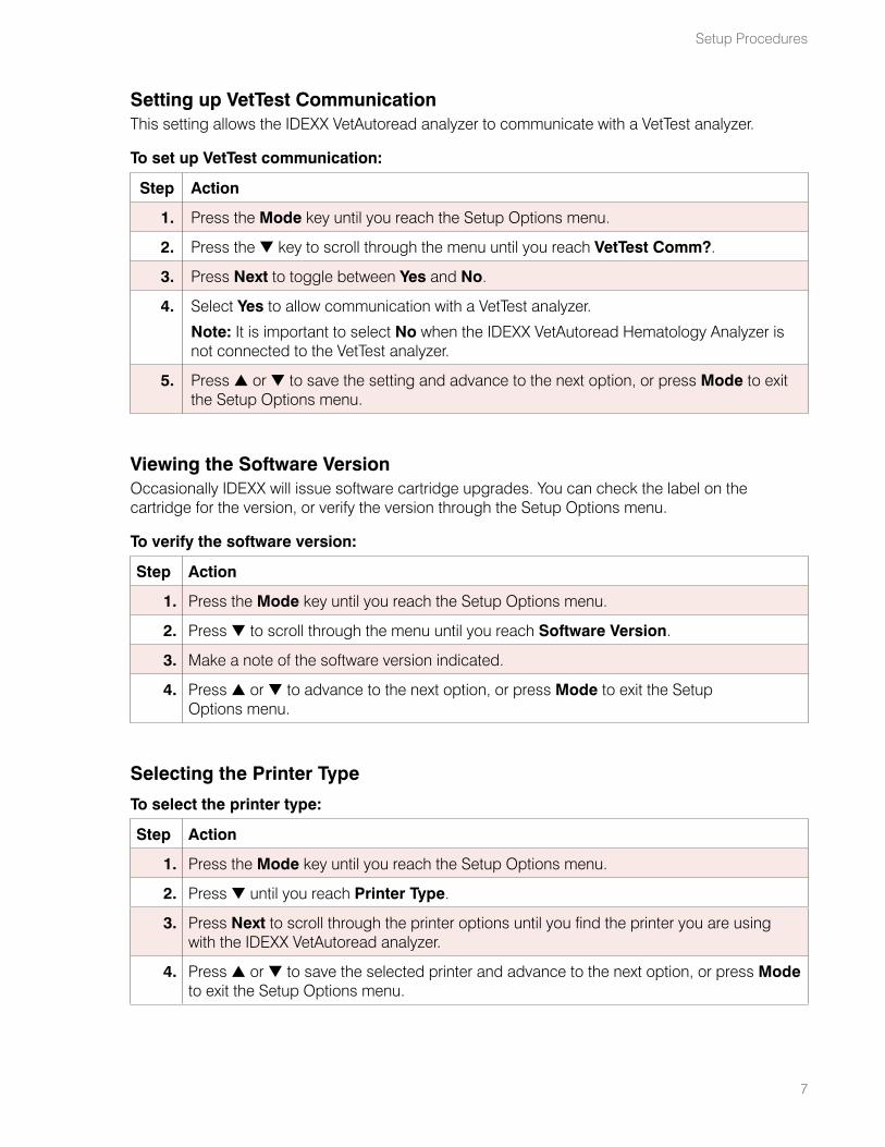
Setting up VetTest CommunicationThis setting allows the IDEXX VetAutoread analyzer to communicate with a VetTest analyzer.
To set up VetTest communication:
Step Action
1. Press the Mode key until you reach the Setup Options menu.
2. Press the t key to scroll through the menu until you reach VetTest Comm?.
3. Press Next to toggle between Yes and No.
4. Select Yes to allow communication with a VetTest analyzer.
Note: It is important to select No when the IDEXX VetAutoread Hematology Analyzer is not connected to the VetTest analyzer.
5. Press s or t to save the setting and advance to the next option, or press Mode to exit the Setup Options menu.
Viewing the Software VersionOccasionally IDEXX will issue software cartridge upgrades. You can check the label on the cartridge for the version, or verify the version through the Setup Options menu.
To verify the software version:
Step Action
1. Press the Mode key until you reach the Setup Options menu.
2. Press t to scroll through the menu until you reach Software Version.
3. Make a note of the software version indicated.
4. Press s or t to advance to the next option, or press Mode to exit the Setup Options menu.
Selecting the Printer Type
To select the printer type:
Step Action
1. Press the Mode key until you reach the Setup Options menu.
2. Press t until you reach Printer Type.
3. Press Next to scroll through the printer options until you find the printer you are using with the IDEXX VetAutoread analyzer.
4. Press s or t to save the selected printer and advance to the next option, or press Mode to exit the Setup Options menu.
Setup Procedures
8
IDEXX VetAutoread Hematology Analyzer Operator’s Guide
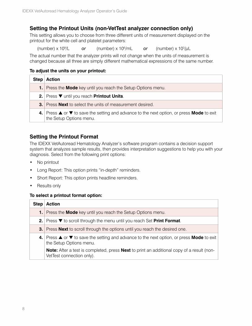
Setting the Printout Units (non-VetTest analyzer connection only)This setting allows you to choose from three different units of measurement displayed on the printout for the white cell and platelet parameters:
(number) x 109/L or (number) x 106/mL or (number) x 103/μL
The actual number that the analyzer prints will not change when the units of measurement is changed because all three are simply different mathematical expressions of the same number.
To adjust the units on your printout:
Step Action
1. Press the Mode key until you reach the Setup Options menu.
2. Press t until you reach Printout Units.
3. Press Next to select the units of measurement desired.
4. Press s or t to save the setting and advance to the next option, or press Mode to exit the Setup Options menu.
Setting the Printout FormatThe IDEXX VetAutoread Hematology Analyzer’s software program contains a decision support system that analyzes sample results, then provides interpretation suggestions to help you with your diagnosis. Select from the following print options:
• No printout
• Long Report: This option prints “in-depth” reminders.
• Short Report: This option prints headline reminders.
• Results only
To select a printout format option:
Step Action
1. Press the Mode key until you reach the Setup Options menu.
2. Press t to scroll through the menu until you reach Set Print Format.
3. Press Next to scroll through the options until you reach the desired one.
4. Press s or t to save the setting and advance to the next option, or press Mode to exit the Setup Options menu.
Note: After a test is completed, press Next to print an additional copy of a result (non-VetTest connection only).
9
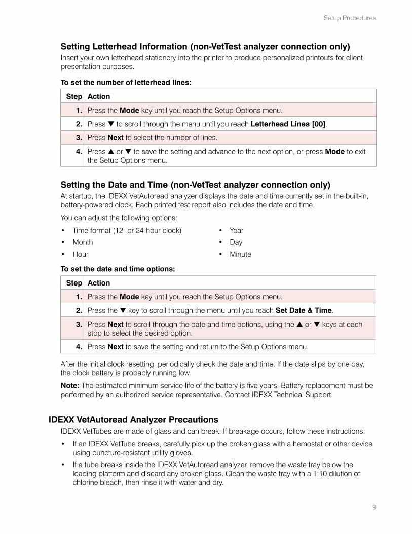
Setting Letterhead Information (non-VetTest analyzer connection only)Insert your own letterhead stationery into the printer to produce personalized printouts for client presentation purposes.
To set the number of letterhead lines:
Step Action
1. Press the Mode key until you reach the Setup Options menu.
2. Press t to scroll through the menu until you reach Letterhead Lines [00].
3. Press Next to select the number of lines.
4. Press s or t to save the setting and advance to the next option, or press Mode to exit the Setup Options menu.
Setting the Date and Time (non-VetTest analyzer connection only)At startup, the IDEXX VetAutoread analyzer displays the date and time currently set in the built-in, battery-powered clock. Each printed test report also includes the date and time.
You can adjust the following options:
• Time format (12- or 24-hour clock) • Year
• Month • Day
• Hour • Minute
To set the date and time options:
Step Action
1. Press the Mode key until you reach the Setup Options menu.
2. Press the t key to scroll through the menu until you reach Set Date & Time.
3. Press Next to scroll through the date and time options, using the s or t keys at each stop to select the desired option.
4. Press Next to save the setting and return to the Setup Options menu.
After the initial clock resetting, periodically check the date and time. If the date slips by one day, the clock battery is probably running low.
Note: The estimated minimum service life of the battery is five years. Battery replacement must be performed by an authorized service representative. Contact IDEXX Technical Support.
IDEXX VetAutoread Analyzer PrecautionsIDEXX VetTubes are made of glass and can break. If breakage occurs, follow these instructions:
• If an IDEXX VetTube breaks, carefully pick up the broken glass with a hemostat or other device using puncture-resistant utility gloves.
• If a tube breaks inside the IDEXX VetAutoread analyzer, remove the waste tray below the loading platform and discard any broken glass. Clean the waste tray with a 1:10 dilution of chlorine bleach, then rinse it with water and dry.
Setup Procedures
10
IDEXX VetAutoread Hematology Analyzer Operator’s Guide
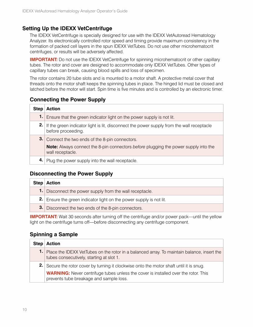
Setting Up the IDEXX VetCentrifugeThe IDEXX VetCentrifuge is specially designed for use with the IDEXX VetAutoread Hematology Analyzer. Its electronically controlled rotor speed and timing provide maximum consistency in the formation of packed cell layers in the spun IDEXX VetTubes. Do not use other microhematocrit centrifuges, or results will be adversely affected.
IMPORTANT: Do not use the IDEXX VetCentrifuge for spinning microhematocrit or other capillary tubes. The rotor and cover are designed to accommodate only IDEXX VetTubes. Other types of capillary tubes can break, causing blood spills and loss of specimen.
The rotor contains 20 tube slots and is mounted to a motor shaft. A protective metal cover that threads onto the motor shaft keeps the spinning tubes in place. The hinged lid must be closed and latched before the motor will start. Spin time is five minutes and is controlled by an electronic timer.
Connecting the Power Supply
Step Action
1. Ensure that the green indicator light on the power supply is not lit.
2. If the green indicator light is lit, disconnect the power supply from the wall receptacle before proceeding.
3. Connect the two ends of the 8-pin connectors.
Note: Always connect the 8-pin connectors before plugging the power supply into the wall receptacle.
4. Plug the power supply into the wall receptacle.
Disconnecting the Power Supply
Step Action
1. Disconnect the power supply from the wall receptacle.
2. Ensure the green indicator light on the power supply is not lit.
3. Disconnect the two ends of the 8-pin connectors.
IMPORTANT: Wait 30 seconds after turning off the centrifuge and/or power pack—until the yellow light on the centrifuge turns off—before disconnecting any centrifuge component.
Spinning a Sample
Step Action
1. Place the IDEXX VetTubes on the rotor in a balanced array. To maintain balance, insert the tubes consecutively, starting at slot 1.
2. Secure the rotor cover by turning it clockwise onto the motor shaft until it is snug.
WARNING: Never centrifuge tubes unless the cover is installed over the rotor. This prevents tube breakage and sample loss.
11
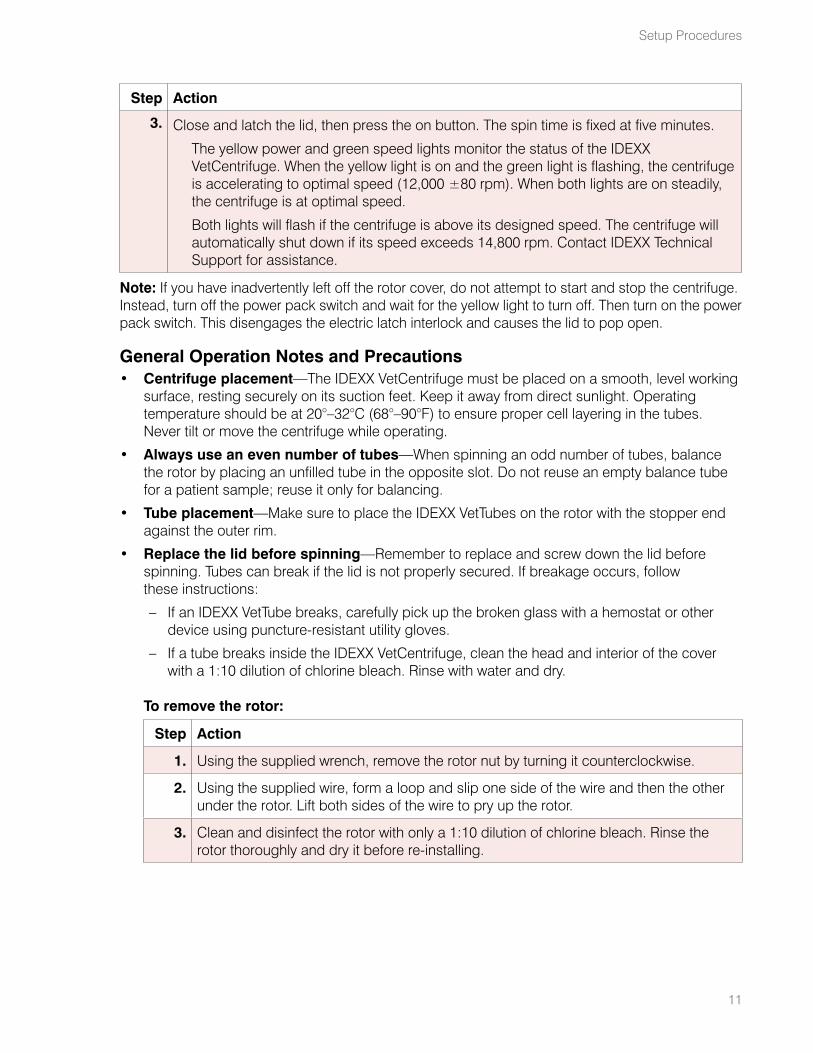
Step Action
3. Close and latch the lid, then press the on button. The spin time is fixed at five minutes.
The yellow power and green speed lights monitor the status of the IDEXX VetCentrifuge. When the yellow light is on and the green light is flashing, the centrifuge is accelerating to optimal speed (12,000 ±80 rpm). When both lights are on steadily, the centrifuge is at optimal speed.
Both lights will flash if the centrifuge is above its designed speed. The centrifuge will automatically shut down if its speed exceeds 14,800 rpm. Contact IDEXX Technical Support for assistance.
Note: If you have inadvertently left off the rotor cover, do not attempt to start and stop the centrifuge. Instead, turn off the power pack switch and wait for the yellow light to turn off. Then turn on the power pack switch. This disengages the electric latch interlock and causes the lid to pop open.
General Operation Notes and Precautions• Centrifuge placement—The IDEXX VetCentrifuge must be placed on a smooth, level working
surface, resting securely on its suction feet. Keep it away from direct sunlight. Operating temperature should be at 20°–32°C (68°–90°F) to ensure proper cell layering in the tubes. Never tilt or move the centrifuge while operating.
• Always use an even number of tubes—When spinning an odd number of tubes, balance the rotor by placing an unfilled tube in the opposite slot. Do not reuse an empty balance tube for a patient sample; reuse it only for balancing.
• Tube placement—Make sure to place the IDEXX VetTubes on the rotor with the stopper end against the outer rim.
• Replace the lid before spinning—Remember to replace and screw down the lid before spinning. Tubes can break if the lid is not properly secured. If breakage occurs, follow these instructions:
– If an IDEXX VetTube breaks, carefully pick up the broken glass with a hemostat or other device using puncture-resistant utility gloves.
– If a tube breaks inside the IDEXX VetCentrifuge, clean the head and interior of the cover with a 1:10 dilution of chlorine bleach. Rinse with water and dry.
To remove the rotor:
Step Action
1. Using the supplied wrench, remove the rotor nut by turning it counterclockwise.
2. Using the supplied wire, form a loop and slip one side of the wire and then the other under the rotor. Lift both sides of the wire to pry up the rotor.
3. Clean and disinfect the rotor with only a 1:10 dilution of chlorine bleach. Rinse the rotor thoroughly and dry it before re-installing.
Setup Procedures
12
IDEXX VetAutoread Hematology Analyzer Operator’s Guide
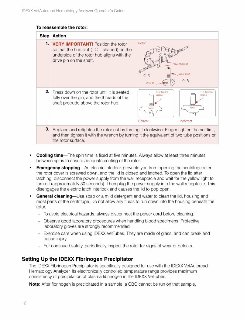
To reassemble the rotor:
Step Action
1. VERY IMPORTANT! Position the rotor so that the hub slot ( shaped) on the underside of the rotor hub aligns with the drive pin on the shaft.
Rotor
Drive pin
Motor shaft
Hub slot
2. Press down on the rotor until it is seated fully over the pin, and the threads of the shaft protrude above the rotor hub.
Correct Incorrect
Rotor
3–5 threads visible
Rotor
1–2 threads visible
3. Replace and retighten the rotor nut by turning it clockwise. Finger-tighten the nut first, and then tighten it with the wrench by turning it the equivalent of two tube positions on the rotor surface.
• Cooling time—The spin time is fixed at five minutes. Always allow at least three minutes between spins to ensure adequate cooling of the rotor.
• Emergency stopping—An electric interlock prevents you from opening the centrifuge after the rotor cover is screwed down, and the lid is closed and latched. To open the lid after latching, disconnect the power supply from the wall receptacle and wait for the yellow light to turn off (approximately 30 seconds). Then plug the power supply into the wall receptacle. This disengages the electric latch interlock and causes the lid to pop open.
• General cleaning—Use soap or a mild detergent and water to clean the lid, housing and most parts of the centrifuge. Do not allow any fluids to run down into the housing beneath the rotor.
– To avoid electrical hazards, always disconnect the power cord before cleaning.
– Observe good laboratory procedures when handling blood specimens. Protective laboratory gloves are strongly recommended.
– Exercise care when using IDEXX VetTubes. They are made of glass, and can break and cause injury.
– For continued safety, periodically inspect the rotor for signs of wear or defects.
Setting Up the IDEXX Fibrinogen PrecipitatorThe IDEXX Fibrinogen Precipitator is specifically designed for use with the IDEXX VetAutoread Hematology Analyzer. Its electronically controlled temperature range provides maximum consistency of precipitation of plasma fibrinogen in the IDEXX VetTubes.
Note: After fibrinogen is precipitated in a sample, a CBC cannot be run on that sample.
13
To set up the IDEXX Fibrinogen Precipitator:
Step Action
1. Place the precipitator on a level surface, label-side up.
2. Insert the power cord into the three-pin jack on the back of the precipitator. Plug the other end of the cord into an AC wall socket.
3. Turn on the precipitator.
The LED indicator light immediately turns yellow. After a warm-up period of 10 to 15 minutes, the LED indicator light changes to green, indicating the precipitator has warmed to the range specified. Periodically, the LED indicator light may change to yellow. This is normal, and indicates the analyzer is in heating mode.
Note: If the LED remains yellow and does not change to green, the precipitator must be serviced.
To run fibrinogen samples:Before you begin: If you are using the IDEXX VetAutoread Hematology Analyzer in conjunction with the IDEXX VetTest Chemistry Analyzer, ensure that the VetTest analyzer is displaying the main menu screen.
Step Action
1. After initial hematology analysis, remove the IDEXX VetTube from the IDEXX VetAutoread Hematology Analyzer.
2. Place a previously spun IDEXX VetTube in one of the four holes located on the face of the precipitator until only the cap is showing. Make a note of the time the tube was placed in the precipitator.
Note: The LED may occasionally turn green and back to yellow; this is normal.
3. Five minutes after placing the tube in the precipitator, remove it and place it in the IDEXX VetCentrifuge for five minutes.
4. On the IDEXX VetAutoread analyzer, press the Mode key until Fibrinogen appears on the screen.
5. Press s or t to select the desired species.
6. Insert the VetTube into the VetAutoread analyzer and close the cover. Results will be sent to the VetTest analyzer.
7. After the fibrinogen results are displayed on the IDEXX VetAutoread screen, select one of the patient identification options on the VetTest analyzer:
• Select 1 to attach only a date and time stamp to the results.
• Select 2 to rename the species and the patient if the current patient ID is not correct. If the patient ID entered matches one of the six most recent patient results stored in the VetTest analyzer, the results will also be merged.
• Select 3 to merge the fibrinogen results with the current patient’s chemistry and/or hematology results.
Setup Procedures
Sample Collection and Preparation
Good sample collection and preparation are necessary for optimum results on your IDEXX VetAutoread™ Hematology Analyzer. We recommend that you familiarize yourself thoroughly with the following guidelines. If you have any questions about sample preparation, call IDEXX Technical Support and we will be happy to help you.
Do not recycle syringes and needles. Dull needles can cause trauma to the vessel, leading to cell destruction, hemolysis and platelet activation, especially in cats.
No matter which type of needle and syringe you choose, refer to these guidelines:
• Choose the appropriate blood vessel and needle size—Use a vessel that allows enough blood collection into the tube or syringe selected. Select the appropriate needle size for the species being drawn.
• Be gentle on the draw—Exceeding the normal blood flow can collapse the vessel, which can be painful to the animal and cause hemolysis.
• Mix the sample with EDTA as soon as possible—When using a syringe and needle, remove the needle from the syringe before dispensing the blood. Remove the cap on the tube and fill the tube to its appropriate level.
IMPORTANT: Immediately mix the EDTA sample at least 10 times by gentle inversion. Remix the EDTA tube very well just before drawing it into an IDEXX VetTube.
• Make sure the sample-to-EDTA ratio is appropriate—Fill the tube to its appropriate level for adequate mixing with EDTA. Overfilling will result in clotting; underfilling will alter the hematocrit and hemoglobin values. Refer to the manufacturer’s package insert for appropriate fill volumes.
• Analyze the sample as soon as possible—For best results, we recommend preparing and testing the IDEXX VetTube within four hours. Even when refrigerated, blood samples will become less viable after four hours.
IMPORTANT: Remix the EDTA tube very well just before drawing it into an IDEXX VetTube. Undermixing the EDTA tube will alter results.
• Discard severely hemolyzed samples—If hemolysis is due to improper sample collection, discard the sample and redraw. If hemolysis is persistent, it may be due to a disease state.
• Discard clotted samples and redraw—The presence of clots will invalidate any results obtained, with the exception of the hematocrit.
Preparing a Canine, Feline or Equine SampleCAUTION: IDEXX VetTubes are made of glass. Be careful when handling and preparing the tubes to prevent breakage and possible injury. Inspect each IDEXX VetTube before using it. Do not used cracked or scratched tubes.
14
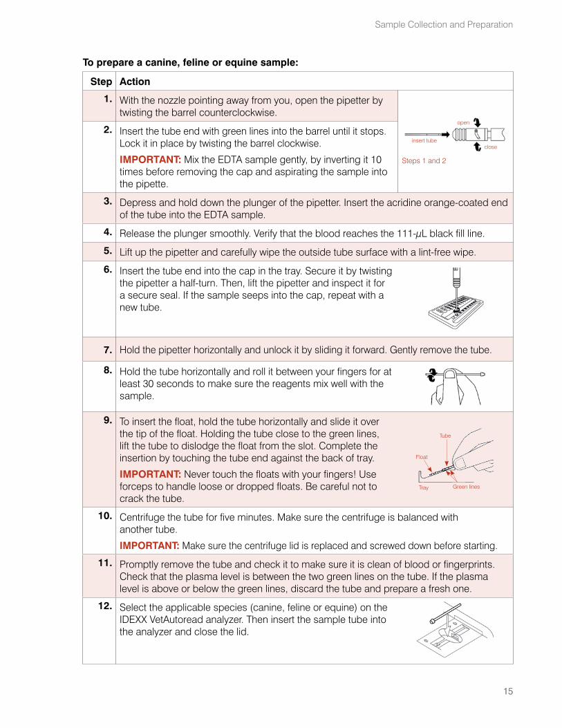
To prepare a canine, feline or equine sample:
Step Action
1. With the nozzle pointing away from you, open the pipetter by twisting the barrel counterclockwise.
insert tube
open
close
Steps 1 and 2
2. Insert the tube end with green lines into the barrel until it stops. Lock it in place by twisting the barrel clockwise.
IMPORTANT: Mix the EDTA sample gently, by inverting it 10 times before removing the cap and aspirating the sample into the pipette.
3. Depress and hold down the plunger of the pipetter. Insert the acridine orange-coated end of the tube into the EDTA sample.
4. Release the plunger smoothly. Verify that the blood reaches the 111-μL black fill line.
5. Lift up the pipetter and carefully wipe the outside tube surface with a lint-free wipe.
6. Insert the tube end into the cap in the tray. Secure it by twisting the pipetter a half-turn. Then, lift the pipetter and inspect it for a secure seal. If the sample seeps into the cap, repeat with a new tube.
7. Hold the pipetter horizontally and unlock it by sliding it forward. Gently remove the tube.
8. Hold the tube horizontally and roll it between your fingers for at least 30 seconds to make sure the reagents mix well with the sample.
9. To insert the float, hold the tube horizontally and slide it over the tip of the float. Holding the tube close to the green lines, lift the tube to dislodge the float from the slot. Complete the insertion by touching the tube end against the back of tray.
IMPORTANT: Never touch the floats with your fingers! Use forceps to handle loose or dropped floats. Be careful not to crack the tube.
Float
Tray
Tube
Green lines
10. Centrifuge the tube for five minutes. Make sure the centrifuge is balanced with another tube.
IMPORTANT: Make sure the centrifuge lid is replaced and screwed down before starting.
11. Promptly remove the tube and check it to make sure it is clean of blood or fingerprints. Check that the plasma level is between the two green lines on the tube. If the plasma level is above or below the green lines, discard the tube and prepare a fresh one.
12. Select the applicable species (canine, feline or equine) on the IDEXX VetAutoread analyzer. Then insert the sample tube into the analyzer and close the lid.
15
Sample Collection and Preparation
15
16
IDEXX VetAutoread Hematology Analyzer Operator’s Guide
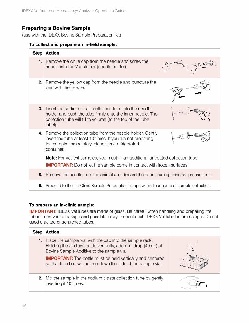
Preparing a Bovine Sample(use with the IDEXX Bovine Sample Preparation Kit)
To collect and prepare an in-field sample:
Step Action
1. Remove the white cap from the needle and screw the needle into the Vacutainer (needle holder).
2. Remove the yellow cap from the needle and puncture the vein with the needle.
3. Insert the sodium citrate collection tube into the needle holder and push the tube firmly onto the inner needle. The collection tube will fill to volume (to the top of the tube label).
4. Remove the collection tube from the needle holder. Gently invert the tube at least 10 times. If you are not preparing the sample immediately, place it in a refrigerated container.
Note: For VetTest samples, you must fill an additional untreated collection tube.
IMPORTANT: Do not let the sample come in contact with frozen surfaces.
5. Remove the needle from the animal and discard the needle using universal precautions.
6. Proceed to the “In-Clinic Sample Preparation” steps within four hours of sample collection.
To prepare an in-clinic sample:IMPORTANT: IDEXX VetTubes are made of glass. Be careful when handling and preparing the tubes to prevent breakage and possible injury. Inspect each IDEXX VetTube before using it. Do not used cracked or scratched tubes.
Step Action
1. Place the sample vial with the cap into the sample rack. Holding the additive bottle vertically, add one drop (40 μL) of Bovine Sample Additive to the sample vial.
IMPORTANT: The bottle must be held vertically and centered so that the drop will not run down the side of the sample vial.
2. Mix the sample in the sodium citrate collection tube by gently inverting it 10 times.
17
Step Action
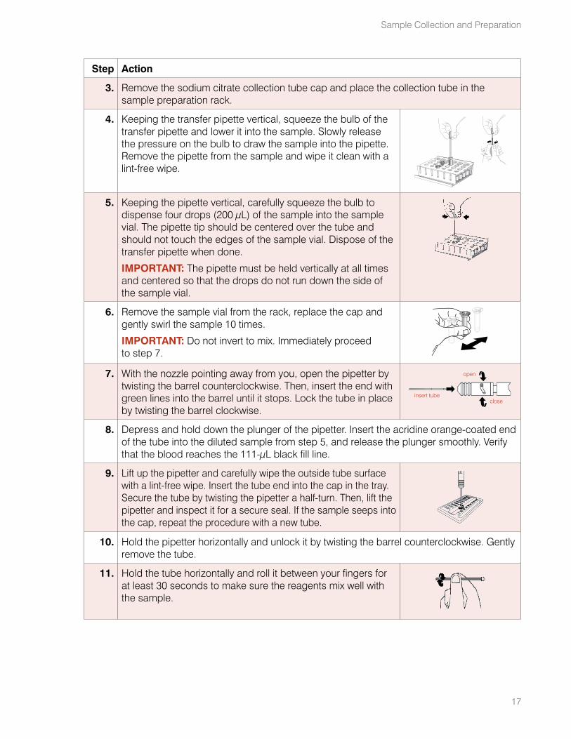
3. Remove the sodium citrate collection tube cap and place the collection tube in the sample preparation rack.
4. Keeping the transfer pipette vertical, squeeze the bulb of the transfer pipette and lower it into the sample. Slowly release the pressure on the bulb to draw the sample into the pipette. Remove the pipette from the sample and wipe it clean with a lint-free wipe.
5. Keeping the pipette vertical, carefully squeeze the bulb to dispense four drops (200 μL) of the sample into the sample vial. The pipette tip should be centered over the tube and should not touch the edges of the sample vial. Dispose of the transfer pipette when done.
IMPORTANT: The pipette must be held vertically at all times and centered so that the drops do not run down the side of the sample vial.
6. Remove the sample vial from the rack, replace the cap and gently swirl the sample 10 times.
IMPORTANT: Do not invert to mix. Immediately proceed to step 7.
7. With the nozzle pointing away from you, open the pipetter by twisting the barrel counterclockwise. Then, insert the end with green lines into the barrel until it stops. Lock the tube in place by twisting the barrel clockwise.
insert tube
open
close
8. Depress and hold down the plunger of the pipetter. Insert the acridine orange-coated end of the tube into the diluted sample from step 5, and release the plunger smoothly. Verify that the blood reaches the 111-μL black fill line.
9. Lift up the pipetter and carefully wipe the outside tube surface with a lint-free wipe. Insert the tube end into the cap in the tray. Secure the tube by twisting the pipetter a half-turn. Then, lift the pipetter and inspect it for a secure seal. If the sample seeps into the cap, repeat the procedure with a new tube.
10. Hold the pipetter horizontally and unlock it by twisting the barrel counterclockwise. Gently remove the tube.
11. Hold the tube horizontally and roll it between your fingers for at least 30 seconds to make sure the reagents mix well with the sample.
Sample Collection and Preparation
18
IDEXX VetAutoread Hematology Analyzer Operator’s Guide
Step Action
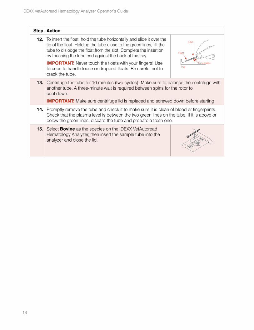
12. To insert the float, hold the tube horizontally and slide it over the tip of the float. Holding the tube close to the green lines, lift the tube to dislodge the float from the slot. Complete the insertion by touching the tube end against the back of the tray.
IMPORTANT: Never touch the floats with your fingers! Use forceps to handle loose or dropped floats. Be careful not to crack the tube.
Float
Tray
Tube
Green lines
13. Centrifuge the tube for 10 minutes (two cycles). Make sure to balance the centrifuge with another tube. A three-minute wait is required between spins for the rotor to cool down.
IMPORTANT: Make sure centrifuge lid is replaced and screwed down before starting.
14. Promptly remove the tube and check it to make sure it is clean of blood or fingerprints. Check that the plasma level is between the two green lines on the tube. If it is above or below the green lines, discard the tube and prepare a fresh one.
15. Select Bovine as the species on the IDEXX VetAutoread Hematology Analyzer, then insert the sample tube into the analyzer and close the lid.
19
Running Tests on the IDEXX VetAutoread™ Analyzer
Before You Begin TestingEach day before you begin testing, perform a calibration check and verify the results against the factory specifications to ensure your system is functioning correctly.
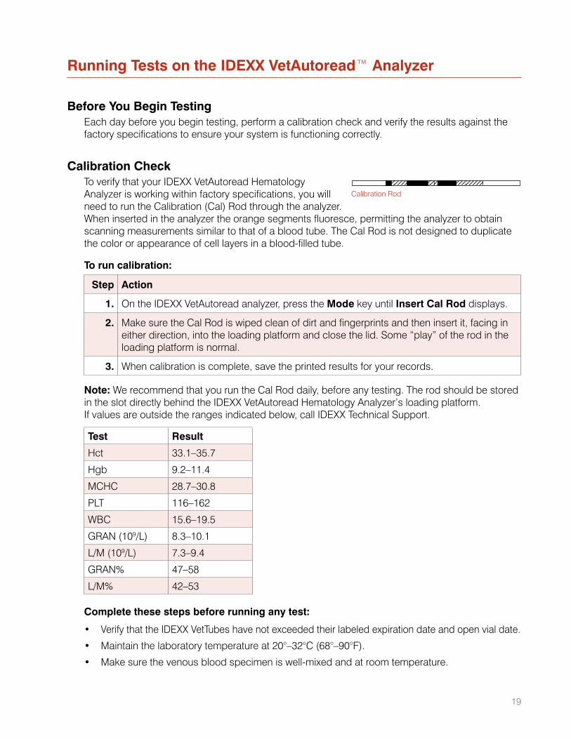
Calibration CheckTo verify that your IDEXX VetAutoread Hematology Analyzer is working within factory specifications, you will need to run the Calibration (Cal) Rod through the analyzer. When inserted in the analyzer the orange segments fluoresce, permitting the analyzer to obtain scanning measurements similar to that of a blood tube. The Cal Rod is not designed to duplicate the color or appearance of cell layers in a blood-filled tube.
To run calibration:
Step Action
1. On the IDEXX VetAutoread analyzer, press the Mode key until Insert Cal Rod displays.
2. Make sure the Cal Rod is wiped clean of dirt and fingerprints and then insert it, facing in either direction, into the loading platform and close the lid. Some “play” of the rod in the loading platform is normal.
3. When calibration is complete, save the printed results for your records.
Note: We recommend that you run the Cal Rod daily, before any testing. The rod should be stored in the slot directly behind the IDEXX VetAutoread Hematology Analyzer’s loading platform. If values are outside the ranges indicated below, call IDEXX Technical Support.
Test Result
Hct 33.1–35.7
Hgb 9.2–11.4
MCHC 28.7–30.8
PLT 116–162
WBC 15.6–19.5
GRAN (109/L) 8.3–10.1
L/M (109/L) 7.3–9.4
GRAN% 47–58
L/M% 42–53
Complete these steps before running any test:
• Verify that the IDEXX VetTubes have not exceeded their labeled expiration date and open vial date.
• Maintain the laboratory temperature at 20°–32°C (68°–90°F).
• Make sure the venous blood specimen is well-mixed and at room temperature.
Calibration Rod
20
IDEXX VetAutoread Hematology Analyzer Operator’s Guide
Test ProcedureBefore placing a prepared tube in the IDEXX VetAutoread Hematology Analyzer, make sure the tube is clean of debris and fingerprints by using a lint-free wipe. Select the desired species, insert the tube into the analyzer loading carriage and close the door. The analyzer automatically begins testing.
Note: The test will abort if you open the door. To repeat an aborted assay, close the door and wait for the tube to return to the loading platform. Remove the tube, re-insert it and close the door.
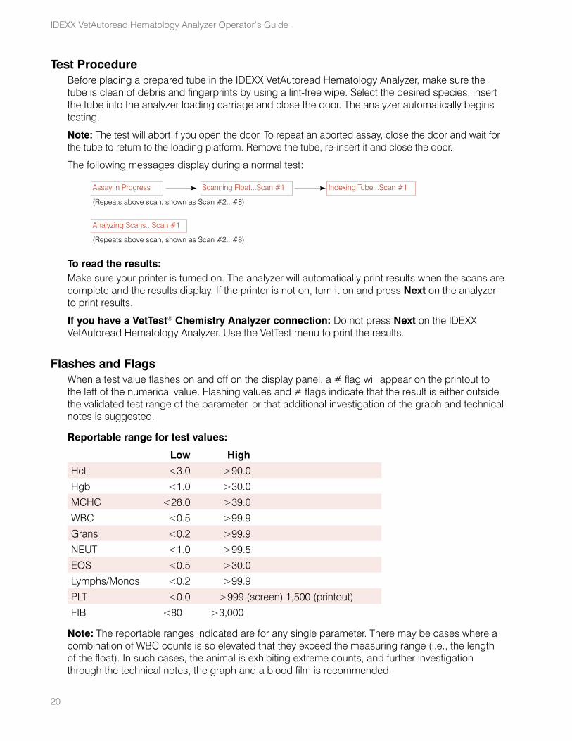
The following messages display during a normal test:
Assay in Progress Scanning Float...Scan #1 Indexing Tube...Scan #1
(Repeats above scan, shown as Scan #2...#8)
Analyzing Scans...Scan #1
(Repeats above scan, shown as Scan #2...#8)
To read the results:Make sure your printer is turned on. The analyzer will automatically print results when the scans are complete and the results display. If the printer is not on, turn it on and press Next on the analyzer to print results.
If you have a VetTest® Chemistry Analyzer connection: Do not press Next on the IDEXX VetAutoread Hematology Analyzer. Use the VetTest menu to print the results.
Flashes and FlagsWhen a test value flashes on and off on the display panel, a # flag will appear on the printout to the left of the numerical value. Flashing values and # flags indicate that the result is either outside the validated test range of the parameter, or that additional investigation of the graph and technical notes is suggested.
Reportable range for test values:
Low High
Hct <3.0 >90.0
Hgb <1.0 >30.0
MCHC <28.0 >39.0
WBC <0.5 >99.9
Grans <0.2 >99.9
NEUT <1.0 >99.5
EOS <0.5 >30.0
Lymphs/Monos <0.2 >99.9
PLT <0.0 >999 (screen) 1,500 (printout)
FIB <80.0 >3,000
Note: The reportable ranges indicated are for any single parameter. There may be cases where a combination of WBC counts is so elevated that they exceed the measuring range (i.e., the length of the float). In such cases, the animal is exhibiting extreme counts, and further investigation through the technical notes, the graph and a blood film is recommended.

21
DashesDashes on the panel and on the printout mean that:
• The computed test value is outside the display range of the instrument.
• A packed cell layer is too small to measure.
• Clumped platelets, missing layers or some other extreme condition exists.
In such cases, dashes instead of test values (or bar graph points) will appear. Refer to the technical notes on the printout for further steps.
Running Tests on the IDEXX VetAutoread Analyzer
Interpreting Test Results
You will gain the most from your IDEXX VetAutoread™ Hematology Analyzer if you understand how it analyzes samples, and if you study the printout carefully, especially the buffy coat profile. Examining the profile is particularly useful because it will help identify several conditions, including platelet clumping, missing layers or blurred boundaries, that will aid in assessing the overall analysis.
Principles of the IDEXX VetAutoread Hematology AnalyzerThe IDEXX VetAutoread Hematology Analyzer is based on the principle that different blood cells have different densities, and that they sort into individual layers when spun in a microhematocrit tube. This principle is why the conventional spun hematocrit tube produces three distinct layers: the red blood cells (which have the greatest density), the buffy coat and the plasma.
The analyzer’s technology expands the buffy coat by means of a molded cylindrical float inserted into a precision-bore capillary tube. The specific gravity of the float is approximately midway between that of the plasma and red cells, causing the buffy coat to expand along the length of the float. Expanded layers of packed white cells and platelets resolve between the float and inner wall of the tube.
The interior of the IDEXX VetTube is coated with acridine orange, a fluorescent dye that stains a variety of cellular components, including nucleoproteins (primarily DNA and RNA), lipoproteins, glycosamines in the granulocytes series, and other cell substances. These cellular components bind the acridine orange and then fluoresce under blue-violet light.
Under this light, normal erythrocytes are unaffected by acridine orange and exhibit a dark red appearance. Granulocytic cells fluoresce orange-yellow, lymphocytes and monocytes fluoresce a brilliant green and platelets fluoresce a pale yellow.
The analyzer’s optics chamber examines the tube and float, and measures fluorescence emitted by the cells in the tube. Software algorithms then delimit the layers in the tube and derive the following measurements:
Hct (%) Hgb (g/dL)
MCHC (g/dL) Total WBC (#)
GRANS (% and absolute) LYMPH/MONOS (% and absolute)
PLT (#) FIB (mg/dL)
Fibrinogen is heat-precipitated and spun to form a layer on top of the float.
In canine and bovine samples only, the analyzer will also delimit neutrophils (absolute) and eosinophils (absolute).
In canine and feline samples only, the analyzer will also delimit reticulocytes (%).
22
23
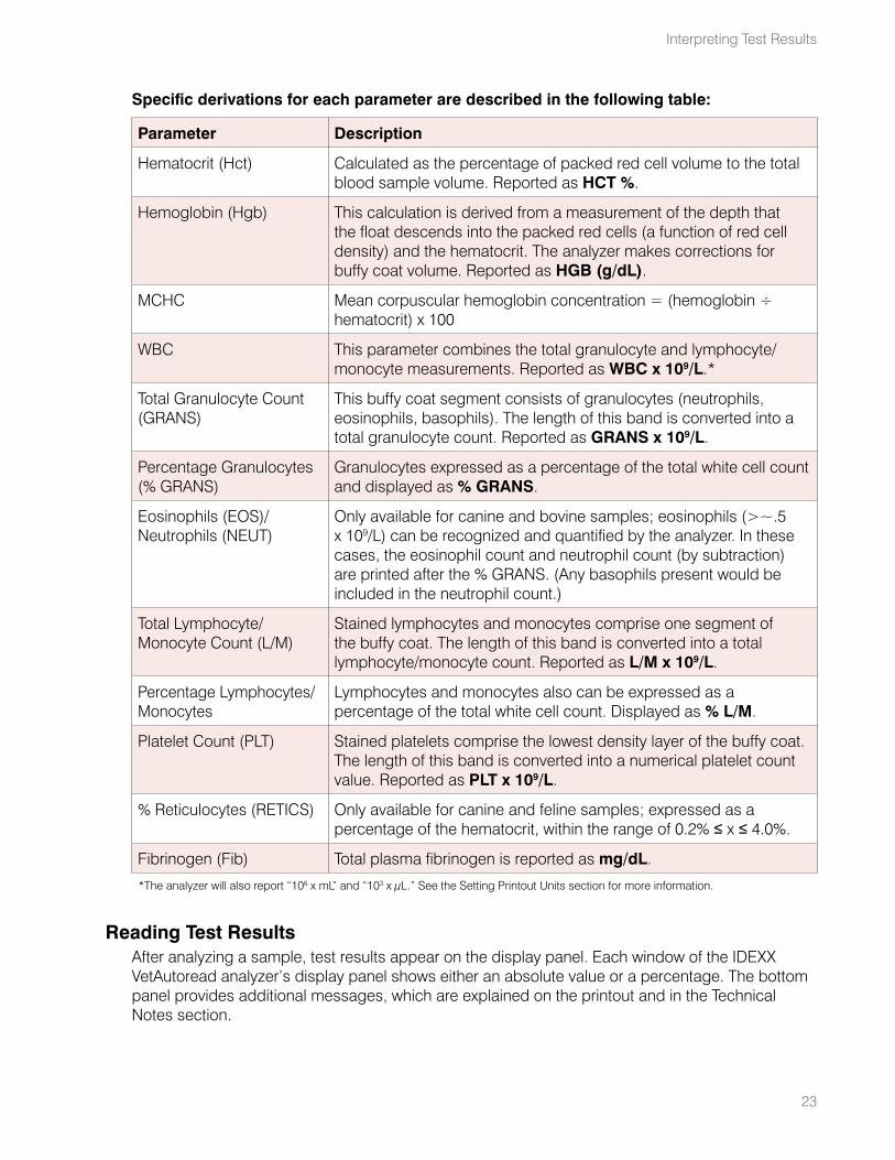
Specific derivations for each parameter are described in the following table:
Parameter Description
Hematocrit (Hct) Calculated as the percentage of packed red cell volume to the total blood sample volume. Reported as HCT %.
Hemoglobin (Hgb) This calculation is derived from a measurement of the depth that the float descends into the packed red cells (a function of red cell density) and the hematocrit. The analyzer makes corrections for buffy coat volume. Reported as HGB (g/dL).
MCHC Mean corpuscular hemoglobin concentration = (hemoglobin ÷ hematocrit) x 100
WBC This parameter combines the total granulocyte and lymphocyte/monocyte measurements. Reported as WBC x 109/L.*
Total Granulocyte Count (GRANS)
This buffy coat segment consists of granulocytes (neutrophils, eosinophils, basophils). The length of this band is converted into a total granulocyte count. Reported as GRANS x 109/L.
Percentage Granulocytes (% GRANS)
Granulocytes expressed as a percentage of the total white cell count and displayed as % GRANS.
Eosinophils (EOS)/Neutrophils (NEUT)
Only available for canine and bovine samples; eosinophils (>~.5 x 109/L) can be recognized and quantified by the analyzer. In these cases, the eosinophil count and neutrophil count (by subtraction) are printed after the % GRANS. (Any basophils present would be included in the neutrophil count.)
Total Lymphocyte/Monocyte Count (L/M)
Stained lymphocytes and monocytes comprise one segment of the buffy coat. The length of this band is converted into a total lymphocyte/monocyte count. Reported as L/M x 109/L.
Percentage Lymphocytes/ Monocytes
Lymphocytes and monocytes also can be expressed as a percentage of the total white cell count. Displayed as % L/M.
Platelet Count (PLT) Stained platelets comprise the lowest density layer of the buffy coat. The length of this band is converted into a numerical platelet count value. Reported as PLT x 109/L.
% Reticulocytes (RETICS) Only available for canine and feline samples; expressed as a percentage of the hematocrit, within the range of 0.2% ≤ x ≤ 4.0%.
Fibrinogen (Fib) Total plasma fibrinogen is reported as mg/dL.
*The analyzer will also report “106 x mL” and “103 x μL.” See the Setting Printout Units section for more information.
Reading Test ResultsAfter analyzing a sample, test results appear on the display panel. Each window of the IDEXX VetAutoread analyzer’s display panel shows either an absolute value or a percentage. The bottom panel provides additional messages, which are explained on the printout and in the Technical Notes section.
Interpreting Test Results
24
IDEXX VetAutoread Hematology Analyzer Operator’s Guide
The IDEXX VetAutoread Hematology Analyzer also provides printed reports. Numerical results appear on the left portion of the report. To the right, an at-a-glance bar graph gives a quick indication of the results.
Messages and Symbols• nRBCs likely
This message displays when an elevated number of nucleated red blood cells is detected.
• Equals Sign (=) An equals sign displays if the parameter’s value equals the number printed.
• Greater Than (>) When the parameter’s value is greater than the number printed, the analyzer’s display panel flashes the value. This occurs in two instances:
– When the number of cells counted exceeds the upper limit of the analyzer
– When platelets are identified on top of the float; the analyzer does not quantify these additional platelets, but indicates that more platelets exist than those quantified in the normal platelet location
• # Sign When further investigation of the buffy coat profile and/or technical notes is recommended, the IDEXX VetAutoread analyzer displays the # sign. In these cases, you should be looking for indication of clumped platelets, missing layers or any other extreme condition that may affect the reported results. As with out-of-range values, the analyzer’s display panel will flash flagged results.
• Dashes (- -) Dashes appear on the printout and on the analyzer’s display panel when the IDEXX VetAutoread analyzer is unable to calculate any value at all. There are two primary reasons for dashes:
– A severely clumped sample. Platelet clumps tend to disperse throughout the sample, interfering with the layer interfaces. Such clumps also will show up on the buffy coat profile as a series of erratic “bumps.” In most cases, you should redraw the sample from the animal and retest. See the Technical Notes section for more information.
– An extremely small or missing layer. In these situations, the analyzer is unable to distinguish the layers, and may have difficulty calculating any values from the buffy coat. In most cases, it will report red blood cell parameters (HCT, Hgb, MCHC). See the Technical Notes section for additional information.
Technical NotesIf the IDEXX VetAutoread Hematology Analyzer detects any irregularities that may affect the sample results, it will provide an explanation and technical notes above the buffy coat profile. The technical notes are labeled Granulocytes (code), Buffy Coat (code), Platelet (code) or HGB (code), depending on the source of the condition. See the Technical Notes section for a complete list of notes and further explanation.
25
Buffy Coat Profile GraphThe buffy coat profile is a reference illustration to verify results and provide a “picture” of the cells’ condition. The IDEXX VetAutoread Hematology Analyzer graphs the fluorescence of these cells using two distinct lines:
• Bold: illustrates fluorescence from dye bound primarily to DNA
• Thin: illustrates fluorescence from dye bound primarily to RNA, lipoproteins (LP) and other cell substances
The analyzer’s optics measure fluorescence from cellular components independently, allowing it to identify the different cell layers in the buffy coat. The buffy coat profile on the printout shows the intensity of the DNA and RNA/LP fluorescence through the buffy coat area, and marks the junctions between the cell layers.
Note: The length of a cell layer in the tube is used to calculate the cell number. The intensity of fluorescence is used by the system to identify the boundaries between layers.
See the Understanding the Buffy Coat Profile Graph section for a complete explanation of the buffy coat profile. Also, refer to the Buffy Coat Profile Reference Graphs section for examples of different results.
Note: Fibrinogen is not included on the buffy coat profile unless same patient results are combined on the VetTest® Chemistry Analyzer.
General hematological reminders, located below the buffy coat profile, provide interpretation text that offers further explanation of potential conditions.
Interpreting Test Results
Understanding the Buffy Coat Profile Graph
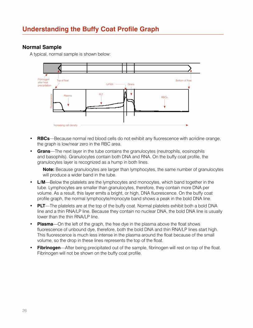
Normal SampleA typical, normal sample is shown below:
• RBCs—Because normal red blood cells do not exhibit any fluorescence with acridine orange, the graph is low/near zero in the RBC area.
• Grans—The next layer in the tube contains the granulocytes (neutrophils, eosinophils and basophils). Granulocytes contain both DNA and RNA. On the buffy coat profile, the granulocytes layer is recognized as a hump in both lines.
Note: Because granulocytes are larger than lymphocytes, the same number of granulocytes will produce a wider band in the tube.
• L/M—Below the platelets are the lymphocytes and monocytes, which band together in the tube. Lymphocytes are smaller than granulocytes, therefore, they contain more DNA per volume. As a result, this layer emits a bright, or high, DNA fluorescence. On the buffy coat profile graph, the normal lymphocyte/monocyte band shows a peak in the bold DNA line.
• PLT—The platelets are at the top of the buffy coat. Normal platelets exhibit both a bold DNA line and a thin RNA/LP line. Because they contain no nuclear DNA, the bold DNA line is usually lower than the thin RNA/LP line.
• Plasma—On the left of the graph, the free dye in the plasma above the float shows fluorescence of unbound dye, therefore, both the bold DNA and thin RNA/LP lines start high. This fluorescence is much less intense in the plasma around the float because of the small volume, so the drop in these lines represents the top of the float.
• Fibrinogen—After being precipitated out of the sample, fibrinogen will rest on top of the float. Fibrinogen will not be shown on the buffy coat profile.
Plasma PLT
Lymps Grans
RBCs
Fibrinogen after heat precipitation
Top of float Bottom of float
Increasing cell density
Fluo
resc
ence
26
27
ReticulocytesThe IDEXX VetAutoread™ analyzer can quantify reticulocytes, as a percentage of the hematocrit, within the following range:
Retics 0.2% ≤ x ≤ 4.0%
The analyzer calculates the reticulocyte quantity in terms of volume, then expresses this as a percentage of the total hematocrit. Because the analyzer is not giving a single-cell count, it uses the ~ symbol:
Retics ~3.0%
If reticulocytes are determined to be greater than 4%, the results will print as:
Retics >4.0%
For zero or less than 0.2% reticulocytes, there will be no reticulocyte indicator on the printout.
The analyzer quantifies reticulocytes whenever it detects them, whether the hematocrit is low, normal or high.
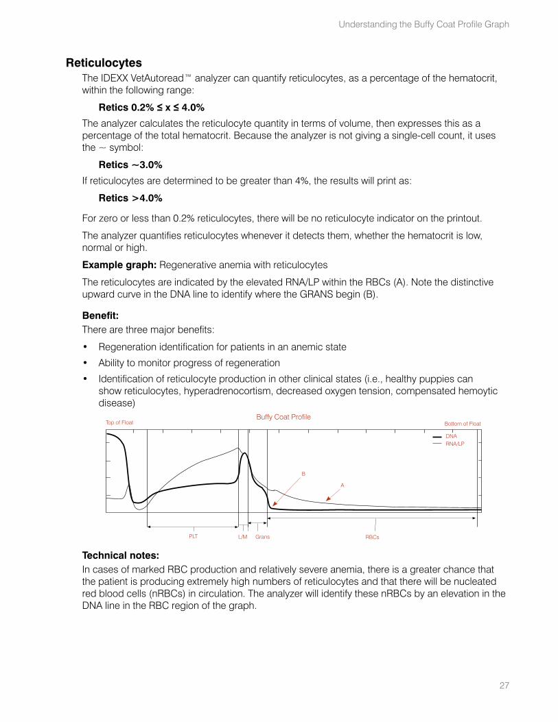
Example graph: Regenerative anemia with reticulocytes
The reticulocytes are indicated by the elevated RNA/LP within the RBCs (A). Note the distinctive upward curve in the DNA line to identify where the GRANS begin (B).
Benefit:There are three major benefits:
• Regeneration identification for patients in an anemic state
• Ability to monitor progress of regeneration
• Identification of reticulocyte production in other clinical states (i.e., healthy puppies can show reticulocytes, hyperadrenocortism, decreased oxygen tension, compensated hemoytic disease)
Technical notes:In cases of marked RBC production and relatively severe anemia, there is a greater chance that the patient is producing extremely high numbers of reticulocytes and that there will be nucleated red blood cells (nRBCs) in circulation. The analyzer will identify these nRBCs by an elevation in the DNA line in the RBC region of the graph.
Buffy Coat Profile
PLT L/M RBCsGrans
DNARNA/LP
B
A
Top of Float Bottom of Float
Understanding the Buffy Coat Profile Graph
28
IDEXX VetAutoread Hematology Analyzer Operator’s Guide
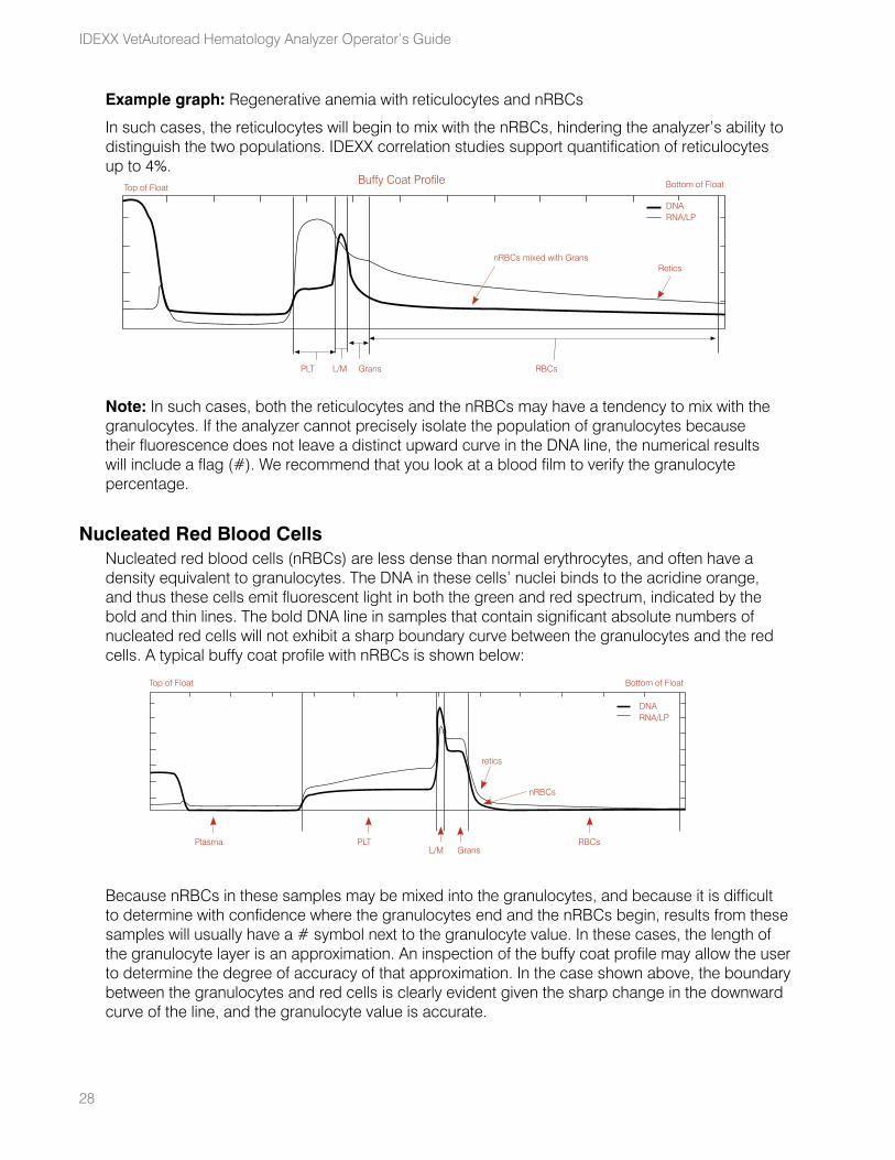
Example graph: Regenerative anemia with reticulocytes and nRBCs
In such cases, the reticulocytes will begin to mix with the nRBCs, hindering the analyzer’s ability to distinguish the two populations. IDEXX correlation studies support quantification of reticulocytes up to 4%.
Note: In such cases, both the reticulocytes and the nRBCs may have a tendency to mix with the granulocytes. If the analyzer cannot precisely isolate the population of granulocytes because their fluorescence does not leave a distinct upward curve in the DNA line, the numerical results will include a flag (#). We recommend that you look at a blood film to verify the granulocyte percentage.
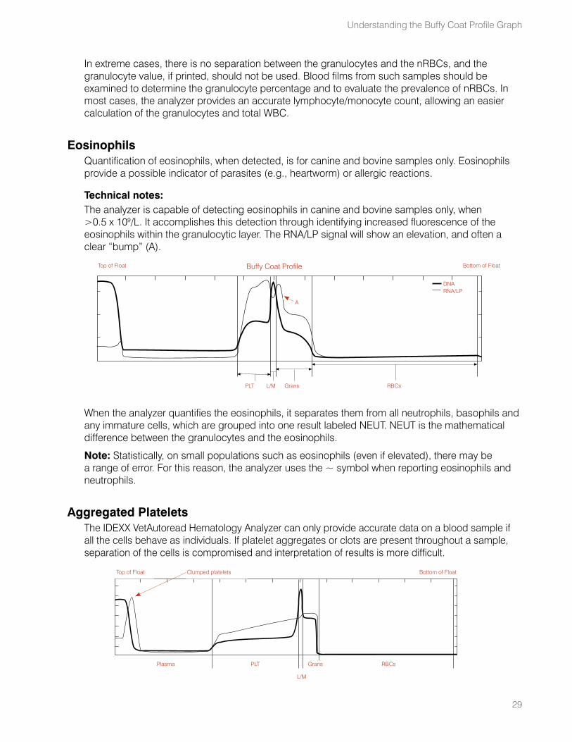
Nucleated Red Blood CellsNucleated red blood cells (nRBCs) are less dense than normal erythrocytes, and often have a density equivalent to granulocytes. The DNA in these cells’ nuclei binds to the acridine orange, and thus these cells emit fluorescent light in both the green and red spectrum, indicated by the bold and thin lines. The bold DNA line in samples that contain significant absolute numbers of nucleated red cells will not exhibit a sharp boundary curve between the granulocytes and the red cells. A typical buffy coat profile with nRBCs is shown below:
Because nRBCs in these samples may be mixed into the granulocytes, and because it is difficult to determine with confidence where the granulocytes end and the nRBCs begin, results from these samples will usually have a # symbol next to the granulocyte value. In these cases, the length of the granulocyte layer is an approximation. An inspection of the buffy coat profile may allow the user to determine the degree of accuracy of that approximation. In the case shown above, the boundary between the granulocytes and red cells is clearly evident given the sharp change in the downward curve of the line, and the granulocyte value is accurate.
Buffy Coat Profile
PLT L/M RBCsGrans
DNARNA/LP
nRBCs mixed with Grans Retics
Top of Float Bottom of Float
PLTPlasmaL/M
RBCsGrans
Top of Float Bottom of Float
retics
nRBCs
DNARNA/LP
29
In extreme cases, there is no separation between the granulocytes and the nRBCs, and the granulocyte value, if printed, should not be used. Blood films from such samples should be examined to determine the granulocyte percentage and to evaluate the prevalence of nRBCs. In most cases, the analyzer provides an accurate lymphocyte/monocyte count, allowing an easier calculation of the granulocytes and total WBC.
EosinophilsQuantification of eosinophils, when detected, is for canine and bovine samples only. Eosinophils provide a possible indicator of parasites (e.g., heartworm) or allergic reactions.
Technical notes:The analyzer is capable of detecting eosinophils in canine and bovine samples only, when >0.5 x 10⁹/L. It accomplishes this detection through identifying increased fluorescence of the eosinophils within the granulocytic layer. The RNA/LP signal will show an elevation, and often a clear “bump” (A).
When the analyzer quantifies the eosinophils, it separates them from all neutrophils, basophils and any immature cells, which are grouped into one result labeled NEUT. NEUT is the mathematical difference between the granulocytes and the eosinophils.
Note: Statistically, on small populations such as eosinophils (even if elevated), there may be a range of error. For this reason, the analyzer uses the ~ symbol when reporting eosinophils and neutrophils.
Aggregated PlateletsThe IDEXX VetAutoread Hematology Analyzer can only provide accurate data on a blood sample if all the cells behave as individuals. If platelet aggregates or clots are present throughout a sample, separation of the cells is compromised and interpretation of results is more difficult.
Buffy Coat Profile
PLT L/M RBCsGrans
DNARNA/LP
A
Top of Float Bottom of Float
PLTPlasma
L/M
RBCsGrans
Top of Float Clumped platelets Bottom of Float
Understanding the Buffy Coat Profile Graph
30
IDEXX VetAutoread Hematology Analyzer Operator’s Guide
Small aggregates of platelets that would interfere with cell counters do not interfere with the IDEXX VetAutoread Hematology Analyzer analysis as long as they accumulate in the platelet layer on the float. Large aggregates will accumulate in the platelet layer, and on top of the float. Usually these can be seen by visual inspection of the tube, and will give a peak in the thin RNA/LP line on the buffy coat profile on the top portion of the float.
The most important point to recognize in this case is that the sample’s platelet mass is greater than the number reported because of the additional platelets unaccounted for at the top of the float. If the rest of the buffy coat profile appears normal, the analyzer’s results contain no distortion caused by the aggregates. Reported results in these cases will show a > (greater than) symbol to indicate recognition of the additional platelets.
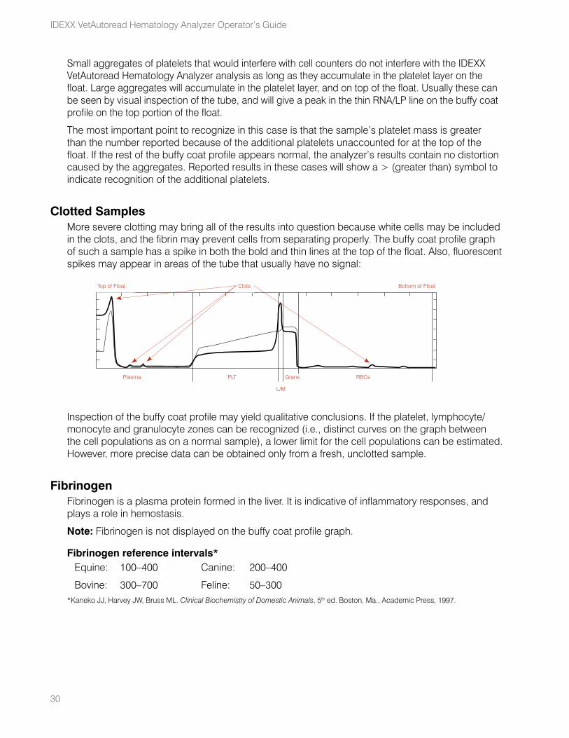
Clotted SamplesMore severe clotting may bring all of the results into question because white cells may be included in the clots, and the fibrin may prevent cells from separating properly. The buffy coat profile graph of such a sample has a spike in both the bold and thin lines at the top of the float. Also, fluorescent spikes may appear in areas of the tube that usually have no signal:
Inspection of the buffy coat profile may yield qualitative conclusions. If the platelet, lymphocyte/monocyte and granulocyte zones can be recognized (i.e., distinct curves on the graph between the cell populations as on a normal sample), a lower limit for the cell populations can be estimated. However, more precise data can be obtained only from a fresh, unclotted sample.
FibrinogenFibrinogen is a plasma protein formed in the liver. It is indicative of inflammatory responses, and plays a role in hemostasis.
Note: Fibrinogen is not displayed on the buffy coat profile graph.
Fibrinogen reference intervals*Equine: 100–400 Canine: 200–400
Bovine: 300–700 Feline: 50–300*Kaneko JJ, Harvey JW, Bruss ML. Clinical Biochemistry of Domestic Animals, 5th ed. Boston, Ma., Academic Press, 1997.
PLTPlasma
L/M
RBCsGrans
Top of Float Clots Bottom of Float
31
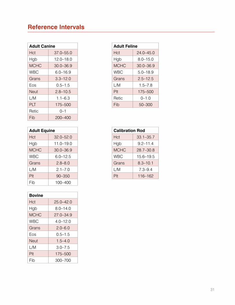
Reference Intervals
Adult Canine Adult Feline
Hct 37.0–55.0 Hct 24.0–45.0
Hgb 12.0–18.0 Hgb 8.0–15.0
MCHC 30.0–36.9 MCHC 30.0–36.9
WBC 6.0–16.9 WBC 5.0–18.9
Grans 3.3–12.0 Grans 2.5–12.5
Eos 0.5–1.5 L/M 1.5–7.8
Neut 2.8–10.5 Plt 175–500
L/M 1.1–6.3 Retic 0–1.0
PLT 175–500 Fib 50–300
Retic 0–1
Fib 200–400
Adult Equine Calibration Rod
Hct 32.0–52.0 Hct 33.1–35.7
Hgb 11.0–19.0 Hgb 9.2–11.4
MCHC 30.0–36.9 MCHC 28.7–30.8
WBC 6.0–12.5 WBC 15.6–19.5
Grans 2.8–8.0 Grans 8.3–10.1
L/M 2.1–7.0 L/M 7.3–9.4
Plt 90–350 Plt 116–162
Fib 100–400
Bovine
Hct 25.0–42.0
Hgb 8.0–14.0
MCHC 27.0–34.9
WBC 4.0–12.0
Grans 2.0–6.0
Eos 0.5–1.5
Neut 1.5–4.0
L/M 3.0–7.5
Plt 175–500
Fib 300–700
Technical Notes
The IDEXX VetAutoread™ Hematology Analyzer is designed to alert the user when certain conditions interfere with the analysis of samples.
Startup Service AlertsIf the Systems Check phase of startup fails, an alert message appears flagging the error condition. Service messages that can occur during startup are:
• NOV/RAM failed initializing
• Checksum error
• RAM test error
• Calibration error backlash
• Filter wheel error (#)
• Lamp test fail
In the event of the NOV/RAM message, the alert will be only momentary, since the analyzer automatically resets certain default or calibration values, then cycles through the startup sequence again.
All other alerts listed abort the startup sequence and prevent testing. In such cases, you should turn the power switch off, wait several seconds, and then turn the power back on. If the alert reappears during the Systems Check phase, contact IDEXX Technical Support.
Test AlertsIf the analyzer detects an irregular condition during a run, an alert message will identify it. A full list of test alert messages is shown below. In all cases, except for the Position Error test alert, the assay is aborted and no hematology results are obtained. As indicated, the IDEXX VetTube must either be wiped clean, or a fresh tube prepared and tested. If the analyzer has a mechanical or optical problem, contact IDEXX Technical Support.
Rotation errorCause: Mode was set to Insert Cal Rod when the tube was inserted.Action: Remove the tube, change Mode to the applicable species and reinsert the tube.
Cause: The Cal Rod did not rotate during scanning.Action: Rerun the Cal Rod. If a second run fails, contact IDEXX Technical Support.
Position error; remove tubeCause: A temporary mechanical problem: the tube was incorrectly positioned in the carrier during its return to the loading platform, the carriage is jammed, the analyzer is cold, or the tube was inserted improperly.Action: Open the door, carefully remove the tube if present, and then follow the instructions on the message display panel to complete the test.
32
33
Error locating meniscus of tubeCause: The fill volume of the tube is incorrect (i.e., either above or below the two green lines), or the tube is dirty.Action: Remove the tube and check it for cleanliness in the meniscus region. Retest, or prepare and test a new tube. Check the pipetter for accuracy where applicable.
Carriage error no sensorCause: The carriage is not moving properly.Action: Turn the analyzer off, wait one minute and then turn it back on. If failure persists, contact IDEXX Technical Support.
Too many bubbles found in tubeCause: The plasma contains excess bubbles, making an accurate linear measurement of the plasma impossible.Action: Prepare and test a new tube.
Cannot identify tube typeCause: The fill volume is incorrect, the fill lines are missing from the tube, the cells clumped on top of the float, the float is missing, the tube lifter is stuck in the load position or the tube is inserted backward.Action: Remove the tube and inspect it. Retest, or prepare and test a new tube.
Error locating float positionCause: One or more of the following: a dirty tube, an optical interference in the tube, a defective float (e.g., wrong length), a float lodged in the wrong part of the blood tube, a missing float, an uncentrifuged sample or a very low hematocrit.Action: Clean the tube and reinsert it into the analyzer. If the failure persists, recentrifuge the tube, or prepare and test a new tube.
Error locating bottom of RBCsCause: The closure is incorrectly seated or defective.Action: Prepare and test a new tube.
Improper fill venous sampleCause: The tube is not filled to the correct level or the tube has a leaky cap.Action: Prepare and test a new tube. Check the pipette for accuracy, where applicable.
Sample AlertsWhen the properties of the sample have affected the reliability of the results, various sample alert messages appear on the display and on the printout.
Affected values have a # sign next to them, and should be verified by inspection of the buffy coat profile or examination of a blood film. The most frequent cause of sample alert messages is platelet aggregation and cell clumping. These samples must be redrawn to obtain accurate values.
Technical Notes
34
IDEXX VetAutoread Hematology Analyzer Operator’s Guide
Granulocytes (1)Sample alert message: “Red cells have not separated cleanly from granulocytes. To confirm granulocyte results, verify proper location of RBC/granulocyte boundary on buffy coat profile, or examine a blood film.”
The red cell layer contains nRBCs, a very high number of reticulocytes or damaged red cells, and thus some cells overlap in density with the granulocyte layer. The granulocyte value has a # flag, indicating that the value needs verification. (Other values that are calculated from the granulocyte value also have the # flag.)
Inspection of the buffy coat profile shows that the boundary between the granulocytes and red cells indicate a gradual change in fluorescence, rather than the sharp change normally seen. In some cases, the probable location of the boundary is evident from the profile, and the resulting uncertainty in the granulocyte value is small.
In extreme cases, no boundary is seen, and no valid estimate of granulocyte value can be made. In these cases, examination of a blood film will probably show a significant number of nRBCs. Because the lymphocyte/monocyte value is valid, an estimate of the granulocyte count can be made by determining the proportion of granulocytes to lymphocytes plus monocytes.
Buffy Coat (1)Sample alert message: “Granulocytes have not separated cleanly from the lymphocyte/monocyte layer and RBC layer. Confirm results with a blood film.”
These samples may have enough nRBCs (or other red cell anomaly) to completely obscure the granulocyte layer and a small, or nonexistent, lymphocyte/monocyte peak. Examination of a blood film is required to assess morphology.
Buffy Coat (3)Sample alert message: “Lymphocyte/Monocyte layer has not separated distinctly from the other cell layers, possibly due to inadequate staining. Confirm differential results with a blood film.”
The boundaries between the lymphocyte/monocyte layer and the adjacent layers are not sharp, and the analyzer has estimated the boundary locations. The lymphocyte/monocyte-to-granulocyte ratio can be confirmed by examining a blood film. In some cases, letting the sample sit for two minutes will allow the cells to take up dye, so the lymphocyte/monocyte peak will become more distinct and readable.
Buffy Coat (4)Sample alert message: “Buffy coat layers inconsistent due to clumped platelets, granulocytes, expired tube or stain on tube exterior. Remove tube, carefully clean exterior, and retest. If situation persists, obtain a fresh sample and retest.”
Five of the eight scans of the buffy coat are significantly different, probably due to aggregation of platelets. If cleaning the tube does not correct the issue, redraw the sample. The presence of platelet clumping will bring into question any buffy coat results.
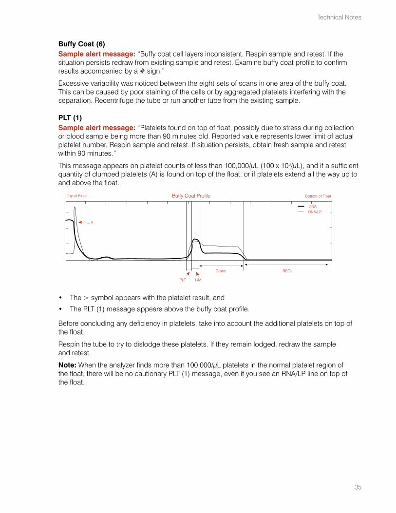
35
Buffy Coat (6)Sample alert message: “Buffy coat cell layers inconsistent. Respin sample and retest. If the situation persists redraw from existing sample and retest. Examine buffy coat profile to confirm results accompanied by a # sign.”
Excessive variability was noticed between the eight sets of scans in one area of the buffy coat. This can be caused by poor staining of the cells or by aggregated platelets interfering with the separation. Recentrifuge the tube or run another tube from the existing sample.
PLT (1)Sample alert message: “Platelets found on top of float, possibly due to stress during collection or blood sample being more than 90 minutes old. Reported value represents lower limit of actual platelet number. Respin sample and retest. If situation persists, obtain fresh sample and retest within 90 minutes.”
This message appears on platelet counts of less than 100,000/μL (100 x 103/μL), and if a sufficient quantity of clumped platelets (A) is found on top of the float, or if platelets extend all the way up to and above the float.
• The > symbol appears with the platelet result, and
• The PLT (1) message appears above the buffy coat profile.
Before concluding any deficiency in platelets, take into account the additional platelets on top of the float.
Respin the tube to try to dislodge these platelets. If they remain lodged, redraw the sample and retest.
Note: When the analyzer finds more than 100,000/μL platelets in the normal platelet region of the float, there will be no cautionary PLT (1) message, even if you see an RNA/LP line on top of the float.
Buffy Coat Profile
PLT L/M
RBCsGrans
DNARNA/LP
A
Top of Float Bottom of Float
Technical Notes
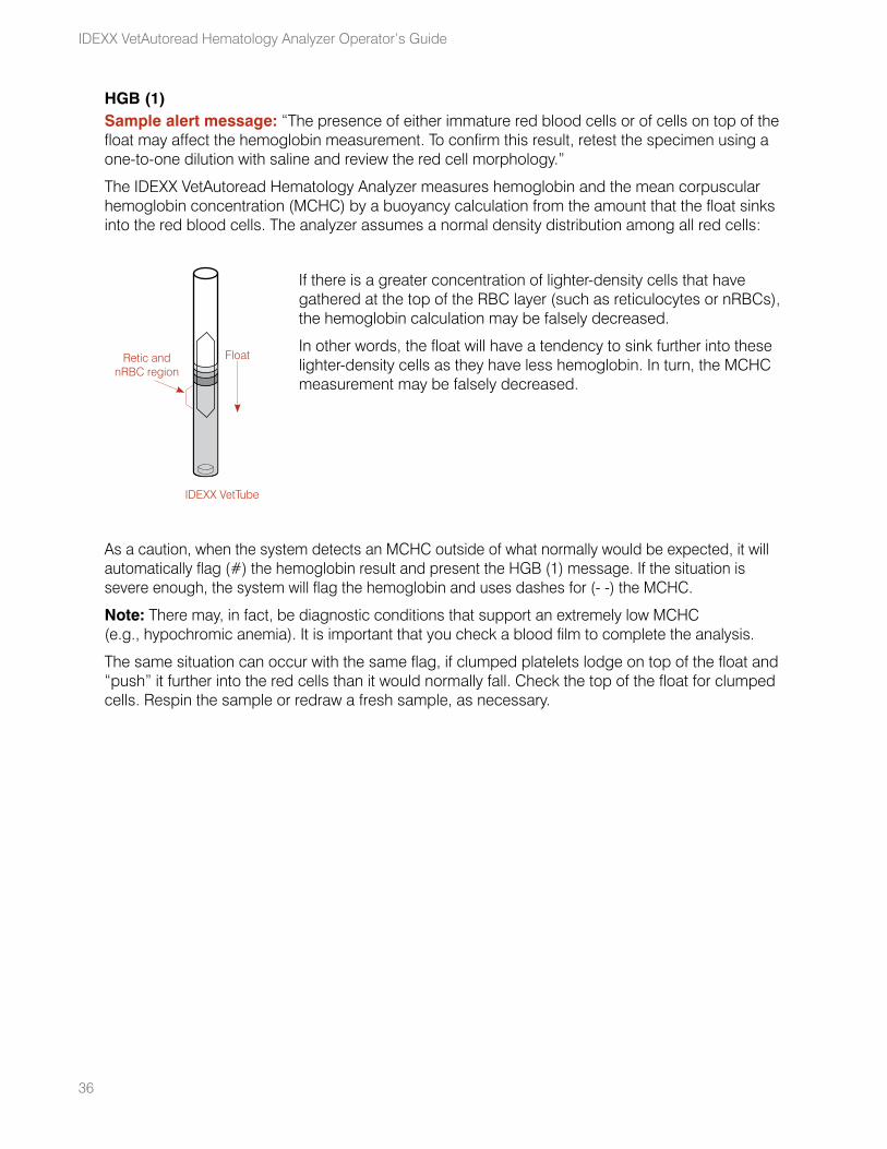
36
IDEXX VetAutoread Hematology Analyzer Operator’s Guide
HGB (1)Sample alert message: “The presence of either immature red blood cells or of cells on top of the float may affect the hemoglobin measurement. To confirm this result, retest the specimen using a one-to-one dilution with saline and review the red cell morphology.”
The IDEXX VetAutoread Hematology Analyzer measures hemoglobin and the mean corpuscular hemoglobin concentration (MCHC) by a buoyancy calculation from the amount that the float sinks into the red blood cells. The analyzer assumes a normal density distribution among all red cells:
If there is a greater concentration of lighter-density cells that have gathered at the top of the RBC layer (such as reticulocytes or nRBCs), the hemoglobin calculation may be falsely decreased.
In other words, the float will have a tendency to sink further into these lighter-density cells as they have less hemoglobin. In turn, the MCHC measurement may be falsely decreased.
As a caution, when the system detects an MCHC outside of what normally would be expected, it will automatically flag (#) the hemoglobin result and present the HGB (1) message. If the situation is severe enough, the system will flag the hemoglobin and uses dashes for (- -) the MCHC.
Note: There may, in fact, be diagnostic conditions that support an extremely low MCHC (e.g., hypochromic anemia). It is important that you check a blood film to complete the analysis.
The same situation can occur with the same flag, if clumped platelets lodge on top of the float and “push” it further into the red cells than it would normally fall. Check the top of the float for clumped cells. Respin the sample or redraw a fresh sample, as necessary.
Float
IDEXX VetTube
Retic and nRBC region
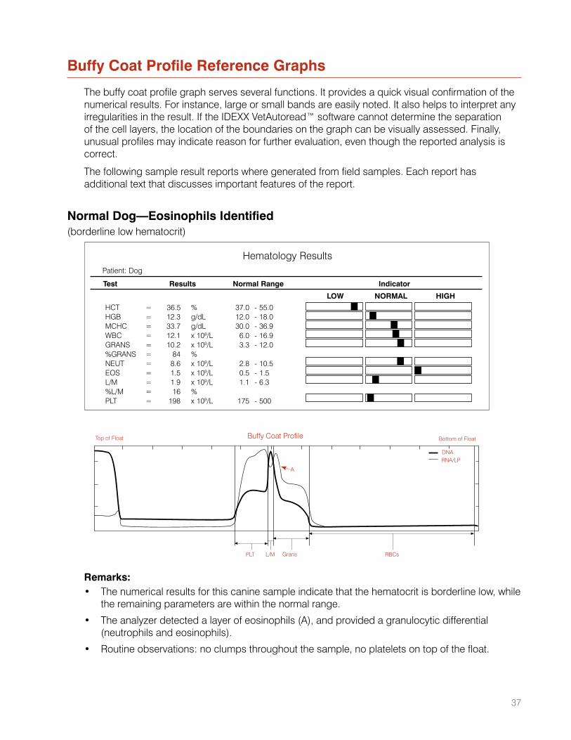
37
Buffy Coat Profile Reference Graphs
The buffy coat profile graph serves several functions. It provides a quick visual confirmation of the numerical results. For instance, large or small bands are easily noted. It also helps to interpret any irregularities in the result. If the IDEXX VetAutoread™ software cannot determine the separation of the cell layers, the location of the boundaries on the graph can be visually assessed. Finally, unusual profiles may indicate reason for further evaluation, even though the reported analysis is correct.
The following sample result reports where generated from field samples. Each report has additional text that discusses important features of the report.
Normal Dog—Eosinophils Identified(borderline low hematocrit)
Remarks:• The numerical results for this canine sample indicate that the hematocrit is borderline low, while
the remaining parameters are within the normal range.
• The analyzer detected a layer of eosinophils (A), and provided a granulocytic differential (neutrophils and eosinophils).
• Routine observations: no clumps throughout the sample, no platelets on top of the float.
Patient: Dog
Hematology Results
Test Results Normal Range Indicator
HCT = 36.5 % 37.0 - 55.0 HGB = 12.3 g/dL 12.0 - 18.0MCHC = 33.7 g/dL 30.0 - 36.9WBC = 12.1 x 10⁹/L 6.0 - 16.9GRANS = 10.2 x 10⁹/L 3.3 - 12.0%GRANS = 84 % NEUT = 8.6 x 10⁹/L 2.8 - 10.5 EOS = 1.5 x 10⁹/L 0.5 - 1.5 L/M = 1.9 x 10⁹/L 1.1 - 6.3%L/M = 16 % PLT = 198 x 109/L 175 - 500
LOW NORMAL HIGH
Buffy Coat Profile
PLT L/M RBCsGrans
DNARNA/LP
A
Top of Float Bottom of Float
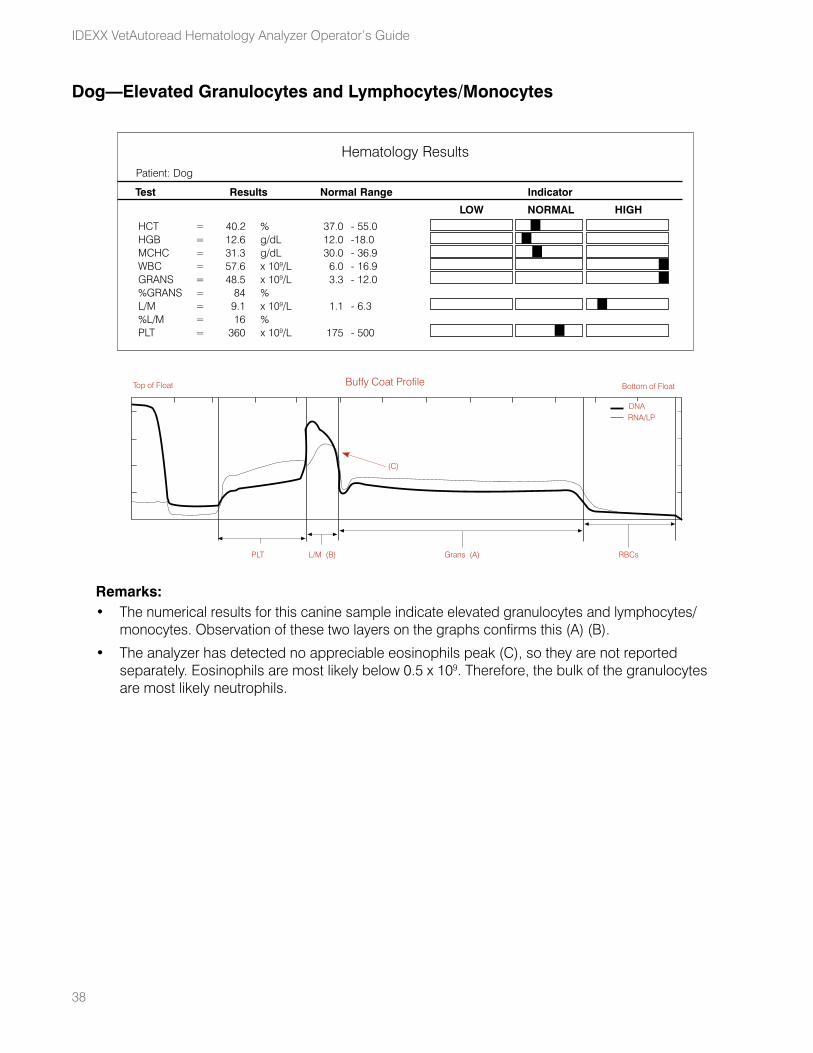
38
IDEXX VetAutoread Hematology Analyzer Operator’s Guide
Dog—Elevated Granulocytes and Lymphocytes/Monocytes
Remarks:• The numerical results for this canine sample indicate elevated granulocytes and lymphocytes/
monocytes. Observation of these two layers on the graphs confirms this (A) (B).
• The analyzer has detected no appreciable eosinophils peak (C), so they are not reported separately. Eosinophils are most likely below 0.5 x 10⁹. Therefore, the bulk of the granulocytes are most likely neutrophils.
Buffy Coat Profile
PLT L/M (B) RBCsGrans (A)
DNARNA/LP
(C)
Top of Float Bottom of Float
HCT = 40.2 % 37.0 - 55.0 HGB = 12.6 g/dL 12.0 - 18.0MCHC = 31.3 g/dL 30.0 - 36.9WBC = 57.6 x 10⁹/L 6.0 - 16.9GRANS = 48.5 x 10⁹/L 3.3 - 12.0%GRANS = 84 % L/M = 9.1 x 10⁹/L 1.1 - 6.3%L/M = 16 % PLT = 360 x 109/L 175 - 500
Patient: Dog
Hematology Results
Test Results Normal Range Indicator
LOW NORMAL HIGH
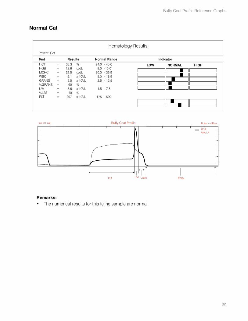
39
Normal Cat
Remarks:• The numerical results for this feline sample are normal.
HCT = 38.3 % 24.0 - 45.0 HGB = 12.6 g/dL 8.0 - 15.0MCHC = 32.5 g/dL 30.0 - 36.9WBC = 9.1 x 10⁹/L 5.0 - 18.9GRANS = 5.5 x 10⁹/L 2.5 - 12.5%GRANS = 60 % L/M = 3.6 x 10⁹/L 1.5 - 7.8%L/M = 40 % PLT = 397 x 109/L 175 - 500
Patient: Cat
Hematology Results
Test Results Normal Range Indicator
LOW NORMAL HIGH
PLT L/M RBCsGrans
DNA
RNA/LP
Top of Float Bottom of FloatBuffy Coat Profile
DNARNA/LP
PLT L/M RBCsGrans
Buffy Coat Profile Reference Graphs
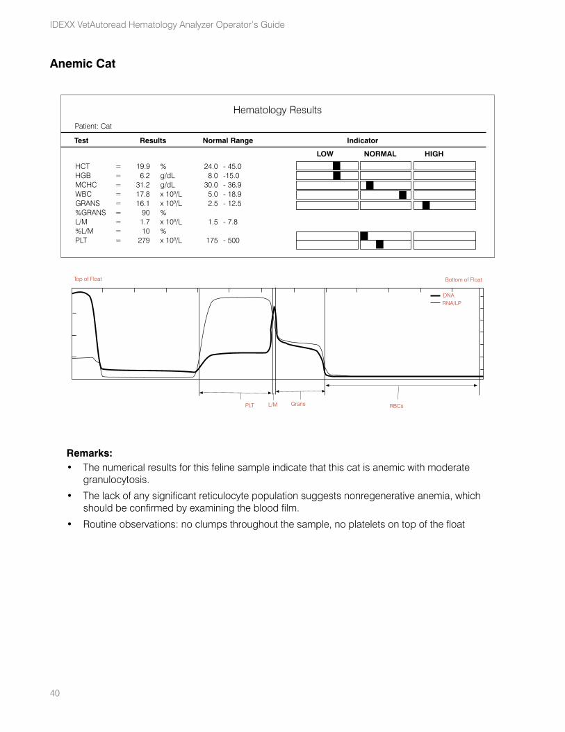
40
IDEXX VetAutoread Hematology Analyzer Operator’s Guide
Anemic Cat
Remarks:• The numerical results for this feline sample indicate that this cat is anemic with moderate
granulocytosis.
• The lack of any significant reticulocyte population suggests nonregenerative anemia, which should be confirmed by examining the blood film.
• Routine observations: no clumps throughout the sample, no platelets on top of the float
HCT = 19.9 % 24.0 - 45.0 HGB = 6.2 g/dL 8.0 - 15.0MCHC = 31.2 g/dL 30.0 - 36.9WBC = 17.8 x 10⁹/L 5.0 - 18.9GRANS = 16.1 x 10⁹/L 2.5 - 12.5%GRANS = 90 % L/M = 1.7 x 10⁹/L 1.5 - 7.8%L/M = 10 % PLT = 279 x 109/L 175 - 500
Patient: Cat
Hematology Results
Test Results Normal Range Indicator
LOW NORMAL HIGH
Buffy Coat Profile
PLT L/M RBCsGrans
DNARNA/LP
Top of Float Bottom of Float
PLT L/M RBCsGrans
DNARNA/LP
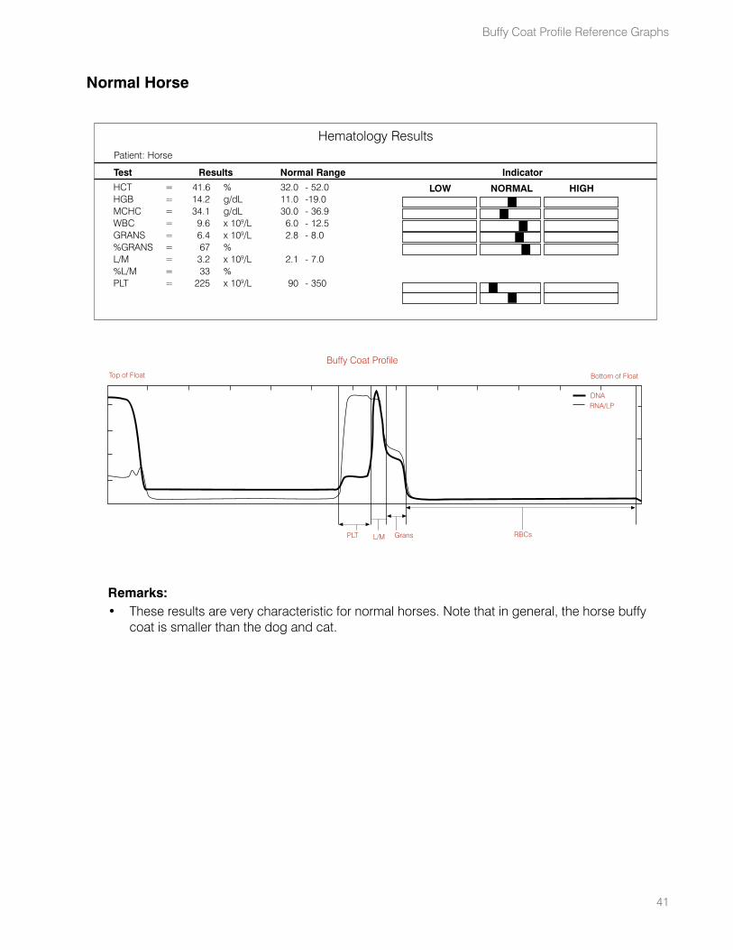
41
Normal Horse
Remarks:• These results are very characteristic for normal horses. Note that in general, the horse buffy
coat is smaller than the dog and cat.
HCT = 41.6 % 32.0 - 52.0 HGB = 14.2 g/dL 11.0 - 19.0MCHC = 34.1 g/dL 30.0 - 36.9WBC = 9.6 x 10⁹/L 6.0 - 12.5GRANS = 6.4 x 10⁹/L 2.8 - 8.0%GRANS = 67 % L/M = 3.2 x 10⁹/L 2.1 - 7.0%L/M = 33 % PLT = 225 x 109/L 90 - 350
Patient: Horse
Hematology Results
Test Results Normal Range Indicator
LOW NORMAL HIGH
Buffy Coat Profile
PLT L/M RBCsGrans
DNARNA/LP
Top of Float Bottom of Float
PLT
DNARNA/LP
L/M RBCsGrans
Buffy Coat Profile Reference Graphs
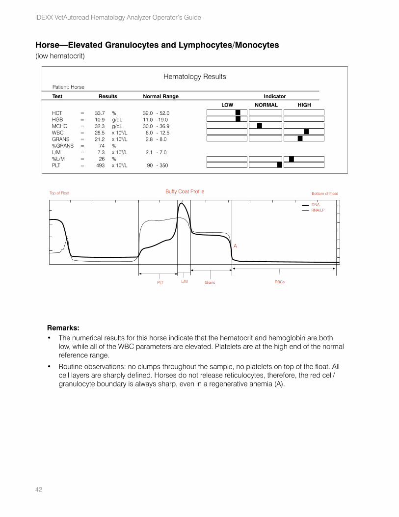
42
IDEXX VetAutoread Hematology Analyzer Operator’s Guide
Horse—Elevated Granulocytes and Lymphocytes/Monocytes(low hematocrit)
Remarks:• The numerical results for this horse indicate that the hematocrit and hemoglobin are both
low, while all of the WBC parameters are elevated. Platelets are at the high end of the normal reference range.
• Routine observations: no clumps throughout the sample, no platelets on top of the float. All cell layers are sharply defined. Horses do not release reticulocytes, therefore, the red cell/granulocyte boundary is always sharp, even in a regenerative anemia (A).
HCT = 33.7 % 32.0 - 52.0 HGB = 10.9 g/dL 11.0 - 19.0MCHC = 32.3 g/dL 30.0 - 36.9WBC = 28.5 x 10⁹/L 6.0 - 12.5GRANS = 21.2 x 10⁹/L 2.8 - 8.0%GRANS = 74 % L/M = 7.3 x 10⁹/L 2.1 - 7.0%L/M = 26 % PLT = 493 x 109/L 90 - 350
Patient: Horse
Hematology Results
Test Results Normal Range Indicator
LOW NORMAL HIGH
Top of Float Bottom of FloatBuffy Coat Profile
PLT L/M Grans
A
DNARNA/LP
RBCs
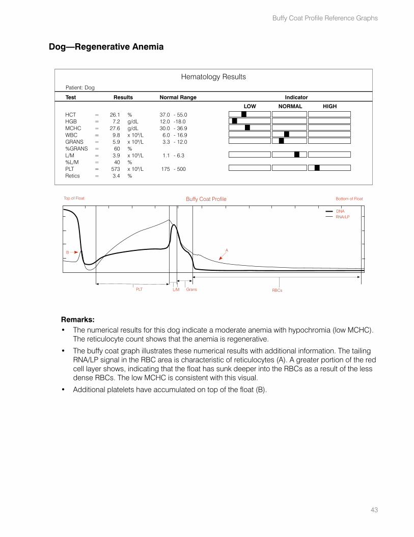
43
Dog—Regenerative Anemia
Remarks:• The numerical results for this dog indicate a moderate anemia with hypochromia (low MCHC).
The reticulocyte count shows that the anemia is regenerative.
• The buffy coat graph illustrates these numerical results with additional information. The tailing RNA/LP signal in the RBC area is characteristic of reticulocytes (A). A greater portion of the red cell layer shows, indicating that the float has sunk deeper into the RBCs as a result of the less dense RBCs. The low MCHC is consistent with this visual.
• Additional platelets have accumulated on top of the float (B).
HCT = 26.1 % 37.0 - 55.0 HGB = 7.2 g/dL 12.0 - 18.0MCHC = 27.6 g/dL 30.0 - 36.9WBC = 9.8 x 10⁹/L 6.0 - 16.9GRANS = 5.9 x 10⁹/L 3.3 - 12.0%GRANS = 60 % L/M = 3.9 x 10⁹/L 1.1 - 6.3%L/M = 40 % PLT = 573 x 109/L 175 - 500Retics = 3.4 %
Patient: Dog
Hematology Results
Test Results Normal Range Indicator
LOW NORMAL HIGH
Buffy Coat Profile
PLT L/M RBCsGrans
DNARNA/LP
B A
Top of Float Bottom of Float
Buffy Coat Profile Reference Graphs
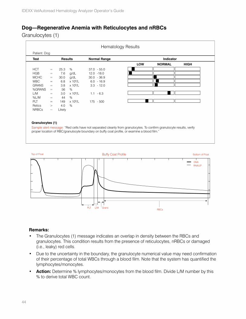
44
IDEXX VetAutoread Hematology Analyzer Operator’s Guide
Dog—Regenerative Anemia with Reticulocytes and nRBCs Granulocytes (1)
Remarks:• The Granulocytes (1) message indicates an overlap in density between the RBCs and
granulocytes. This condition results from the presence of reticulocytes, nRBCs or damaged (i.e., leaky) red cells.
• Due to the uncertainty in the boundary, the granulocyte numerical value may need confirmation of their percentage of total WBCs through a blood film. Note that the system has quantified the lymphocytes/monocytes.
• Action: Determine % lymphocytes/monocytes from the blood film. Divide L/M number by this % to derive total WBC count.
HCT = 25.3 % 37.0 - 55.0 HGB = 7.6 g/dL 12.0 - 18.0MCHC = 30.0 g/dL 30.0 - 36.9WBC = 6.8 x 10⁹/L 6.0 - 16.9GRANS = 3.8 x 10⁹/L 3.3 - 12.0%GRANS = 56 % L/M = 3.0 x 10⁹/L 1.1 - 6.3%L/M = 44 % PLT = 149 x 109/L 175 - 500Retics > 4.0 %NRBCs ~ Likely
Patient: Dog
Hematology Results
Test Results Normal Range Indicator
LOW NORMAL HIGH
Granulocytes (1)Sample alert message: “Red cells have not separated cleanly from granulocytes. To confirm granulocyte results, verify proper location of RBC/granulocyte boundary on buffy coat profile, or examine a blood film.”
Buffy Coat Profile
PLT L/MRBCs
Grans
DNARNA/LP
Top of Float Bottom of Float
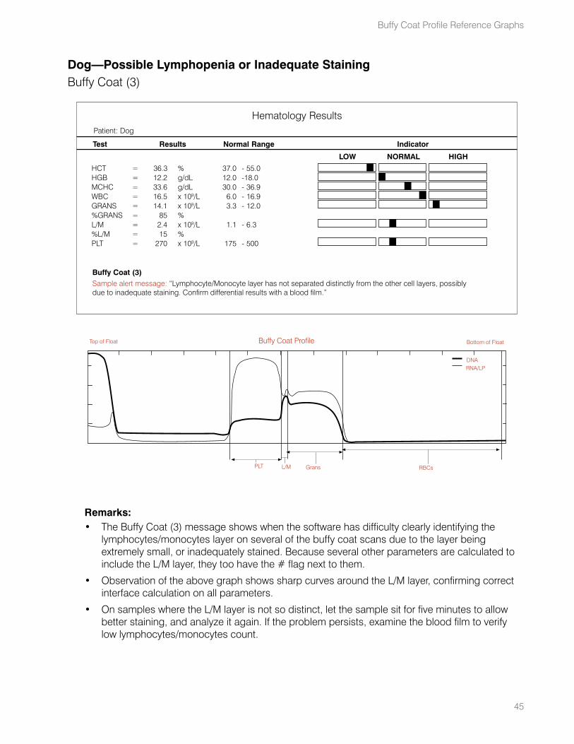
45
Dog—Possible Lymphopenia or Inadequate StainingBuffy Coat (3)
Remarks:• The Buffy Coat (3) message shows when the software has difficulty clearly identifying the
lymphocytes/monocytes layer on several of the buffy coat scans due to the layer being extremely small, or inadequately stained. Because several other parameters are calculated to include the L/M layer, they too have the # flag next to them.
• Observation of the above graph shows sharp curves around the L/M layer, confirming correct interface calculation on all parameters.
• On samples where the L/M layer is not so distinct, let the sample sit for five minutes to allow better staining, and analyze it again. If the problem persists, examine the blood film to verify low lymphocytes/monocytes count.
HCT = 36.3 % 37.0 - 55.0 HGB = 12.2 g/dL 12.0 - 18.0MCHC = 33.6 g/dL 30.0 - 36.9WBC = 16.5 x 10⁹/L 6.0 - 16.9GRANS = 14.1 x 10⁹/L 3.3 - 12.0%GRANS = 85 % L/M = 2.4 x 10⁹/L 1.1 - 6.3%L/M = 15 % PLT = 270 x 109/L 175 - 500
Patient: Dog
Hematology Results
Test Results Normal Range Indicator
LOW NORMAL HIGH
Buffy Coat (3)Sample alert message: “Lymphocyte/Monocyte layer has not separated distinctly from the other cell layers, possibly due to inadequate staining. Confirm differential results with a blood film.”
Buffy Coat Profile
PLT L/M RBCsGrans
DNARNA/LP
Top of Float Bottom of Float
Buffy Coat Profile Reference Graphs
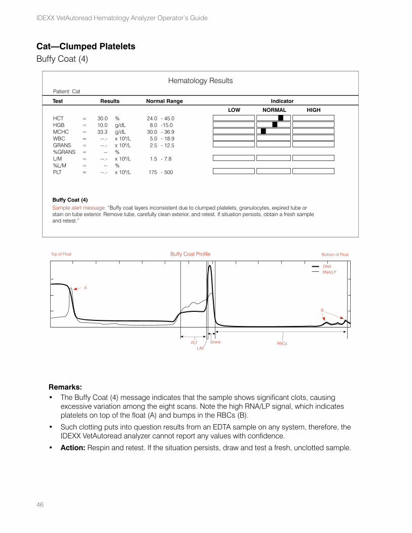
46
IDEXX VetAutoread Hematology Analyzer Operator’s Guide
Cat—Clumped PlateletsBuffy Coat (4)
Remarks:• The Buffy Coat (4) message indicates that the sample shows significant clots, causing
excessive variation among the eight scans. Note the high RNA/LP signal, which indicates platelets on top of the float (A) and bumps in the RBCs (B).
• Such clotting puts into question results from an EDTA sample on any system, therefore, the IDEXX VetAutoread analyzer cannot report any values with confidence.
• Action: Respin and retest. If the situation persists, draw and test a fresh, unclotted sample.
HCT = 30.0 % 24.0 - 45.0 HGB = 10.0 g/dL 8.0 - 15.0MCHC = 33.3 g/dL 30.0 - 36.9WBC = --.- x 109/L 5.0 - 18.9GRANS = --.- x 109/L 2.5 - 12.5%GRANS = -- % L/M = --.- x 109/L 1.5 - 7.8%L/M = -- % PLT = --.- x 109/L 175 - 500
Patient: Cat
Hematology Results
Test Results Normal Range Indicator
LOW NORMAL HIGH
Buffy Coat (4)Sample alert message: “Buffy coat layers inconsistent due to clumped platelets, granulocytes, expired tube or stain on tube exterior. Remove tube, carefully clean exterior, and retest. If situation persists, obtain a fresh sample and retest.”
Buffy Coat Profile
PLTL/M
RBCsGrans
DNARNA/LP
A
B
Top of Float Bottom of Float
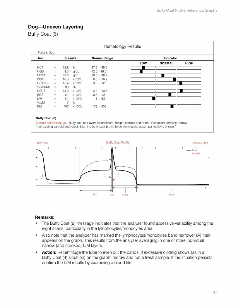
47
Dog—Uneven LayeringBuffy Coat (6)
Remarks:• The Buffy Coat (6) message indicates that the analyzer found excessive variability among the
eight scans, particularly in the lymphocytes/monocytes area.
• Also note that the analyzer has marked the lymphocytes/monocytes band narrower (A) than appears on the graph. This results from the analyzer averaging in one or more individual narrow (and crooked) L/M layers.
• Action: Recentrifuge the tube to even out the bands. If excessive clotting shows (as in a Buffy Coat (4) situation) on the graph, redraw and run a fresh sample. If the situation persists, confirm the L/M results by examining a blood film.
HCT = 28.8 % 37.0 - 55.0 HGB = 9.3 g/dL 12.0 - 18.0MCHC = 32.3 g/dL 30.0 - 36.9WBC = 16.5 x 10⁹/L 6.0 - 16.9GRANS = 15.4 x 10⁹/L 3.3 - 12.0%GRANS = 93 % NEUT = 14.2 x 10⁹/L 2.8 - 10.5 EOS = 1.1 x 10⁹/L 0.5 - 1.5 L/M = 1.1 x 10⁹/L 1.1 - 6.3%L/M = 7 % PLT = 407 x 109/L 175 - 500
Patient: Dog
Hematology Results
Test Results Normal Range Indicator
LOW NORMAL HIGH
Buffy Coat (6)Sample alert message: “Buffy coat cell layers inconsistent. Respin sample and retest. If situation persists; redraw from existing sample and retest. Examine buffy coat profile to confirm results accompanied by a # sign.”
Buffy Coat Profile
PLT L/M RBCsGrans
DNARNA/LP
A
Top of Float Bottom of Float
Buffy Coat Profile Reference Graphs
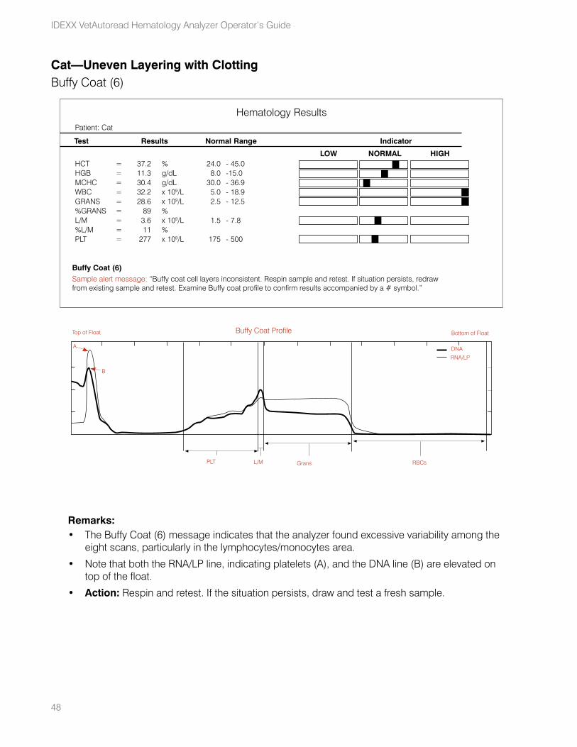
48
IDEXX VetAutoread Hematology Analyzer Operator’s Guide
Cat—Uneven Layering with ClottingBuffy Coat (6)
Remarks:• The Buffy Coat (6) message indicates that the analyzer found excessive variability among the
eight scans, particularly in the lymphocytes/monocytes area.
• Note that both the RNA/LP line, indicating platelets (A), and the DNA line (B) are elevated on top of the float.
• Action: Respin and retest. If the situation persists, draw and test a fresh sample.
HCT = 37.2 % 24.0 - 45.0 HGB = 11.3 g/dL 8.0 - 15.0MCHC = 30.4 g/dL 30.0 - 36.9WBC = 32.2 x 10⁹/L 5.0 - 18.9GRANS = 28.6 x 10⁹/L 2.5 - 12.5%GRANS = 89 % L/M = 3.6 x 10⁹/L 1.5 - 7.8%L/M = 11 % PLT = 277 x 109/L 175 - 500
Patient: Cat
Hematology Results
Test Results Normal Range Indicator
LOW NORMAL HIGH
Buffy Coat (6)Sample alert message: “Buffy coat cell layers inconsistent. Respin sample and retest. If situation persists, redraw from existing sample and retest. Examine Buffy coat profile to confirm results accompanied by a # symbol.”
Buffy Coat Profile
PLT L/M RBCsGrans
DNA
RNA/LP
A
B
Top of Float Bottom of Float
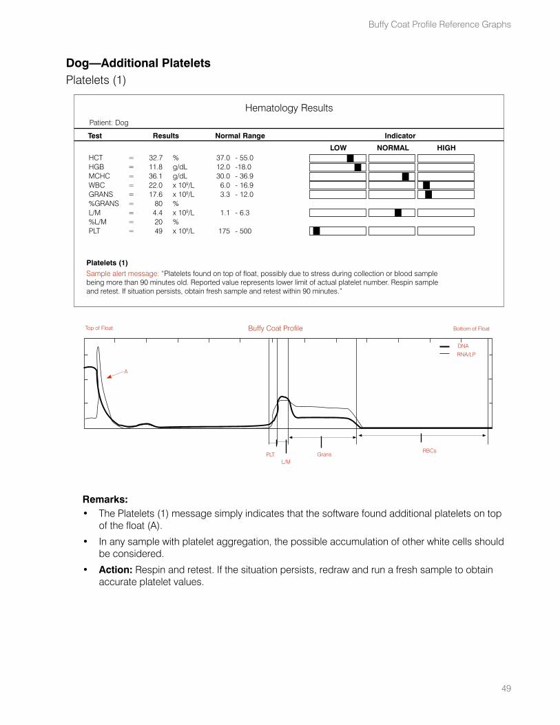
49
Dog—Additional PlateletsPlatelets (1)
Remarks:• The Platelets (1) message simply indicates that the software found additional platelets on top
of the float (A).
• In any sample with platelet aggregation, the possible accumulation of other white cells should be considered.
• Action: Respin and retest. If the situation persists, redraw and run a fresh sample to obtain accurate platelet values.
HCT = 32.7 % 37.0 - 55.0 HGB = 11.8 g/dL 12.0 - 18.0MCHC = 36.1 g/dL 30.0 - 36.9WBC = 22.0 x 10⁹/L 6.0 - 16.9GRANS = 17.6 x 10⁹/L 3.3 - 12.0%GRANS = 80 % L/M = 4.4 x 10⁹/L 1.1 - 6.3%L/M = 20 % PLT = 49 x 109/L 175 - 500
Patient: Dog
Hematology Results
Test Results Normal Range Indicator
LOW NORMAL HIGH
Platelets (1)Sample alert message: “Platelets found on top of float, possibly due to stress during collection or blood sample being more than 90 minutes old. Reported value represents lower limit of actual platelet number. Respin sample and retest. If situation persists, obtain fresh sample and retest within 90 minutes.”
Buffy Coat Profile
PLTL/M
RBCsGrans
DNA
RNA/LP
A
Top of Float Bottom of Float
Buffy Coat Profile Reference Graphs
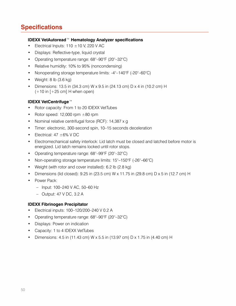
Specifications
IDEXX VetAutoread™ Hematology Analyzer specifications• Electrical Inputs: 110 ±10 V, 220 V AC
• Displays: Reflective-type, liquid crystal
• Operating temperature range: 68°–90°F (20°–32°C)
• Relative humidity: 10% to 95% (noncondensing)
• Nonoperating storage temperature limits: -4°–140°F (-20°–60°C)
• Weight: 8 lb (3.6 kg)
• Dimensions: 13.5 in (34.3 cm) W x 9.5 in (24.13 cm) D x 4 in (10.2 cm) H (+10 in [+25 cm] H when open)
IDEXX VetCentrifuge™• Rotor capacity: From 1 to 20 IDEXX VetTubes
• Rotor speed: 12,000 rpm ±80 rpm
• Nominal relative centrifugal force (RCF): 14,387 x g
• Timer: electronic, 300-second spin, 10–15 seconds deceleration
• Electrical: 47 ±6% V DC
• Electromechanical safety interlock: Lid latch must be closed and latched before motor is energized. Lid latch remains locked until rotor stops.
• Operating temperature range: 68°–99°F (20°–32°C)
• Non-operating storage temperature limits: 15°–150°F (-26°–66°C)
• Weight (with rotor and cover installed): 6.2 lb (2.8 kg)
• Dimensions (lid closed): 9.25 in (23.5 cm) W x 11.75 in (29.8 cm) D x 5 in (12.7 cm) H
• Power Pack:
– Input: 100–240 V AC, 50–60 Hz
– Output: 47 V DC, 3.2 A
IDEXX Fibrinogen Precipitator• Electrical inputs: 100–120/200–240 V 0.2 A
• Operating temperature range: 68°–90°F (20°–32°C)
• Displays: Power on indication
• Capacity: 1 to 4 IDEXX VetTubes
• Dimensions: 4.5 in (11.43 cm) W x 5.5 in (13.97 cm) D x 1.75 in (4.40 cm) H
50
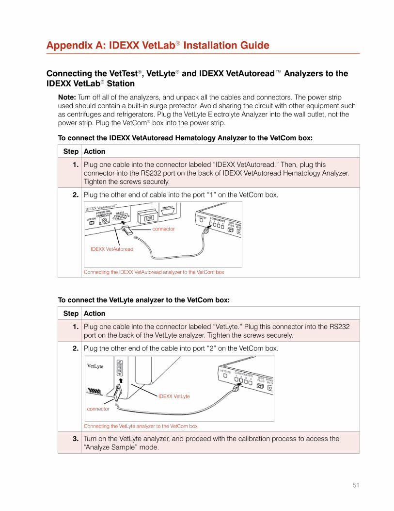
51
Appendix A: IDEXX VetLab® Installation Guide
Connecting the VetTest®, VetLyte® and IDEXX VetAutoread™ Analyzers to the IDEXX VetLab® Station
Note: Turn off all of the analyzers, and unpack all the cables and connectors. The power strip used should contain a built-in surge protector. Avoid sharing the circuit with other equipment such as centrifuges and refrigerators. Plug the VetLyte Electrolyte Analyzer into the wall outlet, not the power strip. Plug the VetCom® box into the power strip.
To connect the IDEXX VetAutoread Hematology Analyzer to the VetCom box:
Step Action
1. Plug one cable into the connector labeled “IDEXX VetAutoread.” Then, plug this connector into the RS232 port on the back of IDEXX VetAutoread Hematology Analyzer. Tighten the screws securely.
2. Plug the other end of cable into the port “1” on the VetCom box.
connector
IDEXX VetAutoread
Connecting the IDEXX VetAutoread analyzer to the VetCom box
To connect the VetLyte analyzer to the VetCom box:
Step Action
1. Plug one cable into the connector labeled “VetLyte.” Plug this connector into the RS232 port on the back of the VetLyte analyzer. Tighten the screws securely.
2. Plug the other end of the cable into port “2” on the VetCom box.
IDEXX VetLyte
connector
Connecting the VetLyte analyzer to the VetCom box
3. Turn on the VetLyte analyzer, and proceed with the calibration process to access the “Analyze Sample” mode.
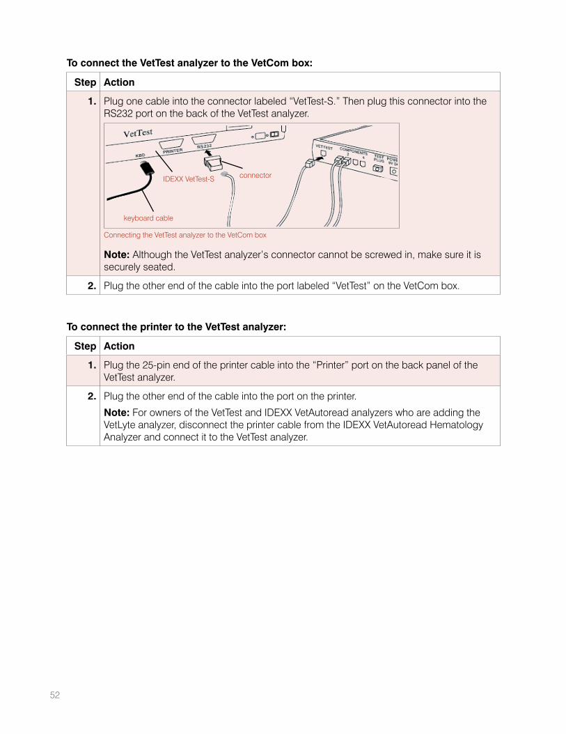
To connect the VetTest analyzer to the VetCom box:
Step Action
1. Plug one cable into the connector labeled “VetTest-S.” Then plug this connector into the RS232 port on the back of the VetTest analyzer.
IDEXX VetTest-S
keyboard cable
connector
Connecting the VetTest analyzer to the VetCom box
Note: Although the VetTest analyzer’s connector cannot be screwed in, make sure it is securely seated.
2. Plug the other end of the cable into the port labeled “VetTest” on the VetCom box.
To connect the printer to the VetTest analyzer:
Step Action
1. Plug the 25-pin end of the printer cable into the “Printer” port on the back panel of the VetTest analyzer.
2. Plug the other end of the cable into the port on the printer.
Note: For owners of the VetTest and IDEXX VetAutoread analyzers who are adding the VetLyte analyzer, disconnect the printer cable from the IDEXX VetAutoread Hematology Analyzer and connect it to the VetTest analyzer.
52
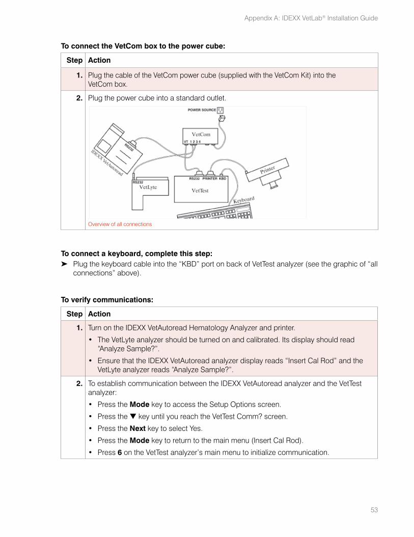
53
To connect the VetCom box to the power cube:
Step Action
1. Plug the cable of the VetCom power cube (supplied with the VetCom Kit) into the VetCom box.
2. Plug the power cube into a standard outlet.
Overview of all connections
To connect a keyboard, complete this step:➤ Plug the keyboard cable into the “KBD” port on back of VetTest analyzer (see the graphic of “all
connections” above).
To verify communications:
Step Action
1. Turn on the IDEXX VetAutoread Hematology Analyzer and printer.
• The VetLyte analyzer should be turned on and calibrated. Its display should read “Analyze Sample?”.
• Ensure that the IDEXX VetAutoread analyzer display reads “Insert Cal Rod” and the VetLyte analyzer reads “Analyze Sample?”.
2. To establish communication between the IDEXX VetAutoread analyzer and the VetTest analyzer:
• Press the Mode key to access the Setup Options screen.
• Press the t key until you reach the VetTest Comm? screen.
• Press the Next key to select Yes.
• Press the Mode key to return to the main menu (Insert Cal Rod).
• Press 6 on the VetTest analyzer’s main menu to initialize communication.
Appendix A: IDEXX VetLab® Installation Guide
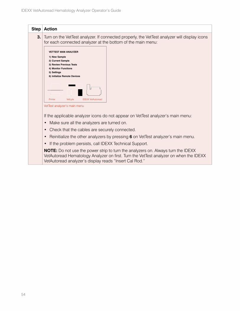
54
IDEXX VetAutoread Hematology Analyzer Operator’s Guide
Step Action
3. Turn on the VetTest analyzer. If connected properly, the VetTest analyzer will display icons for each connected analyzer at the bottom of the main menu:
VETTEST 8008 ANALYZER
1) New Sample
2) Current Sample
3) Review Previous Tests
4) Monitor Functions
5) Settings
6) Initialize Remote Devices
Printer VetLyte IDEXX VetAutoread
VetTest analyzer’s main menu
If the applicable analyzer icons do not appear on VetTest analyzer’s main menu:
• Make sure all the analyzers are turned on.
• Check that the cables are securely connected.
• Reinitialize the other analyzers by pressing 6 on VetTest analyzer’s main menu.
• If the problem persists, call IDEXX Technical Support.
NOTE: Do not use the power strip to turn the analyzers on. Always turn the IDEXX VetAutoread Hematology Analyzer on first. Turn the VetTest analyzer on when the IDEXX VetAutoread analyzer’s display reads “Insert Cal Rod.”


idexx.com
One IDEXX Drive Westbrook, Maine 04092 USA 06-02822-05