Embed Size (px)
Citation preview
© The Ulster Medical Society, 2013. www.ums.ac.uk
Ulster Med J 2013;82(2):100-108
Imaging Centre, Royal Victoria Hospital, Grosvenor Road, Belfast BT12 6BA.
Correspondence to Dr McNamee
Grand Rounds
Imaging in Cauda Equina Syndrome – A Pictorial Review John McNamee, Peter Flynn,Suzanne O’Leary, Mark Love, Barry Kelly
Accepted xx January xxxx
IntroductIon
Cauda Equina Syndrome (CES) is an uncommon condition important because of its catastrophic consequences for the patient and its potential for medico-legal actions against medical staff. The need for prompt intervention makes research into the subject practically and ethically challenging. This article aims to provide an overview of CES with illustrations of its common causes and mimics.
defInItIon
CES results from dysfunction of the sacral and lumbar nerve roots in the vertebral canal producing impairment of bladder, bowel or sexual function and perianal or saddle numbness1. It is a rare condition with a prevalence in the general population estimated between 1:100000 and 1:330002.
AnAtomy
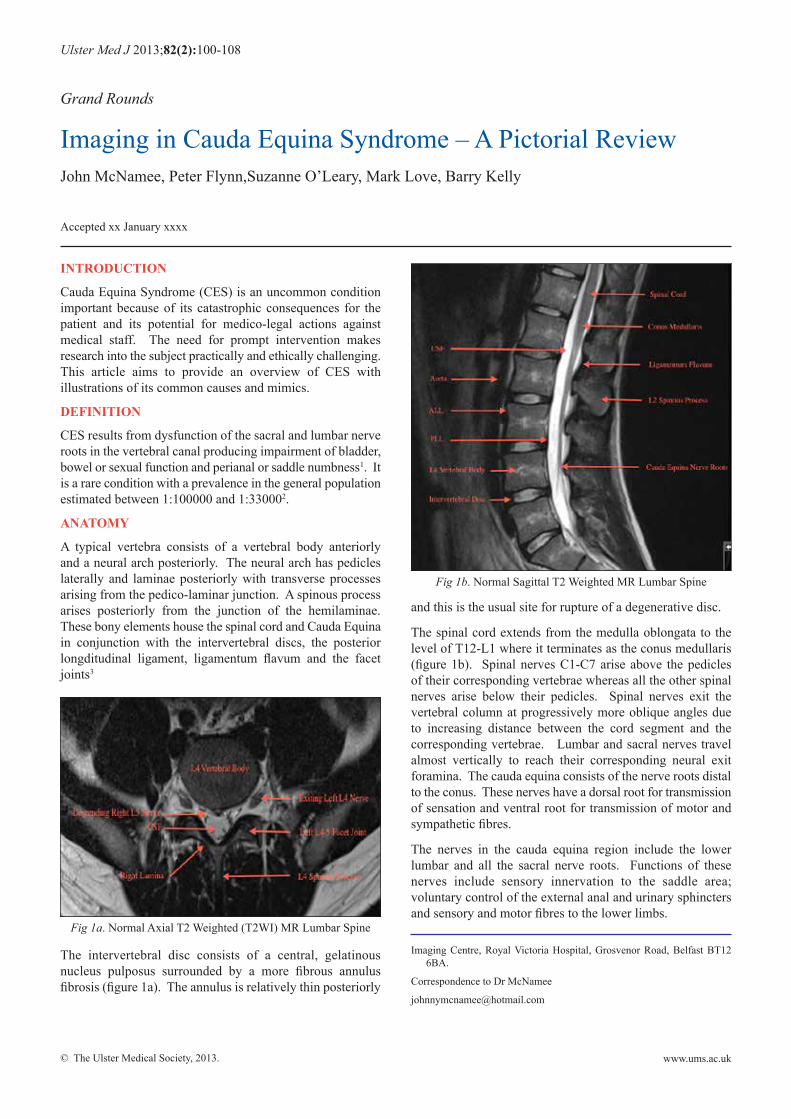
A typical vertebra consists of a vertebral body anteriorly and a neural arch posteriorly. The neural arch has pedicles laterally and laminae posteriorly with transverse processes arising from the pedico-laminar junction. A spinous process arises posteriorly from the junction of the hemilaminae. These bony elements house the spinal cord and Cauda Equina in conjunction with the intervertebral discs, the posterior longditudinal ligament, ligamentum flavum and the facet joints3
The intervertebral disc consists of a central, gelatinous nucleus pulposus surrounded by a more fibrous annulus fibrosis (figure 1a). The annulus is relatively thin posteriorly
and this is the usual site for rupture of a degenerative disc.
The spinal cord extends from the medulla oblongata to the level of T12-L1 where it terminates as the conus medullaris (figure 1b). Spinal nerves C1-C7 arise above the pedicles of their corresponding vertebrae whereas all the other spinal nerves arise below their pedicles. Spinal nerves exit the vertebral column at progressively more oblique angles due to increasing distance between the cord segment and the corresponding vertebrae. Lumbar and sacral nerves travel almost vertically to reach their corresponding neural exit foramina. The cauda equina consists of the nerve roots distal to the conus. These nerves have a dorsal root for transmission of sensation and ventral root for transmission of motor and sympathetic fibres.
The nerves in the cauda equina region include the lower lumbar and all the sacral nerve roots. Functions of these nerves include sensory innervation to the saddle area; voluntary control of the external anal and urinary sphincters and sensory and motor fibres to the lower limbs.
Fig 1a. Normal Axial T2 Weighted (T2WI) MR Lumbar Spine
Fig 1b. Normal Sagittal T2 Weighted MR Lumbar Spine
© The Ulster Medical Society, 2013.
Imaging in Cauda Equina Syndrome – A Pictorial Review 101
www.ums.ac.uk
The spinal cord vasculature is complex and variable. In broad terms the cord is supplied by two posterolateral spinal arteries and a single anterior spinal artery which all arise from the vertebral arteries. The spinal arteries receive further supply along their routes from radiculomedullary arteries arising from the subclavian artery and branches of the intercostal and lumbar arteries such as the artery of Adamkiewicz.
PresentAtIon
CES is defined by impairment of bladder, bowel and sexual function with perianal and saddle numbness1.
Other symptoms that may be present include back pain with or without radicular symptoms; sensory changes or numbness in the lower limbs; lower limb weakness and reduced or absent lower limb reflexes. A thorough history should include any obvious precipitants relating to the aetiologies listed above such as trauma, underlying malignancy or recent surgery.
Saddle anaesthesia and bladder, bowel or sexual dysfunction are the key clinical findings to discriminate between CES and sciatica, which can also present with low back pain and radiculopathy4.
clAssIfIcAtIon
CES may be divided into complete or incomplete4. In complete cauda equina syndrome patients present with saddle anaesthesia and retention/incontinence of bladder or bowel. In incomplete CES there is saddle anaesthesia but bladder and bowel dysfunction has not progressed to full retention or incontinence. Bladder or bowel symptoms that these patients my report include loss of urgency or altered urinary sensation.
AetIologIes
The most common cause of CES is lumbar disc herniation at the L4-L5 and L5-S1 levels1. Multiple other pathologies can damage the anatomical structures involved. An extensive list of causes is given in the table 1 below5:
InvestIgAtIons
Magnetic Resonance Imaging (MRI) is the imaging study of choice for the evaluation of suspected patients with CES due to its ability to accurately depict soft tissue pathology. It can also identify potential mimics such as aortic dissection or spinal infarction. Disadvantages include lack of 24-hour availability and contraindications such as pacemakers and poor patient tolerance due to claustrophobia.
Myelography and CT Myelography can be used as an alternative for patients not suitable for MRI but have the disadvantage of being invasive techniques. Plain films are generally unhelpful in the investigation of a herniated disc but can provide valuable information in the setting of acute trauma. Inflammatory markers and CSF studies should be performed when an inflammatory or infectious aetiology is being considered.
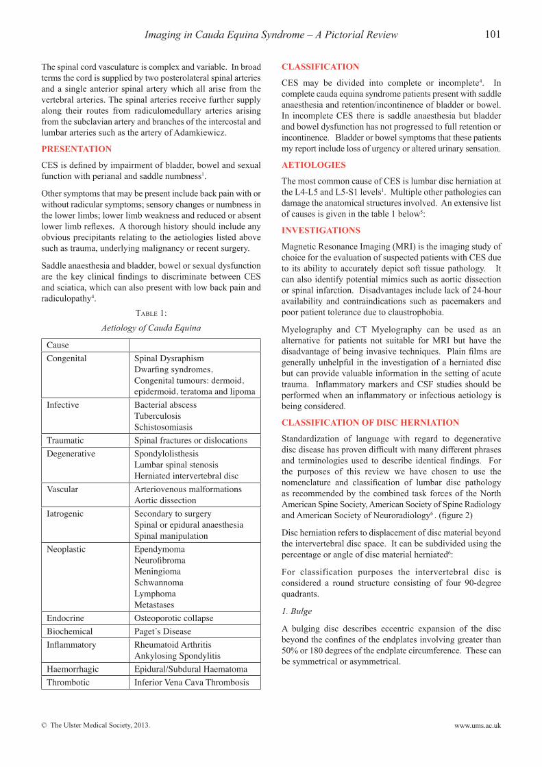
clAssIfIcAtIon of dIsc HernIAtIon
Standardization of language with regard to degenerative disc disease has proven difficult with many different phrases and terminologies used to describe identical findings. For the purposes of this review we have chosen to use the nomenclature and classification of lumbar disc pathology as recommended by the combined task forces of the North American Spine Society, American Society of Spine Radiology and American Society of Neuroradiology6 . (figure 2)
Disc herniation refers to displacement of disc material beyond the intervertebral disc space. It can be subdivided using the percentage or angle of disc material herniated6:
For classification purposes the intervertebral disc is considered a round structure consisting of four 90-degree quadrants.
1. Bulge
A bulging disc describes eccentric expansion of the disc beyond the confines of the endplates involving greater than 50% or 180 degrees of the endplate circumference. These can be symmetrical or asymmetrical.
Table 1: Aetiology of Cauda Equina
CauseCongenital Spinal Dysraphism
Dwarfing syndromes,Congenital tumours: dermoid, epidermoid, teratoma and lipoma
Infective Bacterial abscessTuberculosisSchistosomiasis
Traumatic Spinal fractures or dislocationsDegenerative Spondylolisthesis
Lumbar spinal stenosisHerniated intervertebral disc
Vascular Arteriovenous malformationsAortic dissection
Iatrogenic Secondary to surgerySpinal or epidural anaesthesiaSpinal manipulation
Neoplastic EpendymomaNeurofibromaMeningiomaSchwannomaLymphomaMetastases
Endocrine Osteoporotic collapseBiochemical Paget’s DiseaseInflammatory Rheumatoid Arthritis
Ankylosing SpondylitisHaemorrhagic Epidural/Subdural HaematomaThrombotic Inferior Vena Cava Thrombosis
© The Ulster Medical Society, 2013.
102 The Ulster Medical Journal
www.ums.ac.uk
2. Broad-based herniation
A broad based disc herniation describes expansion of disc beyond the confines of the endplates that involves 25-50% of the endplate circumference.
3. Focal herniations, protrusions and extrusions.
A focal herniation describes expansion of the disc beyond the confines of the endplates involving less than 25% of the endplate circumference. Focal herniations can be subdivided into protrusions and extrusions depending on their shape. An extrusion has a narrow isthmus connecting the displaced disc material to the parent disc and protrusions have a broader base. Sequestration describes complete separation of disc material from the parent disc into the epidural space.
The location of a disc herniation should also be described in the axial plane as either central, subarticular (lateral recess), foraminal or extra foraminal. In the cranio-caudal plane it can be described as suprapedicular, pedicular, infrapedicular or at disc level.
PIctorIAl exAmPles
Detailed pictorial examples of all the causes listed in the table above are beyond the scope of this review. The most common causes of CES in decreasing order are1:
• Disc Herniation• Tumour• Infection• Stenosis• Haematoma• Inflammatory• Vascular
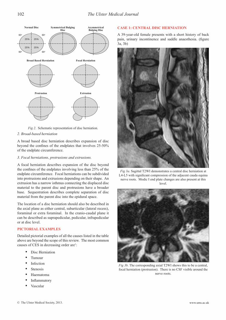
cAse 1: centrAl dIsc HernIAtIon
A 39-year-old female presents with a short history of back pain, urinary incontinence and saddle anaesthesia. (figure 3a, 3b)
Fig 2. Schematic representation of disc herniation.
Fig 3a. Sagittal T2WI demonstrates a central disc herniation at L4-L5 with significant compression of the adjacent cauda equina
nerve roots. Modic I end plate changes are also present at this level.
Fig 3b. The corresponding axial T2WI shows this to be a central, focal herniation (protrusion). There is no CSF visible around the
nerve roots.
25%
90o
normal disc symmetrical Bulging disc
Asymmetrical Bulging disc
90o
90o
90o
25%
25%
25%
Broad Based Herniation
Protrusion
focal Herniation
extrusion
© The Ulster Medical Society, 2013.
Imaging in Cauda Equina Syndrome – A Pictorial Review 103
www.ums.ac.uk
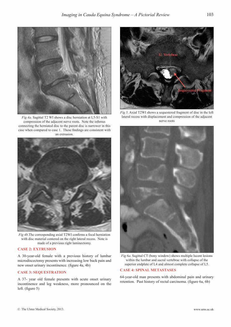
cAse 2: extrusIon
A 30-year-old female with a previous history of lumbar microdiscectomy presents with increasing low back pain and new onset urinary incontinence. (figure 4a, 4b)
cAse 3: sequestrAtIon
A 37- year old female presents with acute onset urinary incontinence and leg weakness, more pronounced on the left. (figure 5)
cAse 4: sPInAl metAstAses
64-year-old man presents with abdominal pain and urinary retention. Past history of rectal carcinoma. (figure 6a, 6b)
Fig 4a. Sagittal T2 WI shows a disc herniation at L5-S1 with compression of the adjacent nerve roots. Note the isthmus
connecting the herniated disc to the parent disc is narrower in this case when compared to case 1. These findings are consistent with
an extrusion.
Fig 4b.The corresponding axial T2WI confirms a focal herniation with disc material centered on the right lateral recess. Note is
made of a previous right laminectomy.
Fig 5. Axial T2WI shows a sequestered fragment of disc in the left lateral recess with displacement and compression of the adjacent
nerve roots
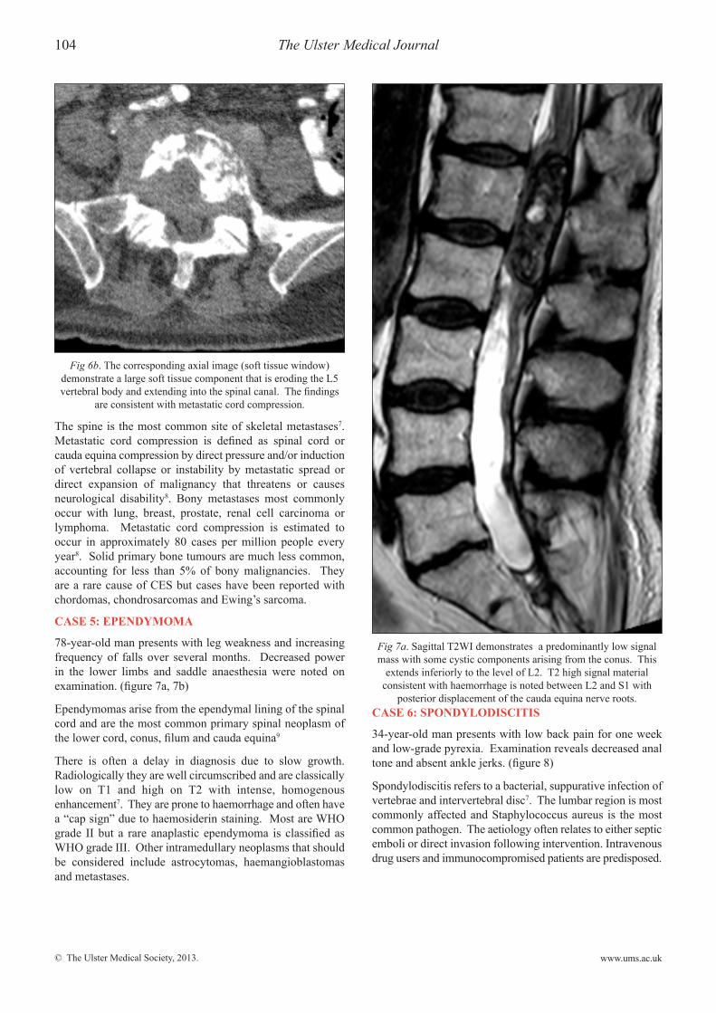
Fig 6a. Sagittal CT (bony window) shows multiple lucent lesions within the lumbar and sacral vertebrae with collapse of the
superior endplate of L4 and almost complete collapse of L5.
© The Ulster Medical Society, 2013.
104 The Ulster Medical Journal
www.ums.ac.uk
The spine is the most common site of skeletal metastases7. Metastatic cord compression is defined as spinal cord or cauda equina compression by direct pressure and/or induction of vertebral collapse or instability by metastatic spread or direct expansion of malignancy that threatens or causes neurological disability8. Bony metastases most commonly occur with lung, breast, prostate, renal cell carcinoma or lymphoma. Metastatic cord compression is estimated to occur in approximately 80 cases per million people every year8. Solid primary bone tumours are much less common, accounting for less than 5% of bony malignancies. They are a rare cause of CES but cases have been reported with chordomas, chondrosarcomas and Ewing’s sarcoma.
cAse 5: ePendymomA
78-year-old man presents with leg weakness and increasing frequency of falls over several months. Decreased power in the lower limbs and saddle anaesthesia were noted on examination. (figure 7a, 7b)
Ependymomas arise from the ependymal lining of the spinal cord and are the most common primary spinal neoplasm of the lower cord, conus, filum and cauda equina9
There is often a delay in diagnosis due to slow growth. Radiologically they are well circumscribed and are classically low on T1 and high on T2 with intense, homogenous enhancement7. They are prone to haemorrhage and often have a “cap sign” due to haemosiderin staining. Most are WHO grade II but a rare anaplastic ependymoma is classified as WHO grade III. Other intramedullary neoplasms that should be considered include astrocytomas, haemangioblastomas and metastases.
cAse 6: sPondylodIscItIs
34-year-old man presents with low back pain for one week and low-grade pyrexia. Examination reveals decreased anal tone and absent ankle jerks. (figure 8)
Spondylodiscitis refers to a bacterial, suppurative infection of vertebrae and intervertebral disc7. The lumbar region is most commonly affected and Staphylococcus aureus is the most common pathogen. The aetiology often relates to either septic emboli or direct invasion following intervention. Intravenous drug users and immunocompromised patients are predisposed.
Fig 7a. Sagittal T2WI demonstrates a predominantly low signal mass with some cystic components arising from the conus. This
extends inferiorly to the level of L2. T2 high signal material consistent with haemorrhage is noted between L2 and S1 with
posterior displacement of the cauda equina nerve roots.
Fig 6b. The corresponding axial image (soft tissue window) demonstrate a large soft tissue component that is eroding the L5 vertebral body and extending into the spinal canal. The findings
are consistent with metastatic cord compression.
© The Ulster Medical Society, 2013.
Imaging in Cauda Equina Syndrome – A Pictorial Review 105
www.ums.ac.uk
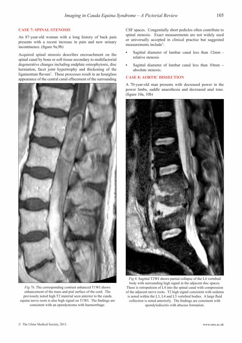
cAse 7: sPInAl stenosIs
An 87-year-old woman with a long history of back pain presents with a recent increase in pain and new urinary incontinence. (figure 9a,9b)
Acquired spinal stenosis describes encroachment on the spinal canal by bone or soft tissue secondary to multifactorial degenerative changes including endplate osteophytosis, disc herniation, facet joint hypertrophy and thickening of the ligamentum flavum7. These processes result in an hourglass appearance of the central canal effacement of the surrounding
CSF spaces. Congenitally short pedicles often contribute to spinal stenosis. Exact measurements are not widely used or universally accepted in clinical practise but suggested measurements include7:
• Sagittal diameter of lumbar canal less than 12mm - relative stenosis
• Sagittal diameter of lumbar canal less than 10mm – absolute stenosis.
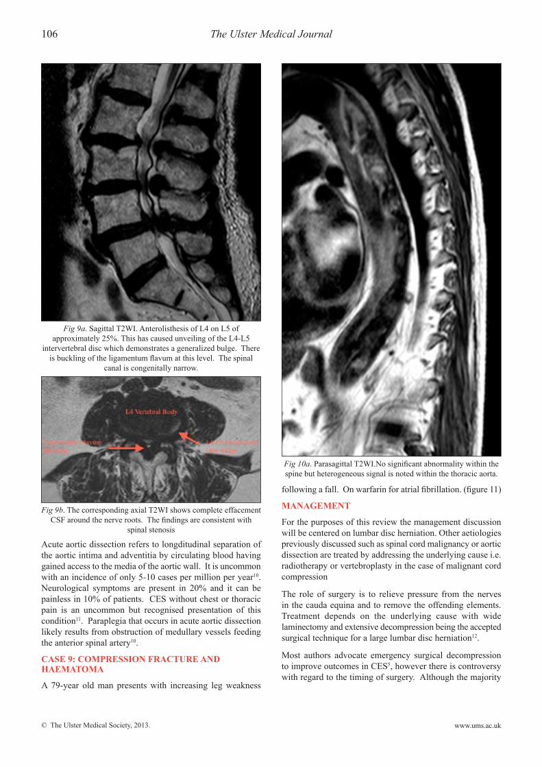
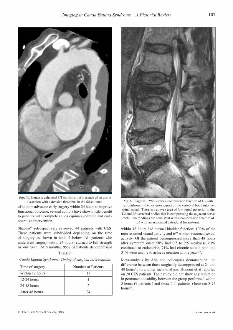
cAse 8: AortIc dIssectIon
A 70-year-old man presents with decreased power in the power limbs, saddle anaesthesia and decreased anal tone. (figure 10a, 10b)
Fig 8. Sagittal T2WI shows partial collapse of the L4 vertebral body with surrounding high signal in the adjacent disc spaces.
There is retropulsion of L4 into the spinal canal with compression of the adjacent nerve roots. T2 high signal consistent with oedema is noted within the L3, L4 and L5 vertebral bodies. A large fluid collection is noted anteriorly. The findings are consistent with
spondylodiscitis with abscess formation.
Fig 7b. The corresponding contrast enhanced T1WI shows enhancement of the mass and pial surface of the cord. The
previously noted high T2 material seen anterior to the cauda equina nerve roots is also high signal on T1WI. The findings are
consistent with an ependymoma with haemorrhage.
© The Ulster Medical Society, 2013.
106 The Ulster Medical Journal
www.ums.ac.uk
Acute aortic dissection refers to longditudinal separation of the aortic intima and adventitia by circulating blood having gained access to the media of the aortic wall. It is uncommon with an incidence of only 5-10 cases per million per year10. Neurological symptoms are present in 20% and it can be painless in 10% of patients. CES without chest or thoracic pain is an uncommon but recognised presentation of this condition11. Paraplegia that occurs in acute aortic dissection likely results from obstruction of medullary vessels feeding the anterior spinal artery10.
cAse 9: comPressIon frActure And HAemAtomA
A 79-year old man presents with increasing leg weakness
following a fall. On warfarin for atrial fibrillation. (figure 11)
mAnAgement
For the purposes of this review the management discussion will be centered on lumbar disc herniation. Other aetiologies previously discussed such as spinal cord malignancy or aortic dissection are treated by addressing the underlying cause i.e. radiotherapy or vertebroplasty in the case of malignant cord compression
The role of surgery is to relieve pressure from the nerves in the cauda equina and to remove the offending elements. Treatment depends on the underlying cause with wide laminectomy and extensive decompression being the accepted surgical technique for a large lumbar disc herniation12.
Most authors advocate emergency surgical decompression to improve outcomes in CES5, however there is controversy with regard to the timing of surgery. Although the majority
Fig 9a. Sagittal T2WI. Anterolisthesis of L4 on L5 of approximately 25%. This has caused unveiling of the L4-L5
intervertebral disc which demonstrates a generalized bulge. There is buckling of the ligamentum flavum at this level. The spinal
canal is congenitally narrow.
Fig 9b. The corresponding axial T2WI shows complete effacement CSF around the nerve roots. The findings are consistent with
spinal stenosis
Fig 10a. Parasagittal T2WI.No significant abnormality within the spine but heterogeneous signal is noted within the thoracic aorta.
© The Ulster Medical Society, 2013.
Imaging in Cauda Equina Syndrome – A Pictorial Review 107
www.ums.ac.uk
of authors advocate early surgery within 24 hours to improve functional outcome, several authors have shown little benefit to patients with complete cauda equina syndrome and early operative intervention.
Shapiro13 retrospectively reviewed 44 patients with CES. These patients were subdivided depending on the time of surgery as shown in table 2 below. All patients who underwent surgery within 24 hours returned to full strength by one year. At 6 months, 95% of patients decompressed
within 48 hours had normal bladder function; 100% of the men resumed sexual activity and 6/7 women resumed sexual activity. Of the patient decompressed more than 48 hours after symptom onset 58% had 0/5 to 2/5 weakness, 63% continued to catheterise, 71% had chronic sciatic pain and 31% were unable to achieve erection at one year4,13.
Meta-analysis by Ahn and colleagues demonstrated no difference between those surgically decompressed at 24 and 48 hours14. In another meta-analysis, Hussain et al reported on 20 CES patients. Their study did not show any reduction in permanent disability between the group performed within 5 hours (9 patients ) and those ( 11 patients ) between 8-24 hours15.
Fig 11. Sagittal T2WI shows a compression fracture of L3 with retropulsion of the posterior aspect of the vertebral body into the spinal canal. There is a convex area of low signal posterior to the L2 and L3 vertebral bodies that is compressing the adjacent nerve roots. The findings are consistent with a compression fracture of
L3 with an associated extradural haematoma.
Fig10b. Contrast enhanced CT confirms the presence of an aortic dissection with extensive thrombus in the false lumen.
Table 2: Cauda Equina Syndrome: Timing of surgical interventions
Time of surgery Number of PatientsWithin 12 hours 1712-24 hours 124-48 hours 2After 48 hours 24
© The Ulster Medical Society, 2013.
108 The Ulster Medical Journal
www.ums.ac.uk
While there is debate about the exact timing of surgery in the literature, the consensus view is that those with incomplete cauda equina syndrome or indeterminate cases should be decompressed immediately as their neurologic and urologic outcomes are clearly improved if the patient does not progress to complete cauda equina syndrome4,5.
conclusIons
We advocate the definition, as proposed by Fraser et al, that CES results from dysfunction of the sacral and lumbar nerve roots within the vertebral canal producing impairment of bladder, bowel or sexual function and perianal or saddle numbness. Elucidation of these findings in the clinical examination is crucial.
The number of potential aetiologies is vast but the most common causes are disc herniation, tumours, infection, spinal stenosis, inflammatory causes and vasculature occlusion.
Standardisation of language regarding lumbar disc pathology has proven difficult. We recommend the nomenclature and classification as recommended by the combined task forces of North American Spine Society, American Society of Spine Radiology and American Society of Neuroradiology.
Early surgical decompression is advocated by most authors to best aid patient recovery and reduce long term disability. There is evidence to suggest intervention within 24 hours significantly improves outcomes.
The authors have no conflict of interest.
references:
1. Fraser S, Roberts L, Murphy E. Cauda equina syndrome: a literature review of its definition and clinical presentation. Arch Phys Med Rehabil. 2009:90(11);1964-68.
2. Mooney V. Differential diagnosis of low back disorders: principles of classification. In: Frymore JW, editor. The adult spine: principles and practice. New York: Raven Press; 1991. p. 1559-60.
3. Ryan S. The spinal column and its contents. In: Ryan S, McNicholas M. Anatomy for diagnostic imaging. 2nd ed. Philadelphia: Saunders. 2004. p. 85-105.
4. Gitelman A, Hishmeh S, Morelli BN, Joseph SA, Casden A, Kuflik P, et al. Cauda equina syndrome: a comprehensive review. Am J Orthop. 2008;37(11):556-62
5. Ma B, Wu H, Jia LS, Yuan W, Shi GD, Shi JG. Cauda equina syndrome: a review of clinical progress. Chin Med J. 2009;122(10):1214-22.
6. Fardon D, Milette P. Nomenclature and classification of lumbar disc pathology. Recommendations of the Combined task Forces of the North American Spine Society, American Society of Spine Radiology and American Society of Neuroradiology. Spine. 2001:26,(5); E93-E113.
7. Ross J. Degenerative disease and inflammatory arthritides. In: Harnsberger HR, Osborn A, Ross J, Macdonald A, editors. Diagnostic imaging spine. Philadelphia: Amirsys. Lippincott, Williams & Wilkins: 2005. p. 60-3.
8. National Institute for Health and Care Excellence Guidelines [NICE]. Metastatic spinal cord compression: diagnosis and management of adults at risk of or with metastatic spinal cord compression. CG75. Cardiff: National Collaborating Centre for Cancer: 2008. Available from: http://www.nice.org.uk/CG75. Last accessed April 2013.
9. Kahan H, Sklar EM, Post MJ, Bruce JH, et al. MR characteristics of histopathologic subtypes of spinal ependymoma. AJNR Am J Neuroradiol. 1996:17(1);143-50.
10. Patel N, Noel CR, Weiner BK. Aortic dissection presenting as an cauda equina syndrome: case report. J Bone Joint Surg. 2002:84-A(8);1430-2.
11. Greenwood WR, Robinson MD. Painless dissection of the thoracic aorta. Am J Emerg Med. 1986:4(4);330-3
12. Shapiro S. Cauda equina syndrome secondary to lumbar disc herniation. Neurosurgery. 1993:32(5);743-6
13. Shapiro S. Medical realities of cauda equina syndrome secondary to lumbar disc herniation. Spine. 2000;25(3):348-51.
14. Ahn UM, Ahn Nu, Buchowski JM, Garrett ES, Siebert AN, Kostuik JP. Cauda equina syndrome secondary to lumbar disc herniation: a meta-analysis of surgical outcomes. Spine. 2000;25(12):1515-22
15. Hussain SA, Gullan RW, Chitnavis BP. Cauda equina syndrome: outcome and implications for management. Br J Neurosurg. 2003:17(2);164-7.