Embed Size (px)
Citation preview

IFA GUIDE TO SUCCESSFUL IMMUNOFLUORESCENCE
FROM CELL SIGNALING TECHNOLOGY | www.cellsignal.com
Immunofluorescence is a powerful tool for elucidating the complex signaling events that underlie biological processes and disease. This guide highlights critical steps in the immunofluorescence protocol and demonstrates how protocol changes can affect the final outcome of your experiment.
2
Immunofluorescence (IF) combines the use of antibodies with fluorescence imaging techniques to visualize target
proteins and other biomolecules within fixed cell or tissue samples. This process can reveal the localization, relative
expression, and even activation states of target proteins. The power of IF is that it provides data that is both graphical
and quantifiable.
When performing IF experiments, proteins of interest can be detected using either primary antibodies covalently
conjugated to fluorophores (direct detection) or a two-step approach with unlabeled primary antibody followed by
fluorophore-conjugated secondary antibody (indirect detection). Either method allows the user to combine multiple
fluorophores (multiplex analysis), making IF ideal for investigating protein co-localization, changes in subcellular
localization, differential activation of proteins within a cell, identification of different cell subsets, and other analyses.
At Cell Signaling Technology (CST), our goal is to provide highly specific antibodies that yield strong, specific
signal with minimal background. Our scientists screen a large number of antibodies and recommend only those
best suited for the application. Our validation efforts include extensive protocol optimization and antibody titration
to determine the best working conditions for each antibody. In addition, our scientists validate supporting reagents,
such as fluorophore-conjugated secondary antibodies, to enhance antigen detection and improve the efficiency of
IF protocols.
In this application guide, we will highlight the critical steps in our protocol for IF, introduce important concepts about
antibody performance and design of controls, and provide supporting data to explain our recommendations.
Introduction
Immunofluorescence techniques referenced in this guide
Technique Starting material CST protocols available
IF-IC (immunocytochemistry) Cultured cell lines or primary cells IF Standard, IF Methanol-fixed, IF Methanol-perm
IF-F (frozen) Frozen tissue IF Standard, IF Methanol-perm
mIHC (multiplexed immunohistochemistry) Formalin-fixed, paraffin-embedded (FFPE) tissue
mIHC/Paraffin

3
9 Step Protocol for a Successful Immunofluorescence Experiment
For more data on experimental controls visit
www.cellsignal.com/ifvalidation
1
7
2
9
8
Prepare Your Cells or Tissue• Tissue• Plating conditions• Optimizing cell density and cell health
3Fixation – Stabilize and Preserve Sample• Choice of fixative• Fixation incubation time
4 Permeablilization• Choice of detergents or alcohols
6Immunostaining• Primary antibody dilution• Washing and secondary incubation
5 Blocking • Choice of blocking reagent
• For paraffin-embedded tissue: deparaffinization/rehydration and antigen retrieval steps
Mount Sample
Counterstain (Optional)
Design Your Experiment• Experimental controls• Selecting antibodies and fluorophores: direct vs. indirect detection methods• Multiplexing
Data Collection and Imaging
4
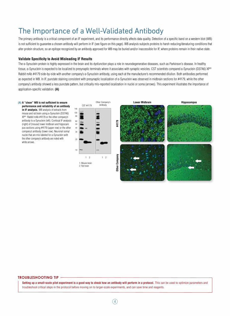
The Importance of a Well-Validated Antibody The primary antibody is a critical component of an IF experiment, and its performance directly affects data quality. Detection of a specific band on a western blot (WB) is not sufficient to guarantee a chosen antibody will perform in IF (see figure on this page). WB analysis subjects proteins to harsh reducing/denaturing conditions that alter protein structure, so an epitope recognized by an antibody approved for WB may be buried and/or inaccessible for IF, where proteins remain in their native state.
Validate Specificity to Avoid Misleading IF Results The α-Synuclein protein is highly expressed in the brain and its dysfunction plays a role in neurodegenerative diseases, such as Parkinson’s disease. In healthy tissue, α-Synuclein is expected to be localized to presynaptic terminals where it associates with synaptic vesicles. CST scientists compared α-Synuclein (D37A6) XP® Rabbit mAb #4179 side-by-side with another company’s α-Synuclein antibody, using each at the manufacturer’s recommended dilution. Both antibodies performed as expected in WB. In IF, punctate staining consistent with presynaptic localization of α-Synuclein was observed in midbrain sections for #4179, while the other company’s antibody showed a less punctate pattern, but critically mis-reported localization in nuclei or soma (arrows). This experiment illustrates the importance of application-specific validation. (A)
www.cellsignal.com/ifvalidation
Lower Midbrain Hippocampus
Othe
r Com
pany
#4
179
Setting up a small-scale pilot experiment is a good way to check how an antibody will perform in a protocol. This can be used to optimize parameters and troubleshoot critical steps in the protocol before moving on to larger-scale experiments, and can save time and reagents.
TROUBLESHOOTING TIP
CST #4179Other Company’s
Antibody
1: Mouse brain2: Rat brain
1 2
kDa
α-Synuclein
140
100
80
6050
40
30
20
10
1 2
(A) A “clean” WB is not sufficient to ensure performance and reliability of an antibody in IF analysis. WB analysis of extracts from mouse and rat brain using α-Synuclein (D37A6) XP® Rabbit mAb #4179 or the other company’s antibody to α-Synuclein (left). Confocal IF analysis (right) of [mouse] lower midbrain and hippocam pus sections using #4179 (upper row) or the other company’s antibody (lower row). Neuronal soma/ nuclei that are mis-labeled for α-Synuclein with the other company’s antibody are noted with white arrows.
5
Critical Steps in the Recommended ProtocolAn optimized protocol is necessary to achieve consistent, reliable IF results. We have tested common variations in fixation, permeabilization, and antibody concentration for many of our IF-approved antibodies. This section of the guide will highlight some of the data we use to support our IF protocol recommendations.
Design Your Experiment Experimental ControlsIncorporation of appropriate controls is important to confirm that the only changes between samples are in the experimental variable(s), and that the reagents – including antibodies – are performing as expected. Such controls could involve pharmacological treatments, addition of extracellular ligands to modulate signaling pathways, or comparison of cells with differential gene expression (knockout, siRNA, etc.). Typically, variables and controls are performed in parallel such that the fixation step and subsequent processing can be performed at the same time. The type of control used is dependent on the type of experiment. In the following sections we provide examples of controls our scientists use to evaluate antibody performance in IF-IC, some of which you may want to incorporate to confirm specificity in your cell type.
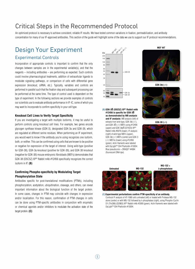
Knockout Cell Lines to Verify Target SpecificityIf you are investigating a target with multiple isoforms, it may be useful to perform controls using knockout cell lines. For example, two genes encode glycogen synthase kinase (GSK-3), designated GSK-3α and GSK-3ß, which are regulated at different serine residues. When performing an IF experiment, you would want to know if the antibody you’re using recognizes one isoform, both, or neither. This can be confirmed using cells that are known to be positive or negative for expression of the target of interest. Using wild-type (positive for GSK-3ß), GSK-3α knockout (positive for GSK-3ß), and GSK-3ß knockout (negative for GSK-3ß) mouse embryonic fibroblasts (MEFs) demonstrates that GSK-3ß (D5C5Z) XP® Rabbit mAb #12456 specifically recognizes the correct isoform in IF. (B)
Confirming Phospho-specificity by Modulating Target Phosphorylation StateAntibodies specific for post-translational modifications (PTMs), including phosphorylation, acetylation, ubiquitination, cleavage, and others, can reveal important information about the biological function of the target protein. In some cases, changes in PTM may coincide with changes in expression and/or localization. For this reason, confirmation of PTM changes in cells can be done using PTM-specific antibodies in conjunction with enzymatic or chemical agonists and/or inhibitors to modulate the activation state of the target protein. (C)
GSK-3α (-/-)
GSK-3ß (-/-)
MEF WT
kDa
GSK-3ß
GSK-3αGSK-3ß
MEF (W
T)
GSK-3α
(-/-)
GSK-3ß
(-/-)
200140
100
80
6050
40
6050
40
30
20
MG-132MG-132 +
λ-phosphataseUntreated
(B) GSK-3ß (D5C5Z) XP® Rabbit mAb #12456 is specific for GSK-3ß as demonstrated by WB analysis and IF analysis: WB analysis (left) of extracts from wild-type, GSK-3α (-/-), and GSK-3ß (-/-) MEFs using #12456 (upper) and GSK-3α/ß (D75D3) XP® Rabbit mAb #5676 (lower). IF analysis (right) of wild-type MEFs (upper), GSK-3α (-/-) MEFs (center) and GSK-3 (-/-) MEFλs (lower) using #12456 (green). Actin filaments were labeled with DyLightTM 554 Phalloidin #13054. Blue pseudocolor = DRAQ5® #4084 (fluorescent DNA dye).
(C) Experimental perturbations confirm PTM-specificity of an antibody. Confocal IF analysis of HT-1080 cells untreated (left) or treated with Protease MG-132 alone (center) or with MG-132 followed by λ-phosphatase (right), using Phospho-Cyclin D1 (Thr286) (D29B3) XP® Rabbit mAb #3300 (green). Actin filaments were labeled with DyLightTM 554 Phalloidin #13054.
6
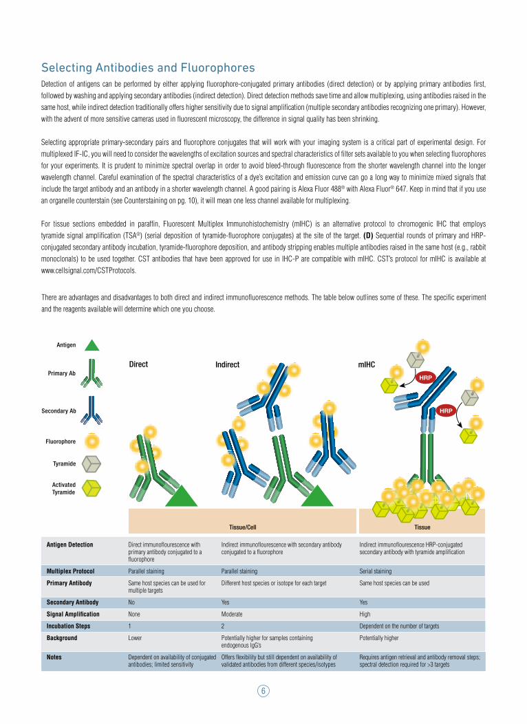
Selecting Antibodies and FluorophoresDetection of antigens can be performed by either applying fluorophore-conjugated primary antibodies (direct detection) or by applying primary antibodies first, followed by washing and applying secondary antibodies (indirect detection). Direct detection methods save time and allow multiplexing, using antibodies raised in the same host, while indirect detection traditionally offers higher sensitivity due to signal amplification (multiple secondary antibodies recognizing one primary). However, with the advent of more sensitive cameras used in fluorescent microscopy, the difference in signal quality has been shrinking.
Selecting appropriate primary-secondary pairs and fluorophore conjugates that will work with your imaging system is a critical part of experimental design. For multiplexed IF-IC, you will need to consider the wavelengths of excitation sources and spectral characteristics of filter sets available to you when selecting fluorophores for your experiments. It is prudent to minimize spectral overlap in order to avoid bleed-through fluorescence from the shorter wavelength channel into the longer wavelength channel. Careful examination of the spectral characteristics of a dye’s excitation and emission curve can go a long way to minimize mixed signals that include the target antibody and an antibody in a shorter wavelength channel. A good pairing is Alexa Fluor 488® with Alexa Fluor® 647. Keep in mind that if you use an organelle counterstain (see Counterstaining on pg. 10), it will mean one less channel available for multiplexing.
For tissue sections embedded in paraffin, Fluorescent Multiplex Immunohistochemistry (mIHC) is an alternative protocol to chromogenic IHC that employs tyramide signal amplification (TSA®) (serial deposition of tyramide-fluorophore conjugates) at the site of the target. (D) Sequential rounds of primary and HRP-conjugated secondary antibody incubation, tyramide-fluorophore deposition, and antibody stripping enables multiple antibodies raised in the same host (e.g., rabbit monoclonals) to be used together. CST antibodies that have been approved for use in IHC-P are compatible with mIHC. CST’s protocol for mIHC is available at www.cellsignal.com/CSTProtocols.
Antigen Detection
Multiplex Protocol
Primary Antibody
Secondary Antibody
Signal Amplification
Incubation Steps
Background
Notes
Direct immunoflourescence with primary antibody conjugated to a fluorophore
Parallel staining
Same host species can be used for multiple targets
No
None
1
Lower
Dependent on availability of conjugated antibodies; limited sensitivity
Indirect immunoflourescence with secondary antibody conjugated to a fluorophore
Parallel staining
Different host species or isotope for each target
Yes
Moderate
2
Potentially higher for samples containing endogenous IgG’s
Offers flexibility but still dependent on availability of validated antibodies from different species/isotypes
Indirect immunoflourescence HRP-conjugated secondary antibody with tyramide amplification
Serial staining
Same host species can be used
Yes
High
Dependent on the number of targets
Potentially higher
Requires antigen retrieval and antibody removal steps;spectral detection required for >3 targets
HRP
HRP
Antigen
Secondary Ab
Primary Ab
Fluorophore
Antigen
Secondary Ab
Primary Ab
Fluorophore
Antigen
Secondary Ab
Primary Ab
Fluorophore
Antigen
Secondary Ab
Primary Ab
Fluorophore
Tyramide
Activated Tyramide
Tissue/Cell Tissue
Direct Indirect mIHC
There are advantages and disadvantages to both direct and indirect immunofluorescence methods. The table below outlines some of these. The specific experiment and the reagents available will determine which one you choose.
7
Prepare Your Cells or TissueTissueTissue sections can be prepared either by quick freezing in optimal cutting temperature (OCT) medium (IF-F) or by embedding in paraffin (IHC-P). Frozen samples should be sectioned using a cryostat and allowed to air dry on the slide 10–15 minutes prior to fixation in the next step. Formalin-fixed, paraffin-embedded (FFPE) tissue blocks are another common preparation in the IHC-P protocol, which require deparaffination and antigen retrieval steps before incubation with antibodies.
Cells: Plating Conditions (IF-IC)Because the IF-IC protocol ends with imaging fixed and stained cells on a fluorescence microscope, it must begin with seeding of cells (either passaged from immortalized cell lines, or isolated primary cells) on a support material compatible with fluorescence microscopy. Typical support formats include glass-bottom cell culture dishes, glass coverslips (kept in plastic culture dishes) prepared with polylysine and/or extracellular matrix components to support adherent cell culture, and commercially available multiwell chambers mounted on glass slides that are compatible with microscopes.
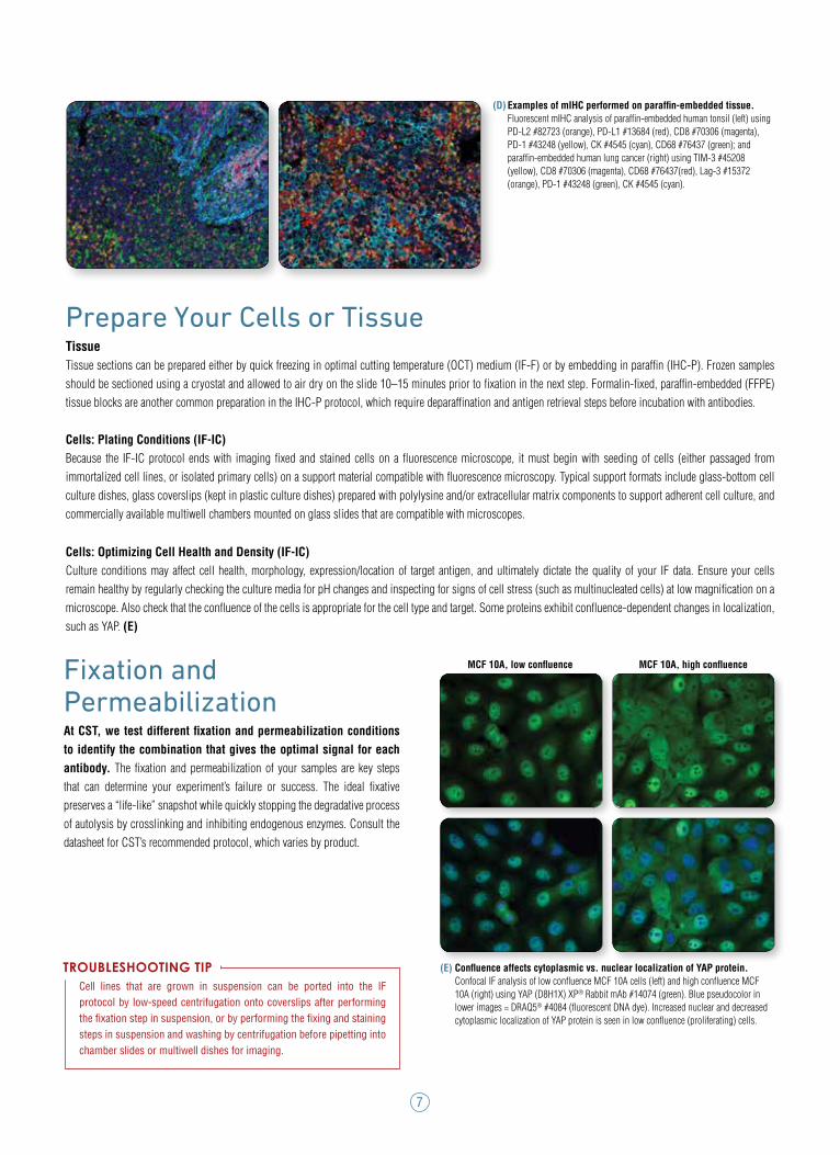
Cells: Optimizing Cell Health and Density (IF-IC)Culture conditions may affect cell health, morphology, expression/location of target antigen, and ultimately dictate the quality of your IF data. Ensure your cells remain healthy by regularly checking the culture media for pH changes and inspecting for signs of cell stress (such as multinucleated cells) at low magnification on a microscope. Also check that the confluence of the cells is appropriate for the cell type and target. Some proteins exhibit confluence-dependent changes in localization, such as YAP. (E)
Cell lines that are grown in suspension can be ported into the IF protocol by low-speed centrifugation onto coverslips after performing the fixation step in suspension, or by performing the fixing and staining steps in suspension and washing by centrifugation before pipetting into chamber slides or multiwell dishes for imaging.
TROUBLESHOOTING TIP
Fixation and PermeabilizationAt CST, we test different fixation and permeabilization conditions to identify the combination that gives the optimal signal for each antibody. The fixation and permeabilization of your samples are key steps that can determine your experiment’s failure or success. The ideal fixative preserves a “life-like” snapshot while quickly stopping the degradative process of autolysis by crosslinking and inhibiting endogenous enzymes. Consult the datasheet for CST’s recommended protocol, which varies by product.
(D) Examples of mIHC performed on paraffin-embedded tissue. Fluorescent mIHC analysis of paraffin-embedded human tonsil (left) using PD-L2 #82723 (orange), PD-L1 #13684 (red), CD8 #70306 (magenta), PD-1 #43248 (yellow), CK #4545 (cyan), CD68 #76437 (green); and paraffin-embedded human lung cancer (right) using TIM-3 #45208 (yellow), CD8 #70306 (magenta), CD68 #76437(red), Lag-3 #15372 (orange), PD-1 #43248 (green), CK #4545 (cyan).
MCF 10A, low confluence MCF 10A, high confluence
(E) Confluence affects cytoplasmic vs. nuclear localization of YAP protein. Confocal IF analysis of low confluence MCF 10A cells (left) and high confluence MCF 10A (right) using YAP (D8H1X) XP® Rabbit mAb #14074 (green). Blue pseudocolor in lower images = DRAQ5® #4084 (fluorescent DNA dye). Increased nuclear and decreased cytoplasmic localization of YAP protein is seen in low confluence (proliferating) cells.
8
Fixation TissueIF-F samples that have been fresh frozen and cryostat sectioned should now be treated with fixative. Alternatively, tissue samples may be preserved by fixing first with transcardial perfusion, post-fixation, and cryo-preservation steps, followed by freezing and sectioning.
For fluorescent IHC, tissue is preserved via perfusion as indicated above for IF-F, followed by paraffin embedding and sectioning. Prior to incubation with antibody, the sections must be deparaffinized with xylene and treated by heat or microwave in an antigen/epitope retrieval step.
CellsIn IF-IC, the samples should be rapidly fixed such that cellular structures and target localization are faithfully preserved, while also allowing antibodies to recognize and report their target. This is accomplished by quickly exchanging media for fixative solution. Different approaches to fixation in IF-IC are available (see below), and the optimal fixation method depends on the cell type, target protein, and the antibody being used.
Choice of FixativeAldehyde-based fixatives such as formaldehyde, formalin (a mixture of dissolved formaldehyde with a lower percentage of methanol), and glutaraldehyde are most commonly used. For most antibodies, CST recommends fixation with 4% formaldehyde. Aldehydes react with and crosslink cellular proteins, stabilizing and hardening the sample. The degree of crosslinking is dependent upon conditions (see below). Aldehydes cross the plasma membrane and fix soluble proteins better than alcohols, but some targets can lose their antigenicity with aldehyde crosslinking.
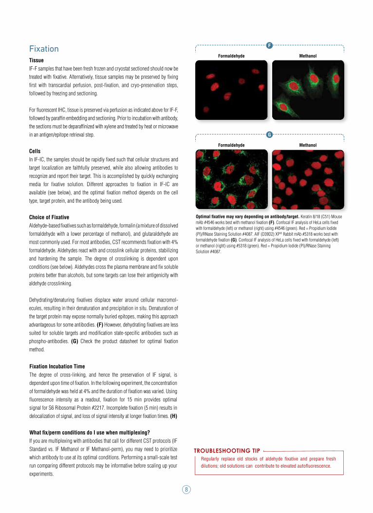
Dehydrating/denaturing fixatives displace water around cellular macromol-ecules, resulting in their denaturation and precipitation in situ. Denaturation of the target protein may expose normally buried epitopes, making this approach advantageous for some antibodies. (F) However, dehydrating fixatives are less suited for soluble targets and modification state-specific antibodies such as phospho-antibodies. (G) Check the product datasheet for optimal fixation method.
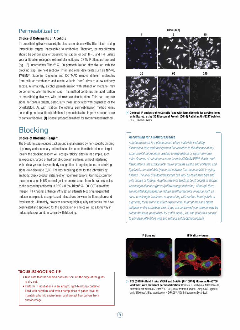
Fixation Incubation TimeThe degree of cross-linking, and hence the preservation of IF signal, is dependent upon time of fixation. In the following experiment, the concentration of formaldehyde was held at 4% and the duration of fixation was varied. Using fluorescence intensity as a readout, fixation for 15 min provides optimal signal for S6 Ribosomal Protein #2217. Incomplete fixation (5 min) results in delocalization of signal, and loss of signal intensity at longer fixation times. (H)
What fix/perm conditions do I use when multiplexing? If you are multiplexing with antibodies that call for different CST protocols (IF Standard vs. IF Methanol or IF Methanol-perm), you may need to prioritize which antibody to use at its optimal conditions. Performing a small-scale test run comparing different protocols may be informative before scaling up your experiments.
Regularly replace old stocks of aldehyde fixative and prepare fresh dilutions; old solutions can contribute to elevated autofluorescence.
TROUBLESHOOTING TIP
Formaldehyde Methanol
F
G
Formaldehyde Methanol
Optimal fixative may vary depending on antibody/target. Keratin 8/18 (C51) Mouse mAb #4546 works best with methanol fixation (F). Confocal IF analysis of HeLa cells fixed with formaldehyde (left) or methanol (right) using #4546 (green). Red = Propidium Iodide (PI)/RNase Staining Solution #4087. AIF (D39D2) XP® Rabbit mAb #5318 works best with formaldehyde fixation (G). Confocal IF analysis of HeLa cells fixed with formaldehyde (left) or methanol (right) using #5318 (green). Red = Propidium Iodide (PI)/RNase Staining Solution #4087.
9
Accounting for AutofluorescenceAutofluorescence is a phenomenon where materials including tissues and cells emit background fluorescence in the absence of any experimental fluorophore, leading to degradation of signal-to-noise ratio. Sources of autofluorescence include NADH/NADPH, flavins and flavoproteins, the extracellular matrix proteins elastin and collagen, and lipofuscin, an insoluble lysosomal polymer that accumulates in aging tissues. The level of autofluorescence can vary by cell/tissue type and with choice of fixative. Autofluorescence tends to be strongest in shorter wavelength channels (green/yellow/orange emission). Although there are reported approaches to reduce autofluorescence in tissue such as short wavelength irradiation or quenching with sodium borohydride or pigments, these will also affect experimental fluorophores and target antigens in the sample as well. If you are concerned your sample may be autofluorescent, particularly for a dim signal, you can perform a control to compare intensities with and without antibody/fluorophore.
Permeabilization Choice of Detergents or AlcoholsIf a crosslinking fixative is used, the plasma membrane will still be intact, making intracellular targets inaccessible to antibodies. Therefore, permeabilization should be performed after crosslinking fixation for both IF-IC and IF-F unless your antibodies recognize extracellular epitopes. CST’s IF Standard protocol (pg. 12) incorporates Triton® X-100 permeabilization after fixation with the blocking step (see next section). Triton and other detergents such as NP-40, TWEEN®, Saponin, Digitonin and DOTMAC remove different molecules from cellular membranes and create variable “pore” sizes to allow antibody access. Alternatively, alcohol permabilization with ethanol or methanol may be performed after the fixation step. This method combines the rapid fixation of crosslinking fixatives with intermediate denaturation. This can improve signal for certain targets, particularly those associated with organelles or the cytoskeleton. As with fixation, the optimal permeabilization method varies depending on the antibody. Methanol permeabilization improves performance of some antibodies. (I) Consult product datasheet for recommended method.
Blocking Choice of Blocking Reagent The blocking step reduces background signal caused by non-specific binding of primary and secondary antibodies to sites other than their intended target. Ideally, the blocking reagent will occupy “sticky” sites in the sample, such as exposed charged or hydrophobic protein surfaces, without interfering with primary/secondary antibody recognition of target epitopes, maximizing signal-to-noise ratio (S/N). The best blocking agent for the job varies by antibody; check product datasheet for recommendations. Our most common recommendation is 5% normal goat serum (or serum from the same species as the secondary antibody) in PBS + 0.3% Triton® X-100. CST also offers Image-IT® FX Signal Enhancer #11932, an alternate blocking reagent that reduces nonspecific charge-based interactions between the fluorophore and fixed sample. Ultimately, however, choosing high-quality antibodies that have been tested and approved for the application of choice will go a long way in reducing background, in concert with blocking.
TROUBLESHOOTING TIP• Take care that the solution does not spill off the edge of the glass or dry out. • Perform IF incubations in an airtight, light-blocking container lined with parafilm, and with a damp piece of paper towel to maintain a humid environment and protect fluorophore from photodamage.
Time (min)
1 5 15
30 60 240
IF Methanol-permIF Standard
(H) Confocal IF analysis of HeLa cells fixed with formaldehyde for varying times as indicated, using S6 Ribosomal Protein (5G10) Rabbit mAb #2217 (white). Blue = Hoescht #4082.
(I) PDI (C81H6) Rabbit mAb #3501 and ß-Actin (8H10D10) Mouse mAb #3700 work best with methanol permeabilization: Confocal IF analysis of NIH/3T3 cells, permeabilized with 0.3% TritonTM X-100 (left) or methanol (right), using #3501 (green) and #3700 (red). Blue pseudocolor = DRAQ5® #4084 (fluorescent DNA dye).
10
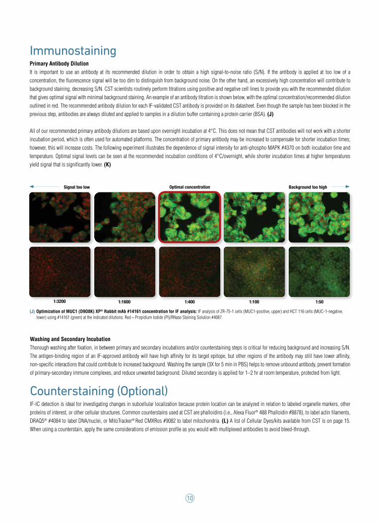
ImmunostainingPrimary Antibody DilutionIt is important to use an antibody at its recommended dilution in order to obtain a high signal-to-noise ratio (S/N). If the antibody is applied at too low of a concentration, the fluorescence signal will be too dim to distinguish from background noise. On the other hand, an excessively high concentration will contribute to background staining, decreasing S/N. CST scientists routinely perform titrations using positive and negative cell lines to provide you with the recommended dilution that gives optimal signal with minimal background staining. An example of an antibody titration is shown below, with the optimal concentration/recommended dilution outlined in red. The recommended antibody dilution for each IF-validated CST antibody is provided on its datasheet. Even though the sample has been blocked in the previous step, antibodies are always diluted and applied to samples in a dilution buffer containing a protein carrier (BSA). (J)
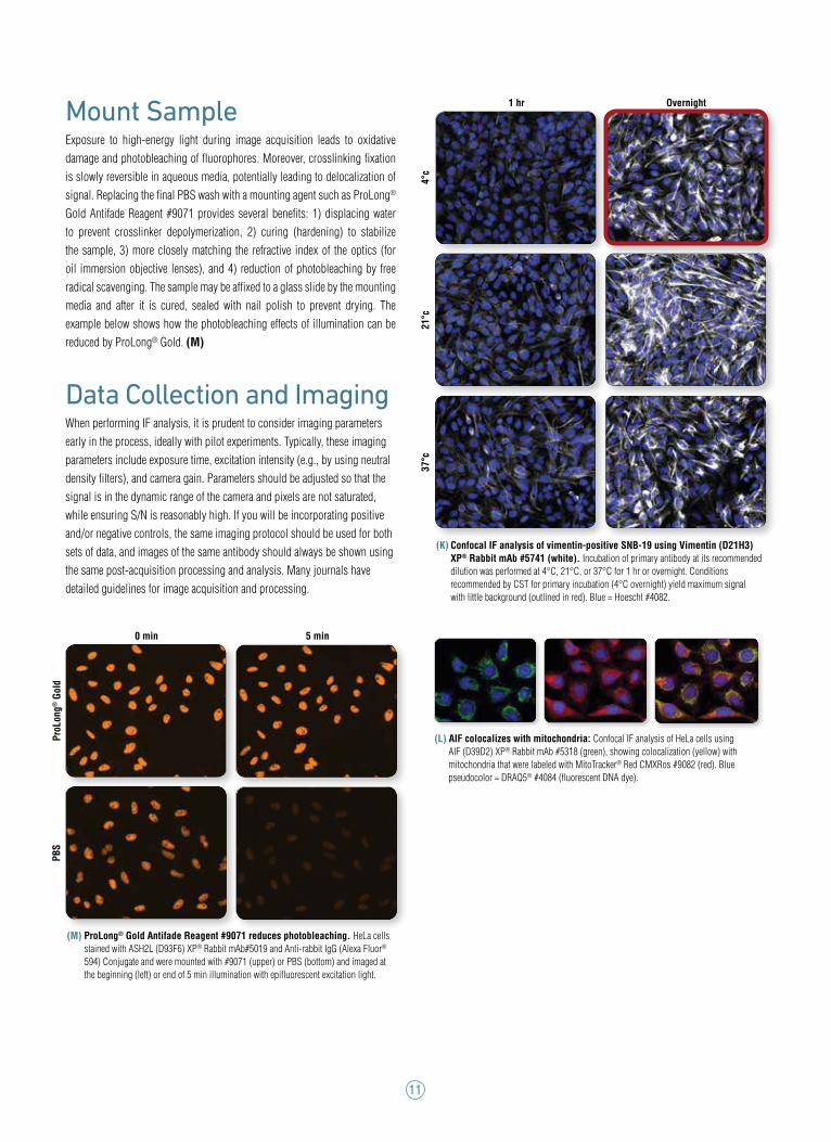
All of our recommended primary antibody dilutions are based upon overnight incubation at 4°C. This does not mean that CST antibodies will not work with a shorter incubation period, which is often used for automated platforms. The concentration of primary antibody may be increased to compensate for shorter incubation times; however, this will increase costs. The following experiment illustrates the dependence of signal intensity for anti-phospho MAPK #4370 on both incubation time and temperature. Optimal signal levels can be seen at the recommended incubation conditions of 4°C/overnight, while shorter incubation times at higher temperatures yield signal that is significantly lower. (K)
Washing and Secondary IncubationThorough washing after fixation, in between primary and secondary incubations and/or counterstaining steps is critical for reducing background and increasing S/N. The antigen-binding region of an IF-approved antibody will have high affinity for its target epitope, but other regions of the antibody may still have lower affinity, non-specific interactions that could contribute to increased background. Washing the sample (3X for 5 min in PBS) helps to remove unbound antibody, prevent formation of primary-secondary immune complexes, and reduce unwanted background. Diluted secondary is applied for 1–2 hr at room temperature, protected from light.
Counterstaining (Optional) IF-IC detection is ideal for investigating changes in subcellular localization because protein location can be analyzed in relation to labeled organelle markers, other proteins of interest, or other cellular structures. Common counterstains used at CST are phalloidins (i.e., Alexa Fluor® 488 Phalloidin #8878), to label actin filaments, DRAQ5® #4084 to label DNA/nuclei, or MitoTracker® Red CMXRos #9082 to label mitochondria. (L) A list of Cellular Dyes/kits available from CST is on page 15. When using a counterstain, apply the same considerations of emission profile as you would with multiplexed antibodies to avoid bleed-through.
1:3200 1:1600 1:400 1:100 1:50
Optimal concentrationSignal too low Background too high
(J) Optimization of MUC1 (D9O8K) XP® Rabbit mAb #14161 concentration for IF analysis: IF analysis of ZR-75-1 cells (MUC1-positive, upper) and HCT 116 cells (MUC-1-negative, lower) using #14161 (green) at the indicated dilutions. Red = Propidium Iodide (PI)/RNase Staining Solution #4087.
11
Mount SampleExposure to high-energy light during image acquisition leads to oxidative damage and photobleaching of fluorophores. Moreover, crosslinking fixation is slowly reversible in aqueous media, potentially leading to delocalization of signal. Replacing the final PBS wash with a mounting agent such as ProLong® Gold Antifade Reagent #9071 provides several benefits: 1) displacing water to prevent crosslinker depolymerization, 2) curing (hardening) to stabilize the sample, 3) more closely matching the refractive index of the optics (for oil immersion objective lenses), and 4) reduction of photobleaching by free radical scavenging. The sample may be affixed to a glass slide by the mounting media and after it is cured, sealed with nail polish to prevent drying. The example below shows how the photobleaching effects of illumination can be reduced by ProLong® Gold. (M)
Data Collection and ImagingWhen performing IF analysis, it is prudent to consider imaging parameters early in the process, ideally with pilot experiments. Typically, these imaging parameters include exposure time, excitation intensity (e.g., by using neutral density filters), and camera gain. Parameters should be adjusted so that the signal is in the dynamic range of the camera and pixels are not saturated, while ensuring S/N is reasonably high. If you will be incorporating positive and/or negative controls, the same imaging protocol should be used for both sets of data, and images of the same antibody should always be shown using the same post-acquisition processing and analysis. Many journals have detailed guidelines for image acquisition and processing.
(L) AIF colocalizes with mitochondria: Confocal IF analysis of HeLa cells using AIF (D39D2) XP® Rabbit mAb #5318 (green), showing colocalization (yellow) with mitochondria that were labeled with MitoTracker® Red CMXRos #9082 (red). Blue pseudocolor = DRAQ5® #4084 (fluorescent DNA dye).
1 hr Overnight
21°c
4°c
37°c
(K) Confocal IF analysis of vimentin-positive SNB-19 using Vimentin (D21H3) XP® Rabbit mAb #5741 (white). Incubation of primary antibody at its recommended dilution was performed at 4°C, 21°C, or 37°C for 1 hr or overnight. Conditions recommended by CST for primary incubation (4°C overnight) yield maximum signal with little background (outlined in red). Blue = Hoescht #4082.
ProL
ong®
Gol
dPB
S
0 min 5 min
(M) ProLong® Gold Antifade Reagent #9071 reduces photobleaching. HeLa cells stained with ASH2L (D93F6) XP® Rabbit mAb#5019 and Anti-rabbit IgG (Alexa Fluor® 594) Conjugate and were mounted with #9071 (upper) or PBS (bottom) and imaged at the beginning (left) or end of 5 min illumination with epifluorescent excitation light.
12
Immunofluorescence General Protocol (IF Standard)
B. Specimen PreparationI. Cultured Cell Lines (IF-IC)
NOTE: Cells should be grown, treated, fixed and stained directly in multi-well plates, chamber slides or on coverslips.
1. Aspirate liquid, then cover cells to a depth of 2–3 mm with 4% formaldehyde diluted in warm PBS.
NOTE: Formaldehyde is toxic, use only in a fume hood.
2. Allow cells to fix for 15 min at room temperature.3. Aspirate fixative, rinse three times in 1X PBS for 5 min each.4. Proceed with Immunostaining (Section C).
II. Frozen/Cryostat Sections (IF-F)1. For fixed frozen tissue proceed with Immunostaining (Section C).2. For fresh, unfixed frozen tissue, fix immediately, as follows: Cover sections with 4% formaldehyde diluted in warm 1X PBS. Allow sections to fix for 15 min at room temperature. Rinse slides three times in PBS for 5 min each. Proceed with Immunostaining (Section C).
C. Immunostaining
NOTE: All subsequent incubations should be carried out at room temperature unless otherwise noted in a humid light-tight box or covered dish/plate to prevent drying and fluorochrome fading.
1. Block specimen in blocking buffer for 60 min.2. While blocking, prepare primary antibody by diluting as indicated on datasheet in antibody dilution buffer.3. Aspirate blocking solution, apply diluted primary antibody.4. Incubate overnight at 4°C.5. Rinse three times in 1X PBS for 5 min each.
NOTE: If using a fluorophore-conjugated primary antibody, then skip to Section C, Step 8.
6. Incubate specimen in fluorochrome-conjugated secondary antibody diluted in antibody dilution buffer for 1–2 hr at room temperature in the dark.7. Rinse three times in 1X PBS for 5 min each.8. Coverslip slides with Prolong® Gold Antifade Reagent (#9071) or Prolong® Gold Antifade Reagent with DAPI (#8961).9. For best results, allow the mounting reagent to cure overnight at room tem- perature. For long-term storage, store slides flat at 4°C protected from light.
Protocols
NOTE: Some CST antibodies work optimally using an alternate protocol. Please see product datasheet for product-specific recommendations.
A. Solutions and Reagents
NOTE: Prepare solutions with reverse osmosis deionized (RODI) or equivalent grade water.
• 1X Phosphate Buffered Saline (PBS): To prepare 1 L 1X PBS pH 8.0, add 100 mL 10X PBS pH 7.4 (1.37M NaCl, 27mM KCl, 101 mM Na2HPO4, 18 mM KH2PO4) to 900 mL dH20, mix. NOTE: adjust pH to 8.0.
• Formaldehyde: 16%, methanol free, Polysciences, Inc. (cat# 18814), use fresh and store opened vials at 4°C in dark. Dilute 1 in 4 in 1X PBS to make a 4% formaldehyde solution.
• Blocking Buffer: (1X PBS/5% normal serum/0.3% Triton® X-100): To prepare 10 ml, add 0.5 ml normal serum from the same species as the secondary antibody (e.g., Normal Goat Serum (#5425) to 9.5 ml 1X PBS) and mix well. While stirring, add 30 µl Triton™ X-100.
• Antibody Dilution Buffer: (1X PBS/1% BSA/0.3% Triton® X-100): To prepare 10 ml, add 30 µl Triton™ X-100 to 10 ml 1X PBS. Mix well then add 0.1g BSA (#9998), mix.
• Fluorophore-conjugated Secondary Antibodies: (Anti-mouse #4408, #4409, #8890, #4410) (Anti-rabbit #4412, #4413, #8889, #4414) (Anti-rat #4416, #4417, #4418).
• Prolong® Gold Antifade Reagent (#9071), Prolong® Gold Antifade Reagent with DAPI (#8961).
• Methanol: 100%, ice-cold (for IF-Methanol-fixed and IF-Methanol- perm protocols)
Proceed to one of the following protocols.
IMPORTANT: Please refer to the APPLICATIONS section on the front page of product datasheet or product webpage to determine if this product is validated and approved for use on cultured cell lines (IF-IC) or frozen tissue sections (IF-F). Alternatively, antibodies that have been approved for IHC can be used in paraffin-embedded sections (see www.cellsignal.com/CSTprotocols for our mIHC/Parrafin protocol). Please see product datasheet or product webpage for appropriate antibody dilution. The following protocols accommodate both indirect and direct IF.
13
Immunofluorescence Protocol with Methanol Fixation (IF Methanol-fixed)
IMPORTANT: Please refer to the APPLICATIONS section on the front page of the datasheet to determine if this product is validated and approved for use with this protocol.
B. Specimen Preparation Cultured Cell Lines (IF-IC)
NOTE: Cells should be grown, treated, fixed and stained directly in multi-well plates, chamber slides or on coverslips.
1. Aspirate liquid, then cover cells to a depth of 2–3 mm with ice-cold 100% methanol.2. Allow cells to fix for 15 min at -20°C.3. Aspirate fixative, rinse three times in PBS for 5 min each.4. Proceed with Immunostaining (Section C).
C. Immunostaining
NOTE: All subsequent incubations should be carried out at room temperature unless otherwise noted in a humid light-tight box or covered dish/plate to prevent drying and fluorochrome fading.
1. Block specimen in Blocking Buffer for 60 min.2. While blocking, prepare primary antibody by diluting as indicated on datasheet in Antibody Dilution Buffer.3. Aspirate blocking solution, apply diluted primary antibody.4. Incubate overnight at 4°C.5. Rinse three times in PBS for 5 min each.
NOTE: If using a fluorochrome-conjugated primary antibody, then skip to Section C, Step 8.
6. Incubate specimen in fluorochrome-conjugated secondary antibody diluted in Antibody Dilution Buffer for 1–2 hours at room temperature in dark.7. Rinse in PBS as in step 5.8. Coverslip slides with Prolong® Gold Antifade Reagent (#9071) or Prolong® Gold Antifade Reagent with DAPI (#8961).9. For best results, examine specimens immediately using appropriate excitation wavelength. For long-term storage, store slides flat at 4°C protected from light.
Immunofluorescence Protocol with Methanol Permeabilization (IF Methanol-perm)
B. Specimen PreparationI. Cultured Cell Lines (IF-IC)
NOTE: Cells should be grown, treated, fixed and stained directly in multiwell plates, chamber slides or on coverslips.
1. Aspirate liquid, then cover cells to a depth of 2–3 mm with 4% formaldehyde in PBS.
NOTE: Formaldehyde is toxic, use only in fume hood.
2. Allow cells to fix for 15 min at room temperature.3. Aspirate fixative, rinse three times in PBS for 5 min each.4. Proceed with Immunostaining (Section C).
II. Frozen/Cryostat Sections (IF-F)1. For fixed frozen tissue proceed with Immunostaining (Section C).2. For fresh, unfixed frozen tissue, please fix immediately, as follows:1. Cover sections with 4% formaldehyde in PBS.
NOTE: Formaldehyde is toxic, use only in fume hood.
2. Allow sections to fix for 15 min at room temperature.3. Rinse slides three times in PBS for 5 min each.4. Proceed with Immunostaining (Section C).
C. Immunostaining
NOTE: All subsequent incubations should be carried out at room temperature unless otherwise noted in a humid light-tight box or covered dish/plate to prevent drying and fluorochrome fading.
1. Methanol Permeabilization Step: Cover cells or tissue sections with ice-cold 100% methanol (use enough to cover completely to a depth of 3–5 mm, DO NOT LET DRY), incubate in methanol for 10 min at -20°C, rinse in PBS for 5 min.2. Block specimen in Blocking Buffer for 60 min.3. While blocking, prepare primary antibody by diluting as indicated on datasheet in Antibody Dilution Buffer.4. Aspirate blocking solution, apply diluted primary antibody.5. Incubate overnight at 4°C.6. Rinse three times in PBS for 5 min each.
NOTE: If using a fluorochrome-conjugated primary antibody, then skip to Section C, Step 9.
7. Incubate specimen in fluorochrome-conjugated secondary antibody diluted in Antibody Dilution Buffer for 1–2 hours at room temperature in dark.8. Rinse in PBS as in step 6.9. Coverslip slides with Prolong® Gold Antifade Reagent (#9071) or Prolong® Gold Antifade Reagent with DAPI (#8961).10. For best results examine specimens immediately using appropriate excitation wavelength. For long term storage, store slides flat at 4°C protected from light.
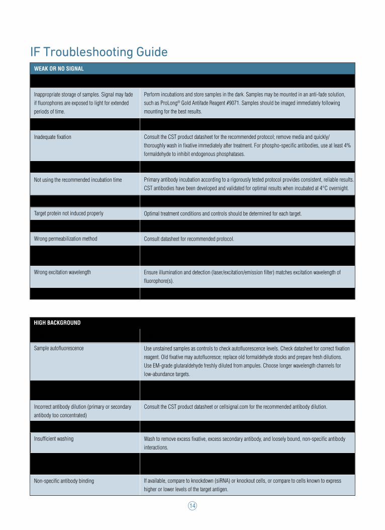
WEAK OR NO SIGNAL
Possible cause Recommendations
14
IF Troubleshooting Guide
Inappropriate storage of samples. Signal may fade if fluorophores are exposed to light for extended periods of time.
Cell/tissue samples stored for too long
Inadequate fixation
Incorrect antibody dilution (antibody too dilute)
Not using the recommended incubation time
Inappropriate testing model
Target protein not induced properly
Low expression of protein of interest
Wrong permeabilization method
Incorrect use of secondary antibody
Wrong excitation wavelength
Low signal in multiplexed IHC
Cause
Sample autofluorescence
Insufficient blocking
Incorrect antibody dilution (primary or secondary antibody too concentrated)
Samples dried out
Insufficient washing
Secondary cross-reactivity
Non-specific antibody binding
Perform incubations and store samples in the dark. Samples may be mounted in an anti-fade solution, such as ProLong® Gold Antifade Reagent #9071. Samples should be imaged immediately following mounting for the best results.
Use freshly prepared slides/plates to avoid loss of antigenicity.
Consult the CST product datasheet for the recommended protocol; remove media and quickly/thoroughly wash in fixative immediately after treatment. For phospho-specific antibodies, use at least 4% formaldehyde to inhibit endogenous phosphatases.
Consult the CST product datasheet or cellsignal.com for the recommended antibody dilution.
Primary antibody incubation according to a rigorously tested protocol provides consistent, reliable results. CST antibodies have been developed and validated for optimal results when incubated at 4°C overnight.
If possible, protein expression should be confirmed by western blot or other means.
Optimal treatment conditions and controls should be determined for each target. Modify detection approach; consider signal amplification, or pair with a brighter fluorophore.
Consult datasheet for recommended protocol.
Use recommended concentration and check secondary antibody is matched to host species of the primary antibody.
Ensure illumination and detection (laser/excitation/emission filter) matches excitation wavelength of fluorophore(s).
Optimization of deparaffination, antigen retrieval and signal amplification methods.
Solution
Use unstained samples as controls to check autofluorescence levels. Check datasheet for correct fixation reagent. Old fixative may autofluoresce; replace old formaldehyde stocks and prepare fresh dilutions. Use EM-grade glutaraldehyde freshly diluted from ampules. Choose longer wavelength channels for low-abundance targets.
Use normal serum from the same species as the secondary antibody used. Consider a charge-based blocker such as Image-iT® FX Signal Enhancer #11932, depending on the source of background.
Consult the CST product datasheet or cellsignal.com for the recommended antibody dilution.
It is vital that sample remains covered in liquid throughout the staining procedure.
Wash to remove excess fixative, excess secondary antibody, and loosely bound, non-specific antibody interactions.
Use isotype control secondary antibodies to determine whether your secondary antibody is cross-reacting.
If available, compare to knockdown (siRNA) or knockout cells, or compare to cells known to express higher or lower levels of the target antigen.
HIGH BACKGROUND
15
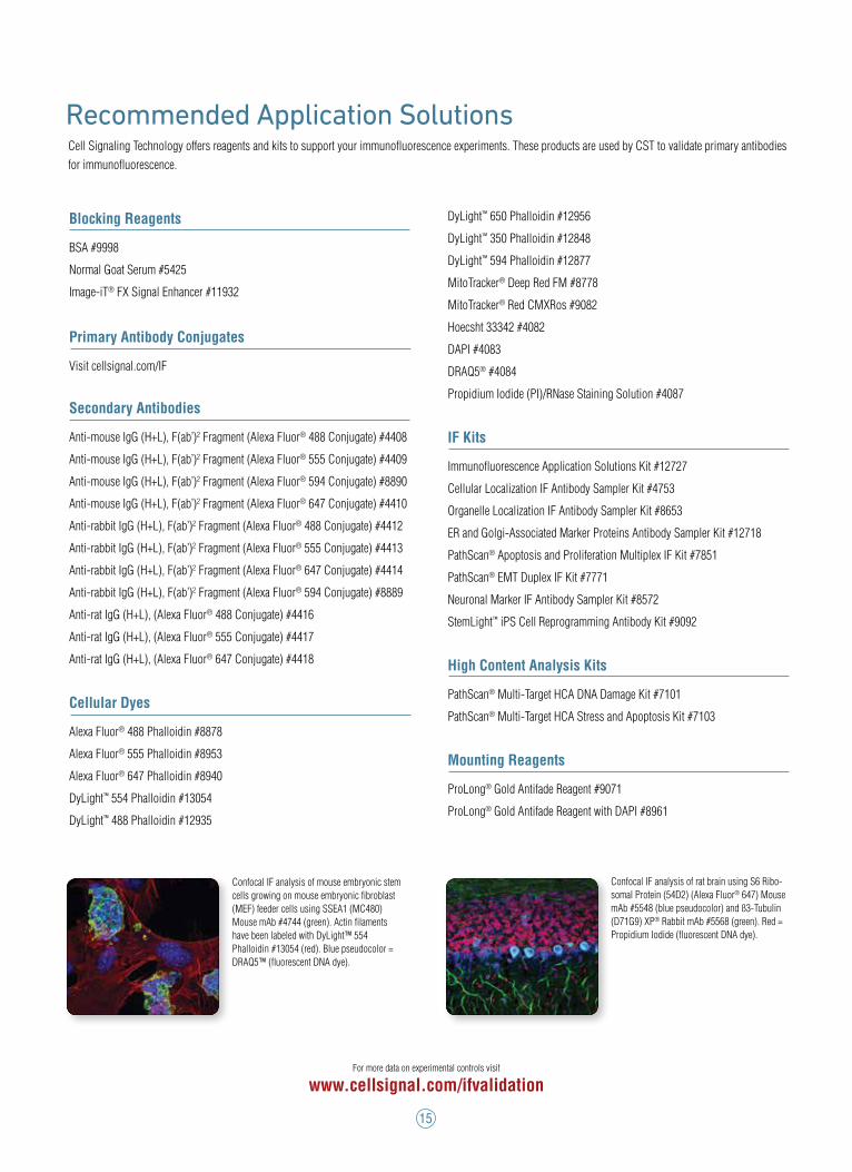
Recommended Application SolutionsCell Signaling Technology offers reagents and kits to support your immunofluorescence experiments. These products are used by CST to validate primary antibodies for immunofluorescence.
Blocking Reagents
BSA #9998
Normal Goat Serum #5425
Image-iT® FX Signal Enhancer #11932
Primary Antibody Conjugates
Visit cellsignal.com/IF
Secondary Antibodies
Anti-mouse IgG (H+L), F(ab’)2 Fragment (Alexa Fluor® 488 Conjugate) #4408
Anti-mouse IgG (H+L), F(ab’)2 Fragment (Alexa Fluor® 555 Conjugate) #4409
Anti-mouse IgG (H+L), F(ab’)2 Fragment (Alexa Fluor® 594 Conjugate) #8890
Anti-mouse IgG (H+L), F(ab’)2 Fragment (Alexa Fluor® 647 Conjugate) #4410
Anti-rabbit IgG (H+L), F(ab’)2 Fragment (Alexa Fluor® 488 Conjugate) #4412
Anti-rabbit IgG (H+L), F(ab’)2 Fragment (Alexa Fluor® 555 Conjugate) #4413
Anti-rabbit IgG (H+L), F(ab’)2 Fragment (Alexa Fluor® 647 Conjugate) #4414
Anti-rabbit IgG (H+L), F(ab’)2 Fragment (Alexa Fluor® 594 Conjugate) #8889
Anti-rat IgG (H+L), (Alexa Fluor® 488 Conjugate) #4416
Anti-rat IgG (H+L), (Alexa Fluor® 555 Conjugate) #4417
Anti-rat IgG (H+L), (Alexa Fluor® 647 Conjugate) #4418
Cellular Dyes
Alexa Fluor® 488 Phalloidin #8878
Alexa Fluor® 555 Phalloidin #8953
Alexa Fluor® 647 Phalloidin #8940
DyLight™ 554 Phalloidin #13054
DyLight™ 488 Phalloidin #12935
DyLight™ 650 Phalloidin #12956
DyLight™ 350 Phalloidin #12848
DyLight™ 594 Phalloidin #12877
MitoTracker® Deep Red FM #8778
MitoTracker® Red CMXRos #9082
Hoecsht 33342 #4082
DAPI #4083
DRAQ5® #4084
Propidium Iodide (PI)/RNase Staining Solution #4087
IF Kits
Immunofluorescence Application Solutions Kit #12727
Cellular Localization IF Antibody Sampler Kit #4753
Organelle Localization IF Antibody Sampler Kit #8653
ER and Golgi-Associated Marker Proteins Antibody Sampler Kit #12718
PathScan® Apoptosis and Proliferation Multiplex IF Kit #7851
PathScan® EMT Duplex IF Kit #7771
Neuronal Marker IF Antibody Sampler Kit #8572
StemLight™ iPS Cell Reprogramming Antibody Kit #9092
High Content Analysis Kits
PathScan® Multi-Target HCA DNA Damage Kit #7101
PathScan® Multi-Target HCA Stress and Apoptosis Kit #7103
Mounting Reagents
ProLong® Gold Antifade Reagent #9071
ProLong® Gold Antifade Reagent with DAPI #8961
For more data on experimental controls visit
www.cellsignal.com/ifvalidation
Confocal IF analysis of mouse embryonic stem cells growing on mouse embryonic fibroblast (MEF) feeder cells using SSEA1 (MC480) Mouse mAb #4744 (green). Actin filaments have been labeled with DyLight™ 554 Phalloidin #13054 (red). Blue pseudocolor = DRAQ5™ (fluorescent DNA dye).
Confocal IF analysis of rat brain using S6 Ribo-somal Protein (54D2) (Alexa Fluor® 647) Mouse mAb #5548 (blue pseudocolor) and ß3-Tubulin (D71G9) XP® Rabbit mAb #5568 (green). Red = Propidium Iodide (fluorescent DNA dye).

For Research Use Only. Not For Use in Diagnostic Procedures.
Printed on recycled paper (25% post-consumer waste fiber) using vegetable inks and processed chlorine free.
© 2017 Cell Signaling Technology, Inc. All rights reserved.
Cell Signaling Technology, CST, PathScan, SignalSlide, SignalStain, StemLight, and XP are trademarks of Cell Signaling Technology, Inc. Alexa Fluor is a registered trademark of Life Technologies Corporation. DRAQ5 is a registered trademark of Biostatus Limited. DyLight is a trademark of Thermo Fisher Scientific, Inc. and its subsidiaries. Image-iT is a registered trademark of Life Technologies Corporation. MitoTracker is a registered trademark of Molecular Probes, Inc. Triton is a trademark of The Dow Chemical Company. TSA is a registered trademark of PerkinElmer.
Alexa Conjugates: This product is provided under an intellectual property license from Life Technologies Corporation. The transfer of this product is contingent on the buyer using the purchased product solely in research, including use with HCS or other automated imaging applications but excluding use in combination with DNA microarrays. The buyer must not sell or otherwise transfer this product or its components for (a) diagnostic, therapeutic or prophylactic purposes; (b) testing, analysis or screening services, or information in return for compensation on a per-test basis; (c) manufacturing or quality assurance or quality control, or (d) resale, whether or not resold for use in research. For information on purchasing a license to this product for purposes other than as described above, contact Life Technologies Corporation, 5791 Van Allen Way, Carlsbad, CA 92008 USA or [email protected].
By RegionUNITED STATESOrders: 877-616-2355 | [email protected] Support: 877-678-8324 | [email protected]
www.cellsignal.com
CHINATel: +86-21-58356288 Support (China): 4006-473287/GreatQ | [email protected] Support (Asia Pacific): [email protected]
www.cst-c.com.cn
EUROPE, MIDDLE EAST & AFRICATel: +31 (0)71 720 0200 Support: [email protected]
www.cellsignal.com
JAPANTel: 03-3295-1630 | Support: [email protected]
www.cstj.co.jp
ORDER INFORMATION Find order information online at www.cellsignal.com/orderinfo
Technical SupportWe hope this guide is a helpful resource for performing immunofluorescence in your own lab. Cell Signaling Technology prides itself in providing you with exceptional customer service and support, and we are happy to share our experience with you. Since all of our antibodies are produced in-house, the same scientists who develop and assay these reagents are available as technical resources for our customers. These scientists can be contacted directly and will personally provide technical assistance to you, our customer.
FRONT COVER PHOTO: Tara, Imaging Specialist, has been with CST since 2008.
17-APS-011-BRO1-E1