Embed Size (px)
Citation preview
�
Abstract²The aim of this study was to enhance the ECG
pre-processing modalities for beat-to-beat QT interval
variability measurement based on template matching. The R-
peak detection algorithm has been substituted and an efficient
baseline removal algorithm has been implemented in existing
computer software. To test performance we used simulated
ECG data with fixed QT intervals featuring Gaussian noise,
baseline wander and amplitude modulation and two alternative
algorithms. We computed the standard deviation of beat-to-
beat QT intervals as a marker of QT interval variability
(QTV). Significantly a lower beat-to-beat QTV was found in
the updated approach compared the original algorithm. In
addition, the updated template matching computer software
outperformed the previous version in discarding fewer beats. In
conclusion, the updated ECG preprocessing algorithm is
recommended for more accurate quantification of beat-to-beat
QT interval variability.
I. INTRODUCTION
The duration of the QT interval represents the total time of ventricular depolarization and repolarization of a cardiac cycle in ECG. The quantification of beat-to-beat QTV has received significant interest for investigating repolarization abnormalities in patients with different cardiac conditions [1-3]. Importantly, elevated beat-to-beat QTV has also been identified as a marker of increased risk for sudden cardiac death [4-6]. Beat-to-beat QTV measurements from standard 12-lead ECGs showed significant differences between leads [7, 8]. Beat-to-beat variability in the QT segment can also be analysed using a vectorcardiographic (VCG) approach, which has shown diagnostic capabilities for identifying cardiac patients [9-11].
The existing measurement techniques for beat-to-beat QT interval in surface ECG are limited in their accuracy due to different reasons. Generally, the first task to be considered for accurate quantification of beat-to-beat QTV is the accurate R-peak detection followed by the detection of Q-wave onset and T-wave offset. If the R-peak detection is faulty then the whole quantification of QTV will be affected. Baseline
M. A. Hasan, D. Abbott and M. Baumert are with the Centre for
Biomedical Engineering (CBME) and the School of Electrical & Electronic
Engineering, University of Adelaide, SA 5005, Australia (phone: 61-8-
8313-4115; fax: 61-8-8303-4360; e-mail:
[email protected], [email protected]
A. Porta is with the Department of Biomedical Sciences for Health,
University of Milan, Milan 20122, Italy (e-mail: [email protected])
V. Starc is with the School of Medicine, The University of Ljubljana,
1000 Ljubljana, Slovenia (e-mail: [email protected])
wander is one of the noises that causes problems for detecting the R-peaks of surface ECG. Due to the baseline wander, the T-peak could be higher than the R-peak and detected as an R-peak instead. In addition, it may also affect the detection of the T-wave offset and thereby affect the measurement of QTV.
The real time QRS detection algorithm proposed by Pan & Tompkins [12] is widely used and also implemented in the computer software for beat-to-beat QT measurements developed by Berger and his co-workers [13]. However, a number of studies have investigated the accuracy of Pan & Tompkins¶ QRS detection algorithm and indicate difficulties when detecting QRS complexes in ECG complicated by cardiac disorders or arrhythmia. In particular, most of the studies reported miss- and false detection of true R-peak with less effective baseline removal in ECG signal [14-20].
Therefore, in this study, we incorporated an alternative R-peak detection technique and baseline removal approach in the existing computer software, which was developed by Berger and his co-workers [13]. The performance of the updated approach has been compared with the existing method [13] and two other methods (conventional method [21], template time shift method [22]) on simulated ECG as described in a previous article [23].
II. METHODS AND METHODOLOGY
A. Data
In this study, we considered the same simulated ECG data that was reported in an earlier article [23]. In brief, the original ECG (a normal noise-free cardiac cycle) was obtained from lead II with sampling frequency 1000 Hz and then digitized (12 bit resolution) using an A/D board. Ten cardiac beats were computed from the original one by decreasing T-wave amplitudes by factor k from 1.0 to 0.1 (where k = 1.0 represents the original cardiac cycle). Finally, ten synthetic signals with 500 cardiac cycles were obtained by repeating each of the ten beats 500 times. All ECG are characterized by no variability in heart period and ventricular repolarization duration.
B. ECG pre-processing
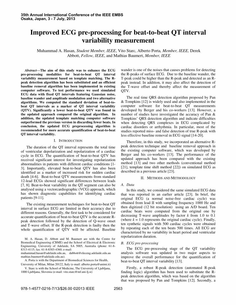
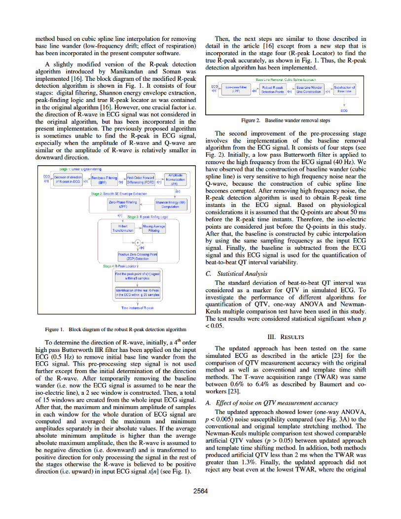
The ECG pre-processing stage of the QT variability analysis software was updated in two major aspects to improve the overall performance for the quantification of beat-to-beat QT interval variability [13].
Firstly, a robust R-peak detection (automated R-peak finding logic) algorithm has been used to substitute the R-peak detection algorithm, which was based on the algorithm that was proposed by Pan and Tompkins [12]. Secondly, a
Improved ECG pre-processing for beat-to-beat QT interval
variability measurement
Muhammad A. Hasan, Student Member, IEEE, Vito Starc, Alberto Porta, Member, IEEE, Derek
Abbott, Fellow, IEEE, and Mathias Baumert, Member, IEEE
35th Annual International Conference of the IEEE EMBSOsaka, Japan, 3 - 7 July, 2013
978-1-4577-0216-7/13/$26.00 ©2013 IEEE 2563
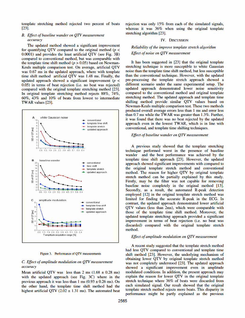
method highly depends on the threshold based R-peak detection [12] where it fails to detect the lower amplitude of QRS. As a result, only higher amplitude of QRS-T waves were considered for computing the QT interval in the signal and thereby it showed lower artificial QTV compared with all other techniques. Furthermore, in the original template stretch method [13], it was assumed to be PVC beats (36% beats from each simulated signal) if the algorithm fails to detect the lower amplitude of QRS-T wave due to the amplitude modulation. Average artificial QTV was found less than 2 ms with updated approach where the automated beat rejection was only 15% from each of the simulated signal. In practice, real ECG signals might not be exactly the same as the simulated signals with amplitude modulation and thus the updated approach appears to be more robust.
V. CONCLUSION
In conclusion, the test results suggest that the updated
ECG pre-processing approach outperforms the existing
algorithm when evaluated on the simulated ECGs for
analysing beat-to-beat QTV. Further, the updated approach
seems to perform comparably to the template time shift
method.
REFERENCES
[1] M. Hinterseer, M. B. Thomsen, B.-M. Beckmann, A. Pfeufer, R.
Schimpf, H.-E. Wichmann, G. Steinbeck, M. A. Vos, and S. Kaab,
"Beat-to-beat variability of QT intervals is increased in patients with
drug-induced long-QT syndrome: a case control pilot study,"
European Heart Journal, vol. 29, pp. 185-190, 2008.
[2] M. Baumert, E. Lambert, G. Vaddadi, C. I. Sari, M. Esler, G.
Lambert, P. Sanders, and E. Nalivaiko, "Cardiac repolarization
variability in patients with postural tachycardia syndrome during
graded head-up tilt," Clinical Neurophysiology, vol. 122, pp. 405-409,
2011.
[3] L. G. Tereshchenko, I. Cygankiewicz, S. McNitt, R. Vazquez, A.
Bayes-Genis, L. Han, S. Sur, J.-P. Couderc, R. D. Berger, A. B. de
Luna, and W. Zareba, "Predictive Value of Beat-to-Beat QT
Variability Index Across the Continuum of Left Ventricular
Dysfunction / Clinical Perspective," Circulation: Arrhythmia and
Electrophysiology, vol. 5, pp. 719-727, 2012.
[4] W. L. Atiga, H. Calkins, J. H. Lawrence, G. F. Tomaselli, J. M. Smith,
and R. D. Berger, "Beat-to-beat repolarization lability identifies
patients at risk for sudden cardiac death," Journal of Cardiovascular
Electrophysiology, vol. 9, pp. 899-908, 1998.
[5] G. Piccirillo, D. Magrì, S. Matera, M. Magnanti, A. Torrini, E.
Pasquazzi, E. Schifano, S. Velitti, V. Marigliano, R. Quaglione, and F.
Barillà, "QT variability strongly predicts sudden cardiac death in
asymptomatic subjects with mild or moderate left ventricular systolic
dysfunction: a prospective study," European Heart Journal, vol. 28,
pp. 1344-1350, 2007.
[6] L. G. Tereshchenko, B. J. Fetics, P. P. Domitrovich, B. D. Lindsay,
and R. D. Berger, "Prediction of Ventricular Tachyarrhythmias by
Intracardiac Repolarization Variability Analysis / CLINICAL
PERSPECTIVE," Circulation: Arrhythmia and Electrophysiology,
vol. 2, pp. 276-284, 2009.
[7] M. A. Hasan, D. Abbott, and M. Baumert, "Relation between Beat-to-
Beat QT Interval Variability and T-Wave Amplitude in Healthy
Subjects," Annals of Noninvasive Electrocardiology, vol. 17, pp. 195-
203, 2012.
[8] M. A. Hasan, D. Abbott, and M. Baumert, "Beat-to-beat QT interval
variability in the 12 lead ECG," in Computing in Cardiology,
Hangzhou, China, 2011, pp. 61-64.
[9] M. A. Hasan, D. Abbott, and M. Baumert, "Beat-to-beat
vectorcardiographic analysis of ventricular depolarization and
repolarization in myocardial infarction," PLoS ONE, vol. 7, p. e49489,
2012.
[10] L. G. Tereshchenko, L. Han, A. Cheng, J. E. Marine, D. D. Spragg, S.
Sinha, D. Dalal, H. Calkins, G. F. Tomaselli, and R. D. Berger, "Beat-
to-beat three-dimensional ECG variability predicts ventricular
arrhythmia in ICD recipients," Heart Rhythm, vol. 7, pp. 1606-1613,
2010.
[11] M. A. Hasan, D. Abbott, and M. Baumert, "Beat-to-Beat Spatial and
Temporal Analysis for QRS-T Morphology," in 34th Annual
International Conference of the IEEE EMBS, San Diego, California
USA, 2012, pp. 4193-4195.
[12] J. Pan and W. J. Tompkins, "A Real-Time QRS Detection Algorithm,"
IEEE Transactions on Biomedical Engineering, vol. BME-32, pp.
230-236, 1985.
[13] R. D. Berger, E. K. Kasper, K. L. Baughman, E. Marban, H. Calkins,
and G. F. Tomaselli, "Beat-to-beat QT interval variability - Novel
evidence for repolarization lability in ischemic and nonischemic
dilated cardiomyopathy," Circulation, vol. 96, pp. 1557-1565, Sep
1997.
[14] D. Benitez, P. Gaydecki, A. Zaidi, and A. Fitzpatrick, "A new QRS
detection algorithm based on the Hilbert transform," in Computers in
Cardiology 2000, 2000, pp. 379-382.
[15] M. Adnane, Z. Jiang, and S. Choi, "Development of QRS detection
algorithm designed for wearable cardiorespiratory system," computer
methods and programs in biomedicine, vol. 93, pp. 20-31, 2009.
[16] M. S. Manikandan and K. P. Soman, "A novel method for detecting
R-peaks in electrocardiogram (ECG) signal," Biomedical Signal
Processing and Control, vol. 7, pp. 118-128, 2012.
[17] N. Debbabi, S. El Asmi, and H. Arfa, "Correction of ECG baseline
wander application to the Pan & Tompkins QRS detection algorithm,"
in I/V Communications and Mobile Network (ISVC), 2010 5th
International Symposium on, 2010, pp. 1-4.
[18] M. Paoletti and C. Marchesi, "Discovering dangerous patterns in long-
term ambulatory ECG recordings using a fast QRS detection
algorithm and explorative data analysis," computer methods and
programs in biomedicine, vol. 82, pp. 20-30, 2006.
[19] A. Ruha, S. Sallinen, and S. Nissila, "A real-time microprocessor QRS
detector system with a 1-ms timing accuracy for the measurement of
ambulatory HRV," Biomedical Engineering, IEEE Transactions on,
vol. 44, pp. 159-167, 1997.
[20] Q. Xue, Y. H. Hu, and W. J. Tompkins, "Neural-network-based
adaptive matched filtering for QRS detection," Biomedical
Engineering, IEEE Transactions on, vol. 39, pp. 317-329, 1992.
[21] A. Porta, G. Baselli, F. Lambardi, S. Cerutti, R. Antolini, M. Greco, F.
Ravelli, and G. Nollo, "Performance assessment of standard
algorithms for dynamic R-T interval measurement: comparison
between R-Tapex and R-Tend approach," Medical and Biological
Engineering and Computing, vol. 36, pp. 35-42, 1998.
[22] V. Starc and T. T. Schlegel, "Real-time multichannel system for beat-
to-beat QT interval variability," Journal of Electrocardiology, vol. 39,
pp. 358-367, 2006.
[23] M. Baumert, V. Starc, and A. Porta, "Conventional QT Variability
Measurement vs. Template Matching Techniques: Comparison of
Performance Using Simulated and Real ECG," PLoS ONE, vol. 7, p.
e41920, 2012.
2566