Embed Size (px)
Citation preview
Infected transverse colonic cystic duplicationsimulating pelvic appendicular abscessRamnik V Patel,1,2 Irene Milliken,3 Alistair Dick,3 David Marshall3
1Department of PaediatricUrology, University CollegeLondon Hospitals NHSFoundation Trust, London, UK2Department of PaediatricUrology, Great Ormond StreetChildren Hospital NHS Trust,London, UK3Department of PaediatricSurgery, The Royal BelfastHospital for Sick Children,Belfast, UK
Correspondence toRamnik V Patel,[email protected]
To cite: Patel RV, Milliken I,Dick A, et al. BMJ Case RepPublished online: [pleaseinclude Day Month Year]doi:10.1136/bcr-2013-201459
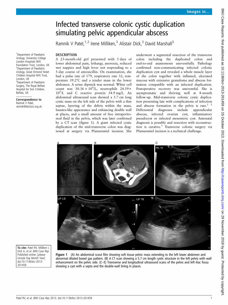
DESCRIPTIONA 23-month-old girl presented with 5 days oflower abdominal pain, lethargy, anorexia, reducedwet nappies and high fever not responding to a5-day course of amoxicillin. On examination, shehad a pulse rate of 179, respiratory rate 32, tem-perature 39.2°C and a tender mass in the lowerabdomen. A urine dipstick was normal. White cellcount was 30.36 × 10⁹/L, neutrophils 24.59×10⁹/L and C reactive protein 34.9 mg/L. Anabdominal ultrasound scan showed a 5.7 cm longcystic mass on the left side of the pelvis with a thinseptae, layering of the debris within the mass,haustra-like appearance and enhancing double wallat places, and a small amount of free intraperito-neal fluid in the pelvis, which was later confirmedby a CT scan (figure 1). A giant infected cysticduplication of the mid-transverse colon was diag-nosed at surgery via Pfannenstiel incision. She
underwent a segmental resection of the transversecolon including the duplicated colon andend-to-end anastomosis uneventfully. Pathologyconfirmed non-communicating infected colonicduplication cyst and revealed a whole muscle layerof the colon together with inflamed, ulceratedmucosa with extensive granuloma and abscess for-mation compatible with an infected duplication.Postoperative recovery was uneventful. She isasymptomatic and thriving well at 8-monthfollow-up. Mid-transverse colonic cystic duplica-tion presenting late with complications of infectionand abscess formation in the pelvis is rare.1 2
Differential diagnoses include appendicularabscess, infected ovarian cyst, inflammatorypseudocyst or infected mesenteric cyst. Antenataldiagnosis is possible and resection with reconstruc-tion is curative.3 Transverse colonic surgery viaPfannenstiel incision is a technical challenge.
Figure 1 (A) An abdominal scout film showing soft tissue pelvic mass extending to the left lower abdomen andabnormal dilated bowel gas pattern. (B) A CT scan showing a 5.7 cm length cystic structure in the left pelvis with wallenhancement on the pelvic side. (C–E) Transverse and longitudinal ultrasound scans of the pelvis and left iliac fossashowing a cyst with a septa and the double-wall lining in places.
Patel RV, et al. BMJ Case Rep 2013. doi:10.1136/bcr-2013-201459 1
Images in…
on 29 Novem
ber 2018 by guest. Protected by copyright.
http://casereports.bmj.com
/B
MJ C
ase Reports: first published as 10.1136/bcr-2013-201459 on 15 O
ctober 2013. Dow
nloaded from
Learning points
▸ Transverse colonic tubular duplications are common butcystic duplications are rare. Since the transverse colon ismobile, cystic duplications of the mid-transverse colon canpresent in the pelvis.
▸ An infected transverse colonic cystic duplication cyst canmimic appendicular abscess with lower abdominal pain andfever associated with tender mass and raised inflammatorymarkers. The classic enhancing mucosa of a duplication cyston ultrasound/CT scan can be destroyed by infection and animportant clue may be lost.
▸ Colonic duplications have a high incidence of complications,such as infection, bleeding, perforation, adenocarcinoma andobstruction, and therefore, even if antenatally diagnosed orincidentally found, should be resected.
Contributors All the authors have actively participated in the clinical managementof this patient and have been involved in the preparation, editing and finalisation ofthe manuscript.
Competing interests None.
Patient consent Obtained.
Provenance and peer review Not commissioned; externally peer reviewed.
REFERENCES1 Banchini F, Delfanti R, Begnini E, et al. Duplication of the transverse colon in an
adult: case report and review. World J Gastroenterol 2013;19:586–9.2 Stefanidis K, Lappas I, Kolofousi C, et al. A rare presentation of colonic duplication
cyst: report of a case and review of literature. JBR-BTR 2012;95:71–3.3 Piolat C, N’Die J, Andrini P, et al. Perforated tubular duplication of the transverse
colon: a rare cause of meconium peritonitis with prenatal diagnosis. Pediatr Surg Int2005;21:110–12.
Copyright 2013 BMJ Publishing Group. All rights reserved. For permission to reuse any of this content visithttp://group.bmj.com/group/rights-licensing/permissions.BMJ Case Report Fellows may re-use this article for personal use and teaching without any further permission.
Become a Fellow of BMJ Case Reports today and you can:▸ Submit as many cases as you like▸ Enjoy fast sympathetic peer review and rapid publication of accepted articles▸ Access all the published articles▸ Re-use any of the published material for personal use and teaching without further permission
For information on Institutional Fellowships contact [email protected]
Visit casereports.bmj.com for more articles like this and to become a Fellow
2 Patel RV, et al. BMJ Case Rep 2013. doi:10.1136/bcr-2013-201459
Images in…
on 29 Novem
ber 2018 by guest. Protected by copyright.
http://casereports.bmj.com
/B
MJ C
ase Reports: first published as 10.1136/bcr-2013-201459 on 15 O
ctober 2013. Dow
nloaded from


![WallFlex Colonic Stent - Boston Scientific- US · WallFlex ™ Colonic Stent Visualization Expertise in combining stent materials has resulted ... (BTS). “The WallFlex™ [Colonic]](https://img.pdfslide.net/doc/110x75/5ae601bc7f8b9a8b2b8ca931/wallflex-colonic-stent-boston-scientific-us-colonic-stent-visualization-expertise.jpg)