Embed Size (px)
Citation preview
INFEeTION WlTH HELICOBACTER PYLORI
1. Exposure Data
1.1 Structure and biology of Helicobacter pylori
1. 1. 1 Taxnomy
The presence of spiral-shaped bacteria on human gastric mucosa was first recognizednearly one hundred years ago (Pel, 1899). These bacteria were isolated for the first time in1982, in cultures of endoscopic biopsy specimens from patients with gastritis and pepticulceration (Marshall, 1983; Warren, 1983). For phenotyic reasons, such as spiral shape,motility, growth under microaerophilic conditions and isolation from the alimentary tract,the organism was classified as a member of the genus Campylobacter and was called Campylo-bacter pyloridis (Marshall et aL., 1987), and then C. pylori (Marshall & Goodwin, 1987). Itbecame clear, however, that C. pyl.ori differed significantly from other members of the genuswith respect to cellular fatty acids, lack of a methylated menaquinone, antimIcrobialsusceptibility and ribosomal ribonucleic acid sequences.
ln 1989, a new genus, Helicobacter, was proposed, and C. pylori was renamed
Helicobacter pylori (Goodwin et al., 1989). The genus now includes a variety of 'gastric' and'non-gastric' Helicobacter species. Classification of bacteria into the new genus was basedmainly on a homology greater than 90% of the nucleotide sequence in the 16S ribosomalRNA molecule (Lee & O'Rourke, 1993). The gastric Helicobacter spp. are H. pylori,H. mustelae (ferrets; Fox et al., 1986, 1988), H. felis (cats and dogs; Lee et aL., 1988, 1990,1992), H. nemestrinae (macaque monkeys; Bronsdonetal., 1991) andH. acinonyx(cheetahs;Eaton et al., 1991a). One non-gastric Helicobacter sp. is H. hepaticus (mouse liver andintestine; Fox et al., 1994). An additional spiral bacterium commonly found in the stomachsof cats, dogs and pigs and infrequently in those of humans, which has not yet been culturedand is knoWn provisionally as 'Gastrospirillum hominis' or 'H. heilmannii', has been proposedfor addition to the genus on the basis of morphological and RNA similarities (Solnick et al.,1993).
1. 1.2 Biology
(a) Morphology; ultrastructural featuresH. pylori is a spiral or slightly curved gram-negative rod with two to six characteristic
unipolar flagella. The bacterium has bluntly rounded ends and measures 2.5-4.0 llm inlength and 0.5- 1.0 llm in width. The cell wall is smooth and may be coated with a prominent
- 1 77-
178 IARC MONOGRAPHS VOLUME 61
glycocalyx with a thickness up to 40 nm (Goodwin et aL., 1989); It is covered with ring-likesubunits with a diameter of 12- 15 nm. Occasionally, bacteria may contain bacteriophages.The flagella measure 2.5 J.m in length and around 30 nm in thickness and have a distinctiveterminal bulb (Goodwin & Worsley, 1993). Each tlagellum consists of a central filamentenveloped by a flagellar sheath. The filament consists mainly of a polymer of a 53-kDa(80 base-pair) flagellin protein (Geis et aL., 1989, 1993); it ends proximally in a basal body,which is associated with the cyoplasmic membrane. The sheath is formed bya lipid bilayer,which extends as a direct continuation from the bacterial outer membrane (Geis et aL., 1993).The bacterium displays remarkable motility in viscous solutions, and the flagella play acentral role in this motility (Hazell et aL., 1986; Suerbaum et al., 1993). H. pylori may changefrom its normal morphological appearence into a range of coccoidal forms, especially in vitroafter prolonged culture or after antibiotic treatment. It is not certain whether the coccoidalforms can resume the spiral, multiplying form. The viability of coccoidal organisms has beenproven by means of acridine orange staining, bromodeoxyridine incorporation and ureaseactivity (Goodwin & Worsley, 1993; Nilius et aL., 1993).
(b) DNA content; genome and plasmids
The DNA of different H. pylori strains contains 34-38 mol % guanine and cytosine(Goodwin & Worsley, 1993). The genome varies in size from 1.6 to 1.73 megabases (Tayloret al., 1992). About 35-50% of H. pylori strains contain plasmids, which have not been asso-ciated with any biological characteristic of the bacteria (Majewski & Goodwin, 1988; Penfoldet al., 1988; Simor et aL., 1990).
A number of specific genes have been cloned, including two structural urease geneswhich encode the subunits of the urease enzye (Labigne et al., 1991), two flagellin genes,called jlaA (Leying et aL., 1992) and jlaB (Suerbaum et al., 1993), a cyotoxin production-associated gene, the cagA gene (Tummuru et al., 1993), the cyotoxic vacA gene (Cover et al.,1994) and a heat-shock protein encoding gene (Macchia et al., 1993).
(c) Growth conditions
H. pylori can be cultured in both solid and liquid media. Basal solid media, su ch as
Colombia blood agar base and brain-heart infusion agar supplemented with serum or char-coal, yield good results (Dent & McNulty, 1988; Goodwin & Worsley, 1993). Brain-heartinfusion (or brucella) broth supplemented with charcoal, serum or cycIodextrins can also beused (Olivieri et al., 1993). Microaerophilic culture conditions are essential, with optimaloxygen concentrations between 2 and 8%. Addition of extra carbon dioxide or 1-5% wholeblood or serum may stimulate culture yields. Bacteria of the genus Helicobacter do notcatabolize carbohydrates (Mégraud et aL., 1985; Goodwin & Worsley, 1993), but H. pylori canuse glucose via the pentose phosphate pathway (Mendz et al., 1993). Maximal growth occursat 37°C and neutral pH (Goodwin & Worsley, 1993). The bacterium is sensitive to almost aIlantibiotics in vitro, with the exception of nalidixic acid, trimethoprim, sulfonamides andvancomycin (Goodwin et al., 1989; Goodwin & Worsley, 1993). Section 1.5 provides furtherinformation about the effcacy of antibiotics in vivo.
INFECTION WITH HELICOBACTER PYLORI 179
(d) Enzymatic activityH. pylori is characterized by strong urease activity, with a Michaelis constant of
0.48 mmollL for urea (Goodwin & Worsley, 1993). The hexameric enzye has a relativemolecular mass of about 600 kDa (909 base pairs) and is composed of six monomers, eachwith two protein subunits of 66 and 31 kDa (100 and 47 base pairs). It is active at pH 4.0- 10.0and has an isoelectric point of 5.93 (Evans et aL., 1992; Goodwin & Worsley, 1993; Mobley &Foxall, 1994). Of the total protein production of the bacterium, 6% consists ofurease (Hu &Mobley, 1990). The urease molecule is associated with a 62-kDa (94-base-pair) heat-shockprotein, the function of which has not been fully elucidated (Evans et al., 1992).
H. pylori is oxidase-positive and produces large amounts of catalase (Goodwin et al.,1989) and superoxide dismutase (Spiegelhalder et al., 1993). The tetrameric catalase, withsubunits of 50 kDa (76 base pairs), has an isoelectric point of 9.0-9.3. H. pylori also producesphospholipase A2 and C, )'-glutamyltranspeptidase, DNase, both acid and alkaline phos-phatase, a mucus-degrading glycosulfatase (Mégraud et al., 1985; Freland & Drugeon, 1988;Slomiany et al., 1992; Ottlecz et al., 1993), alcohol dehydrogenase (Salmela et al., 1993) andleucine aminopeptidase (Mégraud et aI., 1985). lt has significant alcohol dehydrogenase acti-vity at both low and high concentrations of ethanol (Salmela et aI., 1993; Salaspuro, 1994).Hippurate hydrolysis and nitrate reduction do not occur (Goodwin & Worsley, 1993), nordoes H. pylori contain indole or produce hydrogen sulfide (Mégraud et aI., 1985).
1. 1.3 Agent-host relationship
(a) Host and target tissuesNatural infection with H. pylori has been demonstrated only in humans and in nonhuman
primates. Oral challenge under laboratory conditions may lead to colonization in Macacaspecies, gnotobiotic piglets and dogs (Fox et al., 1991). The reasons for this narrow host rangeare unknown but may be related to specific binding capacities for human mucosal antigens(Husson et al., 1993). ln infected humans, H. pylori specifically colonizes the gastrIc mucosa,as it is uniquely adapted to survive the acidic environment. Within the stomach, infection isusually greatest in the antrum (Dixon, 1991); colonization densities in the acid-producingcorpus region of the stomach are lower. For unknown reasons, antral colonization maydecrease and corpus colonization may increase under conditions of lower acid output (Louwet al., 1993). Microscopically, the bacterium can usually be observed withinthe surface mucuslayer, both on the surface epithelium and within the pits. Under the electron microscope, it isusually observed close to intercellular junctions of mucus-secreting cells (Hazell et al., 1986;Caselli et al., 1989). It is not found in areas of intestinal metaplasia (Corre a et al., 1989).Epithelial cell invasion is very rare (Caselli et al., 1989). The specifie affinity of H. pylori forgastric epithelium is exemplified by the occasional demonstration of these bacteria onmetaplastic gastrIc mucosa in the oesophagus (Pauli & Yardley, 1988), in the duodenum, inMeckel's diverticulum or in the rectum (Offerhaus et al., 1990; Kestemberg et aL., 1993).
Interest in possible routes of transmission (see section 1.3) has focused research on thepresence of H. pylori in the mouth and faeces of infected individuals. A1though H. pylori hasbeen detected in both dental plaque and faeces(Thomas et al., 1992; Nguyen et aL., 1993), alimited number of successful isolations have been made, the number of cases studied is small,
180 IARC MONOGRAPHS VOLUME 61
and occasionally the cultured bacteria have been incompletely identified. The bacterium hasbeen found only in the gastrointestinal tract.
(b) Immune response of infected individualsThe presence of H. pylori on the gastric mucosa elicits an inflammatory response in ail
infected individuals. This response is characterized by inflammatory cells in the mucosa (seesections 1.4 and 4.1) and by local and systemic humoral immune responses. The specifieimmunoglobulin (Ig)A response, both locally and systemically, consists mainly of the IgA1subclass (van der Est et aL., 1992). The systemic IgG response involves ail four subdasses.Different subdass responses have been noted in gastritis patients with and without duodenalulcer; it is unknown whether this difference is related to the host or to the bacterial strain(Bontkes et aL., 1992). The IgG response diminishes within 6-12 months after the infectionhas been eradicated with antibiotics (Kosunen et al., 1992). It also appears to diminish afterhistological disappearance of H. pylori due to the development of gastric mucosal atrophy,which is unfavourable to colonization; however, only retrospective evidence is available tosubstantiate this daim (Crabtree et al., 1993a), and long-term follow-up studies have not yetbeen carried out (Kuipers et aL., 1994a).
(c) Colonization factors
A variety of factors play a role in the establishment and maintenance of H. pylori colo-nization in the strongly acidic stomach. Motility makes possible rapid transit through theacidic lumen and penetration into the viscous epithelial mucus layer, which protects againstacid contact. The unipolar flagella are essential for this motility: aflagellated mutants havebeen shown to be immobile (Suerbaum et al., 1993). Adherence to the gastric epithelium isthe next important factor for virulence. Microscopic research has shown adherence toepithelial pedestals (Caselli et aL., 1989), and several investigators have shown specifiebinding capacities for both extracellular matrix proteins and cellular antigens (Borén et al.,1993; Moran et al., 1993). Binding to Lewisb blood group antigens has been reported (Borénet al., 1993).
The production of enzyes, especially urease, is a third factor of importance in Helico-bacter colonization. ln laboratory experiments, a mutant strain of H. pylori with only veryweak urease activity was unable to colonize gnotobiotic piglets (Eaton et al., 1 991b). Ureaseinhibition does not, however, eradicate an established infection. ln vitro, urease-positivebacteria do not survive at pH 1.5 in the absence of urea but can survve when urea is added(Marshall et al., 1990; Ferrero & Lee, 1991). These observations led to the hypothesis thatthe potent urease is required to establish new infections; however, once the bacteria havereached a protected niche deep within the mucus layer, protection is no longer necessary andurease may be needed only for delivery of nitrogen.
(el Pathogenic mechanisms
ln the interaction between H. pylori and the gastric nmcosa, a number of factors havebeen claimed to play a role in the chronic inflammatory reaction and epithelial cell damagewhich, in some cases, lead to overt clinical disease (see section 1.4). Firstly, the bacteriumsecretes several enzymes that can alter the integrity of both the mucus and epithelial cells. It
INFECTION WITH HELICOBACTER PYLORI 181
produces a glycosulfatase that causes loss of mucus viscosity and a diminished capacity toretard hydrogen ion diffusion (Slomiany et al., 1992); mucus secretion is also diminished(Mi cots et al., 1993). Ammonia produced by the potent urease enzye is directly toxIc togastric epithelial cells both in vivo and in vitro (Mégraud et al., 1992; Tsujii et al., 1992). Thephospholipase activity of the bacterium (Daw et aL., 1993) can cause degradation ofmembrane phospholipids, and its alcohol dehydrogenase activity leads to production of thetoxic acetaldehyde in the presence of ethanol (Salmela et al., 1993). The clinical importanceof the latter finding is unknown.
HeIicobacter also produce a variety of substances that may damage the infected host.Shedding of bacterial surface proteins in close proximity to the mucosa may have a chemo-tactic action on leukocyes (Mai et al., 1992). About 50-60% of H. pylori strains can produce acyotoxic protein that causes vacuolization of cultured epithelial cells (Cover et aL., 1990; Foxet al., 1992).
1.2 Methods for detection of infection
1.2.1 Methods based on gastric biopsy specimens
Specimens collected before treatment from both the antrum and the corpus with
standard forceps can be cultured after placing them in either saline (analysis within 4 h) ortransport medium (analysis after up to 24 h) or freezingthem at -70°C or in liquid nitrogen(delayed analysis).
(a) Rapid urease test
The urease in H. pylori breaks down urea into carbon dioxide and ammonia; as ammoniaraises the pH, a positive reaction can be read on a pH indicator within a few minutes(Langenberg et aL., 1984). Urease tests are agar-based, designed for use in hospital and giveresults in less than 1 h; their sensitivity has been reported to be 80-98% and their specificityclose to 100% (Marshall et aL., 1987). Clinical experience indicates, however, that this testmay not be specifie enough to test the success of treatment. A reading at 24 h increases thesensitivity but decreases the specificity.
(b) Histological examination
Sections, which must include the superficial and foveolar epithelium, are fixed informaldehyde or Bouin solution. They can be stained with the standard haematoxylin-eosinstain (Taylor et al., 1987), also used in grading gastritis, but most researchers favour themodified Giemsa stain because better contrast with the background is obtained (Gray et al.,1986). H. pylori is best se en under oil immersion. A positive result is expressed semi-quantitatively according to the histological subclassification of the Sydney system (seepp. 207-208) (Price, 1991)
The sensitivity and specificity ofhistological examination for detecting H. pylori dependon the observer's experience. Specificity can be impaired by the presence of other spiralbacteria or coccoidal bacteria, and interpretation may be difficult when only a small numberof bacteria are present. Histological methods are best for detecting the non-culturableHeIicobacter, H. heilmannii (Heilmann & Borchard, 1991).
182 IARC MONOGRAPHS VOLUME 61
(c) Bacteriological tests
Smears are prepared by scraping a biopsy specimen with the mucus side against the slide.Gram staining allows observation of curved and spiral gram-negative bacteria. This is aquick, simple and inexpensive test with a sensitivity of about 80% (Montgomery et al., 1987).
Culture is the best means of identifyng most infectious agents, because the presence ofeven one bacterium in the specimen can result in the growth of colonies, allowing preciseidentification of the organism. For optimal recovery of H. pylori, biopsy specimens should beground, and fresh media containing blood, preferably of human origin, should be used(Westblom et al., 1991). 2,3,5-Triphenyltetrazol'¡um chloride can be included in the mediumin order to detect early H. pylori colonies (Queiroz et al., 1987). Both selective and non-selective media should be inoculated (Tee et al., 1991), and the culture should be incubated ina microaerobic atmosphere at 37°C for up to 10 days.
H. pylori colonies are identified by microscopic examination and biochemical tests (seeabove). Antimicrobial susceptibility tests and molecular fingerprinting can be undertaken incultures. Since acquired resistance has been noted to four groups of agents used to eradicateH. pylori-nitroimidazoles, macrolides, fluoroquinolones and rifamycins, resistance-mustbe monitored in clinical trials (Glupczyski et al., 1991).
(d) Polymerase chain reaction
The primers used for detection of H. pylori by the polymerase chain reaction (PCR)correspond to genes that encode urease (Labigne-Roussel et al., 1989), 16S ribosomal RNA(Ho et al., 1991), a specific 26-kDa (40-base-pairl protein (Hammar et al., 1992) and anuncharacterized 1.9-kilobase-pair fragment of chromosomal DNA (Valentine et al., 1991).No one pair of primers has proved to be superior to another, but the use of two pairs ofprimers from different genes may increase specificity. PCR can be used to detect specifiegenes of pathogenic relevance, such as the cagA gene (Figura & Crabtree, 1994).
1.2.2 Methods based on gastric juice samples
The techniques used for gastric biopsy specimens can also be used for gastric juicesamples. PCR is equally reliable for gastric juice and biopsy specimens (Westblom et al.,1993a). Culture is less sensitive when performed with gastric juice, probably because viableH. pylori are lost during prolonged contact with acid (Freland & Drugeon, 1988).
1.2.3 Methods based on faecal specimens
Techniques based on faecal specimens are still in an early stage of development. H. pylorihas been cultured from faeces of infants in the Gambia (Thomas et al., 1992) and has beendetected by PCR in faeces (Mapstone et al., 1993), although faecal inhibitors of the reactionremain a problem.
1.2.4 Methods based on dental plaque and saliva samples
H. pylori has also been cultured from dental plaque (Krajden et al., 1989) and saliva(Ferguson et al., 1993). Use of PCR has been reporte d, but these techniques cannot be usedas diagnostic methods.
INFECTION WITH HELICOBACTER PYLORI183
1.2.5 Methods based on blood samples
The systemic immune response present in 98% of infected individuals (Glupczyskiet aL., 1992) can be used for the serological diagnosis of infection (Dooley et al., 1989).Cross-reactions to C. jejuni may occur (Newell, 1987). After infection, IgG antibodies aredetected within a few weeks. Where it has been validated, the sensitivity and specificity of anenzye-linked immunosorbent assay (ELISA) with IgG are greater than 90%. Ideally, suchtests should be standardized in the population under study; however, it may sometimes bediffcult to identify a sufficient number of uninfected people as con trois. When H. pylori hasbeen eradicated, titres decrease consistently after six months (Kosunen et al., 1992). Immu-noblotting allows the detection of a H. pylori-specific 120-128-kDa (182-194-base-pair)cyotoxin-associated protein, the cagA gene product (Crabtree et al., 1991; Tummuru et al.,1993).
1.2.6 Urea breath test
Urea can be hydrolysed by the strong urease of H. pylori. ln the urea breath test, urealabelled with 13C02 is absorbed and subsequently eliminated in the breath. Breath samplesare collected before and 30 min after absorption of labelled urea and analysed by massspectrometry (Graham et al., 1987). A European protocol has been proposed for this test(Logan et al., 1991). Similar tests involve the use of 14C-urea, as 14C02 can be measured easilywith a scintillation counter, but some concern has been expressed over the use of aradioactive isotope. Low-dose tests are being developed to overcome this problem (Bellet aL., 1987).
1.3 Epidemiology of infection
H. pylori infection is long-standing and only rarely resolves spontaneously; it mayoccasionally be influenced by concomitant antimicrobiological treatment. Thus, it is theprevalence of this infection rather than its incidence that is usuaIly estimated In epidemio-logicalstudies (Langenberg et al., 1988; Kuipers et al., 1993a).
1.3.1 Prevalence
The prevalence of H. pylori infection has been estimated in aIl the continents on the basisof the results of serological tests on populations such as blood donors, individuals presentingthemselves to health centres and volunteers recruited in different ways.
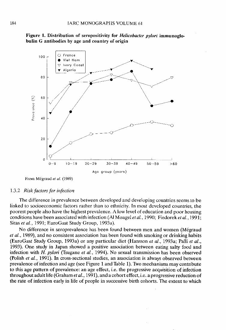
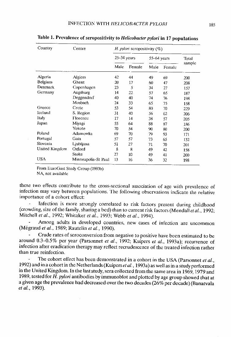
ln developing countries, the prevalence of infection increases rapidly during childhoodand adulthood and is usuaIly 80-90%. The prevalence is substantially lower in developedcountries, especiaIly in childhood (see section 1.3.2). These findings are illustrated in a studyin which the same ELISA technique was used in subjects from four countries with differentgeographical and socioeconomic status (Mégraud et al., 1989) (Figure 1). Similar resultswere reported in the EuroGast study, in which defined populations of two age groups, 25-34and 55-64 years, from 17 geographical areas, mainly European, were studied by the sameprotocol (EuroGast Study Group, 1993a,b; Table 1).
184 IARC MONOGRAPHS VOLUME 61
Figure 1. Distribution of seropositivity for Helicobacter pylori immunoglo-bulIn G antibodies by age and country of origin
100 o France. Vi et Nam\7 Ivory Coast
.. Algeria
~..~--~~. ~~~ /9
~80
..~'- 60
1
o
IDoCID
o,.ID'-
Q.
..40
20
o 10-9 10-19 20-29 30-39 40-49 50-59 ;:60
Age group (years)
From Mégraud et aL. (1989)
1.3.2 Risk factors for infection
The difference in prevalence between developed and developing countries seems to belinked to socioeconomic factors rather than to ethnicity. ln most developed countries, thepoorest people also have the highest prevalence. A low level of education and po or housingconditions have been associated with infection (Al Moagel et al., 1990; Fiedoreket al., 1991;Sitas et aL., 1991; EuroGast Study Group, 1993a).
No difference in seroprevalence has been found between men and women (Mégraudet al., 1989), and no consistent association has been found with smoking or drinking habits(EuroGast Study Group, 1993a) or any particular diet (Hansson et al., 1993a; Palli et al.,1993). One study in Japan showed a positive association between eating salty food andinfection with H. pylori (Tsugane et al., 1994). No sexual transmission has been observed(Polish et aL., 1991). ln cross-sectional studies, an association is always observed betweenprevalence of infection and age (see Figure 1 and Table 1). Two mechanisms may contributeto this age pattern of prevalence: an age effect, i.e. the progressive acquisition of infectionthroughout adult life (Graham et al., 1991), and a cohort effect, I.e. a progressive reduction ofthe rate of infection early in life of people in successive birth cohorts. The extent to which
INFECTION Wirn HELICOBACTER PYLORI 185
Table 1. Prevalence of seropositivity to Helicobacter pylori in 17 populations
Country Centre H pylori seropositivity (%)
25-34 years 55-64 years Totalsample
Male Female Male Female
Algeria Algiers 42 44 49 69 200Belgium Ghent 20 17 60 47 208Denmark Copenhagen 23 5 34 27 157Germany Augsburg 14 22 57 65 187
Deggendod 40 40 74 76 198Mosbach 24 33 65 75 158
Greece Crete 53 54 80 70 229Iceland S. Region 31 40 56 62 206Italy Florence 17 14 38 57 205Japan Miyagi 55 64 88 87 186
Yokote 70 54 90 80 200Poland Adamowka 69 70 79 93 171Portugal Gaia 57 57 73 65 132Slovenia Ljubljana 51 27 71 70 201United Kingdom Oxford 8 8 49 42 158
Stoke 27 10 49 41 200USA Minneapolis-St Paul 13 16 36 32 198
From EuroGast Study Group (1993b)NA, not available
the se two effects contribute to the cross-sectional association of age with prevalence ofinfection may vary between populations. The following observations indicate the relativeimportance of a cohort effect:
Infection is more strongly correlated to risk factors present during childhood(crowding, size of the family, sharing a bed) than to current risk factors (Mendall et al., 1992;Mitchell et al., 1992; Whitaker et al., 1993; Webb et al., 1994).
Among adults in developed countries, new cases of infection are uncommon(Mégraud et al., 1989; Rautelin et aL., 1990).
Crude rates of seroconversion from negative to positive have been estimated to bearound 0.3-0.5% per year (Parsonnet et al., 1992; Kuipers et al., 1993a); recurrence ofinfection after eradication therapy may reflect recrudescence of the treated infection ratherthan true reinfection.
The cohort effect has been demonstrated in a cohort in the USA (Parsonnet et al.,1992) and in a cohort in the Netherlands (Kuipers et aL. , 1993a) as weIl as in a studyperformedin the United Kingdom. ln the last study, sera collected from the same are a in 1969, 1979 and1989, tested for H. pylori antibodies by immunoblot and plotted by age group showed that ata given age the prevalence had decreased over the two decades (26% per decade) (Banatvalaet al., 1993).
186 IARC MONOGRAPHS VOLUME 61
ln some populations, a decrease in seroprevalence has been observed in older people.This finding has been attributed to the disappearance of H. pylori from the gastric mucosa(loss of H. pylori infection) when atrophy develops as a result of long-standing gastritis. Suchloss has been observed in sorne populations (Karneset al., 1991; Kuiperset al., 1994b) but notin another (Guarner et al., 1993). Furthermore, it is still unclear whether a graduai decreasein H. pylori colonization also leads to negative seroconversion. Negative seroconversion wasclaimed in one retrospective study (Crabtree et al., 1993a) but not in two prospective studies(Parsonnet et al., 1992; Kuipers et al., 1994a).
The prevalence of infection is consistently higher in institutionalized children than incontrol groups from the surrounding area (Berkowitz & Lee, 1987; Pérez-Pérez et al., 1990).
For a long time, the stomach was thought to be sterile, and precautions such as the use ofgloves were not taken in performing endoscopies. A higher prevalence of H. pylori infectionhas now been found among gastroenterologists who perform endoscopies than among otherphysicians or dentists (Mitchell et aL., 1989). ln countries with a high prevalence of infection,endoscopists have, nevertheless, a lower prevalence than the general population, probablydue to the fact that they come from the middle and upper classes (Matysiak-Budnik et aL.,1994).
1.3.3 Routes of transmission
Reservoirs of H. pylori are the digestive tracts of humans and sorne primates. Trans-mission.fom reservoirs is considered to be person-to-person. This assumption is supportedby the finding of clustering of similar strains within families, as shown by molecularfingerprinting (Bamford et al., 1993) and by the consistent demonstration of close inter-personal contact as a risk factor for infection. The H. pylori status of mothers ofH. pylori-positive children is significantly different from that of mothers of H. pylori-negativechildren, indicating that the intimate contact between mother and child could be a cause oftransmission (Drumm et aL., 1990). Transmission can exist between couples: 68 % of spousesof H. pylori-infected people were infected, whereas 9% of spouses of uninfected people wereinfected (Malaty et al., 1991). ln another study, the association disappeared in a multiplelogistic regression analysis (Pérez-Pérez et al., 1991). Two modes of transmission have beenproposed: oral-oral and faecal-oral transmission.
(a) Evidence for faecal-oral transmission
H. pylori is eliminated in faeces after turnover of the gastric mucosa. It has been detectedby PCR (Mapstone et aL., 1993) and by culture (Thomas et aL., 1992). Consumption of rawvegetables fertilized with human faeces was found to be a risk factor for infection in Santiago,Chile (Hopkins et al., 1993), and consumption of municipal water was found to be a riskfactor in children in Lima, Peru (Klein et aL., 1991). H. pylori hasbeen detected by PCR insewage water in Peru (Westblom et aL., 1 993b).
(b) Evidence for oral-oral transmission
H. pylori has been detected in the oral cavity (Mapstone et al., 1993) and in the saliva ofone person (Ferguson et al., 1993). Several claims have been made of the detection ofH. pylori by PCR in dental plaque (Krajden et aL., 1989; Majmudar et aL., 1990). When
INFECTION WITH HELICOBACTER PYLORI187
gnotobiotic puppies infected with H. felis were put together with uninfected litter-mates in agerm-free isolator, with continuai oral-oral contact, the agent was transmitted. Transmissiondid not occur between germ-free mice, which are coprophageous, under the same conditions(Lee et aL., 1991).
1.4 Clinical disease in hum ans (other than cancer)
1.4.1 Gastritis
H. pylori is a major cause of gastritis. This inference is based on the following obser-vations: (i) ingestion of H. pylori led to acute gastritis in a sm ail number of case studies(Marshall et aL., 1985a; Morris & Nicholson, 1987; Sobala et al., 1991); (ii) Helicobactercolonization of the stomach is virtually always accompanied by inflammation of the mucosa(Dixon, 1991); (iii) H. pylori infection can be detected in more than 85% of patients withinflammation of the gastric mucosa (Dooley et al., 1989); and (iv) this inflammationdisappears completely within two to three years after eradication of the infection (Rauws etaL., 1988; Genta et al., 1993a).
The infection disappears only as a result of antibiotic therapy, after the development ofunfavourable gastric conditions such as mucosal atrophy or after partial gastrectomy withbile reflux (Karnes et aL., 1991; Kuipers et al., 1993a). 'Spontaneous' clearance of infection isvery rare and may in fact be due to unreported use of antibiotics (Kuipers et al., 1993a). lnsome infected individuals, endoscopic signs of gastritis can be found. The gastritis affectspredominantly the antrum (Tytgat et al., 1993), although corpus involvement is observedhistologically in most infected individuals (see also section 4).
1.4.2 Duodenal ulcer disease
H. pylori infection is the most significant risk factor for duodenal ulcer disease. Afterexclusion of a small subset of cases of duodenal ulcer wIth specific etiology, su ch as use ofnon-steroidal anti-inflammatory drugs, Crohn's disease or ischaemia, the remaining casesare caused by H. pylori (Mégraud & Lamouliatte, 1992). The main arguments for a causalrelationship between H. pylori infection and duodenal ulcer disease are that the infection isseen to precede the disease (Sipponen et al., 1990) and that the disease disappears aftertreatment of the infection. While ulcers have been shown in many studies to relapse wi thin 12months after symptomatic treatment in 50- 100% of patients (Tytgat et al., 1993), eradicationof H. pylori almost totally prevents ulcer recurrence (Marshall et aL., 1988; Graham et al.,1992; Tytgatet al., 1993). It has been estimated that up to 10% ofinfected people will developduodenal ulcer during Iife (Tytgat et al., 1993).
1.4.3 Gastric ulcer disease
H. pylori infection is present in approximately 70% of patients with gastric ulcers(Labenz & Börsch, 1994). A variety of noxious agents such as non-steroidal anti-infIamma-tory drugs and bile reflux are risk factors for the development of gastric ulcers. Afterexclusion of patients with those risk factors, the bacterium is present in more than 95% of theremaining cases. Eradication of the infection significantly prevents ulcer recurrence(Graham et al., 1992; Labenz & Börsch, 1994).
188 IARC MONOGRAPHS VOLUME 61
1.4.4 Hypertrophic protein-losing gastritis
Hypertrophic protein-Iosing gastritis is a rare c1inical disorder characterized by chronicgastritis with giant folds, gastric protein loss and hypoalbuminaemia. The etiology of thisdisorder is unknown. Significant c1inical improvement was seen after H. pylori eradicationtherapy in two studies (Lepore et aL., 1988; Meuwissen et aL., 1992).
1.4.5 Childhood diseases
ln children in developing countries, H. pylori infection has been associated with chronicdiarrhoea and malnutrition (Sullivan et al., 1990). ln developed countries, it has also beenassociated with chronic abdominal pain and growth retardation.
1.5 1reatment and control
1.5.1 Antibiotics and acid suppressive therapy
Since the introduction of H2-blockers and proton pump inhibitors, H. pylori-relateddisorders have been treated with moderate success (Susi et al., 1994). The effects of acidsuppressive medication on H. pylori-related gastritis have not been examined adequately;however, such medication does not cure the infection (Kuipers et al., 1993b). The bacteriumis sensitive to a wide range of antibiotics in vitro, but most are unsuccessful in vivo. Threestrategies have been chosen to overcome this problem: (i) combination of multiplesynergistic antibiotic drugs; (ii) prolongation of drug administration; and (iii) combination ofantibiotics with acid suppressors. A large number of c1inical trials have been carried out tofind an effective treatment regimen. The current preference is for therapy lasting 14 days witheither two antibiotics combined with a bismuth preparation or with one to two antibioticscombined with an acid inhibitor, usually omeprazole (Labenz et al., 1993). With theseregimens, eradication has been achieved in 60-95% of cases, dependingupon the prevalenceof antibiotic-resistant strains and patient compliance.
1.5.2 J1ccination
H. pylori infection is always accompanied by local and systemic immune responses, withno clearance of infection (Bontkes et al., 1992). It is thus uncIear whether immunization canprevent new infections. Successful oral immunization of mice with a sonicated preparation ofH. felis plus adjuvant (cholera toxin) has been achieved (Chen et aL., 1993).
2. Studies of eancer in Humans
2.1 Descriptive studies
2.1.1 Geographical correlations
(a) Gastric carcinoma
Table 2 lists eight studies in which the prevalences of H. pylori infection were comparedin geographical regions with different gastric cancer rates. The presence of infection was
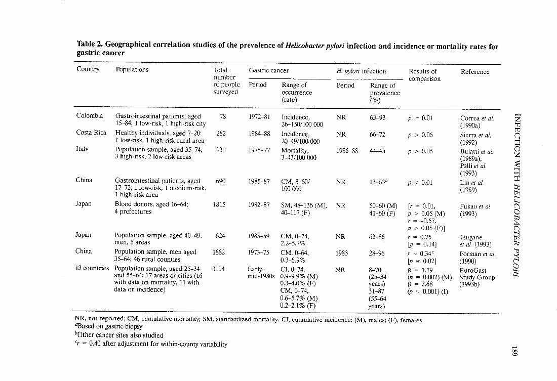
Table 2. Geographical correlation studies of the prevalence of Helicobacter pylori infection and incidence or mortality rates forgastric cancer-Country Populations Total Gastric cancer H. pylori infection Results of Reference
number comparisonof people Period Range of Period Range ofsurveyed ocurrence prevalence
(ra te) (%)
63-93 Correa et al.-Colombia Gastrointestinal patients, aged 78 1972-81 Incidence, NR p = 0.01~15-84; 1 low-risk, 1 high-risk city 26- 150/100 00 (199a) tJCosta Rica Healthy individuals, aged 7-20; 282 1984-88 Incidence, NR 6672 P ? 0.05 Sierra et aL. ("
1 low-risk, 1 high-risk rural area 20-49/100.00 (1992) ~Italy Population sample, aged 35-74; 930 1975-77 Mortality, 1985-88 44-45 P ? 0.05 Buiatti et aL. 0
Z3 high-risk, 2 low-risk areas 3-43/100 00 (1989a);:ePaUi et aL. -
(1993) ~China Gastrointestinal patients, aged 690 1985-87 CM, 8-601 NR 13-63Q p 0: 0.01 Lin et aL.
~17-72; 1Iow-risk, 1 medium-risk, 100 00 (1989)1 high-risk area t'
J apan Bloo donors, aged 16-; 1815 1982-87 SM, 48-136 (M), NR 50-60 (M) (r = 0.01, Fukao et al. (=4 prefectures 40- 11 7 (F) 41-60 (F) P ? 0.05 (M) (1993) C)
r = -0.57, ~p ? 0.05 (F)) ()
Japan Population sample, aged 40-49, 624 1985-89 CM, 0-74, NR 63-86 r = 0.75 Tsugane ~men, 5 areas 2.2-5.7% fp = 0.14) et aL. (1993) ~China Population sample, men aged 1882 1973-75 CM, 0-6, 1983 28-96 r = 0.34c Forman et al. ~
t$35-64; 46 rural counties 0.3-6.9% fp = 0.02) (199)C)13 countries Population sample, aged 25-34 3194 Early- CI, 0-74, NR 8-70 ß = 1.9 EuroGast ~and 55-6; 17 areas or cities (16 mid-1980s 0.9-9.9% (M) (25-34 (p = 0.(02) (M) Study Group ..
with data on mortality, 11 with 0.3-4.0% (F) years) ß = 2.68 (1993b)data on incidence) CM, 0-74, 31-87 (p = 0.(01) (1)
0.6-5.7% (M) (55-60.2-2.1% (F) years)
NR, not reported; CM, cumulative mortality; SM, standardized mortality; CI, cumulative incidence; (M), males; (F), femalesGßased on gastric biopsybOther cancer sites also studiedCr = 0040 after adjustment for within-county variability
l-00\0
190 IARC MONOGRAPHS VOLUME 61
determined in most studies by ELISA for IgG antibodies to H. pylori in serum. ln aIl studies,infection rates were compared with cancer rates in contemporaneous time periods, althougha more appropriate comparison would be between infection prevalence rates and cancerrates several years or even decades later. Su ch a comparison would reflect the time sequenceinvolved if there were a causal relationship between infection and cancer.
Four of the studies were comparisons of regions of high and low risk for gastric cancerwithin a single country; two showed a significant difference between H. pylori prevalencerates, with an increase in the high-risk region (in Colombia, Correa et aL., 1990a; and inChina, Lin et aL., 1989), while the other two showed no significant difference between the tworegions (in Italy, PaIli et aL., 1993; and in Costa Rica, Sierra et aL., 1992). Two studies fromJapan (Fukao et aL., 1993; Tsugane et aL., 1993) compared populations within five and fourareas, respectively; neither showed a significant association between H. pylori seropositivityand gastric cancer mortality.
Forman et al. (1990) examined the prevalence of H. pylori IgG antibodies in 1882residents of 46 rural counties in China and compared them with the gastric cancer mortalityrates in the same counties. The correlation between H. pylori antibody prevalence rate andgastric cancer mortality rate was 0.34 (p = 0.02). The significant positive correlationremained after adjustment for dietary factors associated with risk for gastric cancer (Knelleret al., 1992).
The EuroGast Study Group (1993b) examined the seroprevalence of H. pylori IgGantibodies in 3194 randomly selected subjects resident in 17 centres in 13 countries, chosento reflect the global range in gastric cancer incidence. ln regression analyses, in which the twosexes were combined, there were significant relationships between the prevalence ofH. pylori antibodies and both log-transformed gastric cancer cumulative mortality
(p = 0.002) and incidence (p = 0.001) rates. Exclusion ofthe regions with highest and lowestmortality rates (Japan and the USA, respectively) reduced the strength of the relationshipwith mortality from gastric cancer to a nonsignificant (ß = 0.62;p = 0.3) level (Forman
et al.,
1993 ).
It has been noted (Holcombe, 1992) in Nigeria and other African countries (e.g. Sudan,Uganda and Zimbabwe) that gastric cancer rates are relatively low ( -: 2-3% of aIl malignanttumours) despite a very high prevalence of H. pylori infection. The populations of otherdeveloping countries with low incidence rates of gastric cancer, but for which no estimates ofthe prevalence of infection are available, include Kuwaitis, non-Jews in Israel, Malays inSingapore and those of Ahmedabad, Bangalore, Madras and Bombay in India. Gastríccancer incidence rates in the three population-based cancer registries in Africa (Sétif,A1geria; Bamako, Mali; and the Gambia) range from 3.9 to 19.4 per 100000 in males andfrom 1.5 to 10.3 per 100 000 in females (Parkin et al., 1992). These rates are substantiallybelow those in high-risk regions of the world (e.g. Costa Rica: 46.9 in males and 21.3 Infemales) and are comparable to the rates in US blacks (12.4 in males and 5.6 in females) andin England and Wales (16.9 in males and 6.8 in females).
(b) Gastric lymphoma
Doglioni et al. (1992) compared the incidence of primary gastríc lymphoma, determinedfrom endoscopy c1inic records, in an area of northeastern Italywith that in three communities
INFECTION Wirn HELICOBACTER PYLORI191
in the United Kingdom. ln the Italian city of Peltre, the estimated incidence rate for gastriclymphomas was 66/1 00000 per five years for the period 1986-90 (37 cases). ln three districtsin the United Kingdom, the comparable rates were 6/100 000 (six cases), 4/100000 (sevencases) and 61100 000 (20 cases). The H. pylori infection rate of aH patients undergoingendoscopic biopsy was 87% in Feltre in 1991 and 50-60% in the United Kingdom. (TheWorking Group noted that this was a hospital-based study with no information about thereferral patterns to the local endoscopy unIts. There is, therefore, uncertainty about thedenominator populations used in this study.)
(c) Other cancers
ln the study of Forman et al. (1990) (see above), correlation coefficients were calculatedfor associations between H. pylori IgG antibody prevalence and mortality rates from cancersat 12 sites other than the stomach. None was significant. The correlation with lymphoma (ailtyes) was 0.32 and of borderline significance.
2.1.2 Time trends
Gastric cancer incidence and mortality rates have been declining rapidly in nearly aildeveloped countries for several years. There are few data for developing countries, but thesame trend has generally been observed (Coleman et al., 1993). Secular trends in theprevalence of H. pylori infection have not been investigated extensively, but the oneserological study that has been conducted in the United Kingdom (Banatvala et al., 1993)indIcated that the prevalence has decreased in recent decades. If H. pylori is acquiredpredominantly in childhood (see section 1.3.2), then data on age prevalence (section 1.3.1)can be interpreted as indicating a declining prevalence rate over much of the 20th century.This is also consistent with observed secular trends in duodenal ulcer disease in the USA(Sonnenberg, 1993), the United Kingdom (Susser & Stein, 1962) and Europe (La Vecchiaet al., 1993), a disease strongly associated with H. pylori infection. Data from Japan (Blaser,1993) indicate that mortality from gastrIc cancer in that country has decreased over the past50-80 years, an effect consistent with a secular decrease in exposure to an environmentalagent. The prevalence of gastric cancer of the cardia, in contrast to that of more distal siteswithin the stomach, has been shown to be increasing in a number of populations (Powell &McConkey, 1990; Blot et al., 1991; Hansson et al., 1993b). Gastric cancer of the cardia hasbeen shown in some studies (Talley et al., 1991a; Hansson et al., 1993b) not to be associatedwith H. pylori infection (see sections 2.3 and 2.4).
2.1.3 Socioeconomic trends
Gastric cancer has been shown consistently in several countries to be commoner inpoorer socioeconomIc groups (Howson et al., 1986; Buiatti et al., 1989b; Logan, 1982). Thesame association has been observed consistently for H. pylori infection (see section 1.3.2).
2.2 Case series
2.2.1 Gastric carcinoma
The presence of H. pylori infection has been determined in numerous series of gastriccancer patients, usually by histological examination of biopsy and/or gastrectomy samples
192 IARC MONOGRAPHS VOLUME 61
but also by microbiological culture; in sorne studies, serological tests were used to determinethe presence of specific IgG antibodies to H. pylori. A number of studies were designedspecifically to estimate the prevalence of H. pylori infection in gastric cancer patients; themajority, however, were broader surveys of patients with upper gastrointestinal disease andincluded a small subgroup of patients with gastric cancer. ln the latter studies, it is uncIearwhether adequate mucosa was available to evaluate the presence of H. pylori; there was alsofrequently a subgroup of patients who had dyspeptic symptoms but no lesions in theirstomachs and who were used as a control series. ln a few studies, the control series werehealthy volunteers who had undergone endoscopy. Serologically based studies in which datafrom matched case and control series were available are summarized in sections 2.3 and 2.4.
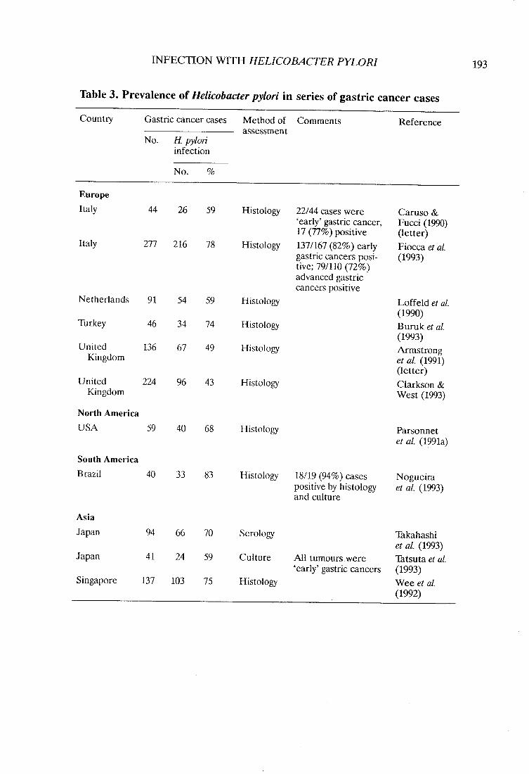
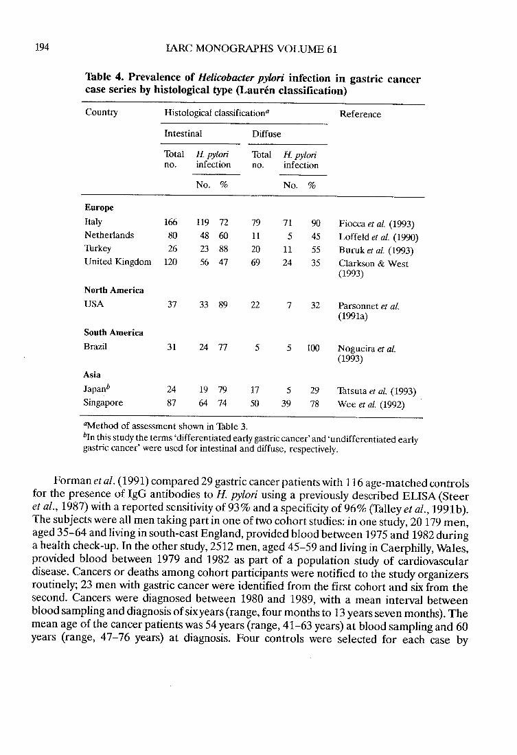
Table 3 lists the Il largest case series. The percentage of gastric cancer patients who hadH. pylori infection varied from 43 to 83%. Particular interest has focused on the Laurénhistological classification of gastric adenocarcinoma into cancers of the intestinal (glandular)tye and cancers of the diffuse tye (Laurén, 1965). It has been reported that the incidence ofthe former varies between populations whereas that of the latter remains relatively constant(Laurén, 1965; Muñoz et aL., 1968; Muñoz & Asvall, 1971; Correa et al., 1973). Environ-mental exposures are thought to be more important in the etiology of intestinal-tye than ofdiffse-tye cancers (Howson et aL., 1986). Table 4 lists eight series in which the cancer caseswere classified into intestinal and diffuse histological categories. ln sorne of these studies, anincreased prevalence of H. pylori infection was seen in association with intestinal-tye
cancers (Parsonnet et al., 1991a; Tatsuta et al., 1993), but this difference was not observedconsistently.
2.2.2 Gastric lymphoma
Wotherspoon et al. (1991) examined 110 patients in the United KIngdom with gastric B-cell mucosa-associated Iymphoid tissue lymphomas, a subset of primary gastric Iymphomas.ln this group, 101/1 10 patients (92%) had histological evidence of H. pylori infection.
A total of 205 surgi cal specimens containing primary malignant B-cell Iymphomas wereinvestigated in Germany. H. pylori colonization was found in 175/178 (98%) cases in whichthe mucosa sorne distance from the tumour could be evaluated (Stolte et al., 1994).
2.3 Cohort studies
2.3.1 Gastric carcinoma
Four prospective studies have been reported in which the relationship between H. pyloriinfection and the subsequent risk of gastric cancer has been assessed. Ali were case-controlcomparisons nested in prospective cohort studies in which blood samples had been takenfrom cancer-free individuals and stored. Specific antibodies to H. pylori were then measuredin blood samples from individuals who subsequently developed gastric cancer, and theproportion of individuals with antibodies was compared wIth that in a matched controlgroup.
194 IARC MONOGRAHS VOLUME 61
Table 4. Prevalence of Helicobacter pylori infection in gastric cancer
case senes by histological type (Laurén classification)
Country Histological classificationa Reference
Intestinal Diffuse
Total H pylori Total H pylorino. infection no. infection
No. % No. %
Europe
Italy 166 119 72 79 71 90 Fiocc et aL. (1993)Netherlands 80 48 60 11 5 45 Loffeld et al. (1990)Thrkey 26 23 88 20 11 55 Buruk et aL. (1993)United Kingdom 120 56 47 69 24 35 Clarkson & West
(1993)
North America
USA 37 33 89 22 7 32 Parsonnet et al.(1991a)
South America
Brazil 31 24 77 5 5 100 Nogueira et al.(1993)
Asia
Japanb 24 19 79 17 5 29 Tatsuta et al. (1993)Singapore 87 64 74 50 39 78 Wee et aL. (1992)
tlethod of assessment shown in Thble 3.bln this study the terms 'differentiated early gastric cancer' and 'undifferentiated early
gastric cancer' were used for intestinal and diffuse, respectively.
Forman et al. (1991) compared 29 gastric cancer patients with 116 age-matched controlsfor the presence of IgG antibodies to H. pylori using a previously described ELISA (Steeret al., 1987) with a reported sensitivity of 93% and a specificity of 96% (Talley et al., 1991b).The subjects were ail men taking part in one of two cohort studies: in one study, 20 179 men,aged 35-64 and living in south-east England, provided blood between 1975 and 1982 duringa health check-up. ln the other study, 2512 men, aged 45-59 and living in Caerphily, Wales,provided blood between 1979 and 1982 as part of a population study of cardiovasculardisease. Cancers or deaths among cohort participants were notified to the study organizersroutinely; 23 men with gastric cancer were identified from the first cohort and six from thesecond. Cancers were diagnosed between 1980 and 1989, with a me an interval betweenblood sampling and diagnosis of six years (range, four months to 13 years seven months). Themean age of the cancer patients was 54 years (range, 41-63 years) at blood sampling and 60years (range, 47-76 years) at diagnosis. Four controls were selected for each case by
INFECTION Wirn HELICOBACTER PYLORI 195
matching on cohort, date ofbirth (within one year), date ofblood sampling (within one year)and number of freeze-thaw cycles the blood sample had undergone. Twenty of the 29 (69%)gastrIc cancer patients and 54 ofthe 116 (47%) controls had antibodies to H. pylori, resultingin a matched odds ratio of 2.8 (95% confidence interval (CI), 1.0-8.0). Stratifyng the casesand corresponding controls into those diagnosed within five years of blood sampling andthose diagnosed five or more years after sampling did not result in a significant difference inthe resulting odds ratios. No information was available on site of cancer within the stomachor on histological subtye.
Parsonnet et al. (1991 b) compared 109 gastric patients with 109 age-, sex- and race-matched con trois for the presence of IgG antibodies to H. pylori using a previously describedELISA (Evans et al., 1989) with a reported sensitivity of 91 % and a specificity of 98%. Thesubjects were taking part in a cohort study in which 128 992 participants living in California,USA, provided blood between 1964 and 1969 during a health check-up. A total of246 gastriccancer registrations and/or hospitalizations for gastric cancer among cohort participantswere notified to the study organizers routinely, and 200 of these were randomly selected.Availability of blood samples resulted in final inclusion of 186 patients with gastric cancer.Cancers were diagnosed between 1964 and 1989, with a mean interval between bloodsampling and diagnosis of 14.2 years (range, 1-24 years). One control was selected for eachcase by matching on age at blood sampling (within one year), sex, race, date of bloodsampling (within 0.5 year) and site of the health check-up. Of the 186 patients, 109 had histo-logically confirmed adenocarcinoma of the stomach; of these, 92 (84%) had antibodies ta H.pylori, as did 66 of the 109 (61 %) controls, resulting in a matched odds ratio of 3.6 (95 % CI,1.8-7.3). When the cases and controls were stratified by sex, the odds ratio for women wasnonsignificantly higher than that for men; when they were stratified byrace, the odds ratio forblacks was nonsignificantly higher than that for whites. Eighty-one patients had the intestinal
tye of adenocarcinoma (Laurén classification), and 67 (83%) of these were seropositive(odds ratio, 3.1; 95% CI, 1.5-6.6); 28 patients had a diffuse tye, and 25 (89%) ofthese wereseropositive (odds ratio, 8.0; 95% CI, 1.0-64). Four patients had an adenocarcinoma at a sitein the cardia; one was seropositive, as was one of the four matched controls. An additional 27patients had adenocarcinoma of the gastroesophageal junction (not included in the mainanalyses above); of these, 17 (63 %) were seropositive, as were 19 (70%) con trois (odds ratio,0.8; 95% CI, 0.3-2.1).
Nomura et al. (1991) compared 109 patients with gastric carcinoma with 109 age-matched con trois for the presence of IgG antibodies to H. pylori using a commercial ELISA.The subjects were all men taking part in a cohort study in which 7498 Japanese-Americansliving in Oahu, Hawaii, USA, provided blood between 1967 and 1970 as part of a populationstudy of heart disease. A total of 137 gastric cancer registrations and/or hospital dischargesfor gastric cancer among cohort participants were notified to the study organizers routinely,all with histologically confirmed gastric cancer. As insufficient serum was available from 26men and the results of the ELISA were indeterminate for two, a total of 109 were included inthe study. Cancers were diagnosed between 1968 and 1989, with a mean interval betweenblood sampling and diagnosis of 13 years (standard deviation, five years). The me an age ofthe cancer patients at recruitment was 59 years. One control was selected for each case bymatching for age at recruitment and date of blood collection. Excluded from the control
196 IARC MONOGRAPHS VOLUME 61
series were men who had had a gastrectomy before blood sampling or who had had adiagnosis of peptic ulcer at any time. The exclusion criteria reduced the pool of availablecontrols by 13%. (The Working Group noted that the exclusion criteria would be likely toreduce the prevalence of H. pylori infection in the control group and, hence, bias theestimated odds ratio upwards. i A1so excluded were men with cardiovascular disease or anyother tye of cancer diagnosed at any time. These exclusions reduced the control pool by
33%. Controls had to be alive when the cancer cases with which they were matched werediagnosed. Of the 109 gastric cancer patients, 103 (94%) had antibodies to H. pylori, as did 83of the 109 (76%) controls, resulting in a matched odds ratio of 6.0 (95% CI,2.1-17).Stratification of the cases into three groups (26, 40 and 43 pairs) on the basis of time betweenblood sampling and cancer diagnosis resulted in odds ratios of 1.5 (95% CI, 0.3-9.0) for lessthan 10 years, 6.0 (1.3-27) for 10-14 years and indeterminate (1.7-97) for 15 years or more.Stratification into two birth cohorts resulted in odds ratios of 3.0 (0.8-11) for those born in1900-09 and 15 (2.0-114) for those born in 1910-19. Eighty-one patients had an intestinaltye of carcinoma, and 75 (93%) ofthese were seropositive (odds ratio, 4.5; 95% CI, 1.5-13);23 patients had a diffuse tye, and ail were seropositive (odds ratio, indeterminate; 1.1-64).Five patients had cancer at the cardia, and two were seropositive; after exclusion of thesepatients, the ove rail odds ratio was 12 (95% CI, 2.8-51). ln this study, a trend was observed (p= 0.0009) of an increasing odds ratio with an increase in the quantitative antibody level.(The Working Group estimated that, had the exclusion criteria relating to con trois with ahistory of gastrectomy or peptic ulcer not been used, the prevalence of H. pylori infection inthe controls would have been increased by approximately 4% and the overall odds ratiowould have been decreased by about 20%, i.e. from 6.0 to 4.8. i
ln a combined analysis of the three nested case-control studies described above,Forman et al. (1994) showed that, overall, 215/247 (87%) gastric cancer patients and 203/334(61 %) controls were seropositive for IgG antibodies to H. pylori, resulting in a matched oddsratio of 3.8 (95% CI, 2.3-6.2). When these results were stratified by time between samplecollection and cancer diagnosis into four periods-fewer than five years, 5-9 years, 10-14years and 15 years or more-there was a significant trend (p = 0.049) towards an increasedodds ratio with increasing time intervaI. The odds ratio changed from 2.1 (95% Ci, 0.6-8.7)to 2.3 (0.9-6.5), 4.4 (1.8-13) and 8.7 (2.7-45) over the four periods, respectively. There were20/25 (80%), 37/46 (80%), 70/78 (90%) and 88/98 (90%) seropositive cases and 34/58(59%),46/85 (54%), 58/93 (62%) and 65/98 (66%) seropositive controls in the four strata,respectively. This trend was interpreted by the authors as indicating that false-negativeassessments of H. pylori status may have occurred more frequently among cancer cases thanamong matched controls, especially among those diagnosed soon after providing blood.False-negative assessments were believed to derive from the precancerous conditions, severeatrophie gastritis and intestinal metaplasia, from loss of H. pylori colonization and loss ofseropositivity.
Lin et al. (1993a (abstract)) compared 29 gastrIc cancer patients in Taiwan, China, with220 con trois matched by age, sex and are a of residence, for the presence of H. pylori IgGantibodies by an ELISA. The subjects were participants in a cohort study in which 9777people in Taiwan had provided blood since 1984. The mean interval between blood samplingand diagnosis of cancer was 3.1 years. Sixt-nine percent of the gastric cancer patients and
INFECTION WITH HELICOBACTER PYLORI 197
59% of the con trois were seropositive for antibodies to H. pylori, resulting in an odds ratio of1.6 (95% CI, 0.68-2.6).
The four prospective studies are summarized in Table 5.
2.3.2 Gastric lymphoma
Parsonnet et al. (1994) compared 33 patients with gastric non- Hodgkin's Iymphoma with134 age- and sex-matched con trois for the presence of H. pylori IgG antibodies using anELISA with a reported sensitivity of96% and a specificity of76% for active gastric infection.The subjects were taking part in one of two cohort studies, one in California, USA, describedabove (Parsonnet et al., 1991 b), and the other of 170 000 participants living in Norway whoprovided blood between 1973 and 1991 during blood donation and health screeningprogrammes. Cancer registrations between 1973 and 1990 among the Norwegian cohortwere notified to the study organizers. Twenty gastric Iymphomas were identified from the UScohort and 13 from the Norwegian cohort, with median intervals between blood samplingand diagnosis of 14 and 13 years, respectively. The median ages of the Iymphoma patients atdiagnosis were 66 and 55 years, and 40 and 69% patients in the two cohorts were men,respectively. Four cancer-free con troIs were selected for each case and matched on cohort,date of birth, age (five-year groups in the USA; within six months in Norway), sex, date andlocation of blood collection and ethnic group (only in the USA). Twenty-eight of the 33(85%) gastric lymphoma patients were seropositive for antibodies to H. pylori, as were 74 ofthe 134 (55%) controls, resulting in a matched odds ratio of 6.3 (95% CI, 2.0-20). There wasno significant difference between odds ratios when the cases and corresponding con troiswere stratified on the basis of cohort, se x, age at diagnosis (~ 65 or :; 65 years) or time
between blood sampling and diagnosis (~ 14 or:; 14 years). ln a separa te analysis of 31patients with non-gastric non-Hodgkin's Iymphoma and 61 matched controls, 20 patients(65%) and 36 con troIs (59%) were seropositive, resulting in a matched odds ratio of 1.2 (95%CI, 0.5-3.0).
2.4 Case-control studies
2.4.1 Gastric carcinoma
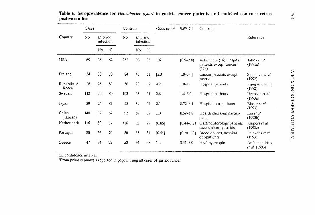
Nine case-control studies have been carried out in which serological assessment of
infection was done retrospectively in cancer patients after diagnosis.Talley et aL. (199 la) compared 69 patients with gastric adenocarcinoma with 252 controls
for the presence of IgG antibodies to H. pylori using a previously described ELISA(Pérez-Pérez et al., 1988) with a reported sensitivity of 96% and a specificity of 94% (Talleyet al., 1991b). The cases of cancer had been confirmed histologically and diagnosed between1982 and 1989 at a single hospital in Minnesota, USA. The median age of the patients was63 years (25th and 75th percentiles, 56.5 and 71 years), and 52% were men. The controlscomprised 76 asymptomatic volunteers with no history of gastrointestinal disease and 176patients who were treated between 1976 and 1989 at the same hospital as the cancer patientsfor a variety of non-malignant conditions: 67 for benign musculoskeletal problems, 52 forbenign oesophageal disease and 57 for benign lung diseases. The me
di an age of the controls
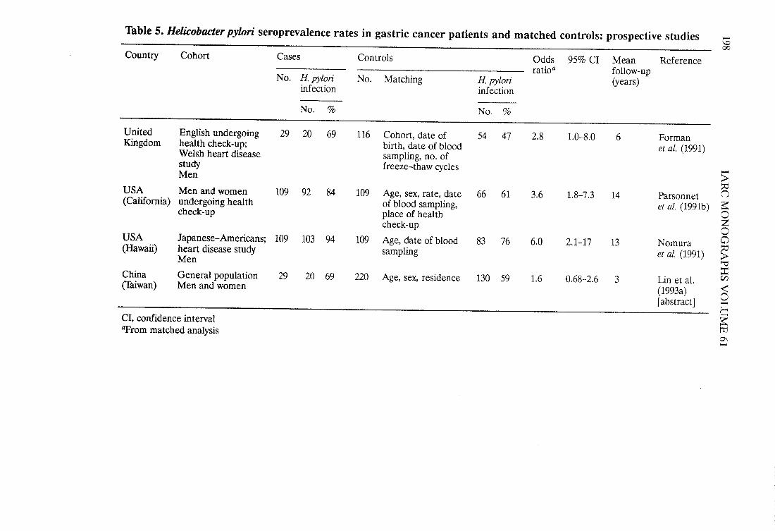
Table 5. Helicobacter pylori seroprevalence rates in gastric cancer patients and matched con troIs: prospective studies ..'-00
Country Cohort Cases Con troIs Odds 95% CI Mean Referenceratioa follow-up
No. H. pylori No. Matching H. pylori (years)infection infection
No. % No. %
United English undergoing 29 20 69 116 Cohort, date of 54 47 2.8 1.0-8.0 6 FormanKigdom health check-up; birth, date of blood et aL. (1991)Welsh heart disease sampling, no. ofstudy freeze-thaw cycles -Men ~::USA Men and women 109 92 84 109 Age, sex, rate, date 66 61 3.6 1.8-7.3 14 Parsonnet n
(Califomia) undergoing health of blood sampling, et al. (1991b) ~check-up place of health 0Zcheck-up 0
USA Japanese- Americans; 109 103 94 109 Age, date of blood 83 76 6.0 2.1-17 13 Nomura a(Hawaii) heart disease study sampling et aL. (1991) ~Men 'i
::China General population 29 20 69 220 Age, sex, residence 130 59 1.6 0.68-2.6 3 Lin et aL. C/(Iiwan) Men and women
(1993a) ~0(abstract J l"
CCI, confidence interval ~tTIlrom matched analysis0\..
INFECTION WITH HELICOBACTER PYLORI199
was 61 years (25th and 75th percentiles, 54 and 67 years), and 50% were men. Of the 69gastric cancer patients, 36 (52%) had antibodies to H. pylori, as did 96 (38%) of the con
trois.The odds ratio, after adjustment for age and sex, was 1.6 (99% CI, 0.79-3.4). Adjustment forlength of storage of the blood samples had no substantial effect on the results. The odds ratiofor gastric cancers at sites other than the cardia (n = 37)was 2.7 (99% CI, 1.0-7.1), while thatfor cancers at sites in the cardia (n = 32) was 0.94 (99% CI, 0.34-2.6). For the intestinal tyeof gastric cancer, according to the Lauren classification (n = 32), the odds ratio was 1.9 (99%CI, 0.67-5.1), while for cancers of the diffuse histological tye (n = 22) it was 2.5 (99% CI,0.73-8.2). After the cancers of the cardia had been excluded, the odds ratios were 4.6 (99%CI, 0.78-27) for the intestinal tye (n = 13) and 2.3 (99% CI, 0.63-8.1) for the diffuse tye(n = 19). There were five additional groups of patients in this study. The proportions withantibodies to H. pylori were 44% of ni
ne with benign gastric lesions, 89% of ni ne with gastriccancers other than adenocarcinoma, 51 % of 80 with colorectal cancer, 49% of 41 withoesophageal cancer and 56% of 79 with lung cancer. ln comparisons with the cancer-freecontrol group, as used in the study of gastric adenocarci.noma, the odds ratios, afteradjustment for age and sex, were 1.5 (99% CI, 0.23-9.1) for benign gastric neoplasms, 13(99% CI, 0.77-203) for other gastric cancers, 1.8 (99% CI, 0.86-3.4) for colorectal cancer,1.4 (99% CI, 0.58-3.4) for oesophageal cancer and 1.8 (99% CI, 0.91-3.6) for lung cancer.
Sipponen et al. (1992) compared 54 patients with gastric adenocarcinoma with 84controls for the presence of IgG, IgA and IgM antibodies to H. pylori using a previouslydescribed ELISA (Kosunen et al., 1989). The cases of gastric cancer were confirmed histolo-gically and occurred in a consecutive series of patients diagnosed in 1988 and 1989 at a singlehospital in Finland. Patients with cancers of the region of the cardia were excIuded, as werepatients who had previously undergone gastric surgery. The mean age of the patients whowere incIuded was 65 years (SD, 16 years), and 48% were men. The controls were 35 patientswith cancers at gastrointestinal sites other than the stomach (6 in the oesophagus, 7 in thepancreas and 22 in the colon) and 48 patients with cancers at sites other than the gastro-intestinal tract. The mean ages of these two groups of controls were 65 years (SD, 12 years)and 66 years (SD, 12 years), respectively, and 57 and 71 %, respectively, were men. IgGantibodies to H. pylori were found in 38/54 (70%) of the gastric cancer patients and 43/84(51 %) of the patients with other cancers. (The unadjusted odds ratio was calculated by theWorking Group to be 2.3 (95% CI, 1.0-5.0).) IgA antibodies to H. pylori were found in 76%of the gastric cancer patients and 58% of the controls (the unadjusted odds ratio was 2.3(95% CI, 1.1-4.8); IgM antibodies were found in 6% of the cases and 5% of the controls.When the gastric cancer patients were stratified into three age groups, IgG antibodies werefound in 8110 (80%) aged 30-49 years, 13/19 (68%) of those aged 50-69 years and 17/25(68%) of those aged 70 years or more. For the patients with other cancers, the respectiveproportions were 5/9 (56%), 22/38 (58%) and 16/37 (43%), resulting in odds ratios for thethree strata of (3.2 (95% CI, 0.3-45.4)), (1.6 (0.4-6.2)) and (2.8 (0.9-9.4)), respectively.Thirty-one gastric cancer patients had tumours of the intestinal tye, and 22 (71 %) of themwere seropositive; 21 gastric cancer patients had tumours of the diffuse tye, and 15 (71 %) ofthem were seropositive.
Kang and Chung (1992) compared 28 patients with gastric adenocarcinoma in theRepublic of Korea with 30 age- and sex-matched controls for the presence oflgG antibodies
20 IARC MONOGRAHS VOLUME 61
to H. pylori, using a commercial ELISA kit. The gastric cancer patients had aIl undergoneresection, had histological confirmation of their disease and had been diagnosed in 1991.The mean age of the cases was 50 years (range, 29-67 years), and 66% were men. Con troiswere hospital patients with a variety of diagnoses other than gastrointestinal disease andincluded nine patients with non-gastrointestinal cancer. The mean age of the controls was 52years (range, 28-69 years), and 67% were men. Twenty-five (89%) of the gastrIc cancerpatients had antibodies to H. pylori, as did 20 (67%) of the control patients. A matchedanalysis resulted in an odds ratio of 4.2 (95 % CI, 1.0-17). Ten of the patients had intestinal-tye cancers, and eight (80%) of these were seropositive; 18 patients had diffse-tyecancers, and 17 (94%) of these were seropositive. AIl nine gastric cancer patients who had'early gastric cancer' were seropositive; of the 19 who had advanced cancer, 16 (84%) wereseropositive.
Hansson et al. (1993a) compared 112 gastric adenocarcinoma patients with 103 controlsfor the presence of IgG antibodies to H. pylori using a commercial ELISA kit wi th a reportedsensitivity of98.7% and a specificity of 100% (Evans et al., 1989). The cases were confirmedhistologicallyand occurred in a consecutive series of patients diagnosed between 1989 and1991 at eight hospitals in central and northern Sweden. Patients over 79 years of age and withadvanced disease (20% of study base) were excluded, as were patients who refused (3%) orwere unable (14%) to give blood. The mean age of the gastric cancer patients was 67 years,and 63% were men. Controls were patients admitted to the same hospitals with a variety ofnon-gastrointestinal diseases, who were frequency matched to the cases by lO-year agegroup, sex and hospital. The me an age of the controls was 67 years, and 66% were men.Antibodies to H. pylori were found in 90/1 12 (80%) of the gastric cancer patients and 631103(61 %) of the controls (odds ratio, 2.6; 95% CI, 1.4-5.0). Wh en the analysis was stratified intothree age groups, the odds ratios were 9.3 (1.4-101) for patients aged less than 60 years, 4.3(1.3- 15) for those 60-69 years and 1.2 (0.44-3.0) for those aged 70 or more. The interactionbetween age and H. pylori seropositivity was significant. There was a higher odds ratio in menthan in women, but the effect was of borderline significance. The multivariate odds ratio forH. pylori seropositivity, estimated in a multiple regression model with adjustment foroccupation, diet, smoking and alcohol consumption (multivariate odds ratio, 2.7; 95% CI,1.3-5.8) showed little difference from the univariate odds ratio. Of patients with gastriccancers at sites other than the cardia, 77/93 (83%) were seropositive (odds ratio, 3.1;1.5-6.3), while 13/19 (68%) patients with cancers of the cardia were seropositive (1.4;0.44-4.8). Of patients with intestinal-tye gastric cancer, 60/75 (80%) were seropositive (2.5;1.2-5.4), while 22/28 (79%) of patients with diffuse-tye cancer were seropositive (2.3;0.82-7.6).
Blaser et al. (1993) compared 29 gastric adenocarcinoma patients with 58 age- (withinone year) and sex-matched controls for the presence of IgG antibodies to H. pylori, using apreviously described ELISA (Pérez-Pérez et al., 1988) with a reported sensitivity of96% anda specificity of 94% (Talley et al., 1991b). The cases were confirmed histologically and hadbeen diagnosed between 1990 and 1992 in one city, Ichikawa, in Japan. The median age ofpatients was 63 years (range, 46-82 years), and 62% were men. Controls were out-patientsattending the same hospital as the gastric cancer patients for a variety of iInesses, excluding'known stomach disease' and chronic liver disease. Twenty-four of the 29 (83%) gastric
INFECTION WITH HELICOBACTER PYLORI 201
cancer patients and 39/58 (67%) controls had antibodies to H. pylori (matched odds ratio,2.1; 95% CI, 0.72-6.4). Exclusion of the three gastric cancer patients with cancers of thecardia and the corresponding controls, justified because of the previously identifiedspecificity of association with cancer other than of the cardia (No mura et al., 1991; Talleyet al., 1991a), resulted in an odds ratio of2.8 (95% CI, 0.82-9.6) for the patients with cancersat sites other than the cardia. Exclusion of non-cardia gastric cancer patients aged 70 years orover (and corresponding controls), justified because of the previously identified reducedassociation in the elderly (Nomura et aL., 1991), resulted in an odds ratio of 6.0 (95% CI,1.1-34). Comparisons of cases on the basis of stage or severity of pathological lesions werereported not to affect the odds ratio. (The Working Group noted that the exclusion ofpatients with known stomach disease from the control group would be likely to reduce theprevalence of H. pylori infection in the group and, hence, bias the estimated odds ratioupwards.)
Lin et al. (1993b,c) compared 148 gastric adenocarcinoma patients with two series ofcontrols (n = 92 and 823) for the presence of IgG antibodies to H. pylori, using a commercialELISA kit with a reported sensitivity of 96% and a specificity of 93%. The cases wereconfirmed histologically and occurred in a consecutive series of patients diagnosed in 1992 ata single hospital in Taiwan, China. The mean age was 59 years (range, 24-87 years), and 61 %were men. The first control series were part of a group of asymptomatic subjects who had hadan endoscopic examination with negative results during a routine health check in 1992. Theirmean age was 52 years (range, 22-77 years), and 59% were men. The second control serieswere randomly selected from household registry files in one precinct and three townships inTaiwan. The subjects included people of ail ages, from -c 10 years to :: 70 years, and 50%were men. (The Working Group noted that the two reports of the study had slightly differentnumbers of cases: 148 (Lin et al., 1993b) and 143 (Lin et aL., 1993c). ln the results reportedbelow, the larger number was used, except where stated. The Working Group also noted thatthe selection of controls for the first series, excluding volunteers who did not haveendoscopically normal stomachs, would be likely to reduce the estimated prevalence of
H. pylori infection in the control group and, hence, bias the estimated odds ratio upwards.)Ninety-two of the 148 (62%) gastric cancer patients and 57/92 (62%) controls in the firstseries had antibodies to H. pylori (age- and sex-adjusted odds ratio, 1.0; 95 % CI, 0.59- 1 .8), asdid 448/823 (54%) controls in the second series (unadjusted odds ratio, 1.4 (95% CI,1.0-2.0); after exclusion of controls from the second series who were aged less than 20 years,347/527 (65%) were seropositive, giving a calculated unadjusted odds ratio of 0.85 (95% CI,0.58-1.2)). Among subjects below the age of 60 years, 44/64 (69%) of gastric cancer cases,40/66 (61 %) of the first series of con trois and 280/436 (64 %) of the second series of controls(20-59 years) were seropositive; among those 60 years of age or more, 48/84 (57%) of thecancer patients, 17/26 (65%) of the first series of controls and 67/91 (74%) of the secondseries of controls were seropositive. Twenty-six of the cancer patients had their tumour in theregion of the cardia, and 17 of these (65 %) were seropositive; 114 cancer patients had theirtumour in regions other than the cardia, and 71 of these (62%) were seropositive. Of the 52patients who had cancers of the intestinal tye, 31 (60%) were seropositive, whereas of 96patients with cancers of the diffuse tye, 61 (64%) were seropositive. Of 26 'early' gastric
202 IARC MONOGRAPHS VOLUME 61
cancer patients, 16 (62%) were seropositive, and of 122 patients with advanced cancers, 76(62 %) were seropositive.
Kuipers et al. (1993c) compared 116 gastric adenocarcinoma patients with 116 age- andsex-matched con trois for the presence of IgG antibodies to H. pylori using a previouslydescribed ELISA (Peña et al., 1989). The cases were confirmed histologically; the patientswere resident in the Netherlands and had a median age of 67 years (range, 23-92 years); 56%were men. Controls were subjects undergoing upper gastrointestinal investigations,excluding those with endoscopic and histological abnormalities such as peptic ulcer, atrophiegastritis and intestinal metaplasia. Antibodies to H. pylori were found in 89/1 16 (77%) gastrIccancer patients and 92/116 (79%) controls (resulting in an unadjusted and unmatched oddsratio of 0.86 (95% CI, 0.44-1.7)). Stratification into five age groups (0( 50,50-59,60-69,70-79 and :/ 79 years) did not significantly change the odds ratios for gastric cancer withinany strata (figures not available). Of the 67 gastric cancer patients who had tumours of theintestinal tye, 51 (76%) were seropositive; of the 36 patients with tumours of the diffusetye, 28 (78%) were seropositive. (The Working Group noted that, despite the exclusionsfrom the control series, the use of symptomatic gastrointestinal disease patients would beIikely to increase the estimated prevalence of H. pylori infection among the contrais and,hence, bias the odds ratio downwards.)
Estevens et al. (1993) compared 80 gastric adenocarcinoma patients with 80 age- andsex-matched controls for the presence of IgG antibodies to H. pylori using an ELISAdeveloped in their laboratory on the basis of a previously described assay (Evans et al., 1989).The cases were confirmed histologicaIly and occurred in a consecutive series diagnosed in1990-91 at a single hospital in Lisbon, Portugal. The mean age was 66 years (SD, 11.9years),and 58% were men. Con trois were blood donors and hospital out-patients attending traumaand orthopaedic c1inics. Antibodies to H. pylori were found in 56/80 (70%) gastric cancerpatients and 65/80 (82%) controls, resulting in an odds ratio oQO.54 (95% CI, 0.24-1.2)). Ofthe gastric cancer patients with tumours of the cardia, 67% were seropositive; of the patientswith tumours at other sites, 70% were seropositive. Of the patients with tumours of theintestinal tye, 64% were seropositive, whereas of those with tumours of the diffuse tye,50% were seropositive.
Archimandritis et al. (1993) compared 47 gastric adenocarcinoma patients with 50control s, matched for age, sex, socioeconomic status and area of residence. The presence ofIgG antibodies to H. pylori was assessed using a commercial ELISA kit. The cases wereconfirmed histologically; patients with tumours of the cardia were excluded. Patients werefrom ail over Greece, their mean age was 62 years (SD, 12.6 years) and 62% were men.Con trois were healthy people from aIl over Greece with 'no evidence of peptic ulcer ornon-ulcer dyspepsia'; their mean age was 62 years (SD, 14.1 years), and 54% were men. Ofthe 47 gastric cancer patients, 34 (72%) were seropositive for H. pylori antibodies, as were34/50 (68%) controls (odds ratio, 1.2; 95% CI, 0.51-3.0). When the analysis was stratified byage, the odds ratio for subjects aged 0( 60 years was 1.5 (0.42-5.0) and that for subjects :; 60
was 0.87 (0.23-3.3). Of the 31 gastric cancer patients with tumours of the intestinal tye, 22
(71 %) were seropositive (1.2; 0.43-3.1); of ni ne patients with tumours of the diffuse tye,seven (78%) were seropositive (0.83; 0.13-5.3). (The Working Group noted that theinformation provided about control selection was inadequate to allow a judgement about the
INFECTION WITH HELICOBACTER PYLORI 203
adequacy of the control group. The exclusion of controls with peptic ulcer or non-ulcerdyspepsia would be likely to reduce the prevalence of H. pylori infection in the control groupand, hence, bias the esimated odds ratio upwards.)
The studies are summarized in Table 6.
2.4.2 Other cancers
No case-control studies of cancers other than gastric cancer have been reported,although the study of Talley et aL. (1991a) (see above) compared patients with lung, oeso-phageal and large bowel cancers.
2.5 Intervention studies
Wotherspoon et al. (1993) gave H. pylori eradication therapy to six patients (three men,aged 37, 76 and 42, and three women, aged 75,60 and 57) with histological and moleculargenetic evidence of primary gastric low-grade B-cell mucosa-associated lymphoid tissuelymphoma with concomitant H. pylori infection. H. pylori was eradicated in ail six patients,and repeated biopsies, 4-10 months after eradication, in five patients showed no evidence ofIymphoma.
Stolte et al. (1994a) treated 16 patients with low-grade mucosa-associated lymphoidtissue Iymphomas, H. pylori infection and gastritis with H. pylori eradication therapy. Thepatients were followed up with repeated endoscopic biopsies 3- 12 months after treatment;12 patients showed regression of the lymphoma. ln six of the 12, sparse residuallymphomatissue was found.
The gastric Iymphomas that respond to H. pylori eradication therapy, the well-differen-tiated mucosa-assocated lymphoid tissue Iymphomas, were previously called 'pseudo-Iymphomas'. They are known to remain localized for many years before invading othertissues.
3. Studies of Cancer in Experimental animais
3.1 Infection with Helicobacter pylori alone
No data were available to the Working Group.
3.2 Infection with Helicobacter pylori in combination with administration of knowncarcinogens
Rat: A total of 90 male Wistar WKY /Std rats, eight weeks of age, received 50 mg/LN-methyl-N'-nitro-N-nitrosoguanidine (MMNG) in the drinking-water for 40 weeks. Onegroup of 30 rats received MNNG al one; a second group of 30 rats was given MNNG plus oralintubations of 0.2 ml brucella broth three times a week for the 40 weeks; the third group of30 rats received MNNG and brucella broth containing 106-108 colony-forming units/ml ofculture of fresh isolates of H. pylori three times a week for 40 weeks, since permanent
::.:..;,:.;~--~
Table 6. Seroprevalence for Helicobacter pylori in gastric cancer patients and matched con troIs: retros-~pective studies
Cases ContraIs Odds ratioa 95% CI ContraIs
Country No. H. pylori No. H. pylori Referenceinfection infection-No. % No. %
USA 69 36 52 252 96 38 1.6 (0.9-2.8) Volunteers (76), hospital Thlley et al.patients except cancer (1991a)(176) -
;iFinland 54 38 70 84 43 51 (2.3 1.0-5.0) Cancer patients except Sipponen et aL. ~gastric (1992) ()
Republic of 28 25 89 30 20 67 4.2 1.0-17 Hospital patients Kang & Chung ~0Korea (1992) ZSweden 112 90 80 103 63 61 2.6 1.4-5.0 Hospital patients Hansson et aL. 0
(1993a)0~
Japan 29 24 83 58 39 67 2.1 0.72-6.4 Hospital out-patients Blaser et al. ;i'"
(1993) ::China 148 92 62 92 57 62 1.0 0.59- 1.8 Health check-up partici- Lin et aL.
C/
(Tiwan) pants (1993b).(0
Netherlands 116 89 77 116 92 79 (0.86) (0.44-1.7) Gastraenteralogy patients Kuipers et aL. t"Cexcept ulcer, gastritis (1993c) ~
Portugal 80 56 70 80 65 81 (0.54) (0.24- 1.2) Blood donors, hospital Estevens et aL. mout-patients (1993 ) 0\..
Greece 47 34 72 50 34 68 1.2 0.51-3.0 Healthy people Archimandritiset al. (1993)
CI, confidence intervalIlrom priary analysis reported in paper, using ail cases of gastric cancer
INFECTION WITH HELICOBACTER PYLORI 205
colonization of the rat gastric mucosa by the H. pylori is not achieved. AlI rats survved 35 ormore weeks. Mer the 40 weeks of treatment, the two control groups had very similarnumbers of gastroduodenal tumours (adenomatous polyps, adenocarcinomas and carcI-nomas): 7/30 of those given MNNG alone and 6/30 of those given MNNG pius brucellabroth; a slight reduction in the number of gastroduodenal tumours was seen in the groupgiven MNNG plus the living cultures of H. pylori (4/30). No difference in the incidence ofgastritis was seen among the three groups (Kawauraet al., 1991). (The Working Group notedthat exposure to H. pylori was intermittent in this model, th us unlike the conditions of humanexposure. )
3.3 Infection with other Helicobacter species
Mouse: ln a study reported as an abstract (Enno et al., 1994),260 specific pathogen-freeBALB/c mice were infected with H. felis. Groups of 20 mice were killed at 2-3-monthintervals up to 26 months. Up to 18 months after infection, minimal gastritis was observed;however, at 22-26 months after infection, 51/80 H. felis-infected animais and 4/48 un-infected controls had large lymphoid aggregates in the cardia. Lymphoepithelial lesions thatwere not seen in control animais and which, according to the authors, are similar to thoseobserved in association with human gastric low-grade B-ceIl Iymphomas, were observed in27/80 infected animais.
3.4 Infection with other Helicobacter species in combination with administration of knowncarcinogens
Ferret: A group of ni ne female ferrets (Mustela putorius Jura), four to five months of age,ovariohysterectomized and naturaIly infected with H. mustelae, received single oral doses of50 mg/kg bw MNNG in 3 ml of olive oil. One additional four-month-old ferret received100 mg/kg bw MNNG, and five controls animais received olive oil only. Mucosal punchbiopsies were obtained by endoscopy from the same region of the stomach at 6- 12-monthintervals; no adenocarcinoma was seen in the limited samples taken. Seven of
the ni ne ferretsdosed with 50 mg/kg bw MNNG were killed between 51 and 55 months after treatment; oneother ferret died, and one was killed at 25 months. At necropsy, two ferrets had pyloric ulcersand two had obvious nodules on the mucosal surface of the pylorus. The single ferret thatreceived 100 mg/kg bw and was kiled at 29 months had clinical gastrointestinal disease. Ithad a grossly thickened pyloric area with al-cm ulcer at the pyloric-duodenal junction.
Histopathological examination of aIl the stomachs revealed that aIl ferrets, control andtreated, had marked chronic gastritis with the major characteristics of multifocal atrophicgastritis. One or more foci of neoplasia were seen in 9 of the 10 MNNG-treated ferrets. Twohad well-defined invasive adenocarcinomas, and four had multiple independent primaryadenocarcinomas. The neoplasms were concentrated in the pyloric antrum at the transitionzone between the corpus and antral mucosa. Metastasis to regional lymph nodes wasobserved in one animaL. The five control animaIs were killed 47-67 months after dosing wItholive oil; two that were killed had chronic renal failure, while the other three wereasymptomatic when they were kiled. No gross lesion was seen in the stomachs of the controlferrets; the only histopathological change observed was mild to moderate gastritis in the
20 IARC MONOGRAHS VOLUME 61
antrum with small foci of gland loss. Adenocarcinomas were not observed in the stomachs ofhundreds of untreated laboratory ferrets examined at routine necropsy (Fox et al., 1993a).(The Working Group noted that the study did not include a group uninfected withH. mustelae but given MNNG.)
4. Other Data Relevant for Evaluation of Carcinogenicity and its Mechanisms
4.1 Pathology of infection
Cross-sectional and longitudinal observations in human populations indicate that aseries of alterations of the gastric mucosa pre~ede gastric carcinoma (Siurala et aI., 1985;Correa et aI., 1990b; Kuipers et aL., 1994a): They follow a sequential presentation of chronicnonatrophic gastritis, atrophic gastritis, intestinal metaplasia and dysplasia. Atrophy (Joss ofgastric glands) is a pivotai change in the precancerous process. It radically alters the gastricmicroenvironment by reducing acid secretion, elevating the gastric luminal pH and resultingin an overgrowth of anaerobic bacteria. Many such bacteria produce reductases which act onnitrate molecules (from food and other sources) and result in elevated concentrations ofN02 - in the gastric lumen. Dietary factors that are important in the progression of theprecancerous process include high salt (NaCI) intake and low consumption of fresh fruits andvegetables (Nomura et aL., 1982; Fontham et aL., 1986; Buiatti et al., 1989b, 1990; Chen et al.,1990; Forman, 1991).
4.1.1 Humans
The anatomIcal substratum resulting from H. pylori infection is chronic gastritis.A1though the association between the bacterium and gastritis was recognized only in 1983(Warren, 1983; Marshall, 1983), the pathological manifestations of chronic gastritis andseveral nosological entities of gastritis had been described previously.
(a) Specifie lesions
Colonies of H. pylori are characteristically located extracellularly in the mucus layerimmediately adjacent to the gastric surface epithelium. They are prominently concentratedin front of the intercellular junctions of the epithelial cells. Most bacteria float freely withinthe mucus layer; a few adhere to pedestals formed by the epithelial cyoplasmic membrane.They may sometimes penetrate the intercellular spaces and, rarely, the ductules of theparietal cells (Chen et al., 1986; Fiocca et al., 1987; Hessey et al., 1990).
H. pylori infection is associated with degenerative changes in the cyoplasm of thesurface epithelial cells, identified on haematoxylin-eosin staining as loss of the superficialportion of the cyoplasm, resulting in microerosions of the surface epithelium (Chan et al.,1991). Under the electron microscope, partial loss and stunting of the microvilli andnumerous intracellular phagolysosomes may be seen (Chen et al., 1986; Fiocca et al., 1987;Hessey et al., 1990).
H. pylori infection results in infiltration of leukocyes into the gastrIc mucosa. The mostabundant are B lymphocyes, which occupy the lamina propria and may lead to formation of
INFECTION WITH HELICOBACTER PYLORI 207
Iymphoid follicles (Genta et al., 1993a,b). Polymorphonuclear neutrophiIs, although lessabundant than lymphocyes, are cornmon in H. pylori infections. They are seen in the laminapropria, in the space between the epithelial cells and in the gastric lumen; they tyicallyaggregate in the neck area of the gastric glands. Other inflammatory cells identified inH. pylon-infected mucosa are plasma cells, T lymphocyes, macrophages and eosinophils(Marshall et aL., 1985b; Dixon et al., 1988; Wyatt & Rathbone, 1988; Genta et al., 1993b).
The lesions associated with acute (new) infection are similar to those described above,except that the polymorphonuclear infiltrate is prominent and precedes the Iymphocyicinfiltrate (Marshall et al., 1985a; Morris & Nicholson, 1987; Graham et al., 1988).
(b) Nosological entities
ln early stages of H. pylori infection, gastritis is nonatrophic. Later, it leads to gland loss(atrophic gastritis) and is frequently followed by intestinal metaplasia.
Atrophic gastritis in patients with the pernicious anaemia syndrome diffusely involvesthe oxytic mucosa while sparing the antrum. This gastritis is called tye A or autoimmune(Strickland & Mackay, 1973; Correa, 1980). ln populations at low risk for perniciousanaemia, atrophic gastritis is multifocal and involves both the antrum and the corpus. Thisgastritis is called tye B (Strickland & McKay, 1973) or multifocal atrophie (Lambert, 1972;Corre a, 1980). The two entitiescoexIst in a few patients, leading to the denomination tyeAB gastritis (Glass & Pitchumoni, 1975). A frequent form of nonatrophic gastritis is locatedpredominantly in the antrum, with mild or no involvement of the oxytic mucosa. Suchlesions have been called diffuse antral (Correa, 1988), interstitial (Cheli et al., 1980), pre-atrophic (Cheli & Testing, 1993) or hypertrophic gastritis (Schindler, 1969). This tye of
gastritis is seen most frequently in conjunction with duodenal ulcer, while rnultifocal atrophiegastritis is associated particularly with gastric ulcer or gastric carcinoma (Schindler, 1969;Lambert, 1972).
Once the pro minent role of H. pylori in chronic gastritis had been recognized, a gradingof gastritis, the Sydney system, was designed (Price, 1991), which is intended to includemicroscopic, gastroscopic and etiological factors, including H. pylori infection. The systemallows the grading of inflammatory and atrophic changes in the corpus and antrum on aserniquantitative scale of 0-3. The name 'pangastritis' is proposed for lesions covering boththe antrum and the corpus, which can be atrophic or nonatrophic (Sipponen et aL., 1991).
H. pylori infection has a prominent role in diffuse antral (nonatrophic) gastritis and inmultifocal atrophic gastritis (Siurala et al., 1985). It has no role in corpus-limited (tye A orautoimmune) atrophic gastritis, or in other specific forms of gastritis such as those associatedwith bile reflux or use of nonsteroidal anti-inflammatory drugs, known as 'reflux', 'reactive'or 'chemical irritational' gastritis (Dixon et al., 1988; Flejou et aL., 1989), or in 'lymphocyicgastritis' (Haot et aL., 1986).
H. pylori infection has also been associated with other, less frequent tyes of gastritis,such as that characterized by prominent hyperplastic foveola, also called 'hypertrophie' or'focal foveolar' hyperplasia (Stolte et aL., 1 994b).
208 IARC MONOGRAPHS VOLUME 61
(c) Epidemiology of chronic gastritis
H. pylori infection is very prevalent in sorne populations of low socioeconomic status(Holcombe, 1992; Sierra et al., 1992). ln a few, gastric biopsy specimens and pepsinogenlevels indicate that the gastritis is not of the atrophic tye (Sierra et al., 1992; Shousha et al.,1993). ln populations at high risk for gastric cancer, atrophic forms of gastritis predominate.Atrophie gastritis associated with the pernicious anaemic syndrome, not usually related to H.pylori infection, is strongly related to genetic susceptibility and affects mainly populations ofnorthern European extraction. ln other populations at high risk for gastric cancer, such asthose of the Andean regions of Latin America, those of China and Japan, and US blacks,atrophic gastritis is multifocal and linked in part to dietary factors (Fontham et aI., 1986;Nomura et aI., 1982).
People of each sex are equally affected, and the prevalence of gastritis is highly age-dependent. Nonatrophic gastritis is more frequent in people under the age of 50, whereasatrophic gastritis and intestinal metaplasia are more frequent among people over that age(Siurala et al., 1985). ln samples from 500 blood donors in Finland, the prevalences of bothH. pylori antibodies (IgG cIass in particular, but also IgA and IgM) and gastritis were shownto increase with age (Kosunen et al., 1989).
(d) Relation of infection to gastritis
The first demonstration of an association between H. pylori infection and human diseasewas the result of two experiments in which H. pylori organisms were ingested voluntarily.Acute gastritis was seen in biopsy specimens from both subjects (Marshall et al., 1985a;Momms & Nicholson, 1987), and one of the volunteers developed chronic gastritis. Anepidemic of hypochlorhydric gastritis (epidemic achlorhydria) described in 1979 was latershown to be due to transmission of H. pylori infection via endoscopy. Acute granulocyicgastritis, lasting some weeks, developed into chronic gastritis within 74 days to two years inthese cases (Ramsey et al., 1979; Graham et aI., 1988). Successful treatment of H. pyloriinfection leads to healing of gastritis (Rauws et al., 1988; Valle et al., 1991; Kosunen et al.,1992; Genta et al., 1993a).
A positive relationship exists between H. pylori infection and gastritis, i.e. with regard tothe degree of mucosal inflammation by mononucIear inflammatory cells, polymorpho-nuclear neutrophils and eosinophils, particularly in the antrum (Stolte et al., 1990; Satohet al., 1991; McGovern et al., 1991; Stol te et aI., 1994b). Specific cyotoxic strains are shownto enhance the inflammatory response, and their occurrence differs between populations.
ln a random sample of gastric biopsy specimens from the antrum, corpus or both inFinland, up to 91 % of people with nonatrophic (superficial) gastritis, up to 41% withadvanced atrophie gastritis but none with normal stomachs or severe atrophie gastritis of theautoimmune (tye A, or corpus-limited) tye contained H. pylori (Siurala et al., 1988). ln asubset of patients with advanced atrophie gastritis, the estimated prevalence of H. pyloriinfection was higher when assessed by both serological and histological methods than when itwas assessed by histology alone (Kames et al., 1991). ln populations at high risk for gastriecancer, such as In Colombia, the prevalence ofH. pylori is close to 100% (Correaetal., 1989).
INFECTION Wirn HELICOBACTER PYLORI 209
(e) Atrophie gastritis and intestinal metaplasia
ln a 3-16-year (average, 5.1 years) follow-up study (7290 person-years) of people in
Narino, Colombia, the rate of transition from normal histological appearance or superficialgastritis to atrophie gastritis or more advanced lesions was 3.3% per year, corresponding to1. 7% for atrophic gastritis, 0.9% for intestinal metaplasia and 0.7% for dysplasia (Correaet al., 1990b).
Mathematical modelling of cross-sectional data on gastritis in Finland and Estoniaindicated a slow, stepwise transition from nonatrophic gastritis to atrophie gastritis over time(Kekki & Villako, 1981; Kekki et al., 1983). The fractional transition rate from the pool ofnonatrophic to the pool of atrophic gastritis was estimated to be 2.1-2.6% per year forpeople aged 25-75 (Kekki & Villako, 1981; Villako et al., 1982).
ln an 11.5-year (range, 10-13) follow-up of 113 patients with and without H. pylorigastritis in the Netherlands, significant progression of nonatrophic gastritis to atrophicgastritis was demonstrated endoscopically (Kuipers et aL., 1994a). Fifteen of 56 (27%)patients with H. pylori infection and nonatrophic gastritis developed atrophic gastritis,whereas only two of 49 (4%) patients without H. pylori infection, ail of whom had normalgastric mucosa at the beginning of follow-up, developed the atrophic stage. The differencewas significant (p -( 0.001).
An endoscopic follow-up of 377 subjects in Finland for 30-34 years (Ihamäki et al., 1985)revealed that progression of atrophic gastritis occurs in the gastric corpus and regression mayoccur in the antrum in the long term.
Since nonatrophic gastritis involves predominantly the antrum and multifocal atrophicgastritis compromises to a large degree both the antrum and the corpus, it is important tostudy the dynamics of involvement of H. pylori in different regions of the stomach. Thelocation and severity of gastritis vary in different disease manifestations of H. pylori infection.Thus, inflammation in duodenal ulcer patients is generally restricted to the antrum, while inthose with gastric ulcer and gastric cancer the gastritis is more widely distributed in thecorpus of the stomach (Stolte et al., 1990). Observations on patients with different acidoutputs may be relevant. ln patients given the anti-acid secretory drug omeprazole, gastritisin the antrum is reduced, while inflammation in the corpus increases, i.e. pangastritis isobserved (Solciaetal., 1994). ln a studyon the long-term effects of omeprazole in 91 patients,only 1 % had atrophic gastritis at the beginning of therapy, but on follow-up (mean, 48months; range, 36-64 months), 25% had atrophic gastritis (Klinkenberg-Knol et al., 1994).Other studies of prolonged omeprazole treatment show lower rates of transition to atrophicgastritis (Lambert et aL., 1993).
Studies conducted before identification of H. pylori also showed changes in thedistribution and intensity of gastritis after acid suppression. After vagotomy, a surgicalprocedure to reduce acid output in duodenal ulcer patients, a marked increase in both theextent and severity of proximal gastritis was seen, but the distal gastritis remained unchanged
(Meikhle et al., 1976).The development of atrophic gastritis depends on factors in addition to H. pylori
infection (Correa, 1992; Fukao et al., 1993). Genetic susceptibility to atrophie gastritis wasseen in segregation analysis in Narino, Colombia, suggesting that expression of a single
210 IARC MONOGRAPHS VOLUME 61
autosomal recessive gene, with age-dependent penetrance, is involved (Bonney et al., 1986;see also section 1.1.2(b)).
if Atrophie gastritis and gastric cancer
ln a meta-analysis of six independent follow-up studies (Varis, 1983), 58 cases of gastriccancer (severe corpus-limited atrophic gastritis) were recorded among 843 patients withpernicious anaemia who were followed up for 7.8-15 years (mean, 11 years; 8990 person-years), providing an estimate of 0.6% for the annual cancer risk and suggesting that theoccurrence of cancer is approximately five times higher among patients with severe atrophiccorpus gastritis than in the population at large. An 1 1-14-year follow-up of three populationsamples in Finland (over 800 people) with and without gastritis indicated that the risk fordeveloping gastric cancer was two to three times higher than that expected in people who hadadvanced atrophiè gastritis. AJI 10 patients with gastric malignancy had had gastritis at thebeginning of follow-up, and none without it developed advanced disease (Ihamäki et al.,1991).
Estimates of cancer risk in association with multifocal atrophic gastritis have been basedon the results of case-control studies (Sipponen et al., 1985, 1994a), which suggest that theage- and sex-adjusted relative risk for gastric cancer is increased by up to 18 fold. The riskrises to 90 fold in patients with severe pangastric atrophy (Sipponen et al., 1985).
The risk for gastric cancer and, in particular, intestinal-tye gastric cancer, is increased in
the presence of intestinal metaplasia and atrophic gastritis (Correa, 1992; Sipponen et al.,1992), especially if the intestinal metaplasia is of
tye III (Jass & Filipe, 1980), also called thecolonic or incomplete tye (Jass & Filipe, 1979; Jass, 1980; Sipponen et al., 1980).Precancerous lesions of various tyes and nature (polyps, dysplasia) have been shown to beassociated with atrophie gastritis and intestinal metaplasia (Laxén et al., 1983; Correa et al.,1990b). The risk for gastric cancer associated with different tyes of intestinal metaplasia wasinvestigated in a cohort of 1525 Siovenian patients. The standardized incidence ratio forstomach cancer was 2.2. When tye 1 metaplasia was used as the reference category, the riskwas 2.1 for tye II and 4.6 for tye III (Filipe et al., 1994).
There is some evidence of a relationship between the occurrence of intestinal metaplasiaand atrophic gastritis and tumours at the same anatomical site in the stomach (Sipponenet al., 1983).
(g) Nonatrophic gastritis and gastric cancerln a case-control study, the age- and sex-adjusted risk for gastric cancer was slightly but
significantly increased (two to three fold) in patients with nonatrophic gastritis over that insubjects with normal, uninfected stomachs (Sipponen et al., 1994).
(h) Mucosal-associated lymphoid tissue
B-Cell lymphoid follicles and aggregates resembling intestinal Peyer's patchesappearing mainly in the gastric antrum and sm aIl curvature of the stomach are a characte-ristic feature of H. pylori-related gastritis; they represent acquired mucosa-associatedIymphoid tissue in the stomach (Isaacson, 1992). These follicles do not occur in uninfectedsubjects or in special forms of gastritis (Stolte & Eidt, 1989; Genta et al., 1993a), whereas they
INFECTION WITH HELICOBACTER PYLORI 211
have been reported to occur in 27- 100% of cases with H. pylori-related gastritis (Genta et al.,1993a). Their prevalence increases with the degree of inflammatory reaction (Stolte & Eidt,1989). Treatment of H. pylori infection results in a slow decrease (but not the disappearance)of Iymphoid follicles within 12 months (Genta et al., 1993b).
ln the most comprehensive study, which was designed to determine the frequency anddistribution of gastric Iymphoid follicles in H. pylori infection, mapped gastric biopsyspecimens were obtained from 20 normal, uninfected volunteers, 25 asymptomaticvolunteers with H. pylori infection and no ulcer disease, 21 duodenal ulcer patients, and 16patients with gastric ulcer. None of the uninfected patients had lymphoid follicles, while aIlsubjects infected with H. pylori had follicles. Eradication of the organism with antimicrobialagents resulted in a slow decrease in the prevalence of follicles (Genta et al., 1993a).
4.1.2 Experimental systems
Investigation of animais infected with different Helicobacter species provides the
opportunity to confirm the role of these bacteria in chronic gastritis and also to demonstratethe progression of chronic gastritis to atrophic gastritis.
(a) Non-human primates
Many studies have shown that a number of primate species are colonized with bacteriasimilar to H. pylori. ln a closed colony of rhesus monkeys (Macaca mulatta), chronic gastritiswas found in 8 of Il animais surveyed, and inflammation was correlated with the presence ofH. pylori-like bacteria (Baskerville & NeweIl, 1988). The inflammatory infiltrate wasprimarily mononuclear, and the lamina propria was heavily infiltrated by lymphocytes,plasma cells and histiocyes. Large Iymphoid follicles occurred in most stomachs. Poly-morphonuclear leukocyes were rarely seen. When intense cellular infiltration was present inthe body of the stomach, atrophy of glands containing parietal and chief cells was observed.
Examination of another rhesus monkey colony revealed marked abnormalities in anumber of animais (Euler et al., 1990). There was a noticeable mixed mononucIear cellinflammatory response in 14/35 animaIs examined. H. pylori was cultured from 12/35
animais. A strong correlation was seen with gastritis: inflammation occurred in 83% ofinfected animais and in only 17% of uninfected animais. When two groups of five uninfectedmonkeys without gastritis at the time of screening were inoculated experimentally with eitherhuman or monkey isolates of H. pylori, the human strain did not colonize the animais, but aIlof them became infected with the monkey isolate and aIl had gastritis by 28 days afterinoculation.
Dubois et al. (1991) found H. pylori-like bacteria in 8 of 29 colony-bred rhesus monkeys,and aIl had gastritis; however, of 14/29 infected with 'H. heilmanni', only two had gastritis.U ninfected animais had no gastritis.
The Japanese monkey (Macaca jùscata) has also been used as an experimental model(Shuto etal., 1993). Of 12 animais inoculated with a human isolate of H. pylori, seven becameinfected and inflammation characterized by polymorphonucIear leukocytes and monocyeswas observed. H. pylori-associated gastritis persisted in two animais followed for more th an18 months.
212 IARC MONOGRAPHS VOLUME 61
(b) Gnotobiotic piglets
Krakowka et al. (1987) were able to infect 17 gnotobiotic domestic Yorkshire pigletswith a human isolate of H. pylori. Histopathological lesions indicative of chronic activegastritis were seen in ail infected piglets. A neutrophilic response was present for two weeksbut then resolved, and the gastritis consisted primarily of mononucIear cells and prominentIymphoid follicles. As piglets can be maintained in the gnotobiotic state for only six weeks,the progression of gastritis could not be assessed.
ln a further study, seven pigs were immunized with 109 H. pylori in incomplete Freund'sadjuvant in two doses given subcutaneously seven days apart; these pigs and eight un-immunized control pigs were then infected with a human strain of H. pylori. The gastritis wasmuch more severe in the previously immuni:led than in the unimmunized piglets.Neutrophilic infiltrates and neutrophilic gland abscesses were seen in the immunized but notin the unimmunized piglets (Eaton & Krakowka, 1992).
(c) Dogs
H. pylori has been shown to infect gnotobiotic, germ-free beagle puppies, and significantchronic gastritis was induced in ail infected animais (Radin et al., 1990). A more intensegastritis was induced when the pups were inoculated with pure cultures of H. felis, anorganism commonly seen in dogs (Lee et al., 1992). Ali infected dogs showed extensivemononuclear inflammation, with the appearance of large lymphoid aggregates. As theanimais were kept for only 30 days after infection, no progression of gastritis was observed.
(d) Cats
When kittens were infected with either H. acinonyx, a species of HeIicobacter isolatedfrom a group of cheetahs with gastritis, or 'H. heilmannii', a HeIicobacter-like bacteriumfound in the same groups of cheetahs, both organisms colonized the feline stomachs andinduced a mild lymphofollicular gastritis, which did not change over Il months (Eaton et al.,1993 ).
A c10sed colony of cats bred by a commercial vendor was shown to be infected byH. pylori. The bacterium colonized primarily the antrum and induced antral gastritis (Handtet al., 1994).
(e) Perrets
ln a study in which 11 adult ferrets were extensively examined (Fox et al., 1990),H. mustelae was present in ail animais, and a diffuse antral gastritis similar to that seen inhumans infected with H. pylori was observed. ln sorne animaIs, the changes observed in theproximal antrum and the transitional zone appeared to be similar to the early stages ofmultifocal atrophic gastritis in humans.
(j Rodents
Rodents have not been shown convincingly to become colonized with H. pylori; however,the feline HeIicobacter, H. feIis, readily colonizes both rats and mice for the life of the animal
(Lee et al., 1993). When four-week-old female Swiss- Webster, isolator-reared, axenic micewere given viable H. feIis orally (Lee et aL., 1990), 18/20 mice became infected. The first

INFECTION WITH HELICOBACTER PYLORI 213
evidence of gastritis was seen two weeks after inoculation and was mainly neutrophilic; byfour weeks, the severity of inflammation had increased and there were more lymphocyes. Byeight weeks, aIl mice had a relatively diffuse active chronic gastritis, with a cell infiltratecomposed of approximately equal numbers of mononuclear and polymorphonuclear
leukocyes, with lymphocyes and neutrophils as the predominant cell tyes. Smalllymphoidnodules had formed in the submucosa, and small aggregates of lymphocyes in the
subglandular area displaced or compressed mucosal glands. ln a more extensive study, thecourse of gastritis was followed up to 50 weeks after infection (Fox et al., 1993b). Between 20and 50 weeks, the gastritis became more chronic, although microabscesses were seen in someanimais even at this late stage. A similar study in rats showed the induction of chronic gastritisthat was less florid than that in the mice (Fox et al., 1991).
The only long-term animal study that allows assessment of the severity of gastritis overthe life of infected animais is one in conventional Quackenbush Swiss mice (Lee et al., 1993).A total of 221 seven-week-old female mice were infected with either a living culture ofH. feUs or a gastric homogenate from mice infected with 'H. heilmannii'. The severity ofgastritis was assessed in mice kiled at regular intervals for up to 72 weeks. Ail infected mIceshowed a slowly progressive chronic gastritis, with increasing numbers of infiltratingmononuclear cells and polymorphonuclear leukocyes. After a year and a half, the inftam-matory reaction was so severe that atrophic changes were seen in both the antral and fundicmucosa. Control animais initially showed no inflammatory changes; however, as the animaisaged, the gastric mucosa of sorne animais became infected with a bacterium, H. muridarum,that normally inhabits the small and large bowel of the rodent. The presence of thisbacterium was also associated with gastritis and atrophic changes.
A severe, long-term gastritis was shown in mice infected for more than six months withan 'H. heilmannii', Helicobacter-like organism originating from a cheetah that had gastritis(Eaton et al., 1993). The infected mice had grossly evident gastric mucosal hypertrophy atsacrifice, with severe lymphoplasmacyIc inflammation, lymphoid follicles and microscopicuIcers.
Mice infected with another animal Helicobacter, 'Gastrospirillum suis' from pigs, alsodeveloped gastritis (Moura et al., 1993). Some degree of glandular destruction in the oxynticmucosa due to an inflammatory reaction involving granulocyes and mononuclear cells wasdescribed.
4.2 Other observations relevant to the interpretation of carcinogenicity and mechanisms ofcarcinogenesis
4.2.1 Humans
H. pylori may act In the development of gastric cancer by a number of possiblemechanisms: (i) an increase in the rate of epithelial cell proliferation; (ii) damage to mucussecretion and the cyoplasm of foveolar ceIls; (iii) facilitation of the synthesis and delivery ofcarcinogens at the site, especially N-nitroso compounds; (iv) inhibition of the local effect ofantioxidants, especially L-ascorbic acid; and (v) induction of mutations and other molecularlesions, either directly or through the release of active oxygen species and NO' bypolymorphonuclear cells and macrophages attracted by the bacteria.
214 IARC MONOGRAPHS VOLUME 61
(a) Increased cel! replication
Atrophic gastritis increases the rate of proliferation of the gastric epithelium (Lipkinet al., 1985), as measured by tritiated thymidine incorporation. This effect was found to beassociated with H. pylori infection (Buset et al., 1992; Cahill et al., 1993; Fischbach et al.,1993) in patients with multifocal atrophic gastritis and infected with H. pylori. Gastric biopsyspecimens taken from patients before and after therapy for H. pylori infection were immuno-stained with antibodies against the proliferating cell nuclear antigen (Brenes et al., 1993). lnpatients who cleared the infection, the labelling index was reduced from 19.95 to 14.12(p 0: 0.001), close to the normal index of 13.05. Patients who did not clear the infectionshowed no reduction in labelling index (18.9 before and 17.9 after treatment).Hyperproliferation of the gastric epithelium thus appears to be caused by H. pylori infection.
Both cell proliferation and ploidy have been assessed on the basis of the nucleolarorganizer regions. The number of regions is increased in the gastric epithelium of patientsinfected with H. pylori, but after successful treatment the region count is rapidly reduced tonormal levels (Correa et al., 1994).
(b) Alteration of the mucus barrier
This mechanism is presumed to be important because the gastric microenvironment ofatrophic gastritis patients contains concentrations of NOi- and nitrogen-containing speciesthat can produce carcinogens but may be separated from the target cell bya normal mucusbarrier. The gastric epithelium is thus protected from the acid environment in the gastriclumen by complex mucus glycoproteins. H. pylori organisms produce proteases and lipaseswhich degrade the mucus gel, causing loss of hydrophobicity (Goggin et al., 1992; Go et al.,1993) and viscosity, which induces breaks in the continuity of the mucus layer (Sidebothamet al., 1991; Slomiany & Siomiany, 1992). This change is followed by increased production ofprostaglandin E2 (Oderda et al., 1993). The damage to the mucus is also associated with bilereflux, a common finding in H. pylori-associated gastritis (Sobala et al., 1991).
(c) Facilitation of synth esis of carcinogens in situ
There is an extensive literature on the possible generation of N-nItroso compounds byovergrowing bacteria in the stomachs of patients with atrophic gastritis (Hill, 1986; Correa,1992). Substrates involved in this process may be nitrogen-containing compounds in foods,which can react with nitrite to produce carcinogenic and mutagenic N-nitroso compounds.Examples include indole substances in fava beans (Yang et al., 1984) and Chinese cabbage(Wakabayashi et al., 1985), which are frequently consumed by inhabitants in areas ofhigh riskfor stomach cancer. ln Costa Rican schoolchildren, N-nitrosoproline excretion after pralineintake, measured as a marker of endogenous nitrosation, was slightly higher (about 1.5-fold)in an area ofhigh gastric cancer risk than in a low-risk area (Sierra et al., 1993), although H.pylori infection is very prevalent (around 70%) in both high- and low-risk areas. These resultsindicate either that H. pylori infection is not causally related to nitrosation or that nitrosationis selectively inhibited in the low-risk area. As H. pylori infection is also prevalent in otherareas of low risk for stomach cancer, such as in Mrica, other environmental, social andgenetic factors appear to be involved in the etiology of gastric cancer (Holcombe, 1992).
INFECTION WITH HELICOBACTER PYLORI215
H. pylori contains alcohol dehydrogenase but not aldehyde dehydrogenases. Thebacterium can thus produce acetaldehyde from even low (0.1 %) concentrations of ethanol(Salaspuro, 1994). Acetaldehyde is a highly reactive, toxic substance which has beenclassified as possibly carcinogenic to humans by an IARC working group (lARC, 1987).
(d) DeCleased levels of L-ascorbic acidInfection with H. pylon' interferes with the normal capacity of the gastric mucosa to
concentrate ascorbic acid. This conclusion is inferred from the fact that uninfected patientshave a higher concentration of ascorbic acid in the gastric juice than infected patients (Sobalaet al., 1989; Rood et al., 1994); furthermore, previously infected patients can concentrateascorbic acid at near normal levels after successful antimicrobial therapy (Sobala et al., 1993;Ruiz et al., 1994).
(e) Induction of mutations
H. pylori has no direct mutagenic activity, and reports of differences in the mutagenicityof gastric juice from patients with and without gastritis are equivocal (Montes et al., 1979;Morris et al., 1984; O'Connor et al., 1984; Farinati et al., 1989). Investigations of alterations inthe p53 gene in 10 gastric adenomas and one carcinoma, however, showed that three of theadenomas contained p53 mutations (Tohdo et aL., 1993). ln a study of samples obtained bygastrectomy from 12 patients with gastric cancer in Italy, mutations of the p53 gene werefound in 3/12 normal areas of the stomach, 4/8 areas of metaplasia, 8/12 areas of dysplasiaand 9/12 of the carcinomas. ln five of seven of the samples that were analysed further, themutations were shown to be GC-+AT transitions in exons 5-8 (Shiao et al., 1994). Ampli-fication of the C-erbB.2 gene is related to invasion and nodal involvement. Differentiaiexpression of the ras oncoprotein in diffuse-tye and in poorly differentiated intestinal-tye
gastric carcinomas implies that there are two distinct subtyes of gastric carcinoma (Tehara,1993).
It has been proposed that the activation of polymorphonuclear leukocyes that occurs inthe gastritis induced by H. pylori could result in the production of oxygen and nitrogenradicals (e.g. hydroxy radicals, nitric oxide), which induce DNA damage (Wink et al., 1991;Nguyen et al., 1992). Davies et al. (1994) reported that gastric biopsy specimens fromH. pylori-infected subjects show more production of reactive oxygen metabolites thanspecimens from uninfected individuals. An inducible form of nitric oxide synthetase wasdetected immunohistochemically in epithelial cells of the stomach infected with H. pylori insubjects with chronIc atrophic gastritis (Pignatelli et al., 1994). (The Working Group notedthe inadequate reporting of the data.)
if Cytotoxin and cytotoxin-associated protein
Only one, variable property of H. pylori has been shown to be correlated with the severityof disease. It is the vacuolating cyotoxin, first described by Leunk et al. (1988), who showedthat broth-culture filtrates induced intracellular vacuolation in seven of nine mammaliantissue culture cell lines tested. This toxin was later found to be an 87-kD protein with partialhomology wIth the internai sequences of ion channel proteins (Cover & Blaser, 1992).
Bacterial culture filtrates from a cyotoxin-producing strain of H. pylori were incubatedwith cell cultures. Mter 16 h, cells were harvested and the Na + IK+ -ATPase activity was
216 IARC MONOGRAPHS VOLUME 61
measured. An immediate reduction in enzye activity was observed. Filtrates of a non-cyotoxin-producing strain did not inhibit enzye activity (Ricci et aL., 1993).
Soon after identification of the cyotoxin, it was shown that a greater proportion ofstrains isolated from duodenal uIcer patients were toxigenic (66.6%) than strains isolatedfrom asymptomatic patients (30.1 %) (Figura et al., 1989). A1so, patients with duodenal ulcerwere more likely to have antibodies that neutralize the activity of the toxin in their serumthan asymptomatic patients (Pereira Lage et al., 1993).
The sera from ail six gastric carcinoma patients and three of five sera from peptic ulcerpatients showed neutralizing activity to the cyotoxin, and 21 of 22 stored sera from gastriccancer patients also showed neutralizing activity (Hi rai et aL., 1994). (The Working Groupnoted the small number of sera in the first part of this study and the lack of control sera fromnon-cancer patients in the retrospective study. J
ln a study of 30 H. pylori patients, 47% of the infecting strains were toxin producers.Cytotoxin production in vitro was shown to be associated with increased antral mucosalpolymorphonuclear leukocye infiltration (Cover et al., 1993). Sixt-nine percent (18/26) ofstrains of H. pylori isolated from patients with diffuse antral gastritis and 89% (70/79) ofstrains isolated from patients with chronic atrophic gastritis were toxin producers (p = 0.043)(Fox et al., 1992).
Antibodies against an H. pylori 120-kDa protein were found in gastric biopsy specimensfrom patients infected with H. pylori. The presence of the antibody was correlated stronglywith the presence of peptic uIcer and severe gastritis (Crabtree et aL., 1991). This veryimmunogenic protein is expressed in association with the vacuolating toxin (Crabtree et al.,1992), and the antigen has been named cagA (cytotoxin-associated protein); its gene (cagA)has been sequenced. Clinical isolates that do not produce the antigen do not have the geneand are unable to produce an active vacuolating cyotoxin (Covacci et aL., 1993). An ELISAfor the 120-kDa protein on sera has allowed investigations of the sera of H. pylori-infectedpatients (Crabtree et al., 1992).
Crabtree et al. (1993a) examined the systemic IgG response to H. pylori in 70 gastriccancer patients; 79% were seropositive by ELISA for H. pylori infection. Of these ELISA-positive sera, 91 % recognized the H. pylori 120-kDa cagA protein by western blotting,significantly more than a control group of 47 ELISA-positive patients with non-ulcerdyspepsia (72 % ).
Cytotoxic strains that express the cagA antigen of H. pylori have also been shown toinduce rapid secretion of significantly more IL-8 in gastric epithelial cell lines thannon-cyotoxic strains (Crabtree et al., 1994). IL-8 has been shown to be expressed in vivo inH. pylori-infected people and is known to be a pote nt neutrophil chemotactic and activatingfactor (Crabtree et al., 1993 b; N oach et al., 1994). Increased IL-8 production has also beenseen in neoplastic tissue. ln a further study using immunofluorescence techniques to locateIL-8 in cryosections of gastric and duodenal biopsies and resected gastric tumour tissuesamples, it was found in the epithelium of histologically normal gastric mucosa, with
particularly strong expression in the surface cells. Gastric epithelial IL-8 expression wasincreased in chronic H. pylori-associated gastritis, and expression of IL-8 within the luminapropria was evident. Gastric carcinoma cells also expressed IL-8 (Crabtree et al., 1994).
INFECTION Wirn HELICOBACTER PYLORI217
4.2.2 Experimental systems
Molecular lesions, cell changes and other precancerous markers have not beenmeasured directly in experimental animais, but Helicobacter-induced changes have beenmimicked and the effects measured. Thus, Tsujii et al. (1993) administered ammonia to rats inthe drinking-water for three days a week for one, two, four and eight weeks at a concentration(0.01 %) that was considered to be equivalent to that of gastrIc juice in H. pylori-infectedpeople (reported to be 0.015%, as compared with .. 0.005% in uninfected people) (Trieblinget al., 1991; Neithercut et al., 1993). Con trois were given tap-water alone. After four to eightweeks, the mucosal thickness of the antrum but not of the body of the stomach was
decreased. Epithelial cell migration rates, measured by incorporation of 5-bromo-2'-deoxyridine (BrdU), were significantly increased, particularly in the antrum. The BrdU-Iabelling index was also significantly increased in ail ammonia:-treated groups. The proli-ferative zone in the antrum was significantly enlarged as mucosal atrophy developed,whereas in the corpus mucosa enlargement of the proliferative zone occurred despite theabsence of mucosal atrophy.
ln an investigation by the same groUP of the possible role of ammonia as a promoter, 85male Sprague-Dawley rats, five weeks of age, received MNNG at 83 mg/L in the drinking-water for 24 weeks. Forty treated animaIs were then given tap-water, and 40 were given0.01 % ammonia in the drinking-water. Animais were kept for a further 24 weeks. Ail ratswere killed when moribund or at 48 weeks after the commencement ofMNNG treatment. Asignificantly higher proportion of the rats given MNNG followed by ammonia developedgastric adenocarcinomas (26/37) th an those given MNNG followed by tap-water (12/39;p.. 0.01)
(Tsujii et aL., 1992).
Administration into the stomachs of mice of a sonicated sample of a cyotoxin-producingstrain of H. pylori induced epitheliai vacuolation and limited infiltration of mononuclear cellsinto the lamina propra. A sonicated sample of a non-toxIn-producing strain did not causeepithelial lesions. The H. pylori cytotoxin gene has been cloned into E. coli, where a proteinwas synthesized as a 140-kDa precursor that is processed to a 94-kDa fully active toxIn. Oraladministration of this recombinant toxIn to the mice induced vacuolation but not cellinfiltration (Telford et aL., 1994).
Consistent with the observation of changes in the location of gastritis with reducedgastric acidity, H. felis, which is normally restricted to the antrum, appears in the body of thestomach of rodents given the acid-suppressive drug, omeprazole. Groups of specificpathogen-free BALB/c mice were colonized with H. felis and given omeprazole or notreatment for one mon th; one month after cessation of treatment, H. feIis was seen ¡n ailareas of the stomach in the omeprazole-treated group but only in the cardia and antrum inthe controls (Danon et al., 1994). A similar result was obtained in omeprazole-treatedH. felis-infected rats (Mellgard et al., 1994). (The Working Group noted the incompletereporting of the data.)
A recently identified bacterium, H. hepaticus, was first isolated in association withhepatocellular tumours in mice. Mice infected with this bacterium developed liver lesions,but tumour development has not yet been seen because of the short duration of theexperiments reported (Ward et al., 1994).
218 IARC MONOGRAHS VOLUME 61
5. Summary of Data Reported and Evaluation
5.1 Exposure data
Helicobacter are spiral, flagellated, gram-negative bacteria that colonize the gastro-intestinal tract of human beings and animais. H. pylori is restricted to human gastric mucosaand can infect some other primates. H. pylori strains are genetically heterogeneous, and thisattribute is useful in studies of transmission. H. pylori can be cultured, is sensitive to mostantibiotics in vitro and is characterized by very strong urease activity.
Colonization of the gastric mucosa and subsequent development of gastritis aredependent on bacterial factors, including motility, potent urease activity and specifieadherence to gastric epithelium.
H. pylori can be detected in gastric biopsy specimens and indirectly by serology andanalysis of breath after ingestion of labelled urea. Standard histological and bacteriologicaltechniques, the polymerase chain reaction and indirect tests are highly sensitive. The rapidurease test on biopsy specimens is practical but less sensitive. Epidemiological studiescurrently involve use of serological tests and mainly commercially available enzye-linkedimmunosorbent assay kits.
H. pylori occurs worldwide and causes a chronic infection which rarely resolves sponta-neously. Its prevalence is highest in developing countries and increases rapidly during thefirst two decades of life, such that 80-90% of the population may be infected by earlyadulthood. ln most developed countries, the prevalence of infection is substantially lower atail ages, and especially in childhood. The prevalence increases gradually throughout life upto the age of 70-80 years. The prevalence in both developed and developing countries ishigher among people in lower socioeconomic classes and may be associated with crowding inchildhood. A progressive reduction in the rate of infection early in life of people in successivebirth cohorts has been observed in developed countries. Transmission occurs from oneperson to another; both oral-oral and oral-faecal routes have been postulated.
H. pylori causes gastritis in ail infected people. This is accompanied by a specifie,systemic immunoglobulin G response. Nevertheless, many such infections are asymptomatic.ln some people, the infection gives ri se to duodenal or gastric ulceration. The infection canbe eradicated successfuly with several regimens in which different drugs are combined.Eradication of H. pylori resolves gastritis, prevents recurrence of peptic ulcer disease andleads to a significant decline in immunoglobulin response within six months.
5.2 Human carcinogenicity data
Six studies in which estimates of prevalence of infection by H. pylori were related toestimates of concurrent or earlier incidence of or mortality from cancer of the stomach in fiveor fewer populations show no consistent association between these variables. Significantlypositive geographical correlations were observed, however, in two larger studies in which theranges of cancer incidence and mortality were much wider: one in 46 rural populations inChina and the other in 17 populations in Europe, Japan and the USA. The populations of
INFECTION WITH HELICOBACTER PYLORI 219
certain developing countries, including many in Afica and some in Asia, have low rates ofgastric cancer; the prevalence of H. pylori infection has been studied iri sorne of thesepopulations and is known to be high.
The association between prior seropositivity for H. pylori and subsequent gastric cancerhas been evaluated in three cohort studies, yielding 29-109 cases of gastric cancer. Signi-ficant positive assocIations were observed in aIl three, with estimated relative risks, based oncase-control analyses within the cohorts, varyng from 2.8 to 6.0. ln a pooled analysis of thethree studies, the relative risk was 3.8, which was significant, and there was a significant trendtowards increasing estimated relative risks with increasing length of follow-up. ln thesecohort studies, potential confounding by dietary and other factors that have previously beenassocIated with gastric cancer was not assessed. The extent to which su ch factors could havecontributed to the association between gastric cancer and infection with H. pylori is difficultto estimate in view of the imprecision of assessments of past dietary habits.
Nine retrospective case-control studies have addressed the association between sero-prevalence for H. pylori infection and incidence of gastric cancer. The estimated relative risksfor gastric cancer were elevated in six studies, ranging from 1.2 to 4.2, and were significant inthree studies. ln a number of studies, the control series may not have been representative ofthe population that gave rise to the cases, either because of the method of sampling (e.g.subjects requiring gastrointestinal investigation) or because of exclusions on the basis of ahistory of gastric symptoms or disease.
When appropriate stratifications of the results of the prospective and retrospectivestudies were reported, the association between infection with H. pylori and gastric cancer wasstronger in younger patients and for cancers at sites other than the cardia. The associationwas similarly strong for the intestinal and diffuse histological tyes of cancer.
The association between H. pylori infection and gastric Iymphoma has been investigatedin some studies. ln two series of 110 and 178 patients with gastric B-ceIl mucosa-associatedlymphoid tissue Iymphomas, 92 and 98%, respectively, had histological evidence of H. pyloriinfection. ln two studies of treatment, five of six patients and 12 of 16 patients showed tumourregression after therapy to eradicate H. pylori. Thirty-three cases of gastric non-Hodgkin'sIymphoma were observed in a cohort study of patients with H. pylori infection in the USA andNorway, giving a significant estimated relative risk of 6.3.
5.3 Animal carcinogenicity data
No adequate study on H. pylori was available.
5.4 Other relevant data
The gastric precancerous process is characterized by sequential lesions of the gastricmucosa, namely chronic gastritis, atrophie gastritis, intestinal metaplasia and dysplasia. Thisconstellation of lesions occurs in one major tye of gastric adenocarcinoma, the intestinaltye, the prevalence of which has been declining in developed countries. The other majortye is diffse carcinoma, which is becoming relatively more frequent in those countries andis associated with chronic nonatrophic gastritis.
220 IARC MONOGRAPHS VOLUME 61
H. pylori is the main cause of most tyes of chronic gastritis. This statement is supportedby the observation that gastritis developed after voluntary ingestion ofbacterial cultures, theconsistent association between infection with the bacterium and gastritis throughout theworld and the disappearance of gastritis after successful treatment of the infection.
Three independent cohort studies have shown the progression of gastritis from the non-atrophic to the atrophic form. Epidemiological studies of atrophie gastritis have also shownan association with dietary factors, especiaIly excessive salt intake and inadequate con-sumption of fresh fruits and vegetables.
The bacteria are present in the human gastric stomach as extracellular colonies in thegastric mucus. ln most patients, sorne bacteria adhere to the epithelial ceIls. Atrophicgastritis induced by H. pylori results in overgrowth of other bacteria.
Several Helicobacter species induce gastritis in many domestic and experimental ani-mais. Infection with H. ¡elis induced chronic gastritis followed by atrophy in mice.
The mechanisms by which H. pylori may increase the risk for gastric cancer are unknown.The bacterium has been shown to increase cell replication in the gastric mucosa. Somestrains of H. pylori which induce inflammation of the gastric mucosa produce cyotoxin.Cytotoxin-associated strains are predominant in both gastric cancer patients and patientswith both duodenal ulcer and atrophic gastritis. A protein associated with cyotoxin-positiveH. pylori strains (cagA) induces expression of interleukin 8 in gastric mucosa, which appearsto be correlated with degree of inflammation.
5.5 Evaluation 1
There is suffczent evidence in humans for the carcinogenicity of infection with HeIico-bacter pylori.
There is inadequate evidence in experimental animais for the carcinogenicity of infectionwith Helicobacter pylori.
Overall evaluation2
Infection with Helicobacter pylori is carcinogenic to humans (Group 1).
6. References
AI-Moagel, M.A, Evans, D.G., Abdulghani, M.E., Adam, E., Evans, D.J., Jr, Malaty, H.M. &Graham, D.Y. (1990) Prevalence of Helicobacter (formerly Campylobacter) pylori infection inSaudi Arabia and comparison of those with and without upper gastrointestinal symptoms. Am.J Gastroenterol., 85, 944-948
Archimandritis, A, Bitsikas, J., Tjivras, M., Anastasakou, E., Tsavaris, N., Kalogeras, D., Davaris, P.& Fertakis, A (1993) Non-cardia gastric adenocarcinoma and Helicobacter pylori infection. /ta/.J Gastroenterol., 25, 368-371
IPor definition of the italicized terms, see Preamble, pp. 30-34.20r T. Shirai disassociated himself from the overall evaluation.
INFECTION WITH HELICOBACTER PYLORI 221
Armstrong, e.p, Burton, P.A & Thompson, M.H. (1991) Helicobacter pylon' and gastric carcinoma(Letter to the Editor). Histopathology, 19, 389-390
Bamford, KR, Bickley, J., Collins, J.S.A, Johnston, RT, Potts, S., Boston, V, Owen, RJ. & Sloan,J.M. (1993) Helicobacter pylori: comparison of DNA fingerprints provides evidence forintrafamilial infection. Gut, 34, 1348-1350
Banatvala, N., Mayo, K, Mégraud, E, Jennings, R, Deeks, J.1. & Feldman, RA. (1993) The cohorteffect and Helicobacter pylori. J infect. Dis., 168, 219-221
Baskerville, A & Newell, D.G. (1988) Naturally occurring chronic gastritis and C. pylon' infection inthe rhesus monkey: a potential model for gastritis in man. Gut, 29,465-472
Bell, B.D., Weil, J., Harrison, G., Morden, A, Jones, P.H., Gant, P.W, Trowell, J.E., Yoong, A.K,Daneshmend, TK & Logan, RF.A (1987) 14C-Urea breath analysis, a noninvasive test forCampylobacter pylori in the stomach. Lancet, i, 1367-1368
Berkowitz, J. & Lee, A (1987) Person-to-person transmission of Campylobacter pylori (Letter to theEditor). Lancet, ii, 680-681
Blaser, M.1. (1993) Helicobacter pylori infection and adenocarcinoma of the distal stomach. Eur. JGastroenterol. Hepatol., 5 (Suppl. 1), S99-S102
Blaser, M.1., Kobayashi, K, Clover, TL., Cao, P., Feurer, LD. & Pérez-Pérez, G.I. (1993) Helicobacterpylon' infection in Japanese patients with adenocarcinoma of the stomach. Int. J Cancer, 55,799-802
Blot, WJ., Devesa, S.S., Kneller, RW & Fraumeni, J.E, Jr (1991) Rising incidence of adenocarcinomaof the esophagus and gastric cardia. J Am. med. Assoc., 265, 1287-1289
Bonney, G.E., Elston, Re., Correa, P., Haenszel, W, Zavala, D.E., Zarama, G., Collazos, T. & Cuello,C. (1986) Genetic etiology of gastric carcinoma: 1. Chronic atrophie gastritis. Genet. Epidemiol.,3, 213-224
Bontkes, H.1., Veenendaal, RA, Peña, AS., Goedhard, J.G., van Duijn, W, Kuiper, 1., Meijer, J.L. &Lamers, C.RH.W (1992) IgG subclass response to Helicobacter pylori in patients with chronicactive gastritis and duodenal ulcer. Scand. J Gastroenterol., 27, 129-133
Borén, T, Falk, P., Roth, KA, Larson, G. & Normark, S. (1993) Attachment of Helicobacter pylori tahuman gastric epithelium mediated by blood group antigens. Science, 262, 1892-1895
Brenes, F., Ruiz, R, Correa, P., Hunter, F., Rhamakrishnan, T, Fontham, E. & Shi, T.- Y. (1993)Helicobacter pylori causes hyperproliferation of the gastric epithelium: pre- and post-eradicationindices of proliferating cell nuclear antigen. Am. J Gastroenterol., 88, 1870-1875
Bronsdon, M.A, Goodwin, C.S., Sly, L.I., Chilvers, 1: & Schoenknecht, ED. (1991) Helicobacternemestrinae sp. nov., a spiral bacterium found in the stomach of a pigtailed macaque (Macacanemestrina). ¡nt. J syst. Bacteriol., 41, 148-153
Buiatti, E., Palli, D., Amadori, D., Marubini, E., Puntoni, R, Avellni, e., Bianchi, S., Cipriani, F.,Cocco, P., Decadi, A, Vindigni, e. & Blot, WJ. (1989a) Methodological issues in a multicentricstudy of gastric cancer and diet in ltaly: study design, data sources and quality controls. Tumon',75,410-19
Buiatti, E., Palli, D., Decadi, A, Amadori, D., Avellini, C., Bianchi, S., Biserni, R, Cipriani, F., Cocco,P., Giacosa, A., Marubini, E., Puntoni, R, Vindigni, e., Fraumeni, J., Jr & Blot, W (1989b) Acase-control study of gastric cancer and diet in Italy. Int. J Cancer, 44, 611-616
Buiatti, E., Palli, D., Decadi, A, Amadori, D., Avellni, C., Bianchi, S., Bonaguri, e., Cipriani, F.,Cocco, P., Giacosa, A, Marubini, E., Minacci, e., Puntoni, R, Russo, A., Vindigni, e., Fraumeni,J.F., Jr & Blot, WJ. (1990) A case-control study of gastric cancer and diet in Italy: II. Associationwith nutrients. ¡nt. J Cancer, 45, 896-901
222 IARC MONOGRAPHS VOLUME 61
Buruk, F., Berberoglu, o., Pak, 1., Aksaz, E. & Celen, O. (1993) Gastric cancer and Helicobacter pyloriinfection. Br. 1 Surg., 80, 378-379
Buset, M., De Koster, E., Deprez, c., Nyst, J.F, Deltenre, M. & Brugmann, G.P. (1992) Gastric corpuscell proliferation, corpus gastritis and Helicobacter pylori (Abstract no. 226). Proc. Am. Assoc.Cancer Res., 33,38
Cahil, RJ., Sant, S., Hamilton, H., Beattie, S. & O'Morain, C. (1993) Helicobacter pylori and increasedcell proliferation: a risk factor for cancer (Abstract). Gastroenterology, 104, A1032
Caruso, M.L. & Fucci, L. (1990) Histological identification of Helicobacter pylori in early andadvanced gastric cancer (Letter to the Editor).1 clin. Gastroenterol., 12,601-602
Caselli, M., Figura, N., Trevisani, L., Pazzi, P., Guglielmetti, p., Bovolenta, M.R & Stabellini, G. (1989)Patterns of physical modes of contact between Campylobacter pylori and gastric epithelium:implications about the bacterial pathogenicity. Am. 1 Gastroenterol., 84,511-513
Chan, WY., Hui, P.K., Chan, J.K.c., Cheung, P.S. Y., Ng, C.S., Sham, C.R. & Gwi, E. (1991) Epithelialdamage by Helicobacter pylori in gastric ulcers. Histopathology, 19, 47-53
Cheli, R & Testino, G. (1993) Definition and classification of chronic gastritis. ln: Cheli, R & Testino,G., eds, Chronic Atrophic Gastritis and Cancer, Verona, Cortina International, pp. 1-10
Cheli, R, Simon, L., Aste, H., Figus, I.A., Nicolá, G., Bajtai, A. & Puntoni, R (1980) Atrophie gastritisand intestinal metaplasia in asymptomatic Hungarian and Italian populations. Endoscopy, 12,105-108
Chen, x.G., Correa, P., Offerhaus, J., Rodriguez, E., Janney, F, Hoffmann, E., Fox, J., Hunter, F. &Diavolitsis, S. (1986) Ultrastructure of the gastric mucosa harboring Campylobacter-like
organisms. Am. 1 clin. Pathol., 86, 575-582Chen, VW, Abu-Elyazeed, R.R., Zavala, D.E., Ktsanes, VK, Haenszel, W, Cuello, C., Montes, G. &
Correa, P. (1990) Risk factors of gastric precancerous lesions in a high-risk Colombianpopulation. 1. Salt. Nutr. Cancer, 13, 59-65
Chen, M., Lee, A., Hazell, S., Hu, P. & Li, Y. (1993) Immunisation against gastric infection withHelicobacter species: first step in the prophylaxis of gastric cancer? Zbl. Bakt. Ser. A, 280, 155-165
Clarkson, KS. & West, KP. (1993) Gastric cancer and Helicobacter pylori infection. 1 clin. Pathol., 46,997-999
Coleman, M.P., Estève, J., Damiecki, P., Arslan, A. & Renard, H., eds (1993) Trends in CancerIncidence and Mortality (IARC Scientific Publications No. 121), Lyon, IARC, pp. 193-224
Correa, P. (1980) The epidemiology and pathogenesis of chronic gastritis: three etiologic entities.Front. gastrointest. Res., 6, 98-108
Corre a, P. (1988) A human model of gastric carcinogenesis. Cancer Res., 48, 3554-3560Correa, P. (1992) Human gastric carcinogenesis: a multistep and multifactorial process-first
American Cancer Society award lecture on cancer epidemiology and prevention. Cancer Res., 52,6735-6740
Corre a, P., Sasano, N., Stemmermann, G.N. & Haenszel, W (1973) Pathology of gastric carcinoma InJapanese populations: comparisons between Miyagi Prefecture, Japan, and Hawaii. 1 natl
Cancer Inst., 51, 1449-1459Correa, P., Haenszel, W, Cuello, c., Tannenbaum, S. & Archer, M. (1975) A model for gastric cancer
epidemiology. Lancet, ii, 58-59Correa, P., Muñoz, N., Cuello, C., Fox, J., Zavala, D. & Ruiz, B. (1989) The role of Campylobacter pylori
in gastro-duodenal disease. ln: Fenoglio-Preiser, c., ed., Progress in Surgical Pathology, VoL. 10,Philadelphia, Field & Wood, pp. 191-210
INFECTION WITH HELICOBACTER PYLORI223
Corre a, P., Fox, J., Fontham, E., Ruiz, B., Lin, Y., Zavala, D., Taylor, N., Mackinley, D., de Lima, E.,Portilla, H. & Zarama, G. (1990a) Helicobacter pylori and gastric carcinoma. Serum antibodyprevalence in populations with contrasting cancer risks. Cancer, 66, 2569-2574
Correa, P., Haenszel, W, Cuello, c., Zavala, D., Fontham, E., Zarama, G., Tannenbaum, S., Collazos,T & Ruiz, B. (1990b) Gastric precancerous process in a high risk population: cross-sectionalstudies. Cancer Res., 50, 4731-4736
Correa, P., Ruiz, B., Shi, 'I- Y., Janney, A, Sobhan, M., Torrado, J. & Hunter, F. (1994) Helicobacterpylori and nucleolar organizer regions in the gastric antral mucosa. Am. 1 clin. Pathol., 101,656-6
Covacci, A, Censini, S., Bugnoli, M., Petracca, R, Burroni, D., Macchia, G., Massone, A, Papini, E.,Xiang, Z.- Y., Figura, N. & Rappuoli, R. (1993) Molecular characterization of the 128-kDaimmunodominant antigen of Helicobacter pylori associated with cytotoxicity and duodenal ulcer.Proc. natl Acad. Sei USA, 90,5791-5795
Cover, TL. & Blaser, M.J. (1992) Purification and characterization of the vacuolating toxin fromHelicobacter pylori.l biol. Chem., 267, 10570-10575
Cover, TL., Dooley, c.P. & Blaser, M.J. (1990) Characterization of and human serologie response toproteins in Helicobacter pylori broth culture supernatants with vacuolizing cytotoxin activity.Infect. Immun., 58, 603-610
Cover, 'IL., Cao, P., Lind, C.D., Tham, KT & Blaser, M.J. (1993) Correlation between vacuolatingcytotoxin production by Helicobacterpylori isolates in vitro and in vivo. Infect. Immun., 61,5008-5012
Cover, TL., Tummuru, M.KR, Cao, P., Thompson, S.A & Blaser, M.J. (1994) Divergence of geneticsequences for the vacuolating cytotoxin among Helicobacter pylori strains.l biol. Chem., 269,10566-10573
Crabtree, J.E., Taylor, J.D., Wyatt, J.L, Heatley, RV, Shallcross, 'IM., Tompkins, D.S. & Rathbone,B.J. (1991) Mucosal IgA recognition of Helicobacter pylori 120 kDa protein, peptic ulceration,and gastric pathology. Lan cet, 338, 332-335
Crabtree, J.E., Figura, N., Taylor, J.D., Bugnoli, M., Armellini, D. & Tompkins, D.S. (1992) Expressionof 120 kilodalton protein and cytotoxici ty in H elicobacter pylori. 1 clin. Pathol., 45, 733-734
Crabtree, J.E., Wyatt, J.1., Sobala, G.M., Miler, G., Tompkins, D.S., Primrose, J.N. & Morgan, A.G.(1993a) Systemic and mucosal humoral responses to Helicobacter pylori in gastric cancer. Gut,34, 1339-1343
Crabtree, J.E., Farmery, S., Lindley, IJ.D., Peichl, P. & Tompkins, D.S. (1993b) Cytotoxic strains ofHelicobacter pylori induce IL-8 production by gastric epithelial ce Ils (Abstract). Actagastroenterol. be/g., 56 (SuppI.), 48
Crabtree, J.E., Wyatt, J.I., Trejdosiewicz, L.K, Peichl, P., Nichols, P.H., Ramsay, N., Primrose, J.N. &Lindley, I..D. (1994) lnterlukin 8 expression in Helicobacter pylori infected, normal andneoplastic gastroduodenal mucosa. J clin. Pathol., 47, 61-66
Cuello, c., Correa, P., Haenszel, W, Gordilo, G., Brown, c., Archer, M. & Tannenbaum, S. (1976)Gastric cancer in Columbia. 1. Cancer risk and suspect environmental agents.! natl Cancer Inst.,57, 1015-1020
Danon, S.J., O'Rourke, J.L., Larsson, H. & Lee, A. (1994) The effect of acid suppressants on thedistribution of Helicobacter felis in the mouse stomach. Am. J Gastroenterol. (in press)
Davies, G.R, Simmonds, N.J., Stevens, TRJ., Sheaff, M.T, Banatvala, N., Laurenson, I.F., Blake,D.R & Rampton, D.S. (1994) Helicobacter pylori stimulates antral mucosal reactive oxygenmetabolite production in vivo. Gut, 35, 179-185
224 IAC MONOGRAHS VOLUME 61
Daw, M.A, O'Moore, R. & O'Morain, C. (1993) Detection of phosphoIipases and cytotoxic activitiesof Helicobacter pylori. ln: Pajares, J.M., Peña, AS. & Malfertheiner, P., eds, Helicobacter pyloriand Gastroduodenal Pathology, Berlin, Springer-Verlag, pp. 101-106
Dent, C. & McNulty, C.AM. (1988) Evaluation of a new selective medium for Campylobacter pylori.Eur. 1 clin. Microbiol. infect. Dis., 7, 555-568
Dixon, M.F. (1991) Helicobacter pylori and peptic ulceration: histopathological aspects. 1Gastroenterol. H epatol., 6, 125-130
Dixon, M.F., Wyatt, J.I., Burke, D.A & Rathbone, BJ. (1988) Lymphocytic gastritis-relationship toCampylobacter pylori infection. 1 Pathol., 154, 125-132
Doglioni, c., Wotherspoon, AC., Moschini, A, de Boni, M. & lsaacson, P.G. (1992) High incidence ofprimaiy gastric lymphoma in northeastern Italy. Lancet, 339, 834-835
Dooley, c.P., Cohen, H., Fitzgibbons, P.L., Bauer, M., Appleman, M.D., Pérez-Pérez, G.I. & Blaser,M.J. (1989) Prevalence of Helicobacter pylori infection and histologic gastritis in asymptomaticpersons. New Engl. 1 Med., 321, 1562-1566
Drumm, B., Pérez-Pérez, G., Blaser, M.J. & Sherman, P.M. (1990) Intrafamilial c1ustering ofHelicobacter pylori infection. New Engl. 1 Med., 322,359-363
Dubois, A, Tarnawski, A, Newell, D.G., Fiala, N., Dabros, W, 5tachura, J., Krivan, H. &Heman-Ackah, L.M. (1991) Gastric injuiy and invasion of parietal cells by spiral bacteria inrhesus monkeys. Are gastritis and hyperchlorhydria infectious diseases? Gastroenterology, 100,884-891
Eaton, KA & Krakowka, S. (1992) Chronic active gastritis due to Helicobacter pylori in immunizedgnotobiotic piglets. Gastroenterology, 103, 1580-1586
Eaton, KA, Radin, MJ., Fox, J.G., Paster, B., Dewhirst, F., Krakowka, S. & Morgan, D.R (1991a)Helicobacter acinonyx, a new species of Helicobacter isolated from cheetahs with gastritis(Abstract No. TI-4). Microbiol. Ecol. Health Dis., 4, 5104
Eaton, KA, Brooks, c.L., Morgan, D.R & Krakowka, S. (1991b) Essential role of urease inpathogenesis of gastritis induced by Helicobacter pylori in gnotobiotic piglets. Infect. Immun., 59,2470-2475
Eaton, KA, Radin, M.J. & Krakowka, S. (1993) Animal models of bacterial gastritis-the role ofhost, bacterial species and duration of infection on severity of gastritis. Zbl. Bakt, 280, 28-37
Eidt, S. & Stolte, M. (1993) Prevalence of lymphoid follicles and aggregates in Helicobacter pylorigastritis in antral and body mucosa.l clin. Pathol., 46, 832-835
Enno, A., O'Rourke, J., Lee, A, Jack, A & Dixon, M.F. (1994) Maltoma-like lesions in the stomachresulting from long-standing Helicobacter infection in the mouse.l Pathol. (in press)
van der Est, M.M.C., Veenendaal, RA, Peña, AS., Kuiper, I. & Lamers, C.B.H. W (1992) Local
immunoglobulin A subclass alteration in the gastric mucosa of Helicobacter pylori-infectedpatients. ln: Pajares, J.M., Peña, AS. & Malfertheimer, P., eds, HelIcobacter pyla ri andGastroduodenal Pathology, Berlin, Springer-Verlag, pp. 170-176
Estevens, J., Fidalgo, P., Tendeiro, T., Chagas, c., Ferra, A., Nobre Leitao, C. & Costa Mira, F. (1993)Anti-Helicobacter pylori antibodies prevalence and gastric adenocarcinoma in Portugal: reportof a case-control study. Eur. 1 Cancer Prev., 2, 377-380
Euler, AR., Zurenko, G.E., Moe, J.B., Ulrich, R.G. & Yagi, Y. (1990) Evaluation of two monkeyspecies (Macaca mulatta and Macaca fascicularis) as possible models for hum an Helicobacterpylori disease. 1 clin. Microbiol., 28, 2285-2290
The EuroGast Study Group (1993a) Epidemiology of, and risk factors for, Helicobacter pyloriinfection among 3194 asymptomatic subjects in 17 populations. Gut, 34, 1672-1676
INFECTION Wirn HELICOBACTER PYLORI 225
The EuroGast Study Group (1993b) An international association between Helicobacter pyloriinfection and gastric cancer. Lancet, 341, 1359-1362
Evans, DJ., Jr, Evans, D.G., Graham, D.Y. & Klein, P.D. (1989) A sensitive and specifie serologie testfor detection of Campylobacter pylori infection. Gastroenterology, 96, 1004-1008
Evans, D.J., Jr, Evans, D.G., Engstrand, L. & Graham, D.Y. (1992) Urease-associated heat shockprotein of Helicobacter pylori. Infect. Immun., 60, 2125-2127
Farinati, E, Lima, V, Naccarato, R & Garro, A.J. (1989) Mutagenic activity in gastric juice and urineof subjects with chronic atrophie gastritis, gastric epithelial dysplasia and gastric cancer. CancerLeu., 48, 169-175
Ferguson, D.A, Jr, Li, c., Patel, N.R., Mayberry, WR., Chi, O.S. & Thomas, E. (1993) Isolation ofHelicobacter pylori from saliva. 1. clin. Microbiol., 31, 2802-2804
Ferrero, RL. & Lee, A (1991) The importance of Ufease in acid protection for the gastric-colonisingbacteria Helicobacter pylori and Helicobacter felis sp. nov. Microb. Ecol. Health Dis., 4, 121-134
Fiedorek, S.c., Malaty, H.M., Evans, D.L., Pumphrey, c.L., Casteel, H.B., Evans, DJ., Jr & Graham,D.Y. (1991) Factors influencing the epidemiology of Helicobacter pylori infection in children.Pediatrics, 88, 578-582
Figura, N. & Crabtree, J.E. (1994) H. pylori vacuolating toxin. ln: Hunt, RH. & Titgat, G.N.J., eds,Helicobacter pylori. Basic Mechanisms ta Clinical Cure, Dordrecht, K1uwer AcademiePublishers, pp. 222-231
Figura, N., Guglielmetti, P., Rossolini, A, Barberi, A, Cusi, G., Musmanno, RA, Russi, M. &Quaranta, S. (1989) Cytotoxin production by Campylobacter pylori strains isolated from patientswith peptic ulcers and from patients with chronic gastritis only. J clin. Microbiol., 27,225-226
Filipe, M.I., Muñoz, N., Matko, 1., Kato, 1., Pompe-Kim, V, Jutersek, A., Teuchmann, S., Benz, M. &Prijon, 1: (1994) Intestinal metaplasia types and the risk of gastric cancer: a cohort study inSlovenia. Int. J Cancer, 57, 324-329
Fiocca, R, Vilani, L., Turpini, E, Turpini, R & Solcia, E. (1987) High incidence of Campylobacter-likeorganisms in endoscopic biopsies from patients with gastritis, with or without peptic ulcer.Digestion, 38, 234-244
Fiocca, R, Luinetti, O., Vilani, L., Chiaravalli, A, Cornaggia, M., Stella, G., Perego, M., Trespi, E. &
Solcia, E. (1993) High incidence of Helicobacter pylori colonization in early gastric cancer and thepossible relationship to carcinogenesis. Eur. J Gastroenterol. Hepatol., 5 (SuppI.), S2-S8
Fischbach, W, Burkert, M. & Mössner, J. (1993) Increased cell proliferation in Helicobacter pylori(HP) infection of human gastric mucosa. A flow cytometric study (Abstract). Gastroenterology,104 (SuppL. 4), A 78
Flejou, J.-E, Bahame, P., Smith, A.C., Stockbrugger, R.W, Rode, J. & Priee, AB. (1989) Perniciousanaemia and Campylobacter-like organisms: is the gastric antrum resistant to colonisation? Cut,30, 60-64
Fontham, E., Zavala, D., Correa, P., Rodriguez, E., Hunter, E, Haenszel, W & Tannenbaum, S. (1986)Diet and chronic atrophie gastritis: a case-control study. J natl Cancer Inst., 76,621-627
Forman, D. (1991) The etiology of gastric cancer. ln O'Neil, I.K., Chen, J. & Bartsch, H., eds,Relevance to Human Cancer ofN-Nitroso Compounds, Tobacco Smoke and Mycotoxins (IARCScientific Publications No. 105), Lyon, lARe, pp. 22-32
Forman, D. (1992) Helicobacter pylori infection and gastric carcinogenesis. EuT. J Gastroenterol.Hepatol., 4, S31-S35
226 IARC MONOGRAPHS VOLUME 61
Forman, D., Si tas, E, Newell, D.G., Stacey, A.R, Boreham, J., Peto, R, Campbell, T.c., Li, J. & Chen,J. (1990) Geographie association of He/icobacter py/ori antibody prevalence and gastric cancermortality in rural China. Int. J Cancer, 46,608-611
Forman, D., Newell, D.G., Fullerton, E, Yarnell, J.WG., Stacey, AR, Wald, N. & Sitas, E (1991)Association between infection with He/icobacter py/ori and risk of gastric cancer: evidence from aprospective investigation. Br. med. J., 302, 1302-1305
Forman, D., Møller, H. & Coleman, M. (on behalf of the EuroGast Study Group) (1993) Internationalassociation between He/icobacter py/ori and gastric cancer (Letter to the Editor). Lancet, 342,
120-121
Forman, D., Webb, P. & Parsonnet, J. (1994) He/icobacter py/ori and gastric cancer (Letter to theEditor). Lancet, 343, 243-244
Fox, J.G., Edrise, RM., Cabot, E.R, Beaucage, c., Murphy, J.c. & Prostak, KS. (1986)Campy/obacter-like organisms isolated from gastric mucosa of ferrets. Am. J veto Res., 47,236-239
Fox, J.G., Cabot, E.R, Taylor, N.S. & Laraway, R (1988) Gastric colonization by Campy/obacterpy/orisubsp. muste/ae in ferrets. Infect. Immun., 56, 2994-2996
Fox, J.G., Correa, P., Taylor, N.S., Lee, A, Otto, G., Murphy, J.c. & Rose, R (1990) Helicobactermuste/ae-associated gastritis in ferrets. An animal model of He/icobacter py/ori gastritis inhumans. Gastroenter%gy, 99, 352-361
Fox, J.G., Otto, G., Taylor, N.S., Rosenblad, W & Murphy, J.c. (1991) He/icobacter mustelae-inducedgastritis and elevated gastric pH in the ferret (Muste/a putorius Jura). Infect. Immun., 59,
1875-1880
Fox, J.G., Corre a, P., Taylor, N.S., Thompson, N., Fontham, E., Janney, F., Sobhan, M., Ruiz, B. &Hunter, F. (1992) High prevalence and persistence of cytotoxin-positive He/icobacter py/oristrains in a population with high prevalence of atrophie gastritis. Am. J Gastroenterol., 87,
1554-1560
Fox, J.G., Wishnok, J.S., Murphy, J.c., Tannenbaum, S.R & Correa, P. (1993a) MNNG-Inducedgastric carcinoma in ferrets infected with Helicobacter muste/ae. Carcinogenesis, 14, 1957-1961
Fox, J.G., Blanco, M., Murphy, J.c., Taylor, N.S., Lee, A, Kabok, Z. & Pappo, J. (1993b) Local andsystemic immune responses in murine Helicobacter felis active chronic gastritis. Infect.Immuno/., 61, 2309-2315
Fox, J.G., Dewhirst, F.E., Tuly, J.G., Pastel', RJ., Yan, L., Taylor, N.S., Collins, M.J., Jr, Gorelick, P.L. &Ward, J.M. (1994) He/icobacter hepaticus sp. nov., a microaerophilic bacterium isolated fromliver and intestinal mucosal scrapings from mice. J clin. Microbio/., 32, 1238-1245
Freland, C. & Drugeon, H.R (1988) Campy/obacter py/oridis. Bacteriological study and sensitivity toantibiotics. Sem. Hôp. Paris, 64, 1299-1304 (in French)
Fukao, A., Komatsu, S., Tsubono, Y., Hisamichi, S., Ohori, H., Kizawa, T., Ohsato, N., Fujino, N.,Endo, N. & Iha, M. (1993) Helicobacter py/ori infection and chronic atrophie gastritis amongJapanese blood donors: a cross-sectional study. Cancer Causes Control, 4, 307-312
Geis, G., Leying, H., Suerbaum, S., Mai, U. & Opferkuch, W (1989) Ultra structure and chemicalanalysis of Campylobacter py/ori flagella. J clin. Microbiol., 27, 436-441
Geis, G., Suerbaum, S., Forsthoff, R, Leying, H. & Opferkuch, W (1993) Ultrastructure andbiochemical studies of the flagellar sheath of Helicobacter pylori. J med. Microbio/., 38, 371-377
Genta, RM., Hamner, H.W & Graham, D.Y. (1993a) Gastric lymphoid follcIes in Helicobacter py/oriinfection: frequency, distribution, and response ta triple therapy. Hum. Patho/., 24, 577-583
INFECTION WITH HELICOBACTER PYLORI 227
Genta, RM., Lew, G.M. & Graham, D.Y. (1993b) Changes in the gastric mucosa followingeradication of Helicobacter pylori. Mod. Pathol., 6, 281-289
Glass, G.B. & Pitchumoni, C.S. (1975) Atrophie gastritis. Structural and ultrastructural alterations,exfoliative cytology and enzyme cytochemistry and histochemistry, proliferation kinetics,immunological derangements and other causes, and clinical associations and sequellae. Hum.Pathol., 6, 219-250
Glupczynski, Y., Labbé, M., Hansen, W, Crokaert, G. & Yourassowsky, E. (1991) Evaluation of the Etest for quantitative antimicrobial susceptibility testing of Helicobacter pylori. J clin. Microbiol.,29, 2072-2075
Glupczynski, Y., Burette, A, Deprez, C., Goossens, H., Van den Boore, e. & Butzler, J.P. (1992)Histological severity of gastritis in H. pylori infected people lacking a systemic antibodyresponse
(Abstract no. WI2.6). lnt. J med. Sei., 161 (SuppL. 10), 28
Go, M.F., Lew, G.M., Lichtenberger, L.M., Genta, RM. & Graham, D. y. (1993) Gastric mucosalhydrophobicity and Helicobacter pylori: response to antimicrobial therapy.Am. J Gastroenterol.,
88, 1362-1365
Goggin, P.M., Marrero, lM., Spychal, RT, Jackson, P.A, Corbishley, C.M. & Northfield, T.e. (1992)Surface hydrophobicity of gastric mucosa in Helicobacter pylori infection: effect of clearance anderadication. Gastroenterology, 103, 1486-1490
Goodwin, e.S. & Worsley, B.W (1993) Microbiology of Helicobacter pylori. Gastroenterol. Clin. NAm., 22, 5-19
Goodwin, e.S., Armstrong, J.A, Chilvers, T, Peters, M., Colins, M.D., Sly, L., McConnell, W. &Harper, WE.S. (1989) Transfer of Campylobacter pylori and Campylobacter mustelae toHelicobacter gen. nov. as Helicobacter pylori comb. nov. and Helicobacter mustelae comb. nov.,respectively. lnt. J syst. Racterol., 39, 397-405
Graham, D.Y., Klein, P.D., Evans, D.J., Jr, Evans, D.G., Alpert, L.e., Opekun, AR. & Boutton, TW(1987) Campylobacter pylori detected noninvasively by the 13C-urea breath test. Lancet, i,1174-1177
Graham, D.Y., Alpert, L.e., Smith, J.L. & Yoshimura, H.H. (1988) Iatrogenic Campylobacter pyloriinfection as a cause of epidemic achlorhydria. Am. J Gastroenterol., 83, 974-980
Graham, D.Y., Malaty, H.M., Evans, D.G.,. Evans, D.J., Jr, Klein, P.D. & Adam, E. (1991)Epidemiology of Helicobacter pylori in an asymptomatic population in the United States. Effectof age, race, and socioeconomic status. Gastroenterology, 100, 1495-1501
Graham, D.Y., Lew, G.M., Klein, P.D., Evans, D.G., Evans, D.J., Jr, Saeed, Z.A. & Malaty, H.M.(1992) Effect of treatment of Helicobacter pylori infection on the long-term recurrence of gastricand duodenal ulcer. A randomized, controlled study. Ann. intem. Med., ll6, 705-708
Gray, S.F., Wyatt, J.I. & Rathbone, B.J. (1986) Simplified techniques for identifying Campylobacterpyloridis (Letter to the Editor). J clin. Pathol., 39, 1279
Guarner, J., Mohar, A, Parsonnet, J. & Halperin, D. (1993) The association of Helicobacter pylori withgastric cancer and preneoplastic gastric lesions in Chiapas, Mexico. Cancer, 71, 297-301
Hammar, M., Tyszkiewicz, T, Wadström, T & O'Toole, P.W (1992) Rapid detection of Helicobacterpylori in gastric biopsy material by polymerase chain reaction. J clin. Microbiol., 30, 54-58
Handt, L.K., Fox, J.G., Dewhirst, F.E., Fraser, G.J., Paster, RJ., Yan, L.L., Rozmiarek, H., Rufo, R. &Stalis, I.H. (1994) Helicobacter pylori isolated from the domestic cat: public health implications.Infect. Immun., 62 (in press)
228 IARC MONOGRAPHS VOLUME 61
Hansson, L., Engstrand, L., Nyrén, O., Evand, D.1., Lindgren, A, Bergstrom, R, Andersson, B.,Athlin, L., Bendtsen, O. & Tracz, P. (1993a) Helicobacter pylori infection: independent riskindicator of gastric adenocarcinoma. Castroenterology, 105, 1098-1103
Hansson, L.-E., Sparén, P. & Nyrèn, O. (1993b) lncreasing incidence of carcinoma of the gastriccardia in Sweden from 1970 to 1985. Br. 1 Surg., 80, 374-377
Haot, J., Jouret-Mourin, A, Delos, M., Wallez, L., Melange, M., de Galocsy, C., Boemer, F, Wilette,M. & Mainguet, P. (1986) Anatomoclinical study of a series of chronic gastritis characterized byintraepithelial lymphocytic infiltration. Acta endosc., 16,69-74 (in French)
Hazell, S.L., Lee, A., Brady, L. & Hennessy, W (1986) Campylobacter pyloridis and gastritis:association with intercellular spaces and adaptation to an environ ment of mucus as importantfactors in colonization of the gastric epithelium.1 infect. Dis., 153, 658-663
Heilmann, K.L. & Borchard, F (1991) Gastritis due to spiral-shaped bacteria other than Helicobacterpylori: clinical, histological and ultrastructural findings. Cut, 32, 137-140
Hessey, S.J., Spencer, J., Wyatt, J.I., Sobala, G., Rathbone, B.1., Axon, A.TR. & Dixon, M.F (1990)Bacterial adhesion and disease activity in Helicobacter associated chronic gastritis. Cut, 31,134-138
Hill, M.1. (1986) Microbes and Human Carcinogenesis, London, Edward Arnold, pp. 36-55Hirai, M., Azuma, T, Ito, S., Kato, T, Kohli, Y& Fujiki, N. (1994) High prevalence of neutralizing
activity to Helicobacter pylori cytotoxin in serum of gastric-carcinoma patients. 1 nt. J Cancer, 56,56-60
Ho, S.-A, Hoyle, J.A, Lewis, FA, Secker, AD., Cross, D., Mapstone, N.P., Dixon, M.F, Wyatt, J.I.,Tompkins, D.S., Taylor, G.R & Quirke, P. (1991) Direct polymerase chain reaction test fordetection of Helicobacter pylori in humans and animais. 1 clin. Microbiol., 29, 2543-2549
Holcombe, C. (1992) Helicobacter pylori: the African enigma. Cut, 33, 429-431Hopkins, RJ., Vial, P.A, Ferreccio, c., Ovalle, J., Prado, P., Sotomayer, V, Russell, RG., Wasserman,
S.S. & Morris, J.G., Jr (1993) Seroprevalence of Helicobacter pylori in Chile: vegetables rnay serveas one route of transmission. 1 infect. Dis., 168,222-226
Howson, c.P., Hiyama, T & Wynder, E.L. (1986) The decline in gastric cancer: epidemiology of anunplanned triumph. Epidemiol. Rev., 8, 1-27
Hu, L.-T & Mobley, H.L.T (1990) Purification and N~terminal analysis of urease from Helicobacterpylori. Infect. Immun., 58, 992-998
Husson, M.-O., Legrand, D., Spik, G. & Leclerc, H. (1993) Iron acquisition by Helicobacter pylori:importance of human lactoferrin. Infect. Immun., 61, 2694-2697
IARC (1987) IARC Monographs on the Evaluation ofCarcìnogenic Risks to Humons, Suppl. 7, OverallEvaluations ofCarcinogenicity: An UpdatingoflARC Monographs Volumes 1-42, Lyon, pp. 77-78
Ihamäki, T, Kekki, M., Sipponen, P. & Siurala, M. (1985) The sequelae and course of chronic gastritisduring a 30- to 34-year bioptic follow-up study. Scand. 1 Castroenterol., 20, 485-491
Ihamäki, T, Sipponen, P., Varis, K., Kekki, M. & Siurala, M. (1991) Characteristics of gastric mucosawhich precede occurrence of gastric malignancy: results of long-term follow-up of three familysamples. Scand. 1 Castroenterol., 26 (SuppL. 186), 16-23
Isaacson, P.G. (1992) Extranodal lymphomas: the MALT concept. Verh. Dtsch. Ces. Pathol., 76, 14-23Jass, J.R (1980) Role of intestinal metaplasia in the histogenesis of gastric carcinoIla.1 clin. Pathol.,
33, 801-810
Jass, J.R & Filipe, 1. (1979) A variant of intestinal metaplasia associated with gastric carcinoma. Ahistochemical study. Histopathology, 3, 191-199
INFECTION WITH HELICOBACTER PYLORI229
Jass, J.R & Filipe, M.I. (1980) Sulphomucins and precancerous les ions of the human stomach.H~topathology, 4, 271-279
Kang, H.C. & Chung, I.S. (1992) Helicobacter pylori infection and gastric adenocarcinoma in Korea;prevalence and distribution of Helicobacter pylori in resected specimen of gastric cancer. 1.cathol. med. Coll., 45, 849-862 (in Korean)
Karnes, WE., Jr, Samloff, I.M., Siurala, M., Kekki, M., Sipponen, P, Kim, S.WR & Walsh,
J.H. (1991)Positive serum antibody and negative tissue staining for Helicobacter pylori in subjects withatrophie body gastritis. Gastroenterology, 101, 167-174
Kawaura, A, Yamamoto, 1., Tanida, N., Inouye, Y., Takahashi, A., Tonokatsu, Y., Sawada, Y.,Sawada, K. & Shimoyama, T (1991) Helicobacter pylori is not a co-carcinogen inN-methyl-N' -nitro-N-nitrosoguanidine-induced rat gastric carcinogenesis. Tokushima 1 ex.Med., 38,71-75
Kekki, M. & Villako, K. (1981) Dynamic behaviour of -gastritis in various populations andsubpopulations. Ann. clin. Res., 13, 119-122
Kekki, M., Varis, K., Pohjanpalo, H., lsokoski, M., Ihamäki, T & Siurala, M. (1983) Course of antrumand body gastritis in pernicious anemia families. Dig. Dis. Sei., 28, 698-704
Kestemberg, A, Mariño, G., de Lima, E., Garcia, ET, Carrascal, E. & Arredondo, J.L. (1993) Gastricheterotopic mucosa in the rectum with Helicobacter pylori-like organisms: a rare cause of rectalbleeding. Int. J colorect. Dis., 8, 9-12
Klein, PD., Gastrointestinal Physiology Working Group, Graham, D.Y., Gailour, A., Opekun, A.R.& O'Brian Smith, E. (1991) Water source as risk factor for Helicobacter pylori infection inPeruvian children. Lancet, l, 1503-1506
Klinkenberg-Knol, E.C., Festen, H.PM., Jansen, J.B.M.J., Lamers, C.B.H.W, Nelis, E, Snel, P,Lückers, A, Dekkers, C.PM., Havu, N. & Meuwissen, S.G.M. (1994) Efficacy and safety oflong-term treatment with omeprazole for refractory reflux esophagitis.Ann. intern. Med., 121 (inpress)
Kneller, RW, Guo, W-D., Hsing, AW, Chen, J.-S., Blot, WJ., Li, J.- Y., Forman, D. & Fraumeni, J.E,Jr (1992) Risk factors for stomach cancer in sixty-five Chinese counties. Cancer Epidemiol.
Biomarkers Prev., l, 113-118Kosunen, TU., Höök, J., Rautelin, H.I. & Myllylä, G. (1989) Age-dependent increase of
Campylobacter pylori antibodies in blood donors. Scand. J Gastroenterol., 24, 110-114
Kosunen, TU., Seppälä, K., Sarna, S. & Sipponen, P (1992) Diagnostic value of decreasing IgG, IgA,and IgM antibody titres after eradication of Helicobacter pylori. Lancet, 339, 893-895
Krajden, S., Fuksa, M., Anderson, J., Kempston, J., Boccia, A, Petrea, c., Babida, c., Karmali, M. &Penner, J.L.(1989) Examination of human stomach biopsies, saliva and dental plaque forCampylobacter pylori. J clin. Microbiol., 27, 1397-1398
Krakowka, S., Morgan, D.R, Kraft, WG. & Leunk, RD. (1987) Establishment of gastricCampylobacter pylori infection in the neonatal gnotobiotic piglet. Infect. Immun., 55,2789-2796
Kuipers, E.J., Peña, AS., van Kamp, G., Uyterlinde, AM., Pals, G., Pels, N.EM., Kurz-Pohlmann, E.& Meuwissen, S.G.M. (1993a) Seroconversion for Helicobacter pylori. Lancet, 342, 328-331
Kuipers, E.J., Klinkenberg-Knol, E.C., Festen, H.PM., Lamers, C.B.H.W, Jansen, J.B.M.J., Snel, P,Nelis, E & Meuwissen, S.G.M. (1993b) Long-term omeprazole therapy does not affectHelicobacter pylori status in most patients. Scand. J Gastroenterol., 28, 978-980
Kuipers, E.J., Gracia-Casanova, M., Peña, AS., Pals, G., van Kamp, G., Kok, A, Kurz-Pohlmann, E.,Pels, N.EM. & Meuwissen, S.G.M. (1993c) Helicobacter pylori serology in patients with gastriccarcinoma. Scand. J Gastroenterol., 28, 433-437
230 IARC MONOGRAPHS VOLUME 61
Kuipers, E.J., Uyterlinde, AM., Nelis, G.F., Meijer, C.J.L.M., Peña, A.S. & Meuwissen, S.G.M.(1994a) Long term follow up of Helicobacter pylori associated gastritis (Abstract).Gastroenterology, 106, AI13
Kuipers, E.J., Peña, AS. & Meuwissen, S.G.M. (1994b) H. pylori and gastric cancer: limitations ofretrospective studies (Letter to the Editor). Gastroenterology, 106, 1398-1399
Labenz, J. & Börsch, G. (1994) Evidence for the essential role of Helicobacter pylori in gastric ulcerdisease. Gut, 35, 19-22
Labenz, J., Gyenes, E., Rühl, G.H. & Börsch, G. (1993) Omeprazole plus amoxicilln: efficacy ofvarious treatment regimens to eradicate Helicobacter pylori. Am. J Gastroenterol., 88,491-495
Labigne, A., Cussac, V & Courcoux, P. (1991) Shuttle c10ning and nucleotide sequences ofHelicobacter pylori genes responsible for urease activity. J Bacteriol., 173, 1920-1931
Labigne-Roussel, A., Courcoux, P. & Moyen, E. (989) Development of gene probes for the detectionand characterisation of Campylobacter pylori. ln: Mégraud, F. & Lamouliatte, H., eds,Gastroduodenal Pathology and Campylobacter pylori, Amsterdam, Elsevier, pp. 123-125
Lambert, R. (1972) Chronic gastritis. A critical study of the progressive atrophy of the gastric mucosa.i)igestion, 7, 83-126
Lambert, R., Grentzfeldt, W, Struber, H.G., Brunner, G. & Solcia, E. (1993) Long term omeprazoletherapy in peptic ulcer disease: gastrin, endocrine cell growth, and gastritis. Gastroenterology,
104, 1356-1370Langenberg, M.-L., Tytgat, G.N.J., Schipper, M.E.I., Rietra, P.J.G.M. & Zanen, H.C. (1984)
Campylobacter-like organisms in the stomach of patients and healthy individuals (Letter to theEditor). Lancet, i, 1348
Langenberg, W, Rauws, E.A.J., Houthoff, H.J., Oudbier, J.H., van Bohemen, c.G., Tytgat, G.N.J. &Rietra, P.J.G.M. (1988) Follow-up study of individuals with untreated Campylobacter
pylori-associated gastritis and of noninfected persons with non-ulcer dyspepsia. J infect. Dis.,157, 1245-1249
Laurén, P. (1965) The two histological main types of gastric carcinoma: diffuse and so-calledintestinal-type carcinoma. An attempt at a histo-clinical classification. Acta pathol. microbiol.scand., 64, 31-49
La Vecchia, c., Lucchini, F., Negri, E., Reggi, V & Levi, F. (1993) The impact of therapeuticimprovements in reducing peptic ulcer mortality in Europe. Int. J Epidemiol., 22, 96-106
Laxén, F., Kekki, M., Sipponen, P. & Siurala, M. (1983) The gastric mucosa in stomach with polyps:morphologie and dynamic evaluation. Scand. J Gastroenterol., 18, 503-511
Lee, A & O'Rourke, J. (1993) Gastric bacteria other th an Helicobacter pylori. Gastroenterol. Clin. NAm., 22, 21-42
Lee, A, Hazell, S.L., O'Rourke, J. & Kouprach, S. (1988) Isolation of a spiral-shaped bacterium fromthe cat stomach. Infect. Immun., 56, 2843-2850
Lee, A, Fox, J.G., Otto, G. & Murphy, J. (1990) A small animal model of human Helicobacter pyloriactive chronic gastritis. Gastroenterology, 99, 1315-1323
Lee, A, Fox, J.G., Otto, G., Dick, E.H. & Krakowka, S. (1991) Transmission of Helicobacter spp.A challenge to the dogma of faecal-oral spread. Epidemiol. Infect., 107, 99-109
Lee, A, Krakowka, S., Fox, J.G., Otto, G., Eaton, K.A & Murphy, J.c. (1992) Roleof Helicobacter felisin chronic gastritis of the canine stomach. ~t. Pathol., 29, 487-494
Lee, A, Chen, M., Coltro, N., O'Rourke, J., Hazell, S., Hu, P. & Li, Y. (1993) Long term infection of thegastric mucosa with Helicobacter species does induce atrophie gastritis in an animal model ofHelicobacter pylori infection. Zbl. Bakt., 280,38-50
INFECTION WITH HELICOBACTER PYLORI 231
Lepore, M.J., Smith, FB. & Bonanno, C.A. (1988) Campylobacter-like organisms in patient withMénétrier's disease (Letter to the Editor). Lancet, i, 466
Leunk, RD., Johnson, P.T, David, B.c., Kraft, WG. & Morgan, D.R (1988) Cytotoxic activity inbroth-culture filtrates of Campylobacter pylori. J med. Microbiol., 26, 93-99 .
Leying, H., Suerbaum, S., Geis, G. & Haas, R (1992) Cloning and genetic characterization of aHelicobacter pylori flagellin gene. Mol. Microbiol., 6, 283-2874
Lin, H.Z., Zhang, Yc., Zhang, WF & Bai, X.W (1989) Campylobacter pyloridis (Cp) infection ofgastric mucosa in the high and low risk areas of gastric cancer in Liaoning Province. Chin. JOncol., Il,365-367 (in Chinese)
Lin, J.-T, Wang, L.-Y, Wang, J.-T, Wang, T-H., Yang, c.-S. & Chen c.-J. (1993a) Weak associationbetween Helicobacter pylori infection and gastric cancer risk: epidemiologic evidence fromTaiwan (Abstract). Gastroentology, 104 (SuppI. 4), A421
Lin, J.-T, Wang, J.-T, Wang, T-H., Wu, M.-S., Lee, T-K. & Chen, c.-J. (1993b) Helicobacter pyloriinfection in a randomly selected population, healthy volunteers, and patients with gastric uIeerand gastric adenocarcinoma. A seroprevalence study in Taiwan. Scand. J Gastroenterol., 28,1067-1072
Lin, J.-T, Wang, J.-T, Wang, T-H., Wu, M.-S. & Chen, c.-J. (1993c) Helicobacter pylori infection inearly and advanced gastric adenocarcinoma: a seroprevalence study in 143 Taiwanese patients.Hepato-Gastroenterol., 40, 596-599
Lipkin, M., Correa, P., Mikol, YB., Higgins, P.J., Cuello, c., Zarama, G., Fontham, E. & Zavala, D.(1985) Proliferative and antigenic modifications in human epithelial cells in chronic atrophiegastritis. J natl Cancer Inst., 75, 613-619
Loffeld, RJ.L.F, Willems, 1., Flendrig, J.A & Arends, J.W (1990) Helicobacter pylori and gastriccarcinoma. Histopathology, 17,537-541
Logan, WP.D., ed. (1982) Cancer Mortality by Occupation and Social Class 1851-1971 (lARC ScientificPublications No. 36; Studies on Medical and Population Subjects No. 44), Lyon, IARC, andLondon, Her Majesty's Stationery Office, pp. 29-31, 109-111
Logan, RP.H., Dil, S., Bauer, FE., Misiewicz, J.J., Walker, M.M., Hirchl, AM., Gummett, PA., Good,D. & Mossi, S. (1991) The European 13C-urea breath test for the detection of Helicobacter pylori.EuT. J Gastroenterol. Hepatol., 3, 915-921
Lauw, J.A, Falck, V, van Rensburg, c., Zak, J., Adams, G. & Marks, I.N. (1993) Distribution ofHelicobacter pylori colonisation and associated gastric inflammatory changes: differencebetween patients with duodenal and gastric ulcers. J clin. Pathol., 46, 754-756
Macchia, G., Massone, A, Burroni, D., Covacci, A, Censini, S. & Rappuoli, R (1993) The Hsp60protein of Helicobacter pylori: structure and immune response in patients with gastroduodenaldiseases. MoL. Microbiol., 9, 645-652
Mai, U.E.H., Pérez-Pérez, G.I., Allen, J.B., Wahl, S.M., Blaser, M.J. & Smith, P.D. (1992) Surfaceproteins from Helicobacter pylori exhibit chemotactic activity for human leucocytes and arepresent in gastric mucosa. J exp. Med., 175,517-525
Majewksi, S.I.H. & Goodwin, C.S. (1988) Restriction endonuclease analysis of the genome ofCampylobacter pylori with a rapid extraction method: evidence for considerable genomic
variation. J infect. Dis., 157, 465-471
Majmudar, P., Shah, S.M., Dhunjibhoy, K.R. & Desai, H.G. (1990) Isolation of Helicobacter pylorifrom dental plaques in healthy volunteers. Indian 1 Gastroenterol., 9,271-272
232 IARC MONOGRAPHS VOLUME 61
Malaty, H.M., Graham, D.Y., Klein, P.D., Evans, D.G., Adam, E. & Evans, DJ. (1991) Transmissionof Helicobacter pylori infection. Studies in families of healthy individuals. Scand. JGastroenterol., 26, 927-932
Mapstone, N.P., Lynch, D.AF., Lewis, F.A, Axon, ATR, Tompkins, D.S., Dixon, M.F. & Quirke, P.(1993) PCR identification of Helicobacter pylori in faeces from gastric patients. Lancet, 341, 447
Marshall, BJ. (1983) Unidentified curved bacili on gastric epithelium in active chronic gastritis(Letter to the Editor). Lancet, i, 1273-1275
Marshall, BJ. & Goodwin, C.S. (1987) Revised nomenclature of Campylobacter pyloridis (Note). Int. Jsyst. Bacteriol., 37, 68
Marshall, BJ., Armstrong, J.A, McGechie, D.B. & Glancy, RJ. (1985a) Attempt to fulfil Koch'spostulates for pyloric campylobacter. Med. J Austr., 142,436-439
Marshall, BJ., McGechie, D.B., Rogers, P.A & Glancy, RJ. (1985b) Pyloric campylobacter infectionand gastroduodenal disease. Med. J Aust., 142,439-444
Marshall, BJ., Warren, J.R, Francis, GJ., Langton, S.R, Goodwin, C.S. & Blincow, E.D. (1987)Rapid urease test in the management of Campylobacter pyloridis-associated gastritis. Am. JGastroenterol., 82, 200-210
Marshall, BJ., Goodwin, C.S., Warren, J.R, Murray, R, Blincow, E.D., Blackbourn, S.J., Phillps, M.,Waters, TE. & Sanderson, c.R. (1988) Prospective double-blind trial of duodenal uIeer relapseafter eradication of Campylobacter pylori. Lancet, ii, 1437-1442
Marshall, BJ., Barrett, LJ., Prakash, c., McCallum, RW & Guerrant, RL. (1990) Urea protectsHelicobacter (Campylobacter) pylori from the bactericidal effect of acid. Gastroenterology, 99,697-702
Matysiak-Budnik, T, Gosciniak, G., Brügmann, D., Lubczynska-Kowalska, W, Poniewierka, E.,Knapik, Z. & Mégraud, F. (1994) Seroprevalence of Helicobacter pylori infection in rnedical staffin Poland. Eur. J Gastroenterol. Hepatol., 6,309-311
McGovern, TW, Talley, NJ., Kephart, G.M., Carpenter, H.A & Gleich, GJ. (1991) Eosinophilinfiltration and degranulation in Helicobacter pylori-associated chronic gastritis. Dig. Dis. Sei.,36, 435-440
Mégraud, F. & Lamouliatte, H. (1992) Helicobacter pylori and duodenal uIeer. Evidence suggestingcausation. Dig. Dis. Sei., 37, 769-772
Mégraud, F., Bonnet, F., Garnier, M. & Larnouliatte, H. (1985) Characterization of Campylobacterpyloridis by culture, enzymatic profile and protein content. J clin. Microbiol., 22, 1007-1010
Mégraud, F., Brassens-Rabbé, M.P., Denis, F., Belbouri, A. & Hoa, D.Q. (1989) Seroepidemiology ofCampylobacter pylori infection in various populations. J clin. Microbiol., 27, 1870-1873
Mégraud, F., Neman-Sirnha, V & Brügmann, D. (1992) Further evidence of the toxic effect ofarnmonia produced by Helicobacter pylori urease on human epithelial cells. Infect. Immun., 60,1858-1863
Meikle, D.D., Taylor, K.B., Truelove, S.C. & Whitehead, R (1976) Gastritis duodenitis, and circulatinglevels of gastrin in duodenal ulcer before and after vagotomy. Gut, 17, 719-728
Mellgård, B., Sjöström, J.-E., Kühler, T, Arvidsson, S., Berglund, M.-L., Sarkkinen, J. & Larsson, H.(1994) Growth. characteristics of Helicobacter pylori and felis in vitro and in vivo. Am. JGastroenterol. (in press)
Mendall, M.A, Goggin, P.M., Molineaux, N., Levy, J., Toosy, 1:, Strachan, D. & Northfield, TC. (1992)Childhood living conditions and Helicobacter pylori seropositivity In adult life. Lancet, 339,896897
INFECTION WITH HELICOBACTER PYLORI 233
Mendz, G.L., Hazell, S.L. & Burns, B.P. (1993) Glucose utilization and lactate production byHelicobacter pylori. J gen. Microbiol., 139,3023-3028
Meuwissen, S.G.M., Ridwan, B.U., Hasper, H.J. & Innemee, G. (1992) Hypertrophie protein-losinggastropathy. A retrospective analysis of 40 cases in the Netherlands. Scand. 1. Gastroenterol., 27(SuppI. 194), 1-7
Micots, 1., Augeron, c., Laboisse, c.L., Muzeau, E & Mégraud, E (1993) Mucin exocytosis: a majortarget for Helicobacter pylori. J clin. Pathol., 46, 241-245
Mitchell, H.M., Lee, A & Carrick, J. (1989) Increased incidence of Campylobacter pylori infection ingastroenterologists: further evidence to support person-to-person transmission of C. pylori.Scand. 1. Gastroentero/., 24, 396-400
Mitchell, H.M., Li, Y.Y., Hu, P.J., Liu, Q., Chen, M., Du, G.G., Wang, Z.J., Lee, A & Hazell, S.L.(1992) Epidemiology of Helicobacter pylori in southern China: identification of early childhood asthe critical period for acquisition. 1. infect. Dis., 166, 149-153
Mobley, H.L.T & Foxall, P.A (1994) H. pylori urease. ln: Hunt, RH. & Tytgat, G.N.J., eds,Helicobacter pylori. Basic Mechanisms to Clinical Cure, Dordrecht, Kluwer, pp. 41-58
Montes, G., Cuello, c., Gordilo, G., Pelon, W, Johnson, W & Correa, P. (1979) Mutagenic activity ofgastric juice. Cancer LeU., 7, 307-312
Montgomery, E., Martin, D.E & Peura, D.A. (1987) Rapid diagnosis of pyloric Campylobacter(Abstract). Am. 1. clin. Patho/., 88, 525
Moran, AP., Kuusela, P. & Kosunen, TU. (1993) Interaction of Helicobacter pylori with extracellularmatrix proteins. 1. app/. Bacteriol., 75, 184-189
Morris, A & Nicholson, G. (1987) Ingestion of Campylobacter pyloridis causes gastritis and raisedfasting gastric pH. Am. 1. Gastroenterol., 82, 192-199
Morris, D.L., Youngs, D., Muscroft, TJ., Cooper, J., Rojinski, C., Burdon, D.W & Keighley, M.RB.(1984) Mutagenicity in gastric juice. Gut, 25, 723-727
Moura, S.B., Queiroz, D.M.M., Mendes, E.N., Nogueira, AM.M.E & Rocha, G.A (1993) Theinflammatory response of the gastric mucosa of mice experimentally infected with 'Gastro-spirillum suis', J med. Microbiol., 39, 64-68
Muñoz, N. & Asvall, J. (1971) Time trends of intestinal and diffuse types of gastric cancer in Norway.lnt. J Cancer, 8, 144-157
Muñoz, N., Corre a, P., Cuello, C. & Duque, E. (1968) Histologie types of gastric carcinoma in high-and low-risk areas. Int. 1. Cancer, 5, 809-818
Neithercut, WD., Rowe, P.A, El Nujumi, A.M., Dahil, S. & McColI, K.E.L. (1993) Effect ofHelicobacter pylori infection on intragastric urea and ammonium concentrations in patients withchronic renal failure. J clin. Pathol., 46, 544-547
Newell, D.G. (1987) Identification of the outer membrane proteins of Campylobacter pyloridis andantigenic cross-reactivity between C. pyloridis and C. jejuni. J gen. Microbiol., 133, 163-170
Nguyen, T, Brunson, D., Crespi, c.L., Penman, B.W, Wishnok, J.S. & Tannenbaum, S.R (1992) DNAdamage and mutation in human cells exposed to nitric oxide in vitro. Proc. natl Acad. Sei USA,89,3030-3034
Nguyen, A-M.H., Engstrand, L., Genta, RM., Graham, D.Y. & El-Zaatari, EAK. (1993) Detectionof Helicobacter pylori in dental plaque by reverse transcription-polymerase chain reaction.1. clin. Microbiol., 31, 783-787
NiIius, M., Ströhle, A., Bode, G. & Malfertheiner, P.(1993) Coccoid like forms (CLF) of Helicobacterpylori. Enzyme activity and antigenicity. Zbl. Bakt. SeT. A, 280, 259-272
234 IARC MONOGRAPHS VOLUME 61
Noach, L.A., Bosma, N.B., Jansen, J., Hoek, FJ., van Deventer, S.J.H. & Tytgat, G.N.J. (1994) Mucosaltumor necrosis factor-o:, interleukin-lß, and interleukin-8 production in patients with Helico-bacter pylori infection. Scand. J Gastroenterol., 29, 425-429
Nogueira, AM.M.F, Ribeiro, G.M., Rodrigues, M.AG., Queiroz, D.M.M., Mendes, E.N., Rocha,G.A. & Barbosa, AJ.A. (1993) Prevalence of Helicobacter pylori in Brazilian patients with gastriccarcInoma. Am. J clin. Pathol., 100, 236-239
Nomura, A., Yamakawa, H., Ishidate, T., Kamiyama, S., Masuda, H., Stemmermann, G.H., Heilbrun,L.K. & Hankin, J.H. (1982) Intestinal metaplasia in Japan: association with diet. J natl Cancerlnst., 68,401-405
Nomura, A, Stemmermann, G.N., Chyou, P.-H., Kato, 1., Pérez-Pérez, G.!. & Blaser, M.J. (1991)Helicobacter pylori infection and gastric carcinoma among Japanese Americans in Hawaii. NewEngl. J Med.,325, 1132-1136
O'Connor, H.J., Axon, AT.R, Riley, S.E. & Garner, Re. (1984) Mutagenicity of gastric juice: theimportance of controllng histidine concentration when using Salmonella tester strains.Carcinogenesis,5, 853-856
Oderda, G., D'Alessandro, M., Mariani, P., Lionetti, P., Bonamico, M., Dell'Olio, D. & Ansaldi, N.(1993) Prostaglandin E2 in gastric mucosa of children with Helicobacter pylori gastritis: relationto thickness of mucus gel layer. J clin. Pathol., 46, 836-839
Offerhaus, G.J.A, Molyvas, E.N. & Hoedemaeker, P.J. (1990) Helicobacter pylori infection of gastricmucIn cell metaplasia: the duodenum revisited. J Pathol., 162, 239-243
Olivieri, R, Bugnoli, M., Armellini, D., Bianciardi, S., Rappuoli, R, Bayeli, P.R, Abate, L., Esposito,E., de Gregorio, L., Aziz, J., Basagni, e. & Figura, N. (1993) Growth of Helicobacter pylori inmedia containing cyclodextrins. J clin. Microbiol., 31, 160-162
Ottecz, A., Romero, J.J., Hazell, S.L., Graham, D.Y & Lichtenberger, L.M. (1993) Phospholipaseactivity of Helicobacter pylori and its inhibition by bismuth salts Biochemical and biophysicalstudies. Dig. Dis. Sci., 38, 2071-2080
Palli, D., Decarli, A., Cipriani, F, Sitas, F, Forman, D., Amadori, D., Avellini, C., Giacosa, A, Manca,P., Russo, A, Samloff, I.M., Fraumeni, J.F, Jr, Blot, WJ. & Buiatti, E. (1993) Helicobacter pyloriantibodies in areas of Italy at varying gastric cancer risk. Cancer Epidemiol. Biomarkers Prev., 2,37-40
Parkin, D.M., Muir, e.S., Whelan, S.L., Gao, YT, Ferlay, J. & Powell, J., eds (1992) Cancer Incidence inFive Continents, Volume VI (IARC Scientific Publications No. 120), Lyon, IARC, pp. 182-193
Parsonnet, J., Vandersteen, D., Goates, J;, Sibley, RK., Pritikin, J. & Chang, Y. (1991a) Helicobacterpylori infection in intestinal- and diffuse-type gastric adenocarcinomas. J natl Cancer Inst., 83,640-643
Parsonnet, J., Friedman, G.D., Vandersteen, D.P., Chang, Y., Vogelman, J.H., Orentreich, N. & Sibley,RK. (1991b) Helicobacter pylori infection and the risk of gastric carcinoma. New Engl. J Med.,325, 1127-1131
Parsonnet, J., Blaser, M.J., Pérez-Pérez, G.I., Hargrett-Bean, N. & Tauxe, RV (1992) Symptoms andrisk factors of Helicobacter pylori infection in a cohort of epidemiologists. Gastroenterology, 102,41-46
Parsonnet, J., Hansen, S., Rodriguez, L., Gelb, AB., Warnke, RA, Jellum, E., Orentreich, N.,Vogelman, J.H. & Friedman, G.D. (1994) Helicobacter pylori infection and gastric lymphoma.New Engl. J Med., 330, 1267-1270
Pauli, G. & Yardley, J.H. (1988) Gastric and esophageal Campylobacter pylori in patients withBarretts esophagus. Gastroenterology, 95, 216-218
INFECTION WITH HELICOBACTER PYLORI 235
Pel, :lK (1899) Ziekten van de Maag (Diseases of the stomach), Amsterdam, De Erven F. BohnPeña, AS., Endtz, R.:l, Offerhaus, G.J .A, Roogenboom- Verdegaal, A., van Duijn, W, de Vargas, N.,
den Hartog, G., Kreuning, J., van der Reyden, J. & Mouton, R:l (1989) Value of serology (ELISAand immunoblotting) for the diagnosis of Campy/obacter py/ori infection. Digestion, 44, 131-141
Penfold, S.S., Lastovica, AJ. & Elisha, B.G. (1988) Demonstration of plasmids in Campy/obacterpy/ori (Letter to the Editor). J infect. Dis., 157, 850
Pereira Lage, A., Glupczynski, Y., Goossens, H., Burette, A & Butzler, J.-:l (1993) Neutralisingantibodies to the vacuolating toxin of He/icobacter py/ori in gastritis only and peptic ulcerpatients. Zb/. Bakt., 280, 197-202
Pérez-Pérez, G.I., Dworkin, B.M., Chodos, J.E. & Blaser, M.J. (1988) Campy/obacter py/ori antibodiesin humans. Ann. intem. Med., 109, 11-17
Pérez-Pérez, G.I., Taylor, D.N., Bodhidatta, L., Wongsrichanalai, J., Baze, WB., Dunn, B.E.,Echeverria, :lD. & Blaser, M.J. (1990) Seroprevalence of He/icobacter py/ori infections inThailand. J infect. Dis., 161, 1237-1241
Pérez-Pérez, G.I., 'Witkin, S.S., Decker, M.D. & Blaser, M.J. (1991) Seroprevalence of He/icobacterpy/ori infection in couples. J clin. Microbio/., 29, 642-644
PignateII, B., Bancel, B., Malaveille, c., Calmels, S., Correa, :l, Patricot, L.M. & Ohshima, H. (1994)Defense against oxidative stress in relation to He/icobacter py/ori infection and precancerousconditions of the stomach (Abstract). Eur. J Cancer Prev. (in press)
Polish, L.B., Douglas, J.M., Jr, Davidson, AJ., Pérez-Pérez, G.I. & Blaser, M.J. (1991)
Characterization of risk factors for H e/icobacter py/ori infection among men attending a sexuallytransmitted disease c1inIc: lack of evidence for sexual transmission. J clin. Microbio/., 29,2139-2143
Powell, J. & McConkey, c.c. (1990) Increasing incidence of adenocarcinoma of the gastric cardia andadjacent sites. Br. J Cancer, 62,440-443
Priee, AB. (1991) The Sydney System: histological division. J Gastroenterol. Hepatol., 6, 209-222
Queiroz, D.M.M., Mendes, E.N. & Rocha, G.A (1987) Indicator medium for isolation ofCampy/obacter py/ori. J clin. Microbiol., 25, 2378-2379
Radin, M.J., Eaton, KA, Krakowka, S., Morgan, D.R, Lee, A, Otto, G. & Fox, J. (1990) He/icobacterpy/ori gastric infection in gnotobiotic beagle dogs. Infect. Immun., 58,2606-2612
Ramsey, E.J., Carey, KV, Peterson, WL., Jackson, J.J., Murphy, F.K, Read, N.W, Taylor, K.B., Trier,J.S. & Fordtran, J.S. (1979) Epidemie gastritis with hypochlorhydria. Gastroenter%gy, 76,1449-1457
Rautelin, H., Kosunen, T.u., Schroeder :l & Perasalo, J. (1990) He/icobacter py/ori antibodies instudents (Abstract no. P-60). Rev. Esp. En! Digest., 78 (SuppI. 1),34
Rauws, E.AJ., Langenberg, W, Houthoff, H.J., Zanen, H.C. & Tytgat, G.N.J. (1988) Campy/obacterpy/oridis-associated chronic active antral gastritis. A prospective study of its prevalence and theeffects of antibacterial and antiulcer treatment. Gastroenter%gy, 94, 33-40
Ricci, V, Sommi,:l, Cova, E., Fiocca, R, Romano, M., Ivey, KJ., Solcia, E. & Ventura, U. (1993) Na + ,K+ -ATPase of gastric cells. A target of He/icobacter py/ori cytotoxic activity. FEBS LeU., 334,158-160
Rood, J.c., Ruiz, B., Fontham, ETH., Malcom, G.T., Hunter, F.M., Sobhan, M., Johnson, WD. &Corre a, :l (1994) He/icobacter py/ori-associated gastritis and the ascorbic acid concentration ingastric juice. Nutr. Cancer (in press)
236 IARC MONOGRAPHS VOLUME 61
Ruiz, B., Rood, J.C., Fontham, E.T.H., Malcom, G.T, Hunter, FM., Sobhan, M., Johnson, WD. &Correa, P. (1994) Vitamin C concentration in gastric juice before and after anti-Helicobacterpylori treatment. Am. J Gastroenterol., 89, 533-539
Salaspuro, M. (1994) H. pylori alcohol dehydrogenase. ln: Hunt, RH. & Tytgat, G.N.S., eds,HelIcobacter pylori. Basic Mechanisms to Clinical Cure, Dordrecht, Kluwer AcademiePublishers, pp. 232-242
Salmela, KS., Roine, RP., Koivisto, T., Höök-Nikanne, J., Kosunen, TU. & Salaspuro, M. (1993)Characteristics of Helicobacter pylori alcohol dehydrogenase. Gastroenterology, 105,325-330
Satoh, K, Kimura, K, Yoshida, Y., Kasano, T., Kihira, K & Taniguchi, Y. (1991) A topographicalrelationship between Helicobacter pylori and gastritis: quantitative assessment of Helicobacterpylori in the gastric mucosa. Am. J Gastroenterol., 86, 285-291
Schindler, R (1969) Gastritis. ln: Paulson, M., ed., Gastroenterologic Medicine, Philadelphia, Lea &Filinger, pp. 687-707
Shiao, Y.H., Rugge, M., Correa, P., Lehmann, P. & Sheer, D. (1994) p53 Alterations in gastricprecancerous lesions. Am. 1 Pathol., 144, 511-517
Shousha, S., El-Sherif, AM., EI-Guneid, A, Arnaout, AH. & Murray-Lyon, I.M. (1993) Helicobacterpylori and intestinal metaplasia: comparison between British and Yemeni patients. Am. 1Gastroenterol., 88, 1373-1376
Shuto, R, Fujioka, T., Kubota, T & Nasu, M. (1993) Experimental gastritis induced by Helicobacterpylori in Japanese monkeys. Infect. Immun., 61, 933-939
Sidebotham, RL., Batten, J.J., Karim, Q.N., Spencer, J. & Baron, J.H. (1991) Breakdown of gastricmucus in presence of Helicobacter pylori. J clin. Pathol., 44, 52-57
Sierra, R, Muñoz, N., Peña, AS., Biemond, 1., van Duijn, W, Lamers, C.B.H.W, Teuchmann, S.,Hernandez, S. & Correa, P. (1992) Antibodies to Helicobacter pylori and pepsinogen levels inchildren from Costa Rica: comparison of two areas with different risks for stomach cancer.Cancer Epidemiol. Biomarkers Prev., l, 449-454
Sierra, R, Chinnock, A, Ohshima, H., Pignatell, B., MalaveiUe, c., Gamboa, c., Teuchmann, S.,Muñoz, N. & Bartsch, H. (1993) ln vivo nitrosoproline formation and other risk factors in CostaRican children from high- and low-risk areas for gastric cancer. Cancer EpidemioI. BiomarkersPrev., 2, 563-568
Simor, A.E., Shames, B., Drumm, B., Sherman, P., Low, D.E. & Penner, J.L. (1990) Typing ofCampylobacter pylori by bacterial DNA restriction endonuclease analysis and determination ofplasmid profile. 1 clin. Microbiol., 28, 83-86
Sipponen, P., Seppälä, K, Varis, K, Hjelt, L., Ihamäki, T., Kekki, M. & Siurala, M. (1980) Intestinalmetaplasia with colonie-type sulphomucins in the gastric mucosa: its association with gastriccarcinoma. Acta pathol. microbiol. scand. Sect. A, 88, 217-224
Sipponen, P., Kekki, M. & Siurala, M. (1983) Atrophie chronic gastritis and intestinal metaplasia in
gastric carcinoma. Comparison with a representative population sample. Cancer, 52, 1062-1068Sipponen, P., Kekki, M., Haapakoski, J., Ihamäki, T. & Siurala, M. (1985) Gastric cancer risk in
chronic atrophie gastritis: statistical calculations of cross-section al data. Int. 1 Cancer, 35,173-177
Sipponen, P., Varis, K, Fräki, O., Korri, O.-M., Seppälä, K. & Siurala, M. (1990) Cumulative lO-yearrisk of symptomatic duodenal and gastric ulcer in patients with or without chronic gastritis.. Scand. J Gastroenterol., 25, 966-973
Sipponen, P., Kekki, M. & Siurala, M. (1991) The Sydney system: epidemiology and natural history ofchronic gastritis.l Gastroenterol. Hepatol., 6, 244-251
INFECTION WITH HELICOBACTER PYLORI 237
Sipponen, P., Kosunen, T.U., Valle, J., Riihelä, M. & Seppälä, K. (1992) Helicobacter pylori infectionand chronic gastritis in gastric cancer. 1 clin. Pathol., 45, 319-323
Sipponen, P., Riihelä, M., Hyvärinen, H. & Seppälä, K. (1994) Chronic non atrophie superficialgastritis increases the risk of gastric carcinoma: a case control study. Scand. 1 Gastroenterol. (inpress)
Sitas, F., Forman, D., Yarnell, J.WG., Burr, ML., Elwood, P.c., Pedley, S. & Marks, K.J. (1991)Helicobacter pylori infection rates in relation to age and social cIass in a population of Welsh men.Gut, 32, 25-28
Siurala, M., Sipponen, P. & Kekki, M. (1985) Chronic gastritis: dynamic and clinical aspects. Scand. 1
Gastroenterol., 20 (SuppI. 109), 69-76Siurala, M., Sipponen, P. & Kekki, M. (1988) Campylobacter pylori in a sample of Finnish population:
relations to morphology and functions of the gastric mucosa. Gut, 29, 909-916Slomiany, B.L. & Slomiany, A. (1992) Mechanism of Helicobacter pylori pathogenesis: focus on mucus.
1 clin. Gastroenterol., 14 (SuppI. 1), S114-S121Slomiany, B.L., Murty, V:L.N., Piotrowski, J., Grabska, M. & Slomiany, A (1992) Glycosulfatase
activity of H. pylori toward human gastric mucin: effect of sucralfate. Am. 1 Gastroenterol., 87,1132-1137
Sobala, G.M., Schorah, C.J., Sanderson, M., Dixon, M.F., Tompkins, D.S., Godwin, P. & Axon, A.TR(1989) Ascorbic acid in the human stomach. Gastroenterology, 97, 357-363
Sobala, G.M., Crabtree, J.E., Dixon, M.E, Schorah, C.J., Taylor, J.D., Rathbone, B.J., Heatley, RV: &Axon, AT.R (1991) Acute Heíicobacter pylori infection: clinical features, local and systemicimmune response, gastric mucosal histology, and gastric juice ascorbic acid concentrations. Gut,32, 1415-1418
Sobala, G.M., Schorah, C.J., Shires, S., Lynch, D.AE, Gallacher, B., Dixon, M.F. & Axon, A.T.R(1993) Effect of eradication of Helicobacter pylori on gastric juice ascorbic acid concentrations.Gut, 34, 1038-1041
Solcia, E., Vilani, L., Fiocca, R, Luinetti, O., Boldorini, R, Trespi, E., Perego, M., Alvisi, e,Lazzaroni, M. & Bianchi Porro, G. (1994) Effects of eradication of Helicobacter pylori on gastritisin duodenal uIcer patients. Scand. 1 Gastroenterol., 29 (SuppI. 201), 28-34
Solnick, J.V:, O'Rourke, J., Lee, A, Paster, B.J., Dewhirst, EE. & Tompkins, L.S. (1993) An unculturedgastric spiral organism is a newly identified Helicobacter in humans.l infect. Dis., 168,379-385
Sonnenberg, A (1993) The US temporal and geographic variations of diseases related to Helicobacterpylori. Am. 1 public Health, 83, 1006-1010
Spiegelhalder, C., Gerstenecker, B., Kersten, A, Schiltz, E. & Kist, M. (1993) Purification of
Helicobacter pylori superoxide dismutase and cloning and sequencing of the gene. Infect.Immun., 61, 5315-5325
Steer, H.W, Hawtin, P.R & Newell, D.G. (1987) An ELISA technique for serodiagnosis ofCampylobacter pyloridis infection in patients with gastritis and benign duodenal ulceration.Serodiagn. Immunother., l, 253-259
Stolte, M. & Eidt, S. (1989) Lymphoid follicles in antral mucosa: immune response to Campylobacterpylori? 1 clin. Pathol., 42, 1269-1271
Stolte, M., Eidt, S. & Ohnsmann, M. (1990) Differences in Helicobacter pylori associated gastritis inthe antrum and body of the stomach. Z. Gastroenterol., 28, 229-233
Stolte, M., Eidt, S., Bayerdörffer, E. & Fischer, R (1994a) H. pylori-associated gastric lymphoma. ln:Hunt, RH. & Tytgat, G.N.J., eds, Helicobacter pylori. Basic Mechanisms to Clinical Cure,Dordrecht, Kluwer, pp. 498-503
238 IARC MONOGRAPHS VOLUME 61
Stolte, M., Bätz, c., Eidt, S. & Bayerdörffer, E. (1994b) 'Hypertrophie' gastritis in H. pylori infection.ln: Hunt, RH. & Tytgat, G.N.J., eds, Helicobacter pylori. Basic Mechanisms to Clinical Cure,Dordrecht, Kluwer, pp. 362-371
Strickland, RG. & Mackay, I.R (1973) A reappraisal of the nature and significance of chronicatrophie gastritis. Dig. Dis., 18,426-440
Suerbaum, S., Josenhans, C. & Labigne, A. (1993) Cloning and genetic characterization of theHelicobacter pylori and Helicobacter mustelae llaB flagellin genes and construction of H. pylorillaA- and llaB-negative mutants by electroporation-mediated allelic exchange. J Bacteriol., 175,3278-3288
Sullvan, P.B., Thomas, J.E., Wight, D.G.D., Neale, G., Eastham, E.J., Corrah, T, Lloyd-Evans, N. &Greenwood, B.M. (1990) Helicobacter pylori in Gambian children with chronic diarrhoea andmalnutrition. Arch. Dis. Child., 65, 189-191
Susi, D., Neri, M., Ballone, E., Mezzetti, A & Cuccurullo, F. (1994) Five-year maintenance treatmentwith ranitidine: effects on the natural history of duodenal ulcer disease. Am. J Gastroenterol., 89,26-32
Susser, M. & Stein, Z. (1962) Civilization and peptic ulcer. Lancet, i, 115-119Takahashi, S., Igarashi, H., Ishiyama, N., Nakamura, K., Masubuchi, N., Ozaki, M., Saito, S., Aoyagi,
T., Itoh, T & Hirata, 1. (1993) ls Helicobacter pylori a causal agent in gastric carcinoma? Zbl.Bakteriol.,280, 144-149
Talley, N.J., Zinsmeister, AR, Weaver, A, DiMagno, E.P., Carpenter, H.A, Pérez-Pérez, G.I. &Blaser, M.J. (1991a) Gastric adenocarcinoma and Helicobacter pylori infection. J natl CancerInst., 83, 1734-1739
Talley, N.J., Newell, D.G., Ormand, J.E., Carpenter, H.A, Wilson, WR, Zinsmeister, A.R.,Pérez-Pérez, G.I. & Blaser, M.J. (1991b) Serodiagnosis of Helicobacter pylori: comparison ofenzyme-linked immunosorbent assays. J clin. Microbiol., 29, 1635-1639
Tatsuta, M., lishi, H., Okuda, S., Taniguchi, H. & Yokota, Y. (1993) The association of Helicobacterpylori with differentiated-type early gastric cancer. Cancer, 72, 1841-1845
Taylor, D.E., Hargreaves, J.A, Ng, L.-K., Sherbaniuk, RW & Jewell, L.D. (1987) Isolation andcharacterization of Campylobacter pyloridis from gastric biopsies. Am. J clin. Pathol., 87,49-54
Taylor, D.E., Eaton, M., Chang, N. & Salama, S.M. (1992) Construction of a Helicobacter pylorigenome map and demonstration of diversity at the genome leveL. J Bacteriol., 174, 6800-6806
Tee, W, Fairley, S., Smallwood, R & Dwyer, B. (1991) Comparative evaluation ofthree selective mediaand a nonselective medium for the culture of Helicobacter pylori from gastric biopsies. J clinMicrobiol., 29, 2587-2589
Tehara, E. (1993) Molecular mechanisms of stomach carcinogenesis. J Cancer Res. clin. Oncol., 119,265-272
Telford, J.L., Ghiara, P., Dell'Orco, M., Comanducci, M., Burroni, D., Bugnoli, M., Tecce, M.F:,Censini, S., Covacci, A, Xiang, Z.- Y., Papini, E., Montecucco, c., Parente, L. & Rappuoli, R.(1994) Gene structure of the Helicobacter pylori cytotoxin and evidence of its key role in gastricdisease. J exp. Med., 179, 1653-1658
Thomas, J.E., Gibson, G.R, Darboe, M.K., Dale, A & Weaver, L.T (1992) Isolation of Helicobacterpylori from human faeces. Lancet, 340, 1194-1195
Tohdo, H., Yokosaki, H., Haruma, K., Kajiyama, G. & Tahara, E. (1993) p53 Gene mutations in gastricadenomas. Virchow's Arch. (B), 63, 191-195

INFECTION Wirn HELICOBACTER PYLORI 239
Triebling, AT., Korsten, M.A, Dlugosz, J.W, Paronetto, F. & Lieber, CS. (1991) Severity ofHelicobacter-induced gastric injury correlates with gastric juice ammonia. Dig. Dis. Sei., 36,1089-1096
Tsugane, S., Kabuto, M., Imai, H., Gey, F., Tei, Y, Hanaoka, T., Sugano, K. & Watanabe, S. (1993)Helicobacter pylori, dietary factors, and atrophie gastritis in five Japanese populations withdifferent gastric cancer mortality. Cancer Causes Control, 4, 297-305
Tsugane, S., Tei, Y, Takahashi, T., Watanabe, S. & Sugano, K. (1994) Salt y food intake and risk ofHelicobacter pylori infection. Jpn. 1. Cancer Res., 85, 474-478
Tsujii, M., Kawano, S., Tsuji, S., Nagano, K., Ito, T., Hayashi, N., Fusamoto, H., Kamada, T. & Tamura,K. (1992) Ammonia-a possible promotor in Helicobacter pylori-related gastric carcinogenesis.Cancer Leu., 65, 15-18
Tsujii, M., Kawano, S., Tsuji, S., Ito, T., Nagano, K., Sasaki, Y, Hayashi, N., Fusamoto, H. & Kamada,T. (1993) Cell kinetics of mucosal atrophy in rat stomach induced by long-term administration ofammonia. Gastroenterology, 104, 796-801
Tummuru, M.K.R, Cover, T.L. & Blaser, M.J. (1993) C10ning and expression of ahigh-molecular-mass major antigen of Helicobacter pylori: evidence of linkage ta cytotoxinproduction. Infect. Immun., 61, 1799-1809
Tytgat, G.N.J., Noach, L.A & Rauws, E.AJ. (1993) Helicobacter pylori infection and duodenal ulcerrelapse. Gastroenterol. Clin. NAm., 22, 1270-139
Valentine, J.L., Arthur, RR, Mobley, H.L.T. & Dick, J.D. (1991) Detection of Helicobacter pylori byusing the polymerase chain reaction. J clin. Microbiol., 29, 689-695
Valle, J., Seppälä, K., Sipponen, P. & Kosunen, T. (1991) Disappearance of gastritis after eradication ofHelicobacter pylori. A morphometric study. Scand. 1. Gastroenterol., 26, 1057-1065
Varis, K. (1983) Surveilance of pernicious anemia. ln: Sherlock, P., Morsan, RC., Barbara, L. &Veronesi, U., eds, Precancerous Lesions of the Gastrointestinal Tract, New York, Raven Press,pp. 189-194
Vilako, K., Kekki, M., Tamm, A, Tammur, E., Savisaar, E., Viirsalu, V. & Sipponen, P. (1982)Epidemiology and dynamics of gastritis in a representative sample of an Estonian urbanpopulation. Scand. 1. Gastroenterol., 17, 601-607
Wakabayashi, K., Nagao, M., Ochiai, M., Tahira, T., Yamaizuki, Z. & Sugimura, T (1985) A mutagenprecursor in Chinese cabbage, indole-3-acetonitrite, which becomes mutagenic on nitritetreatment. Mutat. Res., 143, 17-21
Ward, J.M., Fox, J.G., Anwer, M.R, Haines, D.C, George, C.V, Collns, M.J., Jr, Gorelick, P.L.,Nagashima, K., Gonda, M.A, Gilden, RV, Tully, J.G., Russell, RJ., Benveniste, RE., Pas ter,RH., Dewhirst, EE., Donovan, J.c., Anderson, L.M. & Rice, J.M. (1994) Chronic active hepatitisand associated liver tu mors in mice caused by a persistent bacterial infection with a novelHelicobacter species. 1. natl Cancer Inst. (in press)
Warren, J.R (1983) Unidentified curved bacili on gastric epithelium in active chronic gastritis (Letterto the Editor). Lancet, i, 1273
Webb, P.M., Knght, T., Greaves, S., Wilson, A., Newell, D.G., Eider, J. & Forman, D. (1994) Relationbetween infection with Helicobacter pylori and living conditions in childhood: evidence for pers onto pers on transmission in early life. Br. med. J., 308, 750-753
Wee, A., Kang, J.Y & Teh, M. (1992) Helicobacter pylori and gastric cancer: correlation with gastritis,intestinal metaplasia, and tumour histology. Gut, 33, 1029-1032
240 IARC MONOGRAPHS VOLUME 61
Westblom, TU., Gudipati, S., Madan, E. & Midkiff, B.R. (1991) Improved growth of Helicobacterpylori using a liquid medium supplemented with human serum (Abstract no. 121). /tal. JGastroenterol., 23 (SuppI. 2), 48
Westblom, TU., Phadnis, S., Yang, P & Czinn, S.J. (1993a) Diagnosis of Helicobacter pylori infectionby means of a polymerase chain reaction assay for gastric juice aspirates. Clin. infect. Dis., 16,367-371
Westblom, TU., Fritz, S.B., Phadnis, S., Midkiff, B.R., Leon-Barua, R., Recavarren, S., Ramirez, R. &Ramos, A (1993b) PCR analysis of Peruvian sewage water: support for fecal-oral spread ofHelicobacter pylori (Abstract). Acta gastroenterol. belg., Suppl. 56, 47
Whitaker, C.J., Dubiel, Al. & Galpin, O.P (1993) Social and geographical risk factors in Helicobacterpylori infection. Epidemiol. Infect., iii, 63-70
Wink, D.A, Kasprzak, KS., Maragos, C.M., Elespuru, R.K, Misra, M., Dunams, TM., Cebula, TA.,Koch, WH., Andrews, AW, Allen, J.S. & Keefer, L.K (1991) DNA deaminating ability andgenotoxicity of nitric oxide and its progenitors. Science, 254, 1001-1003
Wotherspoon, AC., Ortiz-Hidalgo, c., Falzon, M.R. & Isaacson, PG. (1991) Helicobacter
pylori-associated gastritis and primary B-cell gastric lymphoma. Lancet, 338, 1175-1176Wotherspoon, AC., Doglioni, C., Diss, TC., Pan, L.-X., Moschini, A., de Boni, M. & Isaacson, PG.
(1993) Regression of primary low-grade B-cell gastric lymphoma of mucosa-associated lymphoidtissue type after eradication of Helicobacter pylori. Lancet, 342, 575-577
Wyatt, J.I. & Rathbone, B.J. (1988) Immune response of the gastric mucosa ta Campylobacter pylori.Seand. J Gastroenterol., 23 (SuppI. 142), 44-49
Yang, D., Tannenbaum, S.R., Büchi, G. & Lee, G.C.M. (1984) 4-Chloro-6methoxyindole is theprecursor of a potent mutagen (4-chloro-6-methoxy-2hydroxy-l-nitroso-indolin_3_one oxime)
that forms during nitrosation of the fava bean (Vicia faba). Carcinogenesis, 5, 1219-1224