Embed Size (px)
Citation preview
J. Plant Physiol. Vol. 141. pp. 304-308 (1993)
Introduction
Influence of Horizontal Clinostat Rotation on Plant Proteins: 1. Effects on Ubiquitinated Polypeptides in the Stroma and Thylakoid Membranes of Vicia faba L. Chloroplasts
DORIS WOLF, MARGOT SCHULZ, and HEIDE SCHNABL
Institut fur Landwirtschaftliche Botanik, Universitat Bonn, Meckenheimer Allee 176, 5300 Bonn 1, FRG
Received August 11, 1992 . Accepted October 14, 1992
Summary
Proteins of the stroma and thylakoid membrane fractions of isolated chloroplasts from leaves of Vida faba 1. were investigated after clinostat rotation by SDS-PAGE and Western-immunoblot analysis with anti-ubiquitin antibodies.
Two-week-old plants were rotated 15 and 24 hours on a slow clinostat. After a 24-h rotation, marked quantitative and qualitative changes in the polypeptide profile of thyla
koid membrane proteins were found. Proteins of the stroma fraction (18kDa and 57kDa) revealed qualitative modifications. Thylakoid membrane proteins (15 kDa and about 20 and 30 kDa) exhibited changes in the immunoresponse to ubiquitin antibodies. The most obvious alterations of immunoreactive thylakoid membrane proteins were detected after 24 hours of rotation in the molecular range of about 20 and 30 kDa. The ubiquitination of these membrane-bound proteins is strongly diminished. This phenomenon probably indicates selective degradation of proteins through the ubiquitin pathway or of the appearance of new conjugates with unknown function. Protein samples of the stroma fraction showed an increased ubiquitination of the large subunit of the RuBPCase after either 15 h or 24 h of clinostat rotation. Free ubiquitin could be proved only in the 24 h treated samples.
A changed protein turnover in soluble and membrane bound proteins of the chloroplasts after prolonged rotation on a slow clinostat is assumed.
Key words: Ubiquitin protein conjugates, clinostat rotation, chloroplasts, thylakoid membranes, stroma fraction, protein pattern, Vida /aba L.
Abbreviations: BSA = bovine serum albumin; DTE = dithioerythritol; EDTA = ethylenediaminetetraacetic acid; SDS-PAGE = sodium dodecyl sulfate polyacrylamide gel electrophoresis; Tris = tris(hydroxymethyl)aminoethane; ub = ubiquitin.
The ultrastructural organization of cell organelles, determined by genetic parameters (automorphose), can be influenced by the gravitational field (Brain, 1939). Reduced gravity should therefore alter the orientation of the organelles, which was indeed shown by Sievers et al. (1991), Volkmann et al. (1991) and Volkmann and Sievers (1992 a, b). Microgravity caused a severe damage of cellular ultrastructures not only in the gravity-sensitive tissue but also in me-
ristematic cells (Volkmann et al., 1991). Those alterations were also found after slow, horizontal rotation of Lepidium sativum (Hensel and Sievers, 1980).
It is likely that clinostat treatment of the plants results not only in primary but also in secondary effects, which could influence the gravity-insensitive tissue as well. There is good evidence that clinostat rotation produces stress reactions in the treated plants, indicated by ethylene production, autolysis of statocytes, enhanced respiratory metabolism, altered enzyme activities and modified growth rates of roots (De-
© 1993 by Gustav Fischer Verlag, Stuttgart
dolph et al., 1966; Leather et al., 1972; Volkmann et al., 1991}. The appearance of stress reactions in horizontally rotated plants of Vicia /aba L. was previously demonstrated by Schulz et al. (1992). Here, a decrease in free ubiquitin and an increase in ubiquitin conjugates point to a change of the protein turnover after omnilateral gravistimulation. Although at least the plasma membrane seems to be involved in graviperception, little is known about the effects of microgravity on the biochemical composition and physiological reactions not only of the plasma membrane but also of other cellular membranes (Volkmann and Sievers, 1979; Bjorkman et al., 1988; Busch and Sievers, 1990; Volkmann et al., 1991). Since alterations in the pattern of ubiquitin conjugates mainly localized in the cytosol were found after clinostat rotations, it was interesting to study possible quantitative and qualitative changes in membrane-bound ubiquitin conjugates. Ubiquitin conjugates were already demonstrated in the soluble and membrane fractions of isolated chloroplasts of different plants (Hoffman et al., 1991; Veierskov and Ferguson, 1991 a, b). Here the influence on ubiquitin conjugates localized in the thylakoid membrane and stroma fraction of isolated chloroplasts from Vicia /aba L. leaves was investigated. Thylakoid membranes represent a part of organelles believed not to be involved in graviperception.
Material and Methods
Plant material and clinostat rotation
Vicia faba 1. (cv. WeiBkeimige, Samenhandlung Schmitz, Bonn) was grown as described in Schulz et a!. (1992). Fourteen-day-old plants (4 per pot) were fixed on the clinostat (3 rpm) and controls were vertically rotated (3 rpm).
Plants were treated for 15 h in the dark or for 24 h with a photoperiod cycle of 9/15 h at 19°C and 70 % humidity. Illumination during the light period was provided by SL 18 lamps (Philips), 1500Wm-z.
Isolation of Proteins from Chloroplasts
The primary leaves of 2-week-old plants were harvested after rotations, frozen in liquid N z and stored at - 80°C.
The leaves (20g) were ground to a powder in liquid N z. The powder was mixed with 10 mL ice-cold isolation medium (350 mM sorbitol, 50 mM Hepes-KOH, pH 8.3), 4 mM EDTA, 1 mM MgCIz, 1 mM MnCIz, 4 mM sodium-ascorbate and 0.5 % bovine serum albumin). The mixture was stirred on ice for 10 min and then filtered through 8 layers of cheeze cloth and 1 layer of a nylon net (20 ~m i.d.). The filtrate was centrifuged for 3 min at 2500 g and 4°C. The pelleted chloroplasts were washed three times in 365 mM sorbitol, 35 mM Hepes-KOH (pH 8.3), 10 mM MgClz and 1 mM DTE. The chloroplasts were lysed on ice in distilled water for 10 min and afterwards sonicated for 30 seconds. Thylakoid membranes and the stroma fraction were separated by differential centrifugation. The membranes were washed two times in 50 mM Tricine-NaOH (pH 7.3) previous to solubilization in 1 % SDS, 10 % glycerol and 0.3 M Tris-HCI (pH 8.6) for 15 min at room temperature. Undissolved particles were pelleted by centrifugation at 12,000 g for 15 min and 4°C. The supernatant was diluted 1 : 2 with sample buffer (LaemmIi, 1970) and boiled for 5 min previous to electrophoretical separation.
Chloroplast protein pattern after clinostat rotation 305
Protein determination was carried out according to the assay of Bensadoun et a!. (1976) and chlorophyll was determined by the method of Arnon (1949).
Samples of the stroma fractions were concentrated by ultrafiltration. Protein contents were determined according to the method of Bradford (1976) using BSA as a standard.
Gel Electrophoresis and Immunoblotting
Samples of stroma and thylakoid membrane proteins were separated on 15 % polyacrylamide SDS slab gels (Laemmli, 1970) and blotted on nitrocellulose sheets (Rosenbaum et a!., 1989). Blots were incubated with antiserum raised against ubiquitin protein conjugates. Purification and characterization of these antibodies has been described in detail by Hershko et a!. (1982). After electrotransfer the thylakoid membrane proteins were incubated with monoclonal antibodies raised against LHC-I (a gift of Dr. HoyerHansen). In stroma protein samples, RuBPCase was identified with a RuBPCase antibody after immunoblotting according to Towbin et al., 1979. The antibodies were a gift of Prof. Schneider-Poetsch, Universitat Kaln.
Enzyme measurements and Intactness of isolated Chloroplasts
The thylakoid membrane fraction was checked for mitochondrial, cytosolic and vacuolar contamination by determination of the following enzyme activities: phosphoenolpyruvate carboxylase, acid phosphatase and cyt c oxidase according to the methods of Maruyama et al., 1966, Boller et a!., 1979 and Moore et a!., 1983.
Chloroplast intactness was determined by the ferricyanide method (Zimmermann et a!., 1989). The chloroplast preparations used for experiments exhibited 55-60% intactness.
Results
Clinostat Rotation
After 15 h of horizontal rotation, plants exhibited epinasty, which was drastically enhanced after the 24-h treatment. This phenomenon was already described by Hartling (1964). In our experiments petioles and leaves were bent to the shoot axis after 24 h of horizontal rotation. Additionally, leaves of horizontally rotated plants showed yellowing after 24 h of rotation.
Isolation and Purity 0/ Chloroplasts
Since the purification of intact chloroplasts by linear Percoll gradient was difficult, probably due to a changed surface charge of the envelopes caused by freezing, chloroplasts isolated by differential centrifugation were used for thylakoid membrane preparation. These chloroplasts showed a 55 - 60 % intactness as determined by the ferricyanide test. Marker enzyme activities in thylakoid membrane fractions were not contaminated with acid phosphatase or phosphoenolpyruvate carboxylase, as demonstrated by the absence of vacuolar or cytosolic constituents. Some cytochrome c oxidase activity (97 nM/ mini mg) points to a 3 % contamination with mitochondria compared with the crude extract.
306 DORIS WOLF, MARGOT SCHULZ, and HEIDE SCHNABL
kDa
94 67
43
30
20
14
2 3 4 5
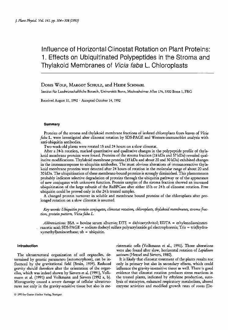
Fig. 1: Coomassie stained electrophoretic pattern of thylakoid membrane proteins from vertically and horizontally rotated Vicia /aha leaves. Lane 1: Molecular weight markers. Lane 2: Thylakoid membrane proteins of 15-h vertically rotated plants as the control for the 15-h horizontally (lane 3) rotated plants. Lane 4: The polypeptide pattern of thylakoid membrane proteins of 24-h vertically compared with the polypeptide pattern of 24-h horizontally (lane 5) rotated plants separated on 15 % SDS-PAGE; 20l1g of each sample were applied on each lane. There are obvious changes in the polypeptide pattern in the molecular weight range of about 20 and 30kDa.
2 3 4 5
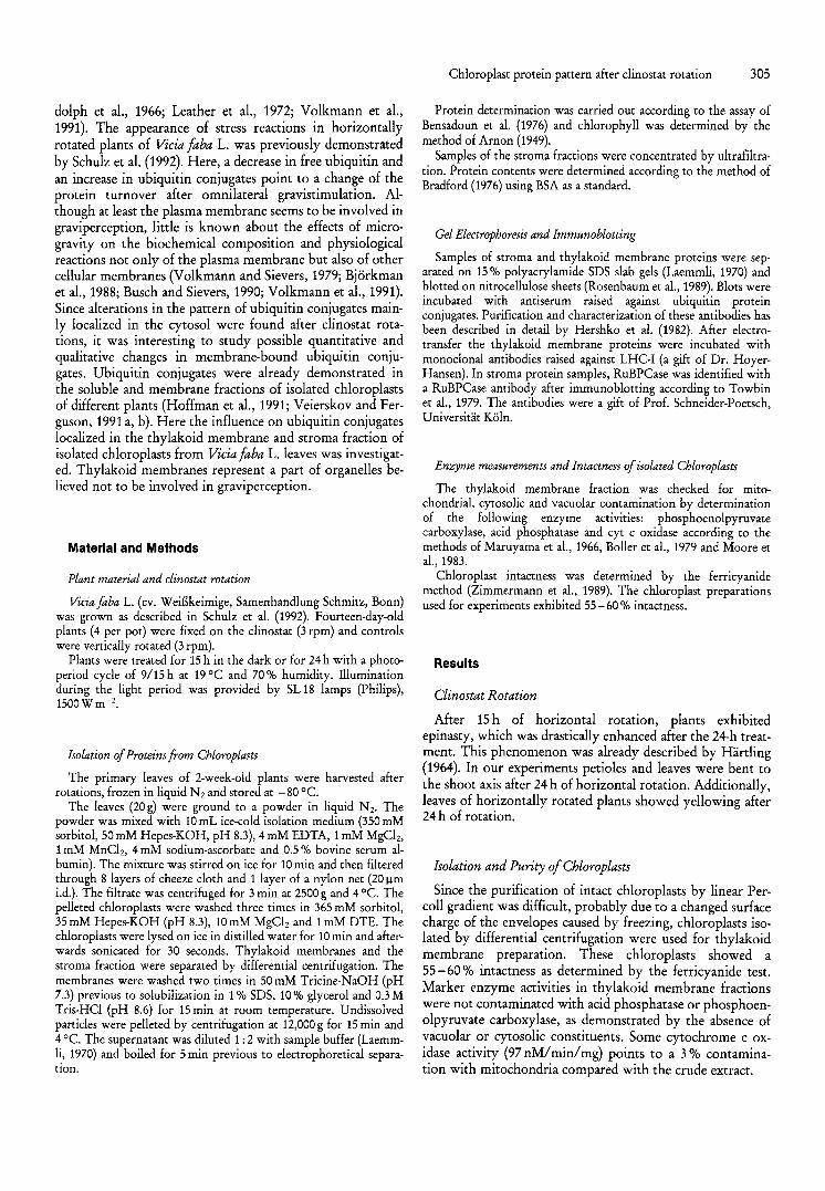
Fig. 2: Immunoblot with polyclonal antibodies raised against ubiquitin. Lane 1: Molecular weight standards. The ubiquitination of thylakoid membrane proteins appear mainly in the molecular weight range of about 30 kDa after 15 h of vertical rotation (lane 2), which slightly decrease during clinostat rotation for 15 h (lane 3) and 24 h of either vertical (lane 4) or 24 h of clinostat rotation (lane 5) see arrow above. The increase in ubiquitinated proteins of about 20 kDa appears after 15 h clinostat (lane 3) and 24 h of vertical rotation (lane 4) and decreased after 24 h of clinostat rotation (lane 5), see arrow.
The polypeptide pattern of tbylakoid membrane proteins after SDS gel electrophoresis
The Coomassie stained gel (Fig. 1) illustrates the protein profile of thylakoid membranes of controls and horizontally rotated plants.
The 15-h clinostat sample showed an increase in polypeptides with molecular weights of 15, 18 and 19 kDa compared with the corresponding control. Samples after prolonged clinostat rotation (24 h), however, exhibited a drastic decrease, mainly in proteins of lower molecular weight (15, 18 and 19kDa) in comparison to the 24h control. The latter control showed an enhancement of the 15, 18 and 19 kDa proteins, similar to the 15-h clinostat sample.
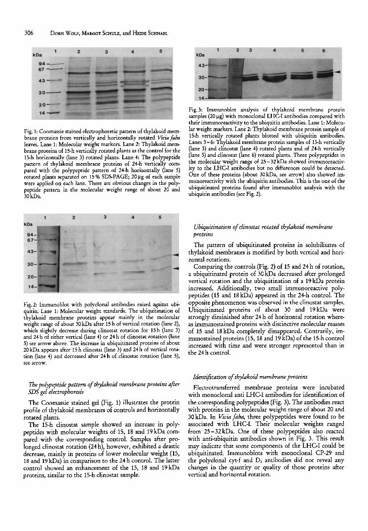
Fig. 3: Immunoblot analysis of thylakoid membrane protein samples (20 11 g) with monoclonal LHC-! antibodies compared with their immunoreactivity to the ubiquitin antibodies. Lane 1: Molecular weight markers. Lane 2: Thylakoid membrane protein sample of 15-h vertically rotated plants blotted with ubiquitin antibodies. Lanes 3-6: Thylakoid membrane protein samples of 15-h vertically (lane 3) and clinostat (lane 4) rotated plants and of 24-h vertically (lane 5) and c1inostat (lane 6) rotated plants. Three polypeptides in the molecular weight range of 25-32kDa showed immunoreactivity to the LHC-! antibodies but no differences could be detected. One of these proteins (about 30kDa, see arrow) also showed immunoreactivity with the ubiquitin antibodies. This is the one of the ubiquitinated proteins found after immunoblot analysis with the ubiquitin antibodies (see Fig. 2).
Ubiquitination of clinostat rotated tbylakoid membrane proteins
The pattern of ubiquitinated proteins in solubilisates of thylakoid membranes is modified by both vertical and horizontal rotations.
Comparing the controls (Fig. 2) of 15 and 24 h of rotation, a ubiquitinated protein of 30 kDa decreased after prolonged vertical rotation and the ubiquitination of a 19 kDa protein increased. Additionally, two small immunoreactive polypeptides (15 and 18 kDa) appeared in the 24-h control. The opposite phenomenon was observed in the clinostat samples. Ubiquitinated proteins of about 30 and 19 kDa were strongly diminished after 24 h of horizontal rotation whereas immunostained proteins with distinctive molecular masses of 15 and 18 kDa completely dissappeared. Contrarily, immunostained proteins (IS, 18 and 19 kDa) of the 15-h control increased with time and were stronger represented than in the 24 h control.
Identification of tbylakoid membrane proteins
Electrotransferred membrane proteins were incubated with monoclonal anti LHC-I antibodies for identification of the corresponding polypeptides (Fig. 3). The antibodies react with proteins in the molecular weight range of about 20 and 30 kDa. In Vicia faha, three polypeptides were found to be associated with LHC-1. Their molecular weights ranged from 25 - 32 kDa. One of these polypeptides also reacted with anti-ubiquitin antibodies shown in Fig. 3. This result may indicate that some components of the LHC-I could be ubiquitinated. Immunoblots with monoclonal CP-29 and the polyclonal cyt-f and DJ antibodies did not reveal any changes in the quantity or quality of those proteins after vertical and horizontal rotation.
2 3 4 5
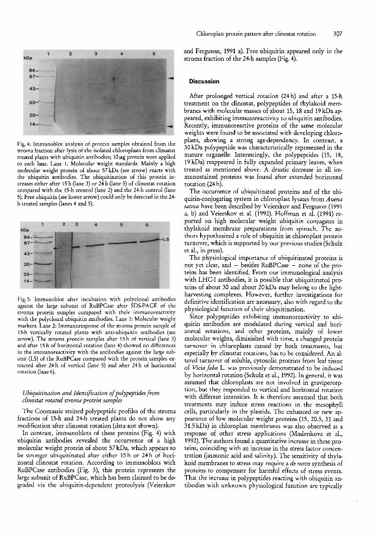
Fig.4: Immunoblot analysis of protein samples obtained from the stroma fraction after lysis of the isolated chloroplasts from clinostat treated plants with ubiquitin antibodies; 10 Ilg protein were applied to each lane. Lane 1: Molecular weight standards. Mainly a high molecular weight protein of about 57 kDa (see arrow) reacts with the ubiquitin antibodies. The ubiquitination of this protein increases either after 15 h (lane 3) or 24 h (lane 5) of clinostat rotation compared with the 15-h control (lane 2) and the 24-h control (lane 5). Free ubiquitin (see lower arrow) could only be detected in the 24-h treated samples (lanes 4 and 5).
2 3 4 5 6
Fig. 5: Immunoblot after incubation with polyclonal antibodies against the large subunit of RuBPCase after SDS-PAGE of the stroma protein samples compared with their immunoreactivity with the polyclonal ubiquitin antibodies. Lane 1: Molecular weight markers. Lane 2: Immunoresponse of the stroma protein sample of 15-h vertically rotated plants with anti-ubiquitin antibodies (see arrow). The stroma protein samples after 15 h of vertical (lane 3) and after 15 h of horizontal rotation (lane 4) showed no differences in the immunoreactivity with the antibodies against the large subunit (LS) of the RuBPCase compared with the protein samples ex· tracted after 24 h of vertical (lane 5) and after 24 h of horizontal rotation (lane 6).
Ubiquitination and Identification of polypeptides from clinostat rotated stroma protein samples
The Coomassie stained polypeptide profiles of the stroma fractions of 15-h and 24-h treated plants do not show any modification after clinostat rotation (data not shown).
In contrast, immunoblots of these proteins (Fig. 4) with ubiquitin antibodies revealed the occurrence of a high molecular weight protein of about 57 kDa, which appears to be stronger ubiquitinated after either 15 h or 24 h of horizontal clinostat rotation. According to immunoblots with RuBPCase antibodies (Fig. 5), this protein represents the large subunit of RuBPCase, which has been claimed to be degraded via the ubiquitin-dependent proteolysis (Veierskov
Chloroplast protein pattern after clinostat rotation 307
and Ferguson, 1991 a). Free ubiquitin appeared only in the stroma fraction of the 24-h samples (Fig. 4).
Discussion
After prolonged vertical rotation (24 h) and after a 15-h treatment on the clinostat, polypeptides of thylakoid membranes with molecular masses of about 15, 18 and 19 kDa appeared, exhibiting immunoreactivity to ubiquitin antibodies. Recently, immunoreactive proteins of the same molecular weights were found to be associated with developing chloroplasts, showing a strong age-dependency. In contrast, a 30 kDa polypeptide was characteristically represented in the mature organelle. Interestingly, the polypeptides (15, 18, 19 kDa) reappeared in fully expanded primary leaves, when treated as mentioned above. A drastic decrease in all immunostained proteins was found after extended horizontal rotation (24 h).
The occurrence of ubiquitinated proteins and of the ubiquitin-conjugating system in chloroplast lysates from Avena sativa have been described by Veierskov and Ferguson (1991 a, b) and Veierskov et al. (1992). Hoffman et a!. (1991) reported on high molecular weight ubiquitin conjugates in thylakoid membrane preparations from spinach. The authors hypothesized a role of ubiquitin in chloroplast protein turnover, which is supported by our previous studies (Schulz et a!., in press).
The physiological importance of ubiquitinated proteins is not yet clear, and - besides RuBPCase - none of the proteins has been identified. From our immunological analysis with LHC-I antibodies, it is possible that ubiquitinated proteins of about 30 and about 20 kDa may belong to the lightharvesting complexes. However, further investigations for definitive identification are necessary, also with regard to the physiological function of their ubiquitination.
Since polypeptides exhibiting immunoreactivity to ubiquitin antibodies are modulated during vertical and horizontal rotations, and other proteins, mainly of lower molecular weights, diminished with time, a changed protein turnover in chloroplasts caused by both treatments, but especially by clinostat rotations, has to be considered. An altered turnover of soluble, cytosolic proteins from leaf tissue of Vicia faba L was previously demonstrated to be induced by horizontal rotation (Schulz et a!., 1992). In general, it was assumed that chloroplasts are not involved in graviperception, but they responded to vertical and horizontal rotation with different intensities. It is therefore assumed that both treatments may induce stress reactions in the mesophyll cells, particularly in the plastids. The enhanced or new appearance of low molecular weight proteins (15, 20.5, 31 and 31.5 kDa) in chloroplast membranes was also observed as a response of other stress applications (Maslenkova et a!., 1992). The authors found a quantitative increase in these proteins, coinciding with an increase in the stress factor concentration Uasmonic acid and salinity). The sensitivity of thylakoid membranes to stress may require a de novo synthesis of proteins to compensate for harmful effects of stress events. That the increase in polypeptides reacting with ubiquitin antibodies with unknown physiological function are typically
308 DORIS WOLF, MARGOT SCHULZ, and HEIDE SCHNABL
found only in developing chloroplasts supports this assumption. Because of the strong age-related appearance, it is unlikely that these ubiquitinated proteins are only marked for degradation, since under normal conditions they are no longer present in 2-week-old thylakoid membranes (Schulz et al., in press). Prolonged stress application may result in a breakdown of compensating mechanisms, which could be an explanation for decreasing quantities of proteins in thylakoid membranes after the 24-h clinostat treatment.
On the other hand, the increased ubiquitination of the RuBPCase after clinostat rotation could arise from an enhanced ubiquitin-dependent proteolysis. The ubiquitindependent breakdown of RuPBCase is possible as shown by the studies of Veierskov and Ferguson (1991 b) using isolated oat chloroplasts. The observation of a stronger ubiquitination of the RuBPCase after horizontal clinostat rotation indicates that the ubiquitin-conjugating system is more active in clinostat treated chloroplasts compared with their controls. The appearance of free ubiquitin in 24-h treated plants revealed changes in the ubiquitin pool of chloroplasts.
Acknowledgements
Financial support (AGRA VIS) by the Bundesminister fur Forschung und Technologie (Bonn) and the Ministerium fUr Wissenschaft und Forschung (Dusseldorf) to H. S. is gratefully acknowledged. We thank Dr. Hoyer-Hansen (Department of Physioloy, Denmark) for the gift of the LHC-I and CP-29 antibodies, Prof. Schneider-Poetsch (Universitat Koln) for the anti-RuBPCase antibody, Mr. U. Johanningmeier (Universitat Bochum) for the Dl antibody and Mr. H. Thomas (Welsh Plant Breeding Station, Scotland) for the cyt-f antibody.
References
ARNON, D. 1.: Copper enzymes in plants: Polyphenoloxidases in Beta vulgaris. Plant Physiol. 24, 1-15 (1949).
BENSADOUN, A. and D. WEINSTEIN: Assay of proteins in the presence of interfering materials. Anal. Biochem. 70,241-250 (1976).
BJORKMAN, T.: Perception of gravity by plants. Adv. Bot. Res. 15, 1-41 (1988).
BOLLER, T. and H. KENDE: Hydrolytic enzymes in the central vacuole of plant cells. Plant Physiol. 63, 1123-1132 (1979).
BRADFORD, M. M.: A rapid and sensitive method for the quantitation of microgram quantities of protein, utilizing the principles of protein-dye binding. Anal. Biochem. 72, 248-254 (1976).
BRAIN, E. D.: Studies in the effects of prolonged rotation of plants on a horizontal clinostat. II. Anatomical structure. New Phytol. 38,240-256 (1939).
BUSCH, M. B. and A. SIEVERS: Hormone treatment of root causes not only a reversible loss of starch but also of structural polarity in statocytes. Planta 181, 358-364 (1990).
DEDOLPH, R. R., B. R. WILSON, W. CHORNEY, and J. J. BREEN: Simulated low-gravity environments and respiratory metabolism in Avena seedlings. Plant Physiol. 41, 1520-1524 (1966).
HARTLING, c.: Untersuchungen uber den EinfluB der Rotation am Klinostaten auf das tropistische Reaktionsvermogen von He· lianthus-Keimlingen. Planta 63, 43 -64 (1964).
HENSEL, W. and A. S1EVERS: Effects of prolonged omnilateral gravistimulation on the ultrastructure of statocytes and on the graviresponse of roots. Planta 150, 338-346 (1980).
HERSHKO, A., E. EYTAN, A. CIECHANOVER, and A. L. HAAs: Immunological analysis of the turnover of ubiquitin-protein conjugates in intact cells. J. BioI. Chern. 257, 13964-13970 (1982).
HOFFMAN, N. E., K. Ko, D. MILKOWSKI, and E. PICHERSKY: Isolation and characterization of tomato cDNA and genomic clones encoding the ubiquitin gene ubi3. Plant Molec. BioI. 17, 1189-1201 (1991).
LAEMMLI, U. K.: Cleavage of structural proteins during the assembly of the head of bacteriophage T4. Nature 227, 680-685 (1970).
LEATHER, G. R., E. FORRENCE, and F. B. ABELES: Increased ethylene production during clinostat experiments may cause leaf epinasty. Plant Physiol. 49,183-186 (1972).
MARUYAMA, H., R. L. EASTERDAY, H.-C. CHANG, and M. D. LANE: The enzymatic carboxylation of phosphoenolpyruvate. J. BioI. Chern. 241, 2405-2412 (1966).
MASLENKOVA, L. T., T. S. MITEVA, and L. P. POPORA: Changes in the polypeptide patterns of barley seedlings exposed to jasmonic acid and salinity. Plant Physiol. 98, 700-707 (1992).
MOORE, A. L. and M. o. PRONDLoVE: Mitochondria and sub-mitochondrial particles. Isolation of membranes. Academic Press, London, 153 -192 (1983).
ROSENBAUM, L. C., G. NILAVER, H. M. HAGMAN, and E. A. NEUWELT: Detection of low-molecular weight polypeptides on nitrocellulose with monoclonal antibodies. Anal. Biochem. 183, 250-257 (1989).
SCHULZ, M., B. SOLSCHEID, and H. SCHNABL: Changes of the soluble protein pattern and evidence for stress reactions in leaf tissue of Vicia /aba L. after clinostat rotation. J. Plant Physiol. 140, 502-507 (1992).
SCHULZ, M., D. WOLF, and H. SCHNABL: Age dependent appearance in chloroplast membranes of Vicia /aba L. J. Plant Physiol. 141, 298-303 (1993).
SIEVERS, A., B. BUCHEN, D. VOLKMANN, and Z. HEJNOWICZ: Role of the cytoskeleton in gravity perception. In: The cytoskeletal basis of plant growth and form p. 169, LLOYD, C. W., ed. Academic Press, London, (1991).
TOWBIN, H., T. STAEHLIN, and J. GORDON: Electrophoretic transfer of proteins from polyacrylamide gels to nitrocellulose sheets: procedure and some applications. Proc. Nat. Acad. Sci. USA 76, 4350-4354 (1979).
VEIERSKOV, B., I. B. FERGUSON, and M. LAY-YEE: Conjugation of ubiquitin to proteins during greening of etiolated oat plants. J. Plant Physiol. 139,749-754 (1992).
VEIERSKOV, B. and 1. B. FERGUSON: Conjugation of ubiquitin to proteins from green plant tissues. Plant Physiol. 96, 4-9 (1991 a).
- - Ubiquitin conjugating activity in leaves and isolated chloroplasts from Avena sativa L. during senescence. J. Plant Physiol. 138,608-613 (1991 b).
VOLKMANN, D. and A. SIEVERS: Forschung unter reduzierter Schwerkraft Tei! I: Grundlagen der Gravitationsbiologie. Naturwissenschaften 79, 68-74 (1992 a).
- - Forschung unter reduzierter Schwerkraft Teil II: Experimente in variierenden Gravitationsfeldern. Naturwissenschaften 79, 118 -124 (1992 b).
VOLKMANN, D., B. BUCHEN, M. TEWINKEL, Z. HEJNOWICZ, and A. SIEVERS: Oriented movement of statoliths studied in a reduced gravitational field during parabolic flights of rockets. Planta 185, 153-161 (1991).
VOLKMANN, D. and A. SIEVERS: Graviperception in multicellular organs. In: Encyclopedia of Plant Physiology, N.S. vol. 7: Physiology of Movements, pp. 573-600, HAUPT, W. and M. E. FEINLEIB, eds. Springer, Berlin, Heidelberg, New York (1979).
ZIMMERMANN, G. M., L. WElL, P. HERZSPRUNG, and K.-E. QUENTIN: Biotest als Summenparameter zur Bestimmung von Herbiziden im Wasser. Methodische Grundlagen. Z. Wasser-AbwasserForsch. 22, 73-77 (1989).