Embed Size (px)
Citation preview
In Vitro Cell. Dev. Biol. 29A:296-309, April 1993 © 1993 Tissue Culture Association 0883-8364/93 $01.50+0.00
INSECT-TRANSMITTED VERTEBRATE VIRUSES: FLAVIVIRIDAE
GEORGE V. LUDWIG p, No LAUREN C. IACONO-CONNORS
Virology Division, United States Army Medical Research Institute of Infectious Diseases, Fort Detrick, Frederick, Maryland 21702
(Received 13 October 1992; accepted 22 October 1992)
SUMMARY
The Flaviviridae include almost 70 viruses, nearly half of which have been associated with human disease. These viruses are among the most important arthropod-borne viruses worldwide and include dengue, yellow fever, and Japanese encephalitis viruses. Morbidity and mortality caused by these viruses vary, but collectively they account for millions of encephalitis, hemorrhagic fever, arthralgia, rash, and fever cases per year. Most of the members of this family are transmitted between vertebrate hosts by arthropod vectors, most commonly mosquitoes or ticks. Transmission cycles can be simple or complex depending on the hosts, vectors, the virus, and the environmental factors affecting both hosts and viruses. Replication of virus in invertebrate hosts does not seem to result in any significant pathology, which suggests a close evolutionary relationship between virus and vector. Another example of this relationship is the ability of these viruses to grow in invertebrate ceil culture, where replication usually results in a steady state, persistent infection, often without cytopathic effect. Yields of virus from insect cell culture vary but are generally similar to yields in vertebrate cells. Replication kinetics are comparable between insect and vertebrate ceil lines, despite differences in incubation tempera- ture. Both vertebrate and insect cell culture systems continue to play a significant role in flavivirus isolation and the diagnosis of disease caused by these agents. Additionally, these culture systems permit the study of flavivirus attachment, penetration, replication, and release from cells and have been instrumental in the production and characterization of live-attenuated vaccines. Both vertebrate and insect cell culture systems will continue to play a significant role in basic and applied flavivirus research in the future.
Key words: Flaviviridae; ecology; mosquito; tick; review; ceil culture.
INTRODUCTION
The family Flaviviridae consists of at least 68 viruses that possess a similar replication strategy and are morphologically, morphogenet- icaily, and biochemicaily similar. Until the advent of modem tech- niques in molecular biology, which are used to elucidate mecha- nisms of replication, the flaviviruses were organized as a separate serogroup (the group B arboviruses) within the family Togaviridae. In 1986, the International Committee for the Taxonomy of Viruses, acting on data summarizing flavivirus biology by Westaway et al. (114), created a separate family, the Flaviviridae.
Members of the family Flaviviridae, although biologically similar, consist of a wide variety of viruses that vary in their pathogenesis and ecology. Some, such as yellow fever, dengue, and Russian spring-summer encephalitis viruses, are often responsible for se- vere disease and significant mortality. These viruses have a multi- farious ecology involving complicated transmission cycles that often include wildlife reservoirs in combination with mosquito or tick vec- tors. Other flaviviruses, like Modoc, Dakar bat, and Cowbone Ridge, fail to produce any detectable disease in humans or domestic animals, and are not transmitted by arthropod vectors. The degree of divergence of the viruses in the family Flaviviridae suggests a long history, and implies that the study of these viruses will require a variety of in vivo and in vitro systems.
When viewed through an electron microscope, flaviviruses are
spherical particles 37 to 50 nm in diameter (85). The virus particles consist of a ribonucleoprotein core surrounded by a lipid bilayer membrane of host-ceil origin. The virion contains three separate structural proteins: the nucleocapsid or core (C) protein, 13 to 14 kDa; the membrane (M) protein, 7 to 8 kDa; and the envelope (E) protein, 50 to 60 kDa. The positive-sense viral RNA codes for seven additional nonstructural proteins, all of" which play a role in virus replication (111,112).
The E protein is the major surface component of the flaviviral envelope. Existing as spikes on the viral membrane, the E protein contains all of the important antigenic determinants responsible for hemagglutination and neutralization and thus plays an important role in immunologic protection from infection. E protein domains are involved in the binding of virions to ceil receptors and probably play a role in intraendosomal fusion at low pH as part of the entry pathway (82).
Antigenic determinants on the E protein serve as the basis for flavivirus classification using serologic tests. For the purposes of classification, a serogroup is defined as two or more viruses, distinct from each other by quantitative serologic standards (4-fold or greater difference between homologous and heterologous titers) in one or more tests, but related to each other or to other viruses by any serologic method. Viruses closely related within a serogroup but distinct from each other are considered to constitute an antigenic
296
FLAVIVIRIDAE 297
TABLE 1
ASSIGNMENT OF FLAVIVIRUSES INTO ANTIGENIC COMPLEXES AS DEFINED BY CROSS REACTIVITY IN CROSS-NEUTRAL-
IZATION TESTS WITH POLYCLONAL ANTISERA FROM HYPERIMMUNE MOUSE ASCITIC FLUID. ~ PRIMARY
HUMAN ILLNESS (IF ANY) ASSOCIATED WITH EACH VIRUS IS INDICATED
Antigenic Complex Viruses
Tick-borne encepbalitits
Rio Bravo
Japanese encephalitis
Tyuleniy Ntaya
Uganda S Dengue
Modoc
Currently unassigned f
(Russian spring-summer encephalitis? Central European eneephalitisb'C), Omsk hemorrhagic fever, 't louping-ill, b Kyasanur forest disease, d (Langat? Carey Island, Phnom-Penh bat), Negishi? Powassan, b Karshi, Royal Farm
Rio Bravo," Entebbe bat, Dakar Bat, Bukalasa bat, Saboya, Apoi
Japanese encephalitis, b Murray Valley encephalitis? Kokobera, Alfuy, Stratford, St. Louis encephalitis, b Usutu, e West Nile, e Kunjin," Kontango
Tyuleniy, Saumarez Reef, Meaban Ntaya, (Israel turkey meningoencephalitis,
Bagaza), (Yokose, Tembusu) Uganda S, Banzi," Bouboui, Edge Hill Dengue 1, ~'e Dengue 2, '~" Dengue 3, a'e
Dengue 4 d'e Modoc, Sal Vieja, Cowbone Ridge, Jutiapa,
San Perlita Aroa, Bussuquara, ~ Cacipacore, Gadgets
Gulley, Ilbeus," Jugra, Kadam, Montana Myotis leukoencephahtis, Naranjal, Roeio, b Sepik, e Sokuluk, Spondweni," Tamana bat, Wesselsbron," yellow fever, ~ Zika"
a Modified from (11) and (82). b Viruses associated primarily with encephalitis syndrome. ° Parentheses indicate viruses more closely related to each other than to
others of the antigenic complex. d Viruses associated primarily with hemorrhagic fever. e Viruses associated primarily with fever, arthralgia, and/or rash. Y Viruses could not be assigned to an antigenic complex because hyper-
immune mouse ascitic fluids made against these viruses did not cross-react significantly with other flaviviruses.
complex (85). By these criteria, the llaviviruses are organized into eight antigenic complexes (Table 1).
This paper pro 4des a general overview of flavivirology, with an emphasis on the importance of vertebrate and invertebrate cell cul- ture systems for the isolation and biological characterization of these agents. In addition, the importance of cell culture for producing flavivirus diagnostic reagents and developing vaccines for current and future flavivirus health threats is discussed.
ECOLOGY
Distribution of flaviviruses into ecologic groups closely parallels their organization based on their antigenic characteristics (Table 1). The members of the Japanese encephalitis, Ntaya, Uganda S, and dengue antigenic groups are transmitted by mosquitoes, whereas members of the tick-borne encephalitis and Tyuleniy antigenic groups are predominantly transmitted by ticks. Members of the Rio Bravo and Modoc antigenic groups have no known invertebrate vectors and are probably transmitted directly from infected animal
to animal. Blok and colleagues (5) have made a detailed comparison of the nucleotide sequences of a variety of flaviviral polymerase and hehcase-like protein coding regions (NS5 and NS3, respectively). Genetic dendograms prepared from these sequence comparisons suggest that the primary division of the flaviviruses is between those transmitted by mosquitoes and those transmitted by ticks. These observations suggest that the evolution of each of the flavivirus anti- genic groups is closely associated with their respective vector spe- cies.
Although the mosquito-borne flaviviruses are ecologically di- verse, two examples will serve to illustrate the complexity of the systems responsible for maintaining, amplifying, and disseminating these viruses in nature.
Yellow fever virus is the type species for the family Flaviviridae. The virus originated in Africa, and the disease caused by the virus has been known for hundreds of years. The virus causes a diversity of symptoms including fever, chills, severe headache, back pain, anorexia, nausea, and vomiting. In severe cases, jaundice appears during a second episode of fever, often associated with generalized hemorrhagic symptoms. Death occurs in 20 to 50% of severe yel- low fever cases (82). In Africa, as well as in the Americas, the virus is maintained in two basic cycles, the sylvatic and the urban forms.
The sylvatic form of yellow fever virus is maintained in cycles involving arboreal monkeys and forest canopy mosquitoes. In Africa, cercopithecid and colobrid monkeys are effective viremic hosts, circulating virus for several days at titers sufficient to infect vector mosquitoes. These hosts are generally resistant to disease, indicating a balanced host-virus relationship. In contrast, most new- worm monkeys are susceptible to disease, and epizootics in howler monkeys (Alouatta sp.), spider monkeys (Ateles sp.), and owl mon- keys (Aotus sp.) are not unknown. Only the capuchin (Cebus sp.) and wooly monkeys (Lagothrix sp.) display a high degree of resis- tance to disease in the Americas. This unstable host-virus relation- ship is probably a result of the more recent introduction of virus into the Western Hemisphere (82).
Canopy-breeding mosquitoes serve as the primary vectors of yel- low fever virus between susceptible monkey hosts. In Africa, the primary mosquito species involved with sylvatic transmission in- clude Aedes furcifer, A. africanus, and A. luteocephalus. The main vectors in tropical America include members of the genus Haema- gogus. Maintenance of virus during extended periods of dry weather may be facilitated by drought-resistant species, such as Sabethes chloropterus, and through vertical transmission in certain African and tropical American vectors (17,28).
The urban form of yellow fever virus is associated with the intro- duction of virus into human populations. Historically, such out- breaks have occurred in the presence of large populations of the container-breeding mosquito, A. aegypti, combined with the intro- duction of sylvatic virus and a large susceptible human population. Since the 1940s, mosquito control measures have regulated and even extirpated vector populations. A. aegypti eradication efforts in tropical America resulted in the disappearance of urban yellow fever since 1942. Within the last decade however, A. aegypti has reinvaded geographic areas where sylvatic yellow fever occurs, sug- gesting that future urban outbreaks in the Americas is possible. The officially reported human cases of yellow fever in Latin America are associated with exposure to infected Haemagogus mosquitoes and do not represent true urban virus ecology. In Africa, on the other hand, large urban epidemics have occurred regularly in recent de-
298 LUDWIG AND IACONO-CONNORS
/ I Moist Dry Savannas and
Rainforest Savannas Urban Areas Enzoollc Zone Zone of Emergence Epidemic Zone
Aedes Sylvatic Ae.
Monkey n Human Human Monkey Human
africanus Aedes aegypti
J
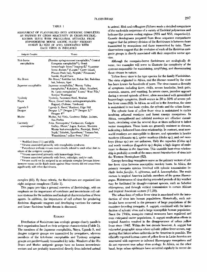
FIc. 1. Transmission cycles of yellow fever virus in Mriea, showing the relationship to vegetational zones. Virus is maintained enzootically in the rainforest zone by A. africanus transmission between susceptible monkeys. Human infections occur, but in an endemic pattern due to low vector populations (see text for exception). In the moist savannahs, a variety of sylvatic vectors can cause intermittent epidemics, with monkeys or humans or both serving as intermediate hosts. In the dry savanna zones, container-breeding Aedes may be responsible for interhuman spread. [Reproduced from ref. (83) with permission.]
cades. Ethiopia experienced a large outbreak in 1960 to 1962 involving an estimated 100 000 cases with approximately 30 000 deaths. A. aegypti seems to be playing a major role in recent urban yellow fever epidemics in Africa, although other species may also play a significant role (101). In eastern Nigeria for instance, A. africanus, the vector responsible for enzootic maintenance, as- sumes the role of epidemic vector in the savannah-rainforest zones, where it is responsible for significant interhuman transmission (83) (Fig. 1). Urban outbreaks in Africa continue to be a major public health concern. An excellent review of yellow fever virus ecology and epidemiology in the Americas and Africa has been published (83).
The tick-borne encephalitis (TBE) virus antigenic group consists of 12 tick-borne viruses that cause a variety of diseases in humans. The severe form of human disease, caused by infection with viruses such as Russian spring-summer encephalitis virus, is characterized by gradual onset of symptoms which include anorexia, fever, head- ache, nausea, vomiting, and photophobia. These symptoms are fol- lowed by variable nettrologie dysfunction which result in death in approximately 20% of cases and mild-to-severe neurologic se- quelae in 30 to 60% of cases (42).
Members of the TBE virus antigenic complex are transmitted between vertebrate hosts by tick vectors. On the European and Asian continents, the main vectors of endemic strains are lxodes
ricinus and L persulcatus. In areas where these tick species do not exist, other tick species of the genera Dermacentor and Haemaphy- sails are probably responsible for the maintenance of TBE viruses (82). Endemnicity of TBE correlates directly with the geographic distribution of the vector species. Tick populations serve as natural reservoirs and provide a mechanism for viral overwintering by verti- cal transmission. Maintenance over extended periods of time is, however, not possible due to inefficient transtadial and transovarial transmission (37).
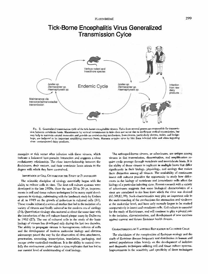
The TBE viruses are maintained, amplified, and disseminated in natural transmission cycles involving the ticks described above and wild vertebrate hosts, particularly small rodents and insectivores (13,82). These cycles are required for endemnicity because of the inefficiency of vertical transmission. Transmission of virus to hu- mans is most common in individuals who work or play in tick-in- fested areas. Domestic animals do not participate in normal trans- mission cycles as a result of low blood titers after infection, but do contribute to the potential for human infection. Lactating sheep, goats, and cattle secrete virus into their milk and may pass virus to humans through contaminated raw dairy products (Fig. 2).
The ecologies of all of the arthropod-borne flaviviruses are equally complex, involving a variety of arthropod vectors, each vary- ing in their ability to transmit virus. No conclusive evidence exists that suggests that any significant pathologic changes occur in the
FLAVIVIRIDAE 299
Tick-Borne Encephalitis Virus Generalized Transmission Cylce
FI¢. 2. Generalized transmission cycle of the tick-borne encephalitis viruses. Ticks from several genera are responsible for transmis- sion between vertebrate hosts. Maintenance by vertical transmission in ticks does not occur due to inefficient vertical transmission, but may help to maintain natural reservoirs and provide an overwintering mechanism, lnsectivores, particularly shrews, moles, and hedge- hogs, are believed to be important amplifying reservoir hosts. Humans acquire virus by bite from infected ticks and when ingesting virus contaminated dairy products.
mosquito or tick vector after infection with these viruses, which indicate a balanced host-parasite interaction and suggests a close evolutionary relationship. The close interrelationship between the ilaviviruses, their vectors, and their vertebrate hosts attests to the degree with which they have co-evolved.
IMPORTANCE OF CELL CULTURE FOR THE STUDY OF FLAVIVIRUSES
The scientific discipline of virology essentially began with the ability to culture cells in vitro. The first cell culture systems were developed in the late 1920s. Over the next 20 to 30 yr, improve- ments in cell and tissue culture techniques led to many rapid devel- opments in virology, culminating with the landmark study by Enders et al. in 1949 on the growth of poliovirus in cultured cells (31). These results initiated a series of studies that led to the isolation of a variety of viruses and finally ushered in the modern eta of virology (34). Quantitative virology also matured at about the same time with the introduction of the cell culture-based plaque assay by Dulhecco in 1952 (27). The use of cultured cells in the study of the basic biology of viruses has developed only during the last two decades. The ability to propagate viruses in homogeneous cultures of cells and the development of modern molecular biology and electron microscopy paved the way for the observation of virus attachment, penetration, uneoating, transcription, translation, packaging, and escape under controlled conditions. It is the ability to control care- fully the environment under which a virus replicates that has led to our current level of understanding of viral biology.
The arthropod-borne viruses, or arboviruses, are unique among viruses in that transmission, dissemination, and amplification re- quire cyclic passage through vertebrate and invertebrate hosts. It is the ability of these viruses to replicate in multiple hosts that differ significantly in their biology, physiology, and ecology that makes them distinctive among all viruses. The availability of continuous insect cell cultures provides the opportunity to study how differ- ences in the biology of vertebrate and invertebrate cells affect the biology of a particular infecting virus. Recent research with a variety of arboviruses suggests that some biological characteristics of a virus are correlated to the host from which the virus was derived (41,59,81,99). Such characteristics may play an important role in the understanding of the mechanisms for attenuation and virulence at the molecular level, and have only recently begun to be studied comparatively in insect and vertebrate cells. Cell culture is essential for the study of flaviviruses, and will continue to play a pivotal role in the isolation, characterization, and development of new vaccines against current and future flavivirus health threats.
CHARACTERISTICS OF FLAVIVIRUS REPLICATION IN CULTURED CELLS
The elucidation of the complexities of flavivirus ecology and the study of flavivirus disease frequency and distribution in human and animal populations relies heavily on the development of isolation and diagnostic techniques utilizing cell mad tissue culture systems. Improvements in the sensitivity and specificity of these techniques
300 LUDWIG AND IACONO-CONNORS
4
• 3
Q.
o ._1 v
$
i - - on 2 2
v~,,, v
I
V e
Incubation Temperature v /
\. , • - 2 8 ° C V~ / • v - 2 0 ° C V
1 , , , ; , i ] , , , , , , ,
0 2 4 6 8 1 0 1 6 2 0 2 4 2 8 3 2 3 6 4 0
Day PI
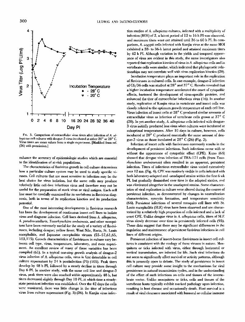
Fro. 3. Comparison of extracellular virus titem after infection of A. al- bopictus cell cultures with dengue-3 virus incubated at either 20 ° or 28 ° C. Virus titers are mean values from a single experiment. [Modified from ref. (26) with permission.]
enhance the accuracy of epidemiologic studies which are essential to the identification of at-risk populations.
The characteristics of flavivirus growth in cell culture determines how a particular culture system may be used to study specific vi- ruses. Cell cultures that are most sensitive to infection may be the best choice for virus isolation, but the same cells may produce relatively little cell-free infectious virus and therefore may not be useful for the preparation of stock virus or viral antigen. Each cell line must be carefully examined for its usefulness in flavivirus diag- nosis, both in terms of its replication kinetics and its production potential.
One of the most interesting developments in flavivirus research has been the development of continuous insect cell lines to isolate virus and diagnose infection. Cell lines derived from A. albopictus, A. pseudoscutellaris, Toxorhynchites amboinesis, and other mosqui- toes have been extremely useful for the study of a variety of flavivi- ruses, including dengue; yellow fever; West Nile, Rocio, St. Louis encephalitis, and Japanese encephalitis viruses (55-57,61,65, 103,115). Growth characteristics of flaviviruses in culture vary be- tween cell type, virus, temperature, laboratory, and even experi- ment. An excellent review of many of these variables has been compiled (61). In a typical one-step growth analysis of dengue-2 virus infection of A. albopictus cells, virus is first detectable in cell culture superuatant by 24 h postinfection (PI) (105). Peak titers develop by 48 h PI, followed by a steady decline in titers through Day 6 PI. In another study, with the same cell line and dengne-3 virus, peak titers were also reached within approximately 48 h, but titers decreased slightly through Day 10 PI, at which time a steady- state persistent infection was established. Over the 42 days the cells were monitored, there was little change in the titer of infectious virus from culture superuatant (Fig. 3) (26). In Kunjin virus infec-
tion studies of A. albopictus cultures, infected with a multiplicity of infection (MOI) of 2, a latent period of 12 to 16 h PI was observed, and maximum titers were not attained until 36 to 60 h PI. In com- parison, A. aegypti cells infected with Kunjin virus at the same MOI exhibited a 28- to 36-h latent period and attained maximum tilers by 42 h PI. Although variation in the yields and temporal appear- ance of virus are evident in this study, the same investigators also reported that replication kinetics of virus in A. albopictus cells and in vertebrate cells were similar, which indicated that phylogenetic rela- tionships may not correlate well with virus replication kinetics (20).
Incubation temperature plays an important role in the replication of flaviviruses in cultured cells. In one example, dengue-2 infection of C6/36 cells was studied at 28 ° and 37 ° C. Results revealed that a higher incubation temperature accelerated the onset of cytopathic effects, hastened the development of virus-specific proteins, and enhanced the titer of extracellular infectious virus (16). In another study, replication of Kunjin virus in vertebrate and insect cells was closely related to the optimum growth temperature of each cell line. Virus infection of insect cells at 28 ° C produced similar amounts of extracellular virus as infection of vertebrate cells grown at 37 ° C (20). In yet another study, A. albopictus cells infected with dengue- 3 virus initially produced less virus when cultures were incubated at suboptimal temperatures. After 10 days in culture, however, cells incubated at 20 ° C produced essentially the same amount of den- gue-3 virus as those incubated at 28 ° C (26) (Fig. 3).
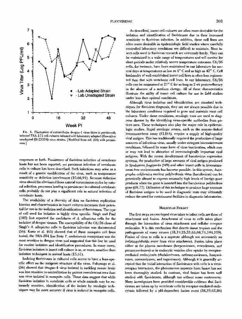
Infection of insect cells with llaviviruses commonly results in the development of persistent infections. Such infections occur with or without the appearance of cytopathic effect (CPE). Kuno (64) showed that dengue virus infection of TRA-171 cells (from Toxo- rhynchites amboinensis) often resulted in an apparent, persistent infection. Titers of infectious extracellular virus varied extensively over 12 mo. (Fig. 4). CPE was routinely visible in cells infected with both laboratory-adapted and -unadapted strains within the first 6 wk PI, but gradually diminished over time in the adapted strains, and was eliminated altogether in the unadapted strains. Some character- istics of viral replication in culture were altered during the course of persistent infection, as demonstrated by changes in neutralization characteristics, syncytia formation, and temperature sensitivity (64). Persistent infections of several mosquito cell lines with St. Louis encephalitis (SLE) virus have been observed and are charac- terized by a relatively high proportion of cells infected and a lack of overt CPE. Unlike dengue virus in A. albopictus cells, titers of SLE virus slowly decrease over time in persistently infected cells (93). These data suggest that there may be significant differences in the regulation and maintenance of persistent flavivirus infections in cell lines of different origins.
Persistent infection of insect-borne flaviviruses in insect cell cul- tures is consistent with the ecology of these viruses in nature. Mos- quitoes or ticks infected with virus, either through horizontal or vertical transmission, are infected for life. Such viral infections do not seem to significantly affect survival or activity patterns, although this is presently open to debate. The study of persistence in insect cell culture may provide some insight to the mechanisms for viral persistence in natural transmission cycles, and in the understanding of the effect of such infections on cells and tissues of the inverte- brate vector. Unlike mosquitoes or ticks, cells and tissues of the vertebrate hosts typically exhibit marked pathology upon infection, resulting in host disease and occasionally death. Host survival is a result of viral clearance associated with humoral or cellular immune
FLAVIVIRIDAE 301
It. v , ~ v - oN N , /
2 - Lab Adapted Strain v - Lab Unadapted Strain
2
1 I I I
0 16 32 48
W e e k PI
Fit;. 4. Fluctuation of cxtraccllular dcngue-I virus fiters in persistently infected TRA- 171 cell cultures infected with laboratory adapted (Hawaii) or unadapted (H-23334) virus strains. [Modified from ref. (64) with permis- sion.]
responses or both. Persistence of flavivirus infection of vertebrate hosts has not been reported, yet persistent infection of vertebrate cells in culture has been described. Such infections may arise as a result of a genetic modification of the virus, such as temperature sensitivity or defective interference (10,66,91). Because defective virus should be eliminated from natural transmission cycles by natu- ral selection, processes leading to persistence in cultured vertebrate cells probably do not play a significant role in natural infection of vertebrate hosts.
The availability of a diversity of data on flavivirus replication kinetics and characteristics in insect cultures increases their poten- tial for use in the isolation and ilentifieation of flaviviruses. The type of cell used for isolation is highly virus specific. Singh and Paul (102) first reported the usefulness of A. albopictus cells for the isolation of dengue viruses. The susceptibility of the C6/36 clone of Singh's A. albopictus cells to flavivirus infection was documented (56). Kuno et al. (65) showed that of three mosquito cell lines tested, the TRA-284 line from T, amboinensis mosquitoes was the most sensitive to dengue virus and suggested that this line be used for routine isolation and identification procedures. In many cases, flavivirus isolation in insect cell culture is as, or more, sensitive than isolation techniques in animal hosts (15,61).
Isolating flaviviruses in cultured cells seems to have a host-spe- cific effect on the antigenic structure of the virus. Fukunaga et al. (36) showed that dengne-4 virus isolated in suckling mouse brain was less sensitive to neutralization by patient convalescent sera than was virus isolated in mosquito cells. These data suggest that while flavivirus isolation in vertebrate cells or whole animals may be ex- tremely sensitive, identification of the isolate by serologic tech- niques may be more accurate if virus is isolated in mosquito cells.
As described, insect cell cultures are often more desirable for the isolation and identification of flaviviruses due to their -increased sensitivity to flavivirus infection. In addition, these cell lines are often more desirable in epidemiologic field studies where carefully controlled laboratory conditions are diflicuh to maintain. Most in- sect cells used in flavivirus research are extremely hardy. They can be maintained in a wide range of temperatures and will survive for short periods under relatively severe temperature extremes. C6/36 cells, for instance, have been maintained in our laboratory for sev- eral days at temperatures as low as 4 ° C and as high as 42 ° C. Cell husbandry of well-established insect cell lines is often less regimen- ted than that with vertebrate cell lines. In our laboratory, C6/36 cells can be maintained at 27 ° C for as long as 2 wk postconfluency in the absence of a medium change. All of these characteristics illustrate the utility of insect cell culture for use in field studies under less than optimal conditions.
Although virus isolation and identification are standard tech- niques for flavivirus diagnosis, they are not always possible due to the laboratory conditions required to grow and maintain most cell cultures. Under these conditions, serologic tests are used to diag- nose disease by the identifying virus-specific antibodies from pa- tient sera. These techniques also play the major role in epidemio- logic studies. Rapid serologic assays, such as the enzyme-linked immunosorbent assay (ELISA), require a supply of high-quality viral antigen. This has traditionally required the production of large amounts of infectious virus, usually under stringent bioeontainment conditions, followed by some form of virus inactivation, which may or may not lead to alteration of immunologically important viral antigens. With the recent development of baculovirus expression systems, the production of large amounts of viral antigen produced in Spodoptera frugiperda (Sf9) and other insect cells in biocontain- ment-free environments has become possible. In this system, Auto- grapha californica nuclear polyhedrosis virus (baculovirus) can be genetically altered to express unusually high levels of foreign gene products when the gene is inserted into the baculovirus polyhedron gene (69,77). Utilization of this technique to produce large amounts of flavivinas antigen to be used in diagnostic tests may ultimately reduce the need for containment facilities in diagnostic laboratories.
MOLECULAR BIOLOGY
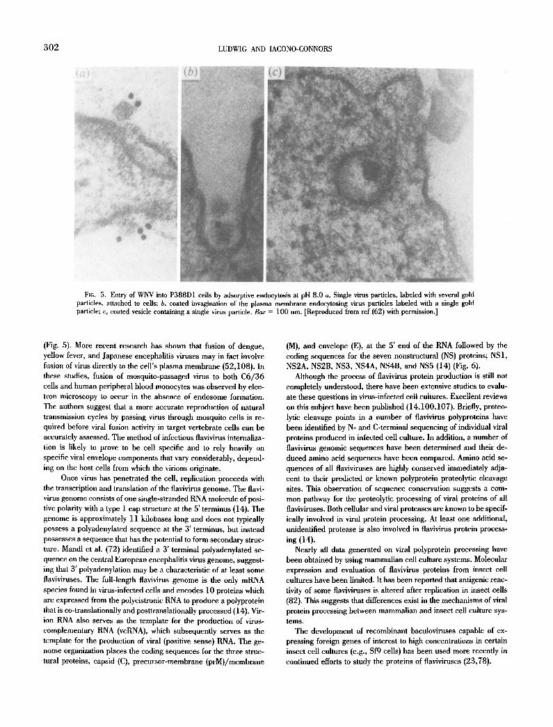
The first steps an enveloped virus takes to infect cells are those of attachment and fusion. Attachment of virus to cells takes place through the interaction of virus attachment and cellular receptor molecules. It is this mechanism that directs tissue tropism and the pathogenesis of many viruses (18,19,25,33,63,68,74,104,109). Fusion of virus to cells is a separate although not necessarily an indistinguishable event from virus attachment. Fusion takes place either at the plasma membrane (herpesviruses, retroviruses, and paramyxoviruses) or in endocytic vesicles after uptake by receptor- mediated endoeytosis (rhabdoviruses, orthomyxoviruses, bunyavi- ruses, coronaviruses, and togaviruses). Although it is generally ac- cepted that the initial interaction of flaviviruses with cells is a virus- receptor interaction, the phenomenon separate from fusion has not been thoroughly studied. In contrast, viral fusion has been well studied with flaviviruses, although not without some controversy. Many investigators have provided considerable evidence that flavi- viruses are taken up by vertebrate cells by receptor-mediated endo- cytosis followed by a pH-dependent fusion event (38,39,62,86)
302 LUDWIG AND IACONO-CONNORS
F1G. 5. Entry of WNV into P388D1 ceils by adsorptive endocytosis at pH 8.0 a, Single virus particles, labeled with several gold particles, attached to cells; b, coated invagination of the plasma membrane endocytosing virus particles labeled with a single gold particle; c, coated vesicle containing a single virus particle. Bar = 100 nm. ]Reproduced from ref (62) with permission.]
(Fig. 5). More recent research has shown that fusion of dengue, yellow fever, and Japanese encephalitis viruses may in fact involve fusion of virus directly to the cell's plasma membrane (52,108). In these studies, fusion of mosquito-passaged virus to both C6/36 cells and human peripheral blood monocytes was observed by elec- tron microscopy to occur in the absence of endosome formation. The authors suggest that a more accurate reproduction of natural transmission cycles by passing virus through mosquito cells is re- quired before viral fusion activity in target vertebrate cells can be accurately assessed. The method of infectious flavivirus internaliza- tion is likely to prove to be cell specific and to rely heavily on specific viral envelope components that vary considerably, depend- ing on the host cells from which the virions originate.
Once virus has penetrated the cell, replication proceeds with the transcription and translation of the flavivirus genome. The flavi- virus genome consists of one single-stranded RNA molecule of posi- tive polarity with a type 1 cap structure at the 5' terminus (14). The genome is approximately 11 kilobases long and does not typically possess a polyadenylated sequence at the 3' terminus, but instead possesses a sequence that has the potential to form secondary struc- ture. Mandl et al. (72) identified a 3' terminal polyadenylated se- quence on the central European encephalitis virus genome, suggest- ing that 3' polyadenylation may be a characteristic of at least some flaviviruses. The full-length tlavivirus genome is the only mRNA species found in virus-infected cells and encodes 10 proteins which are expressed from the polycistronic RNA to produce a polyprotein that is co-translationatly and posttranslationally processed (14). Vir- ion RNA also serves as the template for the production of virus- complementary RNA (vcRNA), which subsequently serves as the template for the production of viral (positive sense) RNA. The ge- nome organization places the coding sequences for the three struc- tural proteins, capsid (C), precursor-membrane (prM)/membrane
(M), and envelope (E), at the 5' end of the RNA followed by the coding sequences for the seven nonstructural (NS) proteins; NS1, NS2A, NS2B, NS3, NS4A, NS4B, and NS5 (14) (Fig. 6).
Although the process of flavivirus protein production is still not completely understood, there have been extensive studies to evalu- ate these questions in virus-infected cell cultures. Excellent reviews on this subject have been published (14,100,107). Briefly, proteo- lyric cleavage points in a number of flavivirus polyproteins have been identified by N- and C-terminal sequencing of individual viral proteins produced in infected cell culture. In addition, a number of flavivirus genomic sequences have been determined and their de- duced amino acid sequences have been compared. Amino acid se- quences of all flaviviruses are highly conserved immediately adja- cent to their predicted or known polyprotein proteolytie cleavage sites. This observation of sequence conservation suggests a com- mon pathway for the proteolytic processing of viral proteins of all flaviviruses. Both cellular and viral proteases are known to be specif- ically involved in viral protein processing. At least one additional, unidentified protease is also involved in flavivirus protein process- ing (14).
Nearly all data generated on viral polyprotein processing have been obtained by using mammalian cell culture systems. Molecular expression and evaluation of flavivirus proteins from insect cell cultures have been limited. It has been reported that antigenic reac- tivity of some flaviviruses is altered after replication in insect cells (82). This suggests that differences exist in the mechanisms of viral protein processing between mammalian and insect cell culture sys- tems.
The development of recombinant baculoviruses capable of ex- pressing foreign genes of interest to high concentrations in certain insect cell cultures (e.g., Sf9 cells) has been used more recently in continued efforts to study the proteins of flaviviruses (23,78).
FLAVIVIRIDAE 303
P R O C E S S I N G O F T H E F L A V I V I R U S P O L Y P R O T E I N
5' 5'NC Cap -I Structural
-It. . I I I I I E~ i~ ~ ~{~ii ~I~iI I I
anchC E prM
V'r'°n Assembly?.
c .N- ~ Virion
Release?
M
"k~t
ORF a'NC I ~ N0nstructural I - ~
IIIi1[! [ " II I NS4A-4B NS5 NS1-2A NS2B NS3
NS1 NS2A 1
Possible YFV N-Linked Glycosylation Site
1.?
NS4A NS4B
,
Signalase, rapid, lumen
Dibasic, rapid, cytoplasm
Dibasic, delayed?, cytoplasm
Dibasic, delayed, lumen
Novel, slightly delayed, lumen
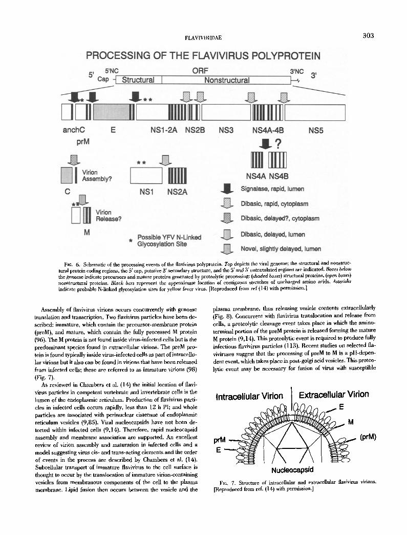
Fie. 6. Schematic of the processing events of the flavivirus polyprotein. Top depicts the viral genome; the structural and nonstruc- tural protein coding regions, the 5' cap, putative 3' secondary structure, and the 5' and 3' untranslated regions are indicated. Boxes below the genome indicate precursors and mature proteins generated by proteolytic processing: (shaded boxes) structural proteins, (open boxes) nonstructural proteins. Black bars represent the approximate location of contiguous stretches of uncharged amino acids, Asterisks indicate probable N-linked glycosylation sites for yellow fever virus. [Reproduced from ref (14) with permission.]
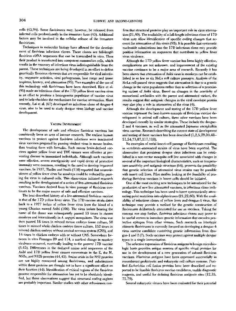
Assembly of flavivirus virions occurs concurrently with genome translation and transcription. Two flavivirus particles have been de- scribed: immature, which contain the precursor-membrane protein (preM), and mature, which contain the fully processed M protein (96). The M protein is not found inside virus-infected cells but is the predominant species found in extracellular virions. The preM pro- tein is found typically inside virus-infected cells as part of intracellu- lar virions but it also can be found in virions that have been released from infected cells; these are referred to as immature virions (98) (Fig. 7).
As reviewed in Chambers et al. (14) the initial location of flavi- virus particles in competent vertebrate and invertebrate cells is the lumen of the endoplasmic reticulum. Production of flavivirus parti- cles in infected cells occurs rapidly, less than 12 h PI; and whole particles are associated with perinuclear cisternae of endoplasmic reticulum vesicles (9,85). Viral nucleocapsids have not been de- tected within infected cells (9,14). Therefore, rapid nucleocapsid assembly and membrane association are supported. An excellent review of virion assembly and maturation in infected cells and a model suggesting virus cis- and trans-aeting elements and the order of events in the process are described by Chambers et al, (14). Subcellular transport o f immature flavivirus to the cell surface is thought to occur by the translocation of immature virion-containing vesicles from membranous components of the cell to the plasma membrane. Lipid fusion then occurs between the vesicle and the
plasma membrane, thus releasing vesicle contents extracellularly (Fig. 8). Concurrent with tlavivirus translocation and release from cells, a proteolytic cleavage event takes place in which the amino- terminal portion of the preM protein is released forming the mature M protein (9,14). This proteolytic event is required to produce fully infectious flavivirus particles (113). Recent studies on selected fla- viviruses suggest that the processing of preM to M is a pH-depen- dent event, which takes place in post-golgi acid vesicles. This proteo- lytic event may be necessary for fusion of virus with susceptible
I Extracellular Virion Intracellular Virion I
Nucleocapsid FIG, 7. Structure of intracellular and extracellular flavivirus virions,
[Reproduced from ref. (14) with permission.]
304 LUDWIG AND IACONO-CONNORS
cells (43,94). Some flaviviruses may, however, be released from infected cells predominantly in the immature form (43). Additional factors may be involved in the cellular release of the immature flavivirions.
Techniques in molecular biology have allowed for the develop- ment of flavivirus infectious clones. These clones are full-length flavivirus cDNA sequences that can be transcribed in vitro. Then their product is transfected into competent mammalian cells, which results in the recovery of infectious virus indistinguishable from the parent. These techniques are being developed in an effort to define genetically flavivirus elements that are responsible for viral infectiv- ity, enzymatic activities, viral pathogenesis, host range and tissue tropisms, latency, and attenuation (95). Two examples of the use of this technology with flaviviruses have been described. Rice et al. (95) made an infectious clone of the 17D yellow fever vaccine virus in an effort to produce a homogenous population of vaccine virus and to help elucidate the mechanisms for vaccine attenuation. More recently, Lai et al. (67) developed an infectious clone of dengue-4 virus, also to be used to study dengue virus biology and vaccine development.
VACCINE DEVELOPMENT
The development of safe and effective flavivirus vaccines has consistently been an area of intense research. The earliest human vaccines to protect against flavivirus infections were inactivated virus vaccines prepared by passing virulent virus in mouse brains, then treating them with formalin. Such mouse brain-derived vac- cines against yellow fever and TBE viruses were effective at pre- venting disease in immunized individuals. Although such vaccines were effective, severe reactigenicity and rapid decay of protective immunity were common, resulting in the need to develop improved vaccines. In 1937, Theiler and Smith (110) reported that neurovir- ulence of yellow fever virus for monkeys could be reduced by pass- ing the virus in cultured cells. This observation initiated research resulting in the development of a series of live-attenuated flavivirus vaccines. Vaccines derived from in vitro passage of flavivirus con- tinues to be the major source of safe and effective vaccines.
The best-described history of a live-attenuated flavivirus vaccine is that of the 17D yellow fever virus. The 17D vaccine strain dates back to a 1927 isolate of yellow fever virus from the blood of a young Ghanian named Asibi (106). The virus isolate bearing the name of the donor was subsequently passed 53 times in rhesus monkeys and intermittently in A. ae~ypti mosquitoes. The virus was then passed 18 times in minced mouse embryo tissue culture, 50 times in minced whole chicken embryo tissue culture, 152 times in minced chicken embryo without central nervous system (CNS), and 14 times in chicken embryo with or without CNS. Somewhere be- tween in vitro Passages 89 and 114, a marked change in monkey virulence occurred, eventually leading to the present 17D vaccine (3,45). Differences in the deduced amino acid sequences of the Asibi and 17D yellow fever viruses concentrate in the E, the M, NS2a, and NS2b proteins (44,45). Amino acids in the NS2 proteins are not highly conserved among flaviviruses, and substitutions within these proteins are thought not to have a significant effect on their function (44). Identification of critical regions of the flavivirus genome responsible for attenuation has yet to be absolutely identi- fied, but these observations suggest that structural coding regions are probably important. Similar studies with other arboviruses con-
firm that structural proteins play an important role in virus attenua- tion (21,40). The availability of a full-length infectious clone of 17D virus may allow identification of specific coding changes that ac- count for attenuation of this strain (95). It is possible that systematic nucleotide substitutions into the 17D infectious clone may provide positive information on sequences that contribute to yellow fever virus virulence.
Although the 17D yellow fever vaccine has been highly effective, complications are not unknown, and improvement of the existing vaccine continues to be a major area of research. Recently it has been shown that attenuation of Asibi virus in monkeys can be estab- lished in as few as six HeLa cell culture passages. Analysis of the HeLa cell-passed virus suggests that attenuation is due to a genetic change in the virus population rather than to selection of a preexist- ing variant of Asibi virus. Based on changes in the reactivity of monoclonal antibodies with the parent and attenuated variant, the results suggest that antigenic changes in the viral envelope protein may also play a role in attenuation of the virus (4).
Although the development and testing of the 17D yellow fever vaccine represent the best-known example of flavivirus vaccine de- velopment in animal cell culture, three other vaccines have been developed recently by similar strategies. These include the dengue- 2 and 4 vaccines, as well as the attenuated Japanese encephalitis virus vaccine. Research describing the current state of development and testing of these vaccines has been described (1,2,5,29,30,48- 51,54,73,87,117,118).
No examples of serial insect cell passage of flaviviruses resulting in vertebrate-attenuated strains of virus have been reported. The observation that persistent dengue virus infections can be estab- lished in a non-vector mosquito cell line associated with changes in several of the important biological characteristics, such as tempera- ture sensitivity and antigenic structure (64), presents the possibility that genetic selection of attenuated virus strains may be possible with insect cell lines. Pilot studies looking at the feasibility of pro- ducing flavivirus vaccines in insect cells should be initiated.
One of the most exciting new techniques to be introduced for the production of new live attenuated vaccines, is infectious clone tech- nology. This technique has been used to insert systematically atten- uating point mutations into alphaviruses (21,22,60). With the avail- ability of infectious clones of yellow fever and dengue-4 virus, this technique may provide a method for the genetic construction of flaviviruses deliberately attenuated for use as vaccines. Taking the concept one step further, flavivirus infectious clones may prove to be useful vectors to introduce genetic information that encodes pro- tective epitopes from other viruses. Such research on intertypic chimeric flaviviruses is currently focused on developing a dengue-4 virus vaccine candidate containing genetic information from den- gue-1 and 2 (7). Such vaccines may protect against multiple dengue types in a single immunization.
The selective expression of llavivirus antigens in benign microbio- logic hosts provides unique sources of specific virual proteins for use in the development of a new generation of subunit flavivirus vaccines. Flavivirus antigens have been expressed successfully in recombinant prokaryotic and eukaryotic cell culture systems. Puri- fied Escherichia coil fusion proteins have been described and re- ported to be feasible flavivirus vaccine candidates, usable diagnostic reagents, and useful for defining tlavivirus antigenic sites (12,35, 75,79).
Several eukaryotic viruses have been evaluated for their potential
FLAVIVIRIDAE 3 0 5
Fit;. 8. Japanese encephalitis virus particles in the peripheral cytoplasm and on the surface of C6/36 cells, 24 h PI. A, one virion each (large arrowheads) is seen on the cell surface and in the lumen of a vacuole (II). One virion (small arrowhead) is recognizable within a vesicle in the peripheral cytoplasm. The rest of the virus particles are seen within the cisternae in the cytoplasm. Bar = 100 nm. B, virus particles (arrowheads) within vesicles beneath the plasma membrane (PM). Bar = 100 nm. C, a virus particle (arrowhead) on the cell surface. Note the clean separation between the viral envelope and the plasma membrane. Bar = 100 nm. [Reproduced trom ref. (53) with permission.]
use as flavivirus vaccine-delivery systems. Vaccinia virus has been developed as a live, infectious expression vector for foreign genes inserted into nonessential virus genome sequences (70 ,71 ,84 ,88) .
Recombinant vaccinia viruses that express flavivirus proteins have been produced and evaluated by numerous investigators in an effort
to develop alternative flavivirus vaccines (6 ,8 ,24 ,32 ,46 ,47 ,76 ,80 ,
306 LUDWIG AND IACONO-CONNORS
89,90,92,116,119). Evidence of protection against a homologous virus challenge is demonstrated in most cases when animals are immunized with vaccinia recombinants expressing selected struc- tural or nonstructural proteins.
In another system, Jacobs et al. (58) have successfully cloned the TBE virus nonstructural protein gene, NS1, into adenovirus under the control of the powerful cytomegalovirus major immediate-early promotor. Under these conditions NS1 is produced in extremely large quantities in cells that do not normally support adenovirus replication. Mice intmunized with the recombinant adenovirus are protected from lethal TBE virus challenge.
Although the live virus vaccines are usually preferable to inacti- vated or subunit vaccines, alternative approaches to vaccinology still play an important role in disease control strategies. Proteins pre- pared in baculovirus expression systems have been used to prepare antigens suitable for vaccination. Most notable is the preparation of a human immunodeficiency virus 1 (HIV1) subunit vaccine from recombinant baculovirus expressed HIV1 antigens (97), which is currently being tested in humans. Recombinant baculovirus-ex- pressed flavivirus proteins have been evaluated for their use as vaccines and can protect appropriate animal models against homolo- gous virus challenge (23,78). Although this expression system has the potential to express recombinant proteins to extremely high levels in cell culture, the problems involved with protein purification currently limit their use in humans.
Each of the approaches to vaccine production listed above is currently being used to develop new and to improve existing flavi- virus vaccines. Each technique totally relies on an appropriate cell culture system for vaccine development, production, and evalua- tion. Future use of these vaccines in humans will depend on the availability of cell lines certified for the development of human use products. Although certified vertebrate cell hues capable of growing virus for human use vaccines are available, no comparable insect cell lines have been developed.
CONCLUSION
The availability of primary and continuous cell culture systems is essential for the study of flavivirus biology, ecology, and epidemiol- ogy, and play a significant role in the development and testing of flavivirus vaccines. Although the importance of cell culture systems for the study of infectious diseases cannot be overstated, the limita- tions of these systems must also be understood. Flavivirus infection of animal hosts is an extremely complex event. Complex organisms are made up of a large number of tissues, each tissue containing many cell types. Each cell type has a different biology reflected by its different functions. In vitro, research of flavivirus infection is conducted most often in clonal populations of cells derived from a single tissue type. Very often the biological, morphologic, and ge- netic characteristics of the cultured cells bear little resemblance to those of the cells of the original tissue. Like the cultures used in these studies, the biology of flaviviruses in cultured cells may boar little resemblance to the biology of the viruses in functioning tissues and organs. As a result, studies on llavivirus biology conducted in cultured cells must be viewed only as a model representing a single event in the complex process contributing to the pathogenesis of viral disease. Cell culture should be used as a tool for studying viral biology in animals and not as a total replacement for studies utilizing intact hosts. With these limitations in mind, further research on
flavivirus biology, pathogenesis, ecology, and epidemiology utilizing mammalian and invertebrate cell cultures should be used to en- hance our understanding of these viruses at the molecular, micro- scopic, and organismal levels.
As previously discussed, the arboviruses are unique in that trans- mission, dissemination, and amplification require cyclic passage through vertebrate and invertebrate hosts. Control of the diseases caused by these viruses can be obtained either at the level of the vertebrate host, in the form of vaccination, or at the level of the invertebrate host. An understanding of the basic biology of these viruses in the cells of the invertebrate host is essential to establish- ing control at the level of the arthropod vector. Such research with flaviviruses is under-represented and is required for a thorough understanding of these viruses in nature.
ACKNOWLEDGEMF.IVrS
The authors thank Drs. Joel Dalrymple, Mike Parker, and Jim LeDuc for critical evaluation of the manuscript.
DISCLAIMER
The views of the authors do not purport to reflect the positions of the U.S. Department of Defense.
REFERENCES
1. Aihara, S.; Rao, C.; Yu, Y-X., et al. Identification of mutations that occurred on the genome of Japanese encephalitis virus during the attenuation process. Virus Genes 5:95-109; 1991.
2. Ao, J.; Yu, Y-X.; Tang, Y. S., et al. Selection of a better and highly attenuated live vaccine virus strain of Japanese encephalitis. II. Safety and immunogenicity of live vaccine SA14-14-2 observed in inoculated children. Chin. J. Microbiol. Immunol. 3:245-253; 1983.
3. Barrett, A. D. T. Yellow fever vaccines. Bull. Inst. Pasteur 85:103- 124; 1987.
4. Barrett, A. D. T., Monath, T. P.; Cropp, C. B., et al. Attenuation of wild-type yellow fever virus by passage in HeEa cells. J. Gen. Virol, 71:2301-2306; 1990.
5. Blok, J.; McWiUiam, S. M.; Buffer, H. C., et ai. Comparison of a dengue-2 virus and its candidate vaccine derivative: sequence rela- tionships with the flaviviruses and other viruses. Virology 187:573- 590; 1992.
6. Bray, M.; Lai, C-J. Dengue virus premembrane and membrane pro- teins elicit a protective immune response. Virology 185:505-508; 1991.
7. Bray, M.; Lai, C-J. Construction ofintertypic chimeric dengue viruses by substitution of structural protein genes. Proc. Nail. Acad. Sci. USA 88:10342-10364; 1991.
8. Bray, M.; Zhao, B. T.; Markoff, L., et al. Mice immunized with recom- binant vaccinia virus expressing dengue-4 virus structural proteins with or without nonstructural protein NS1 are protected against fatal dengue virus encephalitis. J. Virol. 63:2853-2856; 1989.
9. Brinton, M. A. Replication of llaviviruses. In: Schlesinger, S.; Schles- inger, M. J., eds. The Togaviridae and Flaviviridae. New York: Plenum Press; 1986:327-365.
10. Brinton, M. A.; Davis, J.; Schaefer, D. Characterization of West Nile virus persistent infections in genetically resistant and susceptible mouse cells. II. Generation of temperature-sensitive mutants. Virol- ogy 140:152-158; 1985.
11. Calisher, C. H.; Karabatsos, N.; Dalrymple, J. M., et al. Antigenic relationship between flaviviruses as determined by cross-neutraliza- tion tests with polyelonal antisera. J, Gen. Virol. 70:37-43; 1989.
12. Cane, P. A.; Gould, E. A. Reduction of yellow fever virus mouse neurovirulenee by immunization with a bacterially synthesized non- structural protein (NS1) fragment. J. Gen. Virol. 69:1241-1246; 1988.
FLAVIVIRIDAE 307
13. Cerny, V. The role of mammals in natural foci of tick-borne encephali- tis in central Europe. Folia Parasitol. 22:271-273; 1976.
14. Chambers, T. J.; Hahn, C. S.; Gaiter, R., et at. Flavivirus genome organization, expression, and replication. Annu. Rev. Microbiol. 44:649-688; 1990.
15, Chappell, W. A.; Calisher, C. H.; Toole, R. F., et al. Comparison of three methods used to isolate dengue virus type 2. Appl. Microbiol. 22:1100-1103; 1971.
16. Corner, L. C.; Ng, M. L. The influence of higher temperature on dengue-2 virus infected C6/36 mosquito cell line. Can. J. Micro- biol. 33:863-869; 1987.
17. Cornet, M.; Robin, Y.; Heme, G., et al. Une poussre 6pizootique de fi~vre jaune selvatique du Senegal oriental: is31ement du virus de lots de moustiques adultes male et femelles. Med. Malad. Infect. 9:63-66; 1979.
18. Crowell, R. L.; Landau, B. J. Receptors in the initiation of picorna- virus infections. Compr. Virol. 18:1-42; 1983.
19. Dalgleish, A. G,; Beverley, P. C. L.; Clapham, P. R., et al. The CD4 (T4) antigen is an essential component of the receptor for the AIDS retrovirus. Nature 312:763-767; 1984.
20. Davey, M. W.; Bennett, D. P.; Dalgarno, L. The growth of two toga- viruses in cultured mosquito and vertebrate cells. J. Gen. Virol. 20:225; 1973.
21. Davis, N. L.; Powell, N.; Greenwald, G. F., et at. Attenuating muta- tions in the E2 glyeoprotein gene of Venezuelan equine encephalitis virus: construction of single and multiple mutants in a full-length cDNA clone. Virology 183:20-31; 1991.
22. Davis, N. L.; Willis, L. V.; Smith, J. F., et al. In vitro synthesis of infectious Venezuelan equine encephalitis virus RNA from a cDNA clone: analysis of a viable deletion mutant. Virology 171:189-204; 1989.
23. Despr~s, P.; Girard, M.; Bouloy, M. Characterization of yellow fever virus proteins E and NS1 expressed in vero and Spodopterafrugi- perda cells. J. Gen. Virol. 72:1331-1342; 1991.
24. Deubel, V.; Kinney, R. M.; Esposito, J. J., et al. Dengue 2 virus envelope protein expressed by a recombinant vaceinia virus falls to protect monkeys against dengue. J. Gen. Viral. 69:1921-1929; 1988.
25, Dimmock, N. J. Initial stages in infection with animal viruses. J. Gen. Virol, 59:1-22; 1982.
26. Dittmar, D.; Castro, A.; Haines, H. Replication of dengue virus in cultured mosquito cells at suboptimal temperature. Proc. Soc. Exp. Biol. Med. 170:68-74; 1982.
27. Dulbecco, R. Production of plaques in monolayer tissue cultures caused by single particles of an animal virus. Proc. Natl. Acad. Sci. USA 38:747-752; 1952.
28. Dutary, D. E.; LeDuc, J. W. Transovarial transmission of yellow fever virus by a sylvatic vector, Haemagogus equinus. Trans. R. Soc. Trop. Med. Hyg. 75:128; 1992.
29, Eckels, K. H.; Kliks, S. C.; Dubois, D. R., et al. The association of enhancing antibodies with seroconversion in humans receiving a dengue-2 live-virus vaccine. J. Immunol. 135:4201-4203; 1985.
30, Eckels, K. H.; Yu, Y-X.; Dubois, D. R., et al. Japanese encephalitis virus live-attenuated vaccine, Chinese strain SA~4-14-2; adaptation to primary canine kidney cell cultures and preparation of a vaccine for human use. Vaccine 6:513-518; 1988.
31, Enders, J. F.; Welter, T. H.; Robbins, F. C. Cultivation of the Lansing strain of poliomyelitis virus in cultures of various human embryonic tissues. Science 109:85-87; 1949.
32. Falgout, B.; Bray, M.; Schlesinger, J. J., et al. Immunization of mice with recombinant vacciuia virus expressing authentic dengue virus nonstructural protein NS1 protects against lethal dengue virus en- cephalitis. J. Virol. 64:4356-4363; 1990.
33. Fields, B. N.; Greene, M. I. Genetic and molecular mechanisms of viral pathogenesis: implications for prevention and treatment. Na- ture 300:19-23; 1982.
34, Fields, B. N.; Knipe, D. M. Introduction. In: Fields, B. N., ed. Virol- ogy, 2nd ed. New York: Raven Press, Ltd.; 1990:3-7.
35. Fonscca, B. A. L.; Khoshnood, K.; Shope, R. E., et at. Flavivirus type-specific antigens produced from fusions of a portion of the E
protein gene with the Escherichia coli TRPE gene. Am. J. Trop. Med. Hyg, 44:500-508; 1991.
36. Fukunaga, T.; Okuno, Y.; Tadano, M., et al. Neutralization charac- teristics of dengue viruses isolated in mosquito culture and suckling mouse brain from a case of laboratory dengue infection. Biochem. J. 25:139-147; 1982.
37. Goldfarb, L. G. Epidemiologieal models of tick-borne infections (Aeari: Ixodidae and Argasidae). J. Med. Entomol. 23:125-131; 1986.
38. Gollins, S. W.; Porterfield, J. S. Flavivirus infection enhancement in macrophages: an electron microscopic study of viral cellular entry. J. Gen. Virol. 66:1969-1982; 1985.
39. Gollins, S. W.; Porterfield, J. S. The uncoating and infectivity of the flavivirus West Nile virus on interaction with cells: effects of pH and ammonium chloride. J. Gen. Virol. 67:1941-1950; 1986.
40. Gonzalez-Searano, F.; Beaty, B. J.; Sundin, D., et al. Genetic determi- nants of the virulence and infectivity of La Crosse virus. Mierob. Pathog. 4:1-7; 1988.
41. Grady, L. J.; Kinch, W. Two monoelonal antibodies against La Crosse virus show host-dependent neutralizing activity. J. Gen. Viral. 66:2773-2776; 1985.
42. Gresikova, M.; Beran, G. W. Tick-borne encephalitis. In: Beran, G. W., ed. CRC handbook series in zoonoses, section B: viral zoo- noses, vol. 1. Boca Raton, FL: CRC Press; 1981:201-208.
43. Guirakhoo, F.; Heinz, F. X.; Mahdi, C. W., et al. Fusion activity of flaviviruses: comparison of mature and immature (prM-containing) tlck-borue encephalitis virions. J. Gen. Virol. 72:1323-1329; 1991.
44. Hahn, C. S.; Dalrymple, J. M.; Strauss, J. H., et al. Comparison of the virulent Asibi strain of yellow fever virus with the 17D vaccine strain derived from it. Proc. Natl. Acad. Sei. USA 84:2019-2023; 1987:
45. Hahn, C. S.; Rice, C, M.; Strauss, J. H., et al. Comparison of the Asibi and 17D strains of yellow fever virus. Vaccines 87. Cold Spring Harbor, NY: Cold Spring Harbor Laboratory; 1987:316- 321.
46. Hahn, Y. S.; Lenehes, E. M.; Galler, R., et al. Expression of the structural proteins of dengue 2 virus and yellow fever virus by re- combinant vaccinia viruses. Arch. Virul. 115:251-265; 1990.
47. Haishi, S.; Imai, H.; Hirai, K., et al. Expression of envelope glyco- protein (E) of Japanese encephalitis virus by recombinant vaccinia virus. Acta Virol. 33:497-503; 1989.
48. Halstead, S. B,; Diwan, A. R.; Marchette, N. J., et at. Selection of attenuated dengue 4 viruses by serial passage in primary kidney cells. I. Attributes of uncloned virus at different passage levels. Am. J. Trop. Med. Hyg. 33:654-665; 1984.
49. Halstead, S. B.; Eckels, K. H.; Pntvatana, R., et al. Selection of attenuated dengue 4 viruses by serial passage in primary kidney cells. IV. Characterization of a vaccine candidate in fetal rhesus lung cells. Am. J. Trop. Med. Hyg. 33:679-683; 1984.
50. Halstead, S. B.; Marehette, N. J.; Diwan, A. R., et at. Selection of attenuated dengue 4 viruses by serial passage in primary kidney cells. II. Attributes of virus cloned at different dog kidney passage levels. Am. J. Trop. Med. Hyg. 33:666-671; 1984.
51. Halstead, S. B.; Marchette, N. J.; Diwan, A. R., et at. Selection of attenuated dengue 4 viruses by serial passage in primary kidney cells. Ill. Reversion to virulence by passage of cloned virus in fetal rhesus lung cells. Am. J. Trop. Med, Hyg. 33:672-678; 1984.
52. Hase, T.; Summers, P. L.; Eckels, K. H. Flavivirus entry into cul- tured mosquito cells and human peripheral blood monocytes. Arch. Viral. 104:129-143; 1989.
53. Hase, T.; Summers, P. L.; Eckels, K. H., et at. Maturation process of Japanese encephalitis virus in cultured mosquito cells in vitro and mouse brain cells in vivo. Arch. Virol. 96:135-151; 1987.
54. Hokc, C. H., Jr.; Malinoski, F. J.; Eckels, K. H., et al. Preparation of an attenuated dengue 4 (341750 Carib) virus vaccine. 1I. Safety and immunogenicity in humans. Am. J. Trop. Med. Hyg. 43:219- 226; 1990.
55. Hsu, S. H. Growth of arboviruses in arthropod cell cultures: compara- tive studies. I. Preliminary observations on growth of arboviruses in
308 LUDWIG AND IACONO-CONNORS
a newly established mile of mosquito cell (Culex quinquefasciatus Say). Curr. Topics Microbiol. lmmunol. 55:140-148; 1971.
56. Igarashi, A. Isolation ofa Singh's Aedes albopictus cell clone sensitive to dengue and chikungunya viruses. J. Gen. Virol. 40:531-544; 1978.
57. Igarashi, A.; Buei, K.; Ueba, N., et al. Isolation of viruses from female Culex tritaeniorhynchus in Aedes albopictus cell cultures. Am. J. Trop. Med. Hyg. 30:449-460; 1981.
58. Jacobs, S. C.; Stephenson, J. R.; Wilkinson, G. W. G. High-level expression of the tick-borne encephalitis virus NS 1 protein by using an adenovirus-based vector: protection elicited in a marine model. J. Virol. 66:2086-2095; 1992.
59. James, W. S.; Millican, D. Host-adaptive antigenic variation in bun- yaviruses. J. Gen. Virol. 67:2803-2806; 1986.
60. Johnston, R. E.; Smith, J. F. Selection for accelerated penetration in cell culture coselects for attenuated mutants of Venezuelan equine encephalitis virus. Virology 162:437-443; 1988.
61. Karabatsos, N. General characteristics and antigenic relationships. In: Monath, T. P., ed. St. Louis encephalitis. Washington, DC: Amer- ican Public Health Association, Inc.; 1980:105-158.
62. Kimura, T.; Gollins, S. W.; Porterfield, J. S. The effect of pH on the early interaction of West Nile virus with P388D1 cells. J. Gen. Virol. 67:2423-2433; 1986.
63. Klatzmann, D.; Barr~-Sinoussi, F.; Nugeyre, M. T., et at. Selective tropism of lymphadenopathy associated virus (LAV) for helper-in- duced T lymphocytes. Science 225:59-63; 1984.
64. Kuno, G. Persistent infection of a nonvector mosquito cell line (TIL~.- 171) with dengue viruses. Intervirology 18:45-55; 1982.
65. Kuno, G.; Gubler, D. J.; V~lez, M., et at. Comparative sensitivity of three mosquito cell lines for isolation of dengue viruses. Bull. WHO 63:279-286; 1985.
66. Kurane, I.; Kontny, U.; Janus, J., et al. Dengue-2 virus infection of human mononuclear cell lines and establishment of persistent infec- tions. Arch. Virol. 110:91-101; 1990.
67. Lai, C-J.; Zhao, B.; Hori, H., et al. Infectious RNA transcribed from stably cloned full-length cDNA of dengue type 4 virus. Proc. Nail. Acad. Sci. USA 88:5139-5143; 1991.
68. Lentz, T. L.; Burrage, T. G.; Smith, A. L., et at. The acetylcholine receptor as a cellular receptor for rabies virus. Yale J. Biol. Med. 56:315-322; 1983.
69. Luckow, V. A.; Summers, M. D. Trends in the development of bacu- lovirus expression vectors. Bio/Technology 6:47-55; 1988.
70. Mackett, M.; Smith, G. L.; Moss, B. Vaccinia virus: a selectable eukaryotic cloning vector. Proc. Natl. Acad. Sci. USA 79:7415- 74]`9; ],982.
71. Mackett, M.; Smith, G. L.; Moss, B. The construction and character- ization of vaccinia virus recombinants expressing foreign genes. In: Glover, D. M., ed. DNA cloning, vol. 2. Washington, DC: IRL Press; 1985:191-211.
72. Mandl, C. W.; Kunz, C.; Heinz, F. X. Presence of poly(A) in a flavi- virus: significant differences between the 3' noncoding regions of the genomic RNAs of tick-borne encephalitis virus strains. J. Virol. 65:4070-4077; 1991.
73. Marchette, N. J.; Dubois, D. R.; Larsen, L. K., et al. Preparation of an attenuated dengue 4 (341750 Carib) virus vaccine. I. Pre-clini- cal studies. Am. J. Trop. Med. Hyg. 43:212-218; 1990.
74. Marsh, M.; Helenius, A. Virus entry into animal cells. Adv. Virus Res. 36:107-151; 1989.
75. Mason, P. W.; Dalrymple, J. M.; Gentry, M. K., et al. Molecular characterization of a neutralizing domain of the Japanese encephali- tis virus structural glycoprotein. J. Gem Virul. 70:2037-2049; 1989.
76. Mason, P. W.; Pincus, S.; Fouruier, M. J., et al. Japanese encephali- tis virus--vaccinia recombinants produce particulate forms of the structural membrane proteins and induce high levels of protection against lethal JEV infection. Virology 180:294-305; 1991.
77. Matsuura, Y.; Possee, R. D.; Overton, H. A., et al. Baculovirus ex- pression vectors: the requirements for high level expression of pro- teins, including glycoproteins. J. Gen. Virul. 68:1233-1250; 1987.
78. McCown, J.; Cochran, M.; Putnak, R., et at. Protection of mice
against lethal Japanese encephalitis with a recombinant baculovirus vaccine. Am. J. Trop. Med. Hyg. 42:491-499; 1990.
79. Megret, F.; Hugnot, J. P.; Falconar, A., et at. Use of recombinant fusion proteins and monoclonal antibodies to define linear and dis- continuous antigenic sites on the dengue virus envelope glycopro- tein. Virology 187:480-491; 1992.
80. Men, R.; Bray, M.; Lai, C-J. Carboxy-terminally truncated dengue virus envelope glycoproteins expressed on the cell surface and se- creted extracellularly exhibit increased immunogenicity in mice. J. Virol. 65:1400-1407; 1991.
81. Miller, B. R.; Adkins, D. Biological characterization of plaque-size variants of yellow fever virus in mosquitoes and mice. Acta Virol. 32:227-234; 1988.
82. Monath, T. P. Flaviviruses. In: Fields, B. N.; Knipe, D. M., eds. Virology, 2nd ed. New York: Raven Press, Ltd.; 1990:763-814.
83. Monath, T. P. Yellow fever: Victor, Victoria? conqueror, conquest? epidemics and research in the last forty years and prospects for the future. Am. J. Trop. Med. Hyg. 45:1-43; 1991.
84. Moss, B.; Flexner, C. Vaecinia virus expression vectors. Ann. Rev. lmmunol. 5:305-324; 1987.
85. Murphy, F. A. Togavirus morphology and morphogenesis. In: Schle- singer, R. W., ed. The togaviruses: biology, structure, replication. New York: Academic Press; 1980:241-316.
86. Ng, M. L.; Lau, L. C. L. Possible involvement of receptors in the entry of Kunjin virus into vero cells. Arch. Virol. 100:199-211; 1988.
87. Nitayaphan, S.; Grant, J. A.; Chang, G-J. J., et al. Nucleotide se- quence of the virulent SA-14 strain of Japanese encephalitis virus and its attenuated vaccine derivative, 5A-14-14-2. Virology 177:541-552; 1990.
88. Panicali, D.; Paoletti, E. Construction of poxviruses as cloning vec- tors: insertion of the thymidine kinase gene from herpes simplex virus into the DNA of infectious vaccinia virus. Proc. Nail. Acad. Sci. USA 79:4927-4931; 1982.
89. Parrish, C. R.; Cola, G.; Hill, A., et al. Preliminary analysis ofmurine cytotoxic T cell responses to the proteins of the flavivirus Kunjin using vaccinia virus expression. J. Gen. Virol. 72:1645-1653; 1991.
90. Pincus, S.; Mason, P. W.; Konishi, E., et al. Recombinant vaccinia virus producing the prM and E proteins of yellow fever virus protects mice from lethal yellow fever encephalitis. Virology 187:290-297; 1992.
91. Poidinger, M.; Coelen, R. J.; Mackenzie, J. S. Persistent infection of vero cells by the flavivirus Murray Valley encephalitis virus. J. Gen. Virol. 72:573-578; 1991.
92. Putnak, J. R.; Schlesinger, J. J. Protection of mice against yellow fever virus encephalitis by immunization with a vaccinia virus recom- binant encoding the yellow fever virus non-structural proteins, NS1, NS2a, and NS2b. J. Gen. Virol. 71:1697-1702; 1990.
93. Randolph, V. B.; Hardy, J. L. Establishment and characterization of St. Louis encephalitis virus persistent infections in Aedes and Culex mosquito cell lines. J. Gen. ViroL 69:2189-2198; 1988.
94. Randolph, V. B.; Winkler, G.; Stollar, V. Acidotropic amines inhibit proteolytic processing of flavivirus prM protein. Virology 174:450- 458; 1990.
95. Rice, C. M.; Grakoui, A.; Gaiter, R., et at. Transc~ription of infectious yellow fever RNA from full-length cDNA templates produced by in vitro ligation. New Biol. 1:285-296; 1989.
96. Rice, C. R.; Strauss, E. G.; Strauss, J. H. Structure of the flavivirus genome. In: Schlesinger, S.; Schlesinger, M. J., eds. The Togaviri- dae and Flaviviridae. New York: Plenum Publishing Corp.; 1986:279-327.
97. Rusche, J. R.; Lynn, D. L.; Robert-Guroff, M., et al. Humoral im- mune response to the entire human immunodeficieney virus glyco- protein made in insect cells. Proc. Natl. Acad. Sci. USA 84:6924- 6928; 1987.
98. Russell, P. K.; Brandt, W. E.; Dalrymple, J. M. Chemical and anti- genie structure of flaviviruses. In: Schlesinger, R. W., ed. The Toga- viruses: biology, structure, replication. New York: Academic Press; 1980:503-529.
99. Sauter, C. Distinction of influenza viruses of different host cell origin. Experientia 45:594-596; 1989.
FLAVIVIRIDAE 3 0 9
100. Schlesinger, S.; Schlesinger, M. J. Replication of Togaviridae and Flaviviridae. In: Fields, B. N., ed. Virology, 2nd ed. New York: Raven Press; 1990:697-711.
101. S~ri~, C.; Andral, L.; Poirier, A., et al. Studies on yellow fever in Ethiopia. 6. Epidemiological study. Bull. WHO 38:879-884; 1968.
102. Singh, K. R. P.; Paul, S. D. Multiplication of arboviruses in cell lines from Aedes albopictus and Aedes aegypti. Curr. Sci. 37:65-67; 1968.
103. Singh, K. R. P.; Paul, S. D. Isolation of dengue viruses in Aedes albopictus cell cultures. Bull. WHO 40:982-983; 1969.
104. Smith, A. L.; Tignor, G. H. Host cell receptors for two strains of Sindbis virus. Arch. Virol. 66:11-26; 1980.
105. Stevens, T. M.; Schlesinger, R. W. Arbovirus replication in mosquito cell lines (Singh) grown in monolayer or suspension culture. Proc. Soc. Exp. Biol. Med. 134:356-361; 1970.
106. Stokes, A.; Bauer, J. H.; Hudson, N. P. Experimental transmission of yellow fever to laboratory animals. Am. J. Trop. Med. 8:103-164; 1928.
107. Strauss, J. H.; Preugschat, F.; Strauss, E. G. The structure and function of the flavivirus and pestivirus genomes. Viral hepatitis and liver disease: Proceedings of the 1990 International Symposium on Viral Hepatitis and Liver Disease: comtemporary issues and future prospects. New York: Willians & Wilkins; 1991:333-344.
108. Summers, P. L.; Cohen, W. H.; Ruiz, M. M., et al. Flaviviruses can mediate fusion from without in Aedes albopictus mosquito cell cul- tures. Virus Res. 12:383-392; 1989.
109. Tashiro, M.; Homma, M. Pneumotropism of Sendai virus in relation to protease-mediated activation in mouse lungs. Infect. lmmun. 39:879-888; 1983.
110. Theiler, M.; Smith, H. H. The use of yellow fever virus modified by in vitro cultivation for human immunization. J. Exp. Med. 65:787- 800; 1937.
111. Trent, D. W.; Naeve, C. W. Biochemistry and replication. In: Mo- nath, T. P., ed. St. Louis encephalitis. Washington, DC: American Public Health Association; 1980:159-199.
112. Trent, D. W.; Qureshi, A. A. Structural and nonstructural proteins of Saint Louis encephalitis virus. J. Virol. 7:379-388; 1971.
113. Wengler, G.; Wengler, G. Cell-associated West Nile flavivirus is cov- ered with E+Pre-M protein heterodimers which are destroyed and reorganized by proteolytic cleavage during virus release. J. Virol. 63:2521-2526; 1989.
114. Westaway, E. G.; Brinton, M. A.; Gaidamovich, S. Ya., et al. Flavivir- idae. Intervirology 24:183-192; 1985.
115. White, L. A. Susceptibility of Aedes albopictus C6/36 cells to viral infection. J. Clin. Microbiol. 25:1221-1224; 1987.
116. Yasuda, A.; Kimura-Kuroda, J.; Ogimoto, M., et al. Induction of protective immunity in animals vaccinated with recombinant vac- cinia viruses that express preM and E glycoproteins of Japanese encephalitis virus. J. Viral. 64:2788-2795; 1990.
117. Yu, Y-X.; Wu, P. F.; Ao, J., et al. Selection of a better immunogenic and highly attenuated live virus strain of Japanese encephalitis. I. Some biological characteristics of SA14-14-2 mutant. Chin. J. Mi- crobiol, lmmunol. 1:77-86; 1981.
118. Yu, Y-X.; Zhang, G. M.; Guo, Y. P., et al. Safety of a live-attenuated Japanese encephalitis virus vaccine (SA14-14-2) for children. Am. J. Trop. Med. Hyg. 39:214-217; 1988.
119. Zhao, B.; Prince, G.; Horswood, R., et al. Expression of dengue virus structural proteins and nonstructural protein NS 1 by a recombinant vaccinia virus. J. Virol. 61:4019-4022; 1987.