Embed Size (px)
Citation preview
12
Nasopharyngeal Cancer – An Expanding Puzzle – Claiming for Answers
E. Breda1,2, R. Catarino3 and R. Medeiros3,4,5 1Otorrinolaringology Department - Portuguese Institute of Oncology, Porto,
2Health Sciences Department, University of Aveiro, 3Molecular Oncology Unit - Portuguese Institute of Oncology, Porto,
4ICBAS, Abel Salazar Institute for the Biomedical Sciences, Porto, 5Faculty of Health Sciences of Fernando Pessoa University, Porto,
Portugal
1. Introduction
1.1 The Nasopharynx (an unusual perspective of a peculiar structure)
Nasopharynx is a peculiar anatomic structure with an essential role in respiratory, digestive
and auditory systems. It is located between the respiratory and digestive tracts and its
anatomic and physiologic characteristics depend on complex and not totally explained
phylogenic, ontogenic and embryologic factors. Some of these factors demonstrated to be
relevant not only on the pathogenesis, characteristics and development of nasopharyngeal
carcinoma (NPC), but also on therapeutic options and their implications (Breda, Catarino et
al. 2008).
In what concerns to phylogenic aspects, it is important to emphasize that either in chordates
as even in the first aquatic vertebrates (agnate), the upper aerodigestive and respiratory
tracts were associated, providing both the oxygenation and feeding supplies needed in an
aquatic environment. However, according to Everett C. Olson (Rainger 1997), the appraisal
of the oxygen and nourish requirements requested by the tetrapods, demanded the taking
apart of oronasal cavity and the formation of primitive choanae; this was performed by the
expansion of the branchial arches and the development of the mandible. Interestingly, this
original functional ambivalence persists in mammals, and is visible in their anatomic
relationships: nasal cavity is in a dorsal position relatively to the oral cavity but the larynx
diverges ventrally from the pharynx.
In humans, as in most mammalians, pharyngeal and most of nasal cavity mucosa are the
end result of a unique embryologic origin, depending from the cephalic portion of the
primitive gut, a blind ending tube, lined with endodermic tissue, separated from the
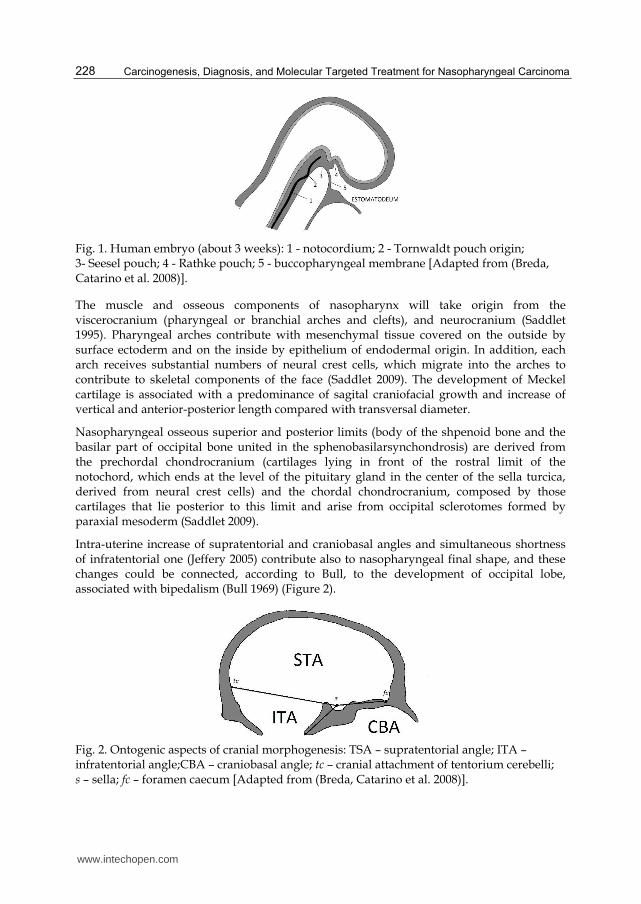
ectodermic stomatodeum by the buccopharyngeal membrane (Beasly 2008). Before the
rupture of buccopharyngeal membrane happens, two recesses will form in this region: the
Rathke pouch, which will origin the adenohypophysis and the Seesel pouch, representing
the primordium of the future nasopharynx (Figure 1).
www.intechopen.com
Carcinogenesis, Diagnosis, and Molecular Targeted Treatment for Nasopharyngeal Carcinoma
228
Fig. 1. Human embryo (about 3 weeks): 1 - notocordium; 2 - Tornwaldt pouch origin; 3- Seesel pouch; 4 - Rathke pouch; 5 - buccopharyngeal membrane [Adapted from (Breda, Catarino et al. 2008)].
The muscle and osseous components of nasopharynx will take origin from the viscerocranium (pharyngeal or branchial arches and clefts), and neurocranium (Saddlet 1995). Pharyngeal arches contribute with mesenchymal tissue covered on the outside by surface ectoderm and on the inside by epithelium of endodermal origin. In addition, each arch receives substantial numbers of neural crest cells, which migrate into the arches to contribute to skeletal components of the face (Saddlet 2009). The development of Meckel cartilage is associated with a predominance of sagital craniofacial growth and increase of vertical and anterior-posterior length compared with transversal diameter.
Nasopharyngeal osseous superior and posterior limits (body of the shpenoid bone and the basilar part of occipital bone united in the sphenobasilarsynchondrosis) are derived from the prechordal chondrocranium (cartilages lying in front of the rostral limit of the notochord, which ends at the level of the pituitary gland in the center of the sella turcica, derived from neural crest cells) and the chordal chondrocranium, composed by those cartilages that lie posterior to this limit and arise from occipital sclerotomes formed by paraxial mesoderm (Saddlet 2009).
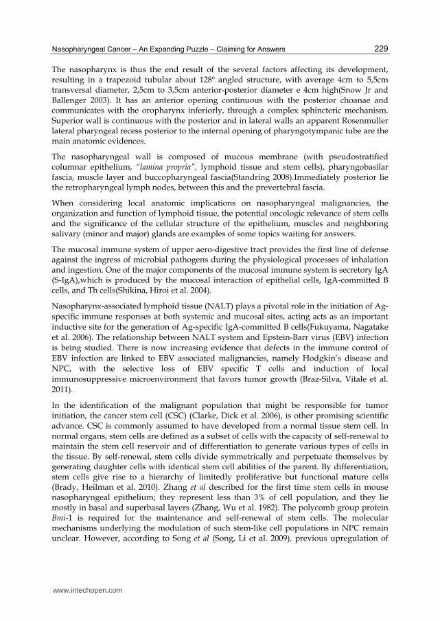
Intra-uterine increase of supratentorial and craniobasal angles and simultaneous shortness of infratentorial one (Jeffery 2005) contribute also to nasopharyngeal final shape, and these changes could be connected, according to Bull, to the development of occipital lobe, associated with bipedalism (Bull 1969) (Figure 2).
Fig. 2. Ontogenic aspects of cranial morphogenesis: TSA – supratentorial angle; ITA – infratentorial angle;CBA – craniobasal angle; tc – cranial attachment of tentorium cerebelli; s – sella; fc – foramen caecum [Adapted from (Breda, Catarino et al. 2008)].
www.intechopen.com
Nasopharyngeal Cancer – An Expanding Puzzle – Claiming for Answers
229
The nasopharynx is thus the end result of the several factors affecting its development, resulting in a trapezoid tubular about 128º angled structure, with average 4cm to 5,5cm transversal diameter, 2,5cm to 3,5cm anterior-posterior diameter e 4cm high(Snow Jr and Ballenger 2003). It has an anterior opening continuous with the posterior choanae and communicates with the oropharynx inferiorly, through a complex sphincteric mechanism. Superior wall is continuous with the posterior and in lateral walls an apparent Rosenmuller lateral pharyngeal recess posterior to the internal opening of pharyngotympanic tube are the main anatomic evidences.
The nasopharyngeal wall is composed of mucous membrane (with pseudostratified columnar epithelium, “lamina propria”, lymphoid tissue and stem cells), pharyngobasilar fascia, muscle layer and buccopharyngeal fascia(Standring 2008).Immediately posterior lie the retropharyngeal lymph nodes, between this and the prevertebral fascia.
When considering local anatomic implications on nasopharyngeal malignancies, the organization and function of lymphoid tissue, the potential oncologic relevance of stem cells and the significance of the cellular structure of the epithelium, muscles and neighboring salivary (minor and major) glands are examples of some topics waiting for answers.
The mucosal immune system of upper aero-digestive tract provides the first line of defense against the ingress of microbial pathogens during the physiological processes of inhalation and ingestion. One of the major components of the mucosal immune system is secretory IgA (S-IgA),which is produced by the mucosal interaction of epithelial cells, IgA-committed B cells, and Th cells(Shikina, Hiroi et al. 2004).
Nasopharynx-associated lymphoid tissue (NALT) plays a pivotal role in the initiation of Ag-
specific immune responses at both systemic and mucosal sites, acting acts as an important
inductive site for the generation of Ag-specific IgA-committed B cells(Fukuyama, Nagatake
et al. 2006). The relationship between NALT system and Epstein-Barr virus (EBV) infection
is being studied. There is now increasing evidence that defects in the immune control of
EBV infection are linked to EBV associated malignancies, namely Hodgkin’s disease and
NPC, with the selective loss of EBV specific T cells and induction of local
immunosuppressive microenvironment that favors tumor growth (Braz-Silva, Vitale et al.
2011).
In the identification of the malignant population that might be responsible for tumor initiation, the cancer stem cell (CSC) (Clarke, Dick et al. 2006), is other promising scientific advance. CSC is commonly assumed to have developed from a normal tissue stem cell. In normal organs, stem cells are defined as a subset of cells with the capacity of self-renewal to maintain the stem cell reservoir and of differentiation to generate various types of cells in the tissue. By self-renewal, stem cells divide symmetrically and perpetuate themselves by generating daughter cells with identical stem cell abilities of the parent. By differentiation, stem cells give rise to a hierarchy of limitedly proliferative but functional mature cells (Brady, Heilman et al. 2010). Zhang et al described for the first time stem cells in mouse nasopharyngeal epithelium; they represent less than 3% of cell population, and they lie mostly in basal and superbasal layers (Zhang, Wu et al. 1982). The polycomb group protein Bmi-1 is required for the maintenance and self-renewal of stem cells. The molecular mechanisms underlying the modulation of such stem-like cell populations in NPC remain unclear. However, according to Song et al (Song, Li et al. 2009), previous upregulation of
www.intechopen.com
Carcinogenesis, Diagnosis, and Molecular Targeted Treatment for Nasopharyngeal Carcinoma
230
Bmi-1 correlates with invasion of NPC and otherwise a possible mechanism of tumorigenesis involves repression of the Ink4a-Arf locus, which encodes the tumor suppressors INK4A and ARF, by direct binding of Bmi-1.
Furthermore, there is an ongoing debate over whether CSCs represent a mature tissue stem cell which has undergone malignant change or whether more differentiated cells re-initiate a ‘stemness’ programme as part of, or following, malignant transformation (Bomken, Fiser et al. 2010).
These and other questions are yet to be answered, namely how to explain the relationship between CSC, NPC and EBV. The recent accomplishment that LMP2A EBV encoded latent protein, induces epithelial–mesenchymal transition and stem-like cell self-renewal in NPC, is a strong evidence of this association (Kong, Hu et al. 2010).
2. The Epstein Barr virus and mechanisms of infection
Many epidemiological studies have been performed concerning NPC and three well-defined etiological factors involved in its pathogenesis have been identified. These include genetic susceptibility, early-age exposure to chemical carcinogens and an association with a latent EBV infection (Chan, Teo et al. 2002; Busson, Keryer et al. 2004).
It is widely accepted that EBV infection plays a major role in the pathogenesis of NPC in
endemic areas, although not entirely clear in non-endemic regions (Henle and Henle 1976;
Pathmanathan, Prasad et al. 1995). The association between NPC and EBV was initially
discovered from serological studies and was later supported by demonstration of EBV DNA
and nuclear antigen proteins (EBNA) in NPC tumor cells (zur Hausen, Schulte-Holthausen
et al. 1970; Henle and Henle 1976). The notion that EBV plays an important role in the
development of NPC is further supported by the observation that early nasopharyngeal
lesions (dysplasia or carcinoma-in situ) are already EBV-positive, harbouring latent and
clonal viral genomes as well as viral oncoproteins such as latent membrane protein (LMP)
(zur Hausen, Schulte-Holthausen et al. 1970; Pathmanathan, Prasad et al. 1995).
In 1958, Denis Burkitt, a British surgeon working in Africa, described a common cancer
primarily affecting children in specific regions of Africa (Burkitt 1958). Burkitt believed that
a virus might be implicated in cancer development, given the climatic and geographic
distribution of the cases (Burkitt 1962). EBV was first identified in 1964 when Anthony
Epstein, Yvonne Barr and Bert Achong detected virus-like particles by electron microscopy
in a cell line that had been established from a Burkitt’s lymphoma biopsy (Epstein, Achong
et al. 1964). The causal link between EBV and BL was corroborated by evidence showing
that BL patient sera had elevated antibodies to EBV antigens (Henle and Henle 1966) . This
group also established a link between primary EBV infection and infectious mononucleosis
and, subsequently, the association of EBV with nasopharyngeal carcinoma (Henle, Henle et
al. 1968; Henle, Henle et al. 1970; zur Hausen, Schulte-Holthausen et al. 1970).
Nasopharyngeal cancer, particularly when showing histological patterns of non-
keratinization (WHO/1978 II or III), is associated with previous EBV infection with
sequential immortalization of the virus in the host’s B-lymphocytes (Niedobitek, Hansmann
et al. 1991).
www.intechopen.com
Nasopharyngeal Cancer – An Expanding Puzzle – Claiming for Answers
231
EBV is a member of the herpesvirus family. As other herpesviruses, EBV is an enveloped virus that contains a DNA core surrounded by an icosahedral nucleocapsid and a tegument. Family members include herpes simplex 1 (HSV-1) and 2 (HSV-2) and varicella-zoster virus (VZV) - alphavirus subfamily; cytomegalovirus (CMV) and human herpesvirus 6 (HHV-6) and 7 (HHV-7) - betaherpesvirus subfamily; and human herpesvirus 8 (HHV-8) and EBV - gammaherpesvirus subfamily. The oncogenic potential of EBV was recognized through the association with numerous human malignancies. In addition to endemic Burkitt’s lymphoma and NPC, EBV was later found in Hodgkin’s lymphoma cases, post-transplant lymphoproliferative diseases, some T-cell lymphomas and a proportion of gastric carcinomas (Young and Rickinson 2004). Research is currently ongoing to determine the role of EBV-encoded gene products in these different cellular environments in an attempt to understand the role that EBV plays in the pathogenesis of these malignancies.
It is now known that EBV infects 90% of the world’s adult population. Although herpesviruses are ubiquitous in nature, humans are the only natural host for EBV. Upon infection, the individual remains a life-long carrier of the virus (Henle and Henle 1976). During acute infection, EBV seems to primarily infect and replicate in the stratified squamous epithelium of the oropharynx, followed by a latent infection of the B lymphocytes. EBV infection of B lymphocytes is thought to occur in the upper aerodigestive tract lymphoid organs and the virus persists in circulating memory B cells (Sixbey, Nedrud et al. 1984; Farrell 1995). Taking in account that EBV infection is a common event in the entire World, the reasons why only some individuals will develop EBV-related malignancies, are interesting aspects yet to clarify.
Much of the known biology of EBV relates to its interaction with B-lymphocytes. This is
mainly a result of the ability of EBV to readily infect and transform normal resting B-
lymphocytes in vitro, which also confirms the B-lymphotropic nature of this virus. EBV
latent gene expression in various EBV-associated malignancies and EBV-derived cell lines
has led to the identification of three different and distinct latency patterns. These latency
patterns characterize a heterogeneous group of diseases and are based on EBV genome
expression arrangements as the result of differential promoter activity influenced by host
cell factors (Thompson and Kurzrock 2004; Young and Rickinson 2004). Latency type I is
generally associated with Burkitt’s lymphoma and is characterized by expression of the
EBV-encoded RNAs (EBERs) and the BamHI-A rightward transcripts (BARTs), in addition
to EBV nuclear antigen-1 (EBNA1) expression. Latency type II is associated with NPC,
gastric cancer and EBV-positive Hodgkin lymphoma and is characterized by expression of
EBERs, BARTs, EBNA1 and the latent membrane proteins (LMP1, LMP2A and LMP2B).
Latency type III is associated with lymphoblastoid cell lines and post-transplant
lymphoproliferative diseases: the full range of latent gene products are expressed, including
EBNAs 1, 2, 3A, 3B, 3C and -LP, the expression of all three latent membrane proteins (LMP1,
LMP2A and LMP2B) and EBERs and BART RNAs. Although these classifications of latency
are useful in characterizing the distinct gene expression patterns, they are by no means
completely definitive (Thompson and Kurzrock 2004). In recent years, there has been
increasing interest in the presence of different viral and cellular micro-RNAs in EBV-
infected B cells and epithelial cells. Future works should investigate the role of EBV-encoded
micro-RNAs and its correlation with transcriptional regulation of both the viral and cellular
genome.
www.intechopen.com
Carcinogenesis, Diagnosis, and Molecular Targeted Treatment for Nasopharyngeal Carcinoma
232
Analysis of EBV in NPC tumors has revealed the presence of clonal EBV genomes,
suggesting that these carcinomas arise from the clonal expansion of a single EBV-infected
progenitor cell (Raab-Traub and Flynn 1986). Similar to most EBV-associated malignancies,
the exact role of EBV in NPC pathogenesis remains poorly understood. Unlike EBV-
associated B cell tumors, where the virus is considered to be an initiating factor in the
oncogenic process, virus infection in the context of NPC pathogenesis seems to act as a
tumor-promoting agent.
Recent studies investigated circulating tumor-derived EBV DNA in the blood of NPC
patients. Using real-time PCR methodology, studies indicate that the level of pre-treatment
EBV DNA has prognostic significance. Furthermore, post-treatment EBV DNA levels seem
to be correlated with treatment response and overall survival of NPC patients (Lo, Chan et
al. 2000; Lo, Leung et al. 2000; Lin, Chen et al. 2001; Chan, Lo et al. 2002). This methodology
should be applied in large-scale trials as an approach for early disease diagnosis.
3. Genetic basis of NPC
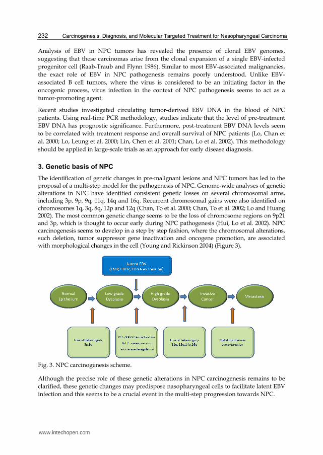
The identification of genetic changes in pre-malignant lesions and NPC tumors has led to the proposal of a multi-step model for the pathogenesis of NPC. Genome-wide analyses of genetic alterations in NPC have identified consistent genetic losses on several chromosomal arms, including 3p, 9p, 9q, 11q, 14q and 16q. Recurrent chromosomal gains were also identified on chromosomes 1q, 3q, 8q, 12p and 12q (Chan, To et al. 2000; Chan, To et al. 2002; Lo and Huang 2002). The most common genetic change seems to be the loss of chromosome regions on 9p21 and 3p, which is thought to occur early during NPC pathogenesis (Hui, Lo et al. 2002). NPC carcinogenesis seems to develop in a step by step fashion, where the chromosomal alterations, such deletion, tumor suppressor gene inactivation and oncogene promotion, are associated with morphological changes in the cell (Young and Rickinson 2004) (Figure 3).
Fig. 3. NPC carcinogenesis scheme.
Although the precise role of these genetic alterations in NPC carcinogenesis remains to be
clarified, these genetic changes may predispose nasopharyngeal cells to facilitate latent EBV
infection and this seems to be a crucial event in the multi-step progression towards NPC.
www.intechopen.com
Nasopharyngeal Cancer – An Expanding Puzzle – Claiming for Answers
233
The development and progression of NPC involves the accumulation of genetic alterations. Both genetic and epigenetic changes can affect the development of NPC, by altering the function of genes that are critical for proliferation, apoptosis and differentiation.
Host factors previously shown to be associated with NPC development include HLA class I and II alleles and polymorphisms of genes responsible for critical cellular functions, as carcinogen metabolism and detoxification, cell cycle control, DNA repair and immune response. Published studies demonstrated that some genetic variations are associated with a higher risk of NPC development (Hildesheim, Anderson et al. 1997; Nazar-Stewart, Vaughan et al. 1999; Cho, Hildesheim et al. 2003; Catarino, Breda et al. 2006; Sousa, Santos et al. 2006). This genetic predisposition to NPC may be modulated by environmental factors and this may have a differential impact in endemic and non-endemic NPC.
Familial clustering of the disease has been widely documented in the Chinese population and several polymorphisms have been associated with NPC development, namely in nitrosamine metabolizing genes cytochrome P450 2A6 (CYP2A6), P450 2E1 (CYP2E1) and P450 2F1 (CYP2F1), Glutathione S- transferase M1 (GSTM1), XRCC1 (codons 194 arg-trp), have been suggested to influence the susceptibility to NPC (Hildesheim and Levine 1993; Pathmanathan, Prasad et al. 1995; Sousa, Santos et al. 2006; Tiwawech, Srivatanakul et al. 2006).
Less in known in non-endemic areas, but it has been suggested that the individual genetic
background may influence the onset of NPC, also in these regions. Regarding cell cycle
control, some studies investigated the role of polymorphisms in cyclin D1 (CCND1) and
TP53 genes in the genetic susceptibility to NPC (Pathmanathan, Prasad et al. 1995; Cho,
Hildesheim et al. 2003; Catarino, Pereira et al. 2008). These genetic variants have been
associated with increased individual risk of NPC development. Regarding the key regulator
of cell cycle, cyclin D1, the study by Catarino et al, demonstrate that individuals carrying the
genetic variant present a 2-fold increased risk for the development of NPC, the proportion
of cases attributable to the genotype being 14.76% (Catarino, Breda et al. 2006). Moreover,
another study indicates that cyclin D1 variants influence the age of onset of oncogenic virus-
associated cancers, namely NPC (Catarino, Pereira et al. 2008). The results of this study
demonstrate that the waiting time for onset of oncogenic virus-associated neoplasia in
patients carrying the cyclin D1 variant was 12 years earlier in comparison with the other
patients. Another study by Sousa et al, focus on the polymorphic variants (Arg/Pro) on
TP53 codon 72 in nasopharyngeal cancer development. This study revealed a three-fold risk
for carriers of Pro/Pro genotype, suggesting that Pro/Pro genotype represents a stable risk
factor for nasopharyngeal carcinoma (Sousa, Santos et al. 2006).
Another study revealed that a MDM2 polymorphism is associated with increased risk to develop NPC adjusted for age and gender, with particular effect in undifferentiated types and early clinical stages. The study also indicates that the median age of onset of NPC was significantly different according to the genetics variants (Sousa, Pando et al. 2011). Regarding the immune response markers, a study revealed an increased frequency of a tumor necrosis factor-alpha (TNF-α) polymorphic variant in patients with NPC. The authors indicate that this variant is associated with a 2-fold increased risk for the development of this disease. Moreover, this effect seems to be stronger in undifferentiated types (Sousa, Breda et al. 2011).
www.intechopen.com
Carcinogenesis, Diagnosis, and Molecular Targeted Treatment for Nasopharyngeal Carcinoma
234
These results performed in low-risk, non-endemic areas indicate that these genetic variations may represent risk markers for NPC and contribute to the definition of genetic susceptibility profiles for the development of this disease. Furthermore, the knowledge of the mechanisms involved in NPC carcinogenesis may help to identify targets for the development of chemoprevention or therapeutic strategies.
4. The Nasopharyngeal Carcinoma (NPC)
4.1 NPC histopathologic classification (final clarification?)
The histopathologic designation of this kind of tumor is matter of debate since the first description of “Primary Carcinoma of Nasopharynx”, performed by Chevalier Jackson (1901) based upon the description of 14 cases(Jackson 1901; Van Hasselt and Gibb 1999). From that time on, several designations have been used to designate this tumor, namely lymphoepithelioma, lymphoepitheliomalike carcinoma, lymphoepithelial carcinoma, Schmincke type lymphoepithelioma, Regaud type lymphoepithelioma, transitional cell carcinoma, intermediate cell carcinoma, anaplastic carcinoma, undifferentiated carcinoma with lymphoid stroma, vesicular nucleus cell carcinoma, and so on.
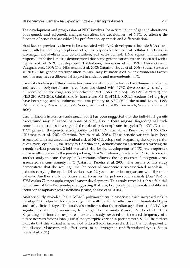
This controversy seems to be solved with the WHO (2003) classification of NPC into three histologic types: keratinizing squamous carcinoma, nonkeratinizing carcinoma and basaloid carcinoma (IARC 2005).
Fig. 4. Keratinizing squamous carcinoma.
In keratinizing squamous carcinoma, the polygonal and stratified tumor cells grow in the form of irregular islands, accompanied by an abundant desmoplastic stroma infiltrated by variable numbers of lymphocytes, plasma cells, neutrophils and eosinophils. The cell borders are distinct and separated by intercellular bridges and possess abundant cytoplasm and small hiperchromatic nucleus. Often keratin pearls can be seen (Figure 4).
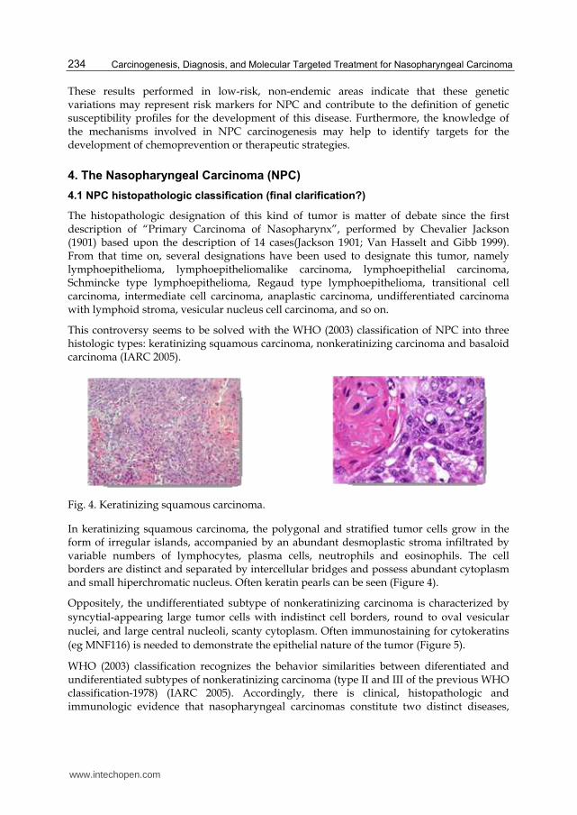
Oppositely, the undifferentiated subtype of nonkeratinizing carcinoma is characterized by
syncytial-appearing large tumor cells with indistinct cell borders, round to oval vesicular
nuclei, and large central nucleoli, scanty cytoplasm. Often immunostaining for cytokeratins
(eg MNF116) is needed to demonstrate the epithelial nature of the tumor (Figure 5).
WHO (2003) classification recognizes the behavior similarities between diferentiated and undiferentiated subtypes of nonkeratinizing carcinoma (type II and III of the previous WHO classification-1978) (IARC 2005). Accordingly, there is clinical, histopathologic and immunologic evidence that nasopharyngeal carcinomas constitute two distinct diseases,
www.intechopen.com
Nasopharyngeal Cancer – An Expanding Puzzle – Claiming for Answers
235
keratinizing squamous carcinoma in one side and nonkeratinizing carcinoma in the other, this later being linked to previous infection by Epstein–Barr virus.
Fig. 5. The undifferentiated subtype of NPC.
However, IARC “WHO Classification of Head and Neck Tumors” (IARC 2005), also describes 6 cases of basaloid squamous cell carcinoma (BSCC) arising as primary tumors of the nasopharynx. These tumors demonstrated to be morphologically identical to the same tumors more commonly occurring in other head and neck sites (larynx, hypopharynx and oropharynx) (Ereno, Gaafar et al. 2008), strongly associated with traditional risk factors, such as tobacco and alcohol abuse.
BSCC of the upper aerodigestive tract was originally described by Wain et al (1986) (Wain, Kier et al. 1986), as a rare variant of squamous cell carcinoma, is characterized by clinically aggressive behavior and worse prognosis, independent of tumor stage. Two major histopathologic features define BSCC. Morphologically, BSCC also possess distinct histological features, presenting basaloid tumor cells (solid, lobular, small, crowded cells with scant cytoplasm, hyperchromatic round nuclei, and small cystic spaces containing PAS-or alcian blue–positive myxoid material) in a festooning growth pattern, interspersed by tumor cells with squamous differentiation (Chernock, Lewis et al. 2010).
These histopathologic features are also very similar with those of oropharyngeal human
papillomavirus (HPV)–related nonkeratinizing squamous cell carcinoma (NK SCC). This
kind of carcinoma occurs predominantly in the tonsillar tissue of the oropharynx, in
younger patients and have unique risk factors related to sexual behavior, including multiple
partners, early age at first intercourse, and oral sex (Chernock, El-Mofty et al. 2009). The
tumor appears to show a lower clinical aggressiveness compared with basaloid squamous
cell carcinoma occurring in other head and neck sites (Begum and Westra 2008).
The analogy between viral etiology and histomorphological characteristics of tumor development site (close relationship between epithelium and lymphoid tissue) in HPV–related”basaloid” NK SCC of oropharynx and NPC nonkeratinizing carcinoma EBV-related, seems to be quite obvious, but not completely clarified, and so are their histopathologic patterns.
4.2 NPC commented epidemiologic data
Considering all nasopharyngeal tumors, the probability of being originated in the epithelial lining varies from 75 to 95% in low-risk populations, and all of them are virtually cancers in high-risk population (Parkin DM 2002). NPC is a patchy worldwide malignant disease, characterized by remarkable divergence in incidences among populations of diverse
www.intechopen.com
Carcinogenesis, Diagnosis, and Molecular Targeted Treatment for Nasopharyngeal Carcinoma
236
geographic regions and races. It is very common in southern Asia (20-30/100 000 inhabitants/year) (Vasef, Ferlito et al. 1997) and quite rare in western countries (<1/100 000 inhabitants/year) (Parkin DM 2002). Intermediate incidence (8-12/100000 inhabitants/year) occurs in certain African and Mediterranean populations (Cvitkovic, Bachouchi et al. 1991) and also in the Inuits from Greenland and Alaska and Malays from Singapore and Malaysia.
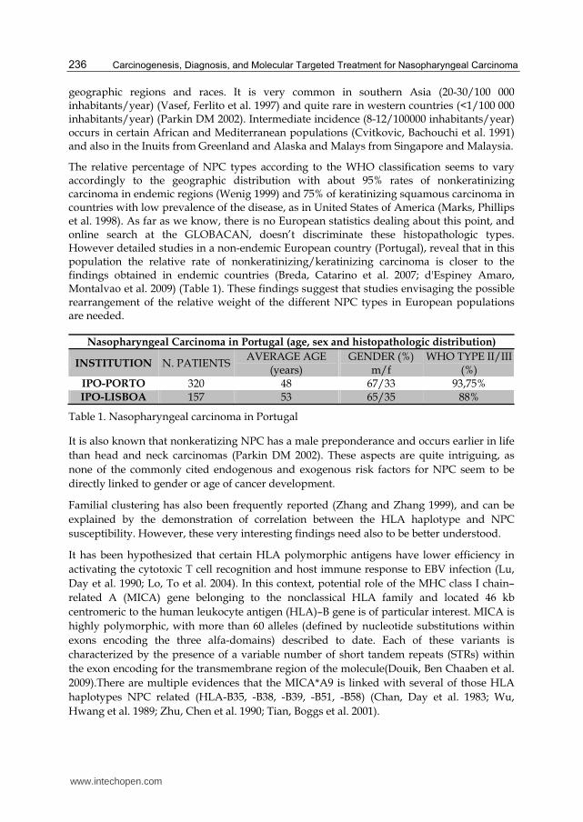
The relative percentage of NPC types according to the WHO classification seems to vary accordingly to the geographic distribution with about 95% rates of nonkeratinizing carcinoma in endemic regions (Wenig 1999) and 75% of keratinizing squamous carcinoma in countries with low prevalence of the disease, as in United States of America (Marks, Phillips et al. 1998). As far as we know, there is no European statistics dealing about this point, and online search at the GLOBACAN, doesn’t discriminate these histopathologic types. However detailed studies in a non-endemic European country (Portugal), reveal that in this population the relative rate of nonkeratinizing/keratinizing carcinoma is closer to the findings obtained in endemic countries (Breda, Catarino et al. 2007; d'Espiney Amaro, Montalvao et al. 2009) (Table 1). These findings suggest that studies envisaging the possible rearrangement of the relative weight of the different NPC types in European populations are needed.
Nasopharyngeal Carcinoma in Portugal (age, sex and histopathologic distribution)
INSTITUTION N. PATIENTSAVERAGE AGE
(years) GENDER (%)
m/f WHO TYPE II/III
(%) IPO-PORTO 320 48 67/33 93,75% IPO-LISBOA 157 53 65/35 88%
Table 1. Nasopharyngeal carcinoma in Portugal
It is also known that nonkeratizing NPC has a male preponderance and occurs earlier in life
than head and neck carcinomas (Parkin DM 2002). These aspects are quite intriguing, as
none of the commonly cited endogenous and exogenous risk factors for NPC seem to be
directly linked to gender or age of cancer development.
Familial clustering has also been frequently reported (Zhang and Zhang 1999), and can be
explained by the demonstration of correlation between the HLA haplotype and NPC
susceptibility. However, these very interesting findings need also to be better understood.
It has been hypothesized that certain HLA polymorphic antigens have lower efficiency in
activating the cytotoxic T cell recognition and host immune response to EBV infection (Lu,
Day et al. 1990; Lo, To et al. 2004). In this context, potential role of the MHC class I chain–
related A (MICA) gene belonging to the nonclassical HLA family and located 46 kb
centromeric to the human leukocyte antigen (HLA)–B gene is of particular interest. MICA is
highly polymorphic, with more than 60 alleles (defined by nucleotide substitutions within
exons encoding the three alfa-domains) described to date. Each of these variants is
characterized by the presence of a variable number of short tandem repeats (STRs) within
the exon encoding for the transmembrane region of the molecule(Douik, Ben Chaaben et al.
2009).There are multiple evidences that the MICA*A9 is linked with several of those HLA
haplotypes NPC related (HLA-B35, -B38, -B39, -B51, -B58) (Chan, Day et al. 1983; Wu,
Hwang et al. 1989; Zhu, Chen et al. 1990; Tian, Boggs et al. 2001).
www.intechopen.com
Nasopharyngeal Cancer – An Expanding Puzzle – Claiming for Answers
237
Wei Tian et al (Tian, Zeng et al. 2006) demonstrated a positive association between MICA*A9 and nasopharyngeal carcinoma, only observed in male subjects, and this finding had statistical significance (P=0.0001). This fact is not well explained, but it should be remembered that HLA genes are sex-linked and transmitted in bloc.
Furthermore, a recent study demonstrated the existence of a binding site in the long intron 1 of the MICA gene for NF-kappaB (Molinero, Fuertes et al. 2004). It has been shown that physiologic concentrations of male hormones (androgens) cause prolonged NF-kappaB DNA binding activities, which are diminished by vitamins C and E (Ripple, Henry et al. 1999)and estrogen inhibits NF-kappa B activity (Ghisletti, Meda et al. 2005; Kalaitzidis and Gilmore 2005). These findings are mostly fascinating when we consider that TRAF is activated by the LMP1 latent EBV protein.
The NPC age distribution is also quite different than usual, and seems to vary according to
the incidence. It is accepted that in low-risk countries, the NPC incidence rises
monotonically with age, but in high-risk regions the age incidence hit the highest point at 55
years(Chang and Adami 2006), showing bimodal age presentation in Northern Africa and
India (Bray, Haugen et al. 2008). These results also need an explanation, namely on the
influence of some genetic polymorphisms that may influence the age of onset of disease
(Catarino, Pereira et al. 2008; Sousa H 2010).
The close relationship between nonkeratinizing NPC and EBV is sustained by “in-situ”
hybridization studies (Wei and Sham 2005) and occurs both in endemic as in non-endemic
countries (Niedobitek, Hansmann et al. 1991; Breda, Catarino et al. 2010). However,
infection with EBV is a worldwide phenomenon and other endogenous and exogenous
factors must be associated with NPC development. The relative weight of these factors is not
yet clearly understood, namely in non-endemic regions.
4.3 NPC stage classification and prognostic evaluation
Whatever its localization, cancer prognosis will conceptually depend on biological aggressiveness of tumor itself, host characteristics and therapeutic options.
The tumor, node, metastasis (TNM) staging system, allows clinicians to categorize tumors of
the head and neck region in a specific manner, to assist with the assessment of disease
status, prognosis and management (Daniel G. Deschler 2008). The introduction of several
clinical staging systems (American Joint Committee on Cancer (AJCC) staging, the
International Union Against Cancer (UICC) staging, Ho’s staging from Hong Kong, Fuzhou
staging from Mainland China, Huang’s or Hsu’s staging from Taiwan), permitted that TNM
staging for NPC remained relatively confused during many years and this was undesirable.
It is accepted that a good TNM classification should fulfill the following criteria:(1) the
subgroups defined by T, N, and M that make up a given group within a grouping scheme
must have similar survival rates (hazard consistency); (2) the survival rates must differ
among the groups (hazard discrimination); (3) the prediction of cure must be high (outcome
prediction); and (4) the distribution of patients among the groups must be balanced
(Groome, Schulze et al. 2001). The association between the AJCC and UICC staging systems
and their succeeding modifications taking into account the criteria previously enunciated,
www.intechopen.com
Carcinogenesis, Diagnosis, and Molecular Targeted Treatment for Nasopharyngeal Carcinoma
238
namely with rearrangements on T and N arrays, allowed a better TNM prognostic
significance in the most recent classifications (Liu, Tang et al. 2008).
However, NPC TNM classification only takes into account the aggressiveness of the
“clinical” tumor; understand all the factors influencing the clinical course of NPC in order
to improve the TNM classification (Greene, Page et al. 2002) and anticipate NPC
prognosis, is still a challenge, recognized by several authors (Lee, Au et al. 2004; Liu, Tang
et al. 2008). Accordingly, other tumor characterystics may confirm relevance on prognosis,
suhc as histological types (WHO type II and III versus type I). Furthermore, taking into
account the relationship between histological types and EBV, it also seems to be very
interesting to know how relevant can be the measurement of EBV related proteins in the
patient’s serum as a prognostic factor of survival. Plasma EBV-DNA measurement is
actually believed to be a more sensitive and specific marker than the serum IgA/VCA
titer for the diagnosis and monitoring of patients with NPC (Shao, Li et al. 2004). Even
more, plasma EBV-DNA findings provide convincing evidence of their usefulness on the
early diagnosis and staging of NPC as well as for monitoring recurrence and metastasis of
this tumor. Some studies indicate that high marker level (>500 copies/ml) at 6 weeks after
radio(chemo)therapy is a powerful prognosticator of recurrence, whereas pretreatment
EBV DNA is a better discriminator of prognosis than conventional TNM classification for
stage II NPC (Haddad 2011).
Patient’s related prognostic factors are seldom referred in literature. Some authors claim
that patients with younger age are associated with improved overall survival. One study
involving 2,054 NPC patients from several European countries also showed that overall
survival declined with age (Jiong, Berrino et al. 1998), and other series from endemic
regions demonstrated similar results (Leung, Tung et al. 2005) (Liu, Tang et al. 2008).
However, not always is disentangled age with better performance status and less
comorbidity characteristics, more often present in younger patients, which might
contribute to better tolerance to radical radio(chemo)therapy, thus resulting in better
survival.
The relationship between gender and prognosis is subject to similar doubts (Leung, Tung et
al. 2005) (Liu, Tang et al. 2008) with controversial results being described. Again, it is
difficult to straighten out gender with lifestyle (including body weight, central adiposity,
physical activity, exposure to smoking, fruit and vegetable intake a.s.o. and these aspects
still wait for clarification.
4.4 NPC treatment, survival rate and quality of life
Radiation therapy (RT) is considered the mainstay treatment for NPC. Recent retrospective analysis including more than two thousand patients treated by RT alone (mostly using 2D technique) showed that the 5-year overall survival (OS) was 85% for stages I–II (Lee, Sze et al. 2005), corroborating that NPC is highly sensitive to ionizing radiation. However, the same study demonstrated an OS of only 66% for stages III–IVB, with metastatic rate remaining high (25% at 5-years) for patients who achieved locoregional control, and these findings supported the need of some kind of systemic treatment, namely for advanced disease.
www.intechopen.com
Nasopharyngeal Cancer – An Expanding Puzzle – Claiming for Answers
239
The advantages of adding chemotherapy (Ch) to RT had already been accomplished by the Intergroup 0099 study, the first prospectively randomized study to demonstrate an improvement in OS from the addition of concurrent chemotherapy and adjuvant chemotherapy to radiation therapy (when compared with radiation therapy alone), without unacceptably increasing the toxicity of treatment (Al-Sarraf, LeBlanc et al. 1998).
From that time on, several trials confirmed not only that the addition of chemotherapy concurrent with radiation therapy has improved the outcomes of patients with NPC, but also that improvements in radiation treatment technology and the adoption of IMRT (Intensity-Modulated Radiation Therapy) have also improved local control, reducing also the morbidity.
Besides the superior dose conformity delivered to tumor, one of the most important promised advantages of IMRT is the achievement of sufficient sparing of critical normal structures adjacent to treated areas, namely spinal cord, parotid glands, temporomandibular joint, middle and inner ears, skin (in the region of the target volumes), and oral cavity, mandible, glottic larynx, brachial plexus, and esophagus (including postcricoid pharynx) (Brady, Heilman et al. 2010) and such sparing has obvious advantages.
However, even with IMRT, minor salivary glands and both palatal and masticator muscles
(medial pterygoid and lateral pterygoid) are often included in target volumes, either gross
tumor volume (GTV), clinical target volume (CTV) or planned target volume (PTV). The
impact of such distresses on Quality of life (QoL) seems not to be disentangled enough
(Bhatia, King et al. 2009; Lee, Harris et al. 2009).
Quality of life refers to the perception of the effects of disease and its impact on the patient’s daily functioning; QoL is a multi-dimensional issue, incorporating physical, psychological, social and emotional domains, and it must be self-reported according to the patient’s own experiences (List and Stracks 2000). These very important mind and corporal approach is not often mentioned in scientific literature; usually, the endpoint of medical care for cancer patients is focused on the survival rate, local control rate, or complication rate. These endpoints were typically assessed from the physician’s points of view and lacked knowledge of patients’ mental or emotional well-being. According to Fang and co-workers (Fang, Tsai et al. 2010), in addition to socioeconomic levels, advanced RT techniques were observed to play a significant role in improving the QoL outcome of NPC survivors. However, the impact size from conventional 2DRT to 3DCRT or IMRT varied on different QoL scales. The therapeutic benefit of IMRT over 2DRT, especially on the swallowing-related QoL scales, is not clear and should be further explored.
One of the main aims of clinical or translational research in cancer is the search for biological factors that could foresee treatment outcomes, in biologic activity and toxic effects. The increasing amount of information regarding the role of genetics in human diseases has led to putative new biomarkers and the development of new fields, as pharmacogenomics (PGx). Pharmacogenomics is a rapidly developing scientific area, especially in oncology. In the most ideal situation, pharmacogenomics will allow oncologists to individualize therapy based on patients' individual germline genetic test results. This can help to improve efficacy, reduce toxicity and predict non-responders in a way that alternative therapy can be chosen or individual dose adjustments can be made. It has been observed that the interpatient variability in response to medications is associated with a spectrum of outcomes, ranging
www.intechopen.com
Carcinogenesis, Diagnosis, and Molecular Targeted Treatment for Nasopharyngeal Carcinoma
240
from failure to demonstrate an expected therapeutic effect, to an adverse reaction resulting in significant patient morbidity and mortality, decreasing quality of life, as well as increasing healthcare costs.
The goal of the emerging disciplines of pharmacogenomics is to personalize therapy based on an individual’s genotype. To date, the success of PGx has spread across all fields of medicine, although little is known about the genetic profile and its correlation with treatment response and overall survival in nasopharyngeal cancer patients. Genetic information has been used in the identification of disease risk, choice of treatment agents and guiding drug dosing. This is particularly important for chemotherapeutic agents, which in general affect both tumor and non-tumor cells and thus have a narrow therapeutic index, with the potential for life-threatening toxicity. Future work should explore the role of genetic variations in nasopharyngeal cancer patients, in a pharmacogenomics approach.
5. References
Al-Sarraf, M, M LeBlanc, PG Giri, KK Fu, J Cooper, T Vuong, AA Forastiere, G Adams, WA Sakr, DE Schuller and JF Ensley (1998). Chemoradiotherapy versus radiotherapy in patients with advanced nasopharyngeal cancer: phase III randomized Intergroup study 0099. J Clin Oncol 16(4): 1310-1317.
Beasly, N, Ed. (2008). Anatomy of the pharynx and oesophagus. Scott-Brown’s Otolaryngology Head and Neck Surgery London.
Begum, S and WH Westra (2008). Basaloid squamous cell carcinoma of the head and neck is a mixed variant that can be further resolved by HPV status. Am J Surg Pathol 32(7): 1044-1050.
Bhatia, KS, AD King, BK Paunipagar, J Abrigo, AC Vlantis, SF Leung and AT Ahuja (2009). MRI findings in patients with severe trismus following radiotherapy for nasopharyngeal carcinoma. Eur Radiol 19(11): 2586-2593.
Bomken, S, K Fiser, O Heidenreich and J Vormoor (2010). Understanding the cancer stem cell. Br J Cancer 103(4): 439-445.
Brady, LW, H-P Heilman, M Molls and C Nieder, Eds. (2010). Nasopharyngeal Cancer Multidisciplinary Management. Medical Radiology diagnostic Imaging and Radiation Oncology, Springer.
Bray, F, M Haugen, TA Moger, S Tretli, OO Aalen and T Grotmol (2008). Age-incidence curves of nasopharyngeal carcinoma worldwide: bimodality in low-risk populations and aetiologic implications. Cancer Epidemiol Biomarkers Prev 17(9): 2356-2365.
Braz-Silva, PH, S Vitale, C Butori, N Guevara, J Santini, M Magalhaes, P Hofman and A Doglio (2011). Specific infiltration of langerin-positive dendritic cells in EBV-infected tonsil, Hodgkin lymphoma and nasopharyngeal carcinoma. Int J Cancer 128(10): 2501-2508.
Breda, E, R Catarino, I Azevedo, T Fernandes, C Barreira da Costa and R Medeiros (2007). [Characterization of the clinical evolution of nasopharyngeal carcinoma in Portuguese population]. Acta Otorrinolaringol Esp 58(5): 191-197.
Breda, E, R Catarino, A Coelho, H Sousa and R Medeiros (2008). [Nasopharyngeal carcinoma study: introduction and multidisciplinary perspective]. Acta Med Port 21(3): 273-284.
www.intechopen.com
Nasopharyngeal Cancer – An Expanding Puzzle – Claiming for Answers
241
Breda, E, RJ Catarino, I Azevedo, M Lobao, E Monteiro and R Medeiros (2010). Epstein-Barr virus detection in nasopharyngeal carcinoma: implications in a low-risk area. Braz J Otorhinolaryngol 76(3): 310-315.
Bull, JW (1969). Tentorium cerebelli. Proc R Soc Med 62(12): 1301-1310. Burkitt, D (1958). A sarcoma involving the jaws in African children. The British journal of
surgery 46(197): 218-223. Burkitt, D (1962). A children's cancer dependent on climatic factors. Nature 194: 232-234. Busson, P, C Keryer, T Ooka and M Corbex (2004). EBV-associated nasopharyngeal
carcinomas: from epidemiology to virus-targeting strategies. Trends in microbiology 12(8): 356-360.
Catarino, R, D Pereira, E Breda, A Coelho, A Matos, C Lopes and R Medeiros (2008). Oncogenic virus-associated neoplasia: a role for cyclin D1 genotypes influencing the age of onset of disease? Biochem Biophys Res Commun 370(1): 118-122.
Catarino, R, D Pereira, E Breda, A Coelho, A Matos, C Lopes and R Medeiros (2008). Oncogenic virus-associated neoplasia: a role for cyclin D1 genotypes influencing the age of onset of disease? Biochemical and biophysical research communications 370(1): 118-122.
Catarino, RJ, E Breda, V Coelho, D Pinto, H Sousa, C Lopes and R Medeiros (2006). Association of the A870G cyclin D1 gene polymorphism with genetic susceptibility to nasopharyngeal carcinoma. Head Neck 28(7): 603-608.
Chan, AS, KF To, KW Lo, M Ding, X Li, P Johnson and DP Huang (2002). Frequent chromosome 9p losses in histologically normal nasopharyngeal epithelia from southern Chinese. International journal of cancer. Journal international du cancer 102(3): 300-303.
Chan, AS, KF To, KW Lo, KF Mak, W Pak, B Chiu, GM Tse, M Ding, X Li, JC Lee and DP Huang (2000). High frequency of chromosome 3p deletion in histologically normal nasopharyngeal epithelia from southern Chinese. Cancer research 60(19): 5365-5370.
Chan, AT, YM Lo, B Zee, LY Chan, BB Ma, SF Leung, F Mo, M Lai, S Ho, DP Huang and PJ Johnson (2002). Plasma Epstein-Barr virus DNA and residual disease after radiotherapy for undifferentiated nasopharyngeal carcinoma. Journal of the National Cancer Institute 94(21): 1614-1619.
Chan, AT, PM Teo and PJ Johnson (2002). Nasopharyngeal carcinoma. Annals of oncology : official journal of the European Society for Medical Oncology / ESMO 13(7): 1007-1015.
Chan, SH, NE Day, N Kunaratnam, KB Chia and MJ Simons (1983). HLA and nasopharyngeal carcinoma in Chinese--a further study. Int J Cancer 32(2): 171-176.
Chang, ET and HO Adami (2006). The enigmatic epidemiology of nasopharyngeal carcinoma. Cancer Epidemiol Biomarkers Prev 15(10): 1765-1777.
Chernock, RD, SK El-Mofty, WL Thorstad, CA Parvin and JS Lewis, Jr. (2009). HPV-related nonkeratinizing squamous cell carcinoma of the oropharynx: utility of microscopic features in predicting patient outcome. Head Neck Pathol 3(3): 186-194.
Chernock, RD, JS Lewis, Jr., Q Zhang and SK El-Mofty (2010). Human papillomavirus-positive basaloid squamous cell carcinomas of the upper aerodigestive tract: a distinct clinicopathologic and molecular subtype of basaloid squamous cell carcinoma. Hum Pathol 41(7): 1016-1023.
Cho, EY, A Hildesheim, CJ Chen, MM Hsu, IH Chen, BF Mittl, PH Levine, MY Liu, JY Chen, LA Brinton, YJ Cheng and CS Yang (2003). Nasopharyngeal carcinoma and genetic
www.intechopen.com
Carcinogenesis, Diagnosis, and Molecular Targeted Treatment for Nasopharyngeal Carcinoma
242
polymorphisms of DNA repair enzymes XRCC1 and hOGG1. Cancer epidemiology, biomarkers & prevention : a publication of the American Association for Cancer Research, cosponsored by the American Society of Preventive Oncology 12(10): 1100-1104.
Clarke, MF, JE Dick, PB Dirks, CJ Eaves, CH Jamieson, DL Jones, J Visvader, IL Weissman and GM Wahl (2006). Cancer stem cells--perspectives on current status and future directions: AACR Workshop on cancer stem cells. Cancer Res 66(19): 9339-9344.
Cvitkovic, E, M Bachouchi and JP Armand (1991). Nasopharyngeal carcinoma. Biology, natural history, and therapeutic implications. Hematol Oncol Clin North Am 5(4): 821-838.
d'Espiney Amaro, C, P Montalvao, P Henriques, M Magalhaes and J Olias (2009). Nasopharyngeal carcinoma: our experience. Eur Arch Otorhinolaryngol 266(6): 833-838.
Daniel G. Deschler, TD, Ed. (2008). Pocket Guide To TNM STAGING OF HEAD AND NECK CANCER AND NECK DISSECTION CLASSIFICATION. , Alexandria, American Academy of Otolaryngology–Head and Neck Surgery Foundation, Inc.
Douik, H, A Ben Chaaben, N Attia Romdhane, HB Romdhane, T Mamoghli, C Fortier, W Boukouaci, L Harzallah, A Ghanem, S Gritli, M Makni, D Charron, R Krishnamoorthy, F Guemira and R Tamouza (2009). Association of MICA-129 polymorphism with nasopharyngeal cancer risk in a Tunisian population. Hum Immunol 70(1): 45-48.
Epstein, MA, BG Achong and YM Barr (1964). Virus Particles in Cultured Lymphoblasts from Burkitt's Lymphoma. Lancet 1(7335): 702-703.
Ereno, C, A Gaafar, M Garmendia, C Etxezarraga, FJ Bilbao and JI Lopez (2008). Basaloid squamous cell carcinoma of the head and neck: a clinicopathological and follow-up study of 40 cases and review of the literature. Head Neck Pathol 2(2): 83-91.
Fang, FM, WL Tsai, TF Lee, KC Liao, HC Chen and HC Hsu (2010). Multivariate analysis of quality of life outcome for nasopharyngeal carcinoma patients after treatment. Radiother Oncol 97(2): 263-269.
Farrell, PJ (1995). Epstein-Barr virus immortalizing genes. Trends in microbiology 3(3): 105-109.
Fukuyama, S, T Nagatake, D-Y Kim, K Takamura, EJ Park, T Kaisho, N Tanaka, Y Kurono and H Kiyono (2006). Cutting Edge: Uniqueness of Lymphoid Chemokine Requirement for the Initiation and Maturation of Nasopharynx-Associated Lymphoid Tissue Organogenesis. J Immunol (177): 4276-4280.
Ghisletti, S, C Meda, A Maggi and E Vegeto (2005). 17beta-estradiol inhibits inflammatory gene expression by controlling NF-kappaB intracellular localization. Mol Cell Biol 25(8): 2957-2968.
Groome, PA, KM Schulze, WJ Mackillop, B Grice, C Goh, BJ Cummings, SF Hall, FF Liu, D Payne, DM Rothwell, JN Waldron, PR Warde and B O'Sullivan (2001). A comparison of published head and neck stage groupings in carcinomas of the tonsillar region. Cancer 92(6): 1484-1494.
Haddad, R, Ed. (2011). Multidisciplinary Management of Head and Neck Cancer, Demos Medical Publishing, LLC.
Henle, G and W Henle (1966). Immunofluorescence in cells derived from Burkitt's lymphoma. Journal of bacteriology 91(3): 1248-1256.
www.intechopen.com
Nasopharyngeal Cancer – An Expanding Puzzle – Claiming for Answers
243
Henle, G and W Henle (1976). Epstein-Barr virus-specific IgA serum antibodies as an outstanding feature of nasopharyngeal carcinoma. International journal of cancer. Journal international du cancer 17(1): 1-7.
Henle, G, W Henle and V Diehl (1968). Relation of Burkitt's tumor-associated herpes-ytpe virus to infectious mononucleosis. Proceedings of the National Academy of Sciences of the United States of America 59(1): 94-101.
Henle, W and G Henle (1976). The sero-epidemiology of Epstein-Barr virus. Advances in pathobiology(5): 5-17.
Henle, W, G Henle, M Scriba, CR Joyner, FS Harrison, Jr., R Von Essen, J Paloheimo and E Klemola (1970). Antibody responses to the Epstein-Barr virus and cytomegaloviruses after open-heart and other surgery. The New England journal of medicine 282(19): 1068-1074.
Hildesheim, A, LM Anderson, CJ Chen, YJ Cheng, LA Brinton, AK Daly, CD Reed, IH Chen, NE Caporaso, MM Hsu, JY Chen, JR Idle, RN Hoover, CS Yang and SK Chhabra (1997). CYP2E1 genetic polymorphisms and risk of nasopharyngeal carcinoma in Taiwan. Journal of the National Cancer Institute 89(16): 1207-1212.
Hildesheim, A and PH Levine (1993). Etiology of nasopharyngeal carcinoma: a review. Epidemiologic reviews 15(2): 466-485.
Hui, AB, KW Lo, PM Teo, KF To and DP Huang (2002). Genome wide detection of oncogene amplifications in nasopharyngeal carcinoma by array based comparative genomic hybridization. International journal of oncology 20(3): 467-473.
IARC, Ed. (2005). Pathology and Genetics of Head and Neck Tumours. Who Health Organization Classification of Tumours, Lyon.
Jackson, C (1901). Primary carcinoma of the nasopharynx: a table of cases. JAMA(37): 371-377.
Jeffery, N (2005). Cranial base angulation and growth of the human fetal pharynx. Anat Rec A Discov Mol Cell Evol Biol 284(1): 491-499.
Jiong, L, F Berrino and JW Coebergh (1998). Variation in survival for adults with Nasopharyngeal cancer in Europe, 1978-1989. Eur J Cancer 34(14): 2162-2166.
Kalaitzidis, D and TD Gilmore (2005). Transcription factor cross-talk: the estrogen receptor and NF-kappaB. Trends Endocrinol Metab 16(2): 46-52.
Kong, QL, LJ Hu, JY Cao, YJ Huang, LH Xu, Y Liang, D Xiong, S Guan, BH Guo, HQ Mai, QY Chen, X Zhang, MZ Li, JY Shao, CN Qian, YF Xia, LB Song, YX Zeng and MS Zeng (2010). Epstein-Barr virus-encoded LMP2A induces an epithelial-mesenchymal transition and increases the number of side population stem-like cancer cells in nasopharyngeal carcinoma. PLoS Pathog 6(6): e1000940.
Lee, AW, WM Sze, JS Au, SF Leung, TW Leung, DT Chua, BC Zee, SC Law, PM Teo, SY Tung, DL Kwong and WH Lau (2005). Treatment results for nasopharyngeal carcinoma in the modern era: the Hong Kong experience. Int J Radiat Oncol Biol Phys 61(4): 1107-1116.
Lee, N, J Harris, AS Garden, W Straube, B Glisson, P Xia, W Bosch, WH Morrison, J Quivey, W Thorstad, C Jones and KK Ang (2009). Intensity-Modulated Radiation Therapy With or Without Chemotherapy for Nasopharyngeal Carcinoma: Radiation Therapy Oncology Group Phase II Trial 0225. Journal of Clinical Oncology 27.
www.intechopen.com
Carcinogenesis, Diagnosis, and Molecular Targeted Treatment for Nasopharyngeal Carcinoma
244
Leung, TW, SY Tung, WK Sze, FC Wong, KK Yuen, CM Lui, SH Lo, TY Ng and SK O (2005). Treatment results of 1070 patients with nasopharyngeal carcinoma: an analysis of survival and failure patterns. Head Neck 27(7): 555-565.
Lin, JC, KY Chen, WY Wang, JS Jan, WM Liang, CS Tsai and YH Wei (2001). Detection of Epstein-Barr virus DNA in the peripheral-blood cells of patients with nasopharyngeal carcinoma: relationship to distant metastasis and survival. Journal of clinical oncology : official journal of the American Society of Clinical Oncology 19(10): 2607-2615.
List, MA and J Stracks (2000). Evaluation of quality of life in patients definitively treated for squamous carcinoma of the head and neck. Curr Opin Oncol 12(3): 215-220.
Liu, MZ, LL Tang, JF Zong, Y Huang, Y Sun, YP Mao, LZ Liu, AH Lin and J Ma (2008). Evaluation of sixth edition of AJCC staging system for nasopharyngeal carcinoma and proposed improvement. Int J Radiat Oncol Biol Phys 70(4): 1115-1123.
Lo, KW and DP Huang (2002). Genetic and epigenetic changes in nasopharyngeal carcinoma. Seminars in cancer biology 12(6): 451-462.
Lo, KW, KF To and DP Huang (2004). Focus on nasopharyngeal carcinoma. Cancer Cell 5(5): 423-428.
Lo, YM, AT Chan, LY Chan, SF Leung, CW Lam, DP Huang and PJ Johnson (2000). Molecular prognostication of nasopharyngeal carcinoma by quantitative analysis of circulating Epstein-Barr virus DNA. Cancer research 60(24): 6878-6881.
Lo, YM, SF Leung, LY Chan, AT Chan, KW Lo, PJ Johnson and DP Huang (2000). Kinetics of plasma Epstein-Barr virus DNA during radiation therapy for nasopharyngeal carcinoma. Cancer research 60(9): 2351-2355.
Lu, SJ, NE Day, L Degos, V Lepage, PC Wang, SH Chan, M Simons, B McKnight, D Easton, Y Zeng and et al. (1990). Linkage of a nasopharyngeal carcinoma susceptibility locus to the HLA region. Nature 346(6283): 470-471.
Marks, JE, JL Phillips and HR Menck (1998). The National Cancer Data Base report on the relationship of race and national origin to the histology of nasopharyngeal carcinoma. Cancer 83(3): 582-588.
Molinero, LL, MB Fuertes, MV Girart, L Fainboim, GA Rabinovich, MA Costas and NW Zwirner (2004). NF-kappa B regulates expression of the MHC class I-related chain A gene in activated T lymphocytes. J Immunol 173(9): 5583-5590.
Nazar-Stewart, V, TL Vaughan, RD Burt, C Chen, M Berwick and GM Swanson (1999). Glutathione S-transferase M1 and susceptibility to nasopharyngeal carcinoma. Cancer epidemiology, biomarkers & prevention : a publication of the American Association for Cancer Research, cosponsored by the American Society of Preventive Oncology 8(6): 547-551.
Niedobitek, G, ML Hansmann, H Herbst, LS Young, D Dienemann, CA Hartmann, T Finn, S Pitteroff, A Welt, I Anagnostopoulos and et al. (1991). Epstein-Barr virus and carcinomas: undifferentiated carcinomas but not squamous cell carcinomas of the nasopharynx are regularly associated with the virus. J Pathol 165(1): 17-24.
Niedobitek, G, ML Hansmann, H Herbst, LS Young, D Dienemann, CA Hartmann, T Finn, S Pitteroff, A Welt, I Anagnostopoulos and et al. (1991). Epstein-Barr virus and carcinomas: undifferentiated carcinomas but not squamous cell carcinomas of the nasopharynx are regularly associated with the virus. The Journal of pathology 165(1): 17-24.
www.intechopen.com
Nasopharyngeal Cancer – An Expanding Puzzle – Claiming for Answers
245
Parkin DM, BF, Ferlay J, Pisani P (2002). Global cancer statistics. CA Cancer J Clin. 2005 : Mar-Apr;55(2): 74-108.
Pathmanathan, R, U Prasad, R Sadler, K Flynn and N Raab-Traub (1995). Clonal proliferations of cells infected with Epstein-Barr virus in preinvasive lesions related to nasopharyngeal carcinoma. The New England journal of medicine 333(11): 693-698.
Raab-Traub, N and K Flynn (1986). The structure of the termini of the Epstein-Barr virus as a marker of clonal cellular proliferation. Cell 47(6): 883-889.
Rainger, R (1997). Everett C. Olson and the development of vertebrate paleoecology and taphonomy. Archives of Natural History 24(3): 373 - 396.
Ripple, MO, WF Henry, SR Schwarze, G Wilding and R Weindruch (1999). Effect of antioxidants on androgen-induced AP-1 and NF-kappaB DNA-binding activity in prostate carcinoma cells. J Natl Cancer Inst 91(14): 1227-1232.
Saddlet, TW, Ed. (2009). Langman's Medical Embriology. Philadelphia, Lippincott Wiliams &Wilkins.
Saddlet T, Ed. (1995). Langman’s Medical Embryology. Shao, JY, YH Li, HY Gao, QL Wu, NJ Cui, L Zhang, G Cheng, LF Hu, I Ernberg and YX Zeng
(2004). Comparison of plasma Epstein-Barr virus (EBV) DNA levels and serum EBV immunoglobulin A/virus capsid antigen antibody titers in patients with nasopharyngeal carcinoma. Cancer 100(6): 1162-1170.
Shikina, T, T Hiroi, K Iwatani, MH Jang, S Fukuyama, M Tamura, H Ishikawa, T Kubo and H Kiyono (2004). IgA Class Switch Occurs in the Organized Nasopharynx- and Gut-Associated Lymphoid Tissue, but Not in the Diffuse Lamina Propria of Airways and Gut. The Journal of Immunology 172: 6259–6264.
Sixbey, JW, JG Nedrud, N Raab-Traub, RA Hanes and JS Pagano (1984). Epstein-Barr virus replication in oropharyngeal epithelial cells. The New England journal of medicine 310(19): 1225-1230.
Snow Jr, JB and J Ballenger, Eds. (2003). Ballenger’s Otorhinolaryngology Head and Neck Surgery. Ontario, BC Decker Inc.
Song, LB, J Li, WT Liao, Y Feng, CP Yu, LJ Hu, QL Kong, LH Xu, X Zhang, WL Liu, MZ Li, L Zhang, TB Kang, LW Fu, WL Huang, YF Xia, SW Tsao, M Li, V Band, H Band, QH Shi, YX Zeng and MS Zeng (2009). The polycomb group protein Bmi-1 represses the tumor suppressor PTEN and induces epithelial-mesenchymal transition in human nasopharyngeal epithelial cells. J Clin Invest 119(12): 3626-3636.
Sousa, H, E Breda, AM Santos, R Catarino, D Pinto and R Medeiros (2011). Genetic risk markers for nasopharyngeal carcinoma in Portugal: tumor necrosis factor alpha -308G >A polymorphism. DNA and cell biology 30(2): 99-103.
Sousa, H, M Pando, E Breda, R Catarino and R Medeiros (2011). Role of the MDM2 SNP309 polymorphism in the initiation and early age of onset of nasopharyngeal carcinoma. Molecular carcinogenesis 50(2): 73-79.
Sousa, H, AM Santos, R Catarino, D Pinto, A Vasconcelos, C Lopes, E Breda and R Medeiros (2006). Linkage of TP53 codon 72 pro/pro genotype as predictive factor for nasopharyngeal carcinoma development. European journal of cancer prevention : the official journal of the European Cancer Prevention Organisation 15(4): 362-366.
Standring, S, Ed. (2008). Gray's Anatomy, Curchill Livingstone Elsevier.
www.intechopen.com
Carcinogenesis, Diagnosis, and Molecular Targeted Treatment for Nasopharyngeal Carcinoma
246
Thompson, MP and R Kurzrock (2004). Epstein-Barr virus and cancer. Clinical cancer research: an official journal of the American Association for Cancer Research 10(3): 803-821.
Tian, W, DA Boggs, WZ Ding, DF Chen and PA Fraser (2001). MICA genetic polymorphism and linkage disequilibrium with HLA-B in 29 African-American families. Immunogenetics 53(9): 724-728.
Tian, W, XM Zeng, LX Li, HK Jin, QZ Luo, F Wang, SS Guo and Y Cao (2006). Gender-specific associations between MICA-STR and nasopharyngeal carcinoma in a southern Chinese Han population. Immunogenetics 58(2-3): 113-121.
Tiwawech, D, P Srivatanakul, A Karalak and T Ishida (2006). Cytochrome P450 2A6 polymorphism in nasopharyngeal carcinoma. Cancer letters 241(1): 135-141.
Van Hasselt, CA and AG Gibb, Eds. (1999). Nasopharyngeal Carcinoma Hong Kong, Greenwich Medical Media Ltd.
Vasef, MA, A Ferlito and LM Weiss (1997). Nasopharyngeal carcinoma, with emphasis on its relationship to Epstein-Barr virus. Ann Otol Rhinol Laryngol 106(4): 348-356.
Wain, SL, R Kier, RT Vollmer and EH Bossen (1986). Basaloid-squamous carcinoma of the tongue, hypopharynx, and larynx: report of 10 cases. Hum Pathol 17(11): 1158-1166.
Wei, WI and JS Sham (2005). Nasopharyngeal carcinoma. Lancet 365(9476): 2041-2054. Wenig, BM (1999). Nasopharyngeal carcinoma. Ann Diagn Pathol 3(6): 374-385. Wu, SB, SJ Hwang, AS Chang, T Hsieh, MM Hsu, RP Hsieh and CJ Chen (1989). Human
leukocyte antigen (HLA) frequency among patients with nasopharyngeal carcinoma in Taiwan. Anticancer Res 9(6): 1649-1653.
Young, LS and AB Rickinson (2004). Epstein-Barr virus: 40 years on. Nature reviews. Cancer 4(10): 757-768.
Zhang, F and J Zhang (1999). Clinical hereditary characteristics in nasopharyngeal carcinoma through Ye-Liang's family cluster. Chin Med J (Engl) 112(2): 185-187.
Zhang, S, Y Wu, Y Zeng, L Zech and G Klein (1982). Cytogenetic studies on an epithelioid cell line derived from nasopharyngeal carcinoma. Hereditas 97(1): 23-28.
Zhu, XN, R Chen, FH Kong and W Liu (1990). Human leukocyte antigens -A, -B, -C, and -DR and nasopharyngeal carcinoma in northern China. Ann Otol Rhinol Laryngol 99(4 Pt 1): 286-287.
zur Hausen, H, H Schulte-Holthausen, G Klein, W Henle, G Henle, P Clifford and L Santesson (1970). EBV DNA in biopsies of Burkitt tumours and anaplastic carcinomas of the nasopharynx. Nature 228(5276): 1056-1058.
www.intechopen.com
Carcinogenesis, Diagnosis, and Molecular Targeted Treatment forNasopharyngeal CarcinomaEdited by Dr. Shih-Shun Chen
ISBN 978-953-307-867-0Hard cover, 246 pagesPublisher InTechPublished online 15, February, 2012Published in print edition February, 2012
InTech EuropeUniversity Campus STeP Ri Slavka Krautzeka 83/A 51000 Rijeka, Croatia Phone: +385 (51) 770 447 Fax: +385 (51) 686 166www.intechopen.com
InTech ChinaUnit 405, Office Block, Hotel Equatorial Shanghai No.65, Yan An Road (West), Shanghai, 200040, China
Phone: +86-21-62489820 Fax: +86-21-62489821
This book is a comprehensive treatise of the potential risk factors associated with NPC development, the toolsemployed in the diagnosis and detection of NPC, the concepts behind NPC patients who develop neuro-endocrine abnormalities and ear-related complications after radiotherapy and chemotherapy, the molecularmechanisms leading to NPC carcinogenesis, and the potential therapeutic molecular targets for NPC.
How to referenceIn order to correctly reference this scholarly work, feel free to copy and paste the following:
E. Breda, R. Catarino and R. Medeiros (2012). Nasopharyngeal Cancer – An Expanding Puzzle – Claiming forAnswers, Carcinogenesis, Diagnosis, and Molecular Targeted Treatment for Nasopharyngeal Carcinoma, Dr.Shih-Shun Chen (Ed.), ISBN: 978-953-307-867-0, InTech, Available from:http://www.intechopen.com/books/carcinogenesis-diagnosis-and-molecular-targeted-treatment-for-nasopharyngeal-carcinoma/nasopharyngeal-cancer-an-expanding-puzzle