Embed Size (px)
Citation preview
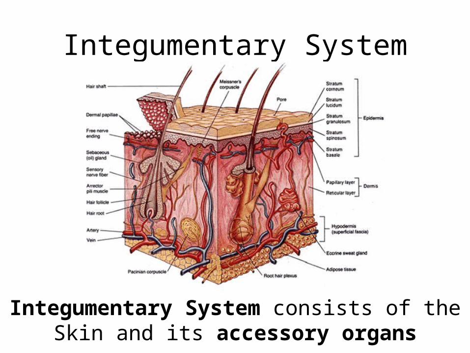
Integumentary System
Integumentary System consists of theSkin and its accessory organs
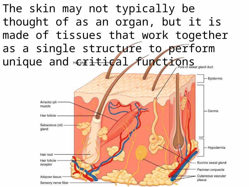
The skin may not typically be thought of as an organ, but it is made of tissues that work together as a single structure to perform unique and critical functions
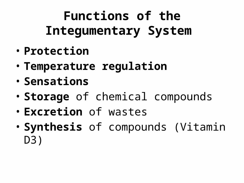
Functions of the Integumentary System
• Protection• Temperature regulation• Sensations• Storage of chemical compounds• Excretion of wastes• Synthesis of compounds (Vitamin D3)
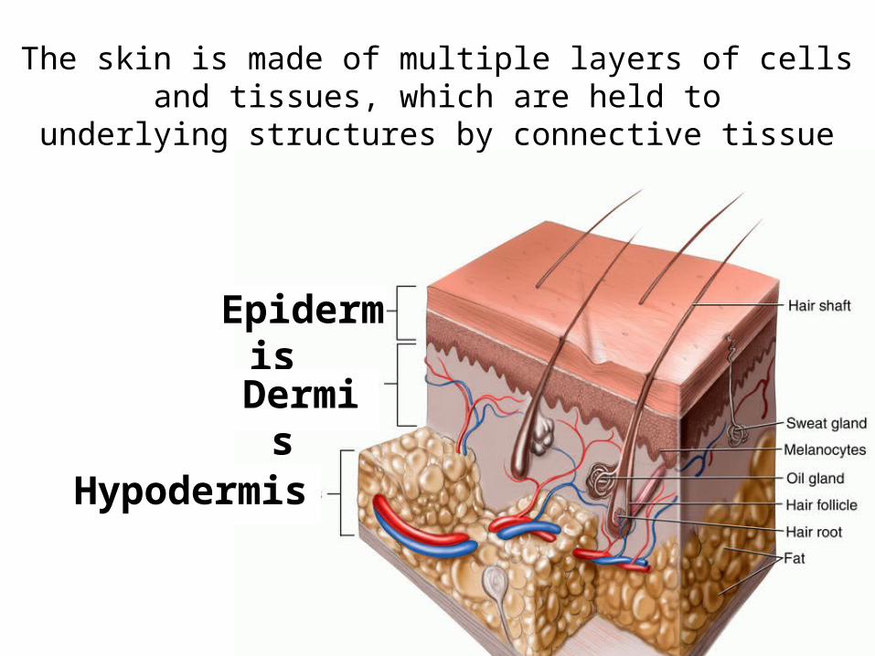
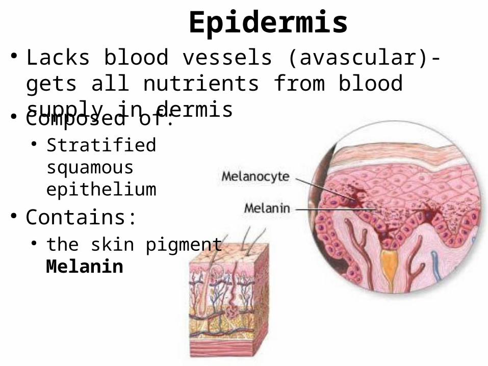
Epidermis
Dermis
Hypodermis
The skin is made of multiple layers of cells and tissues, which are held to
underlying structures by connective tissue
Epidermis
● Composed of:● Stratified squamous
epithelium● Contains:
● the skin pigment Melanin
● Lacks blood vessels (avascular)- gets all nutrients from blood supply in dermis
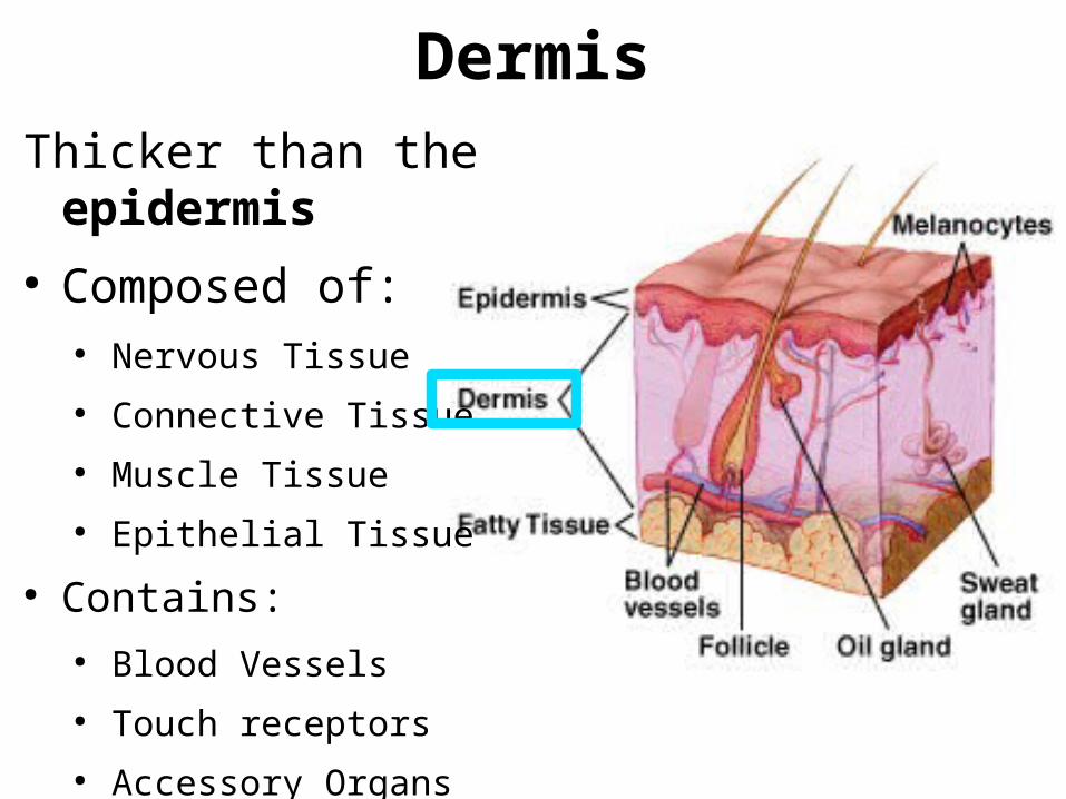
DermisThicker than the epidermis ● Composed of:
● Nervous Tissue● Connective Tissue● Muscle Tissue● Epithelial Tissue
● Contains:● Blood Vessels● Touch receptors● Accessory Organs
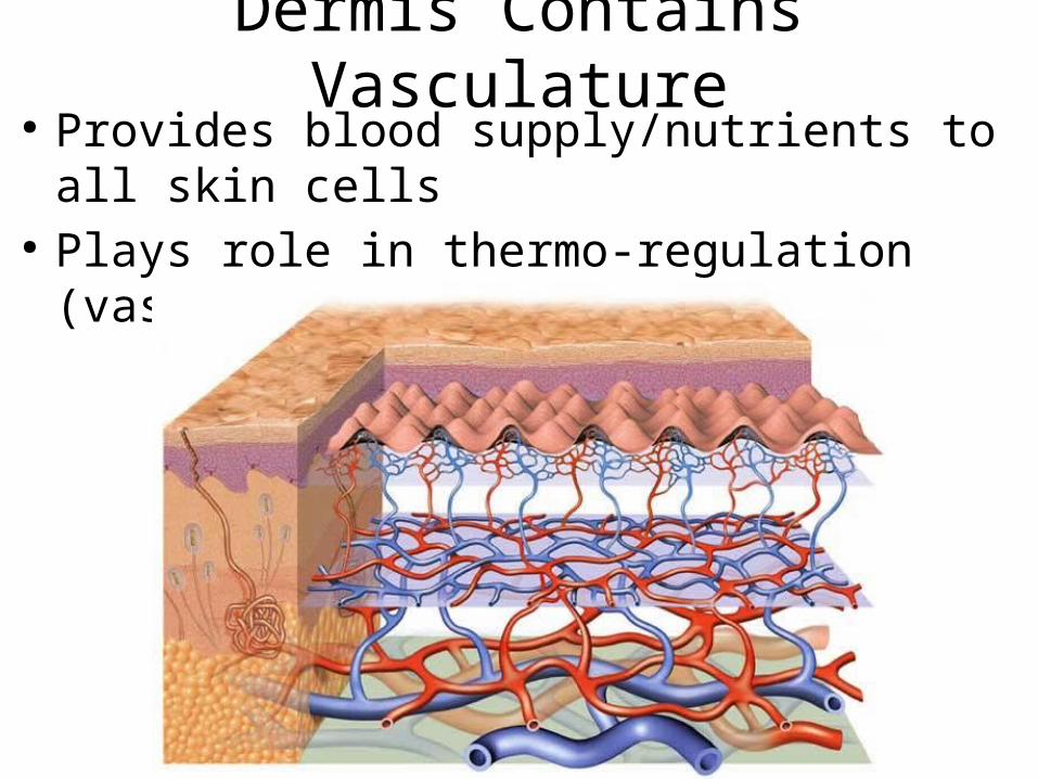
Dermis Contains Vasculature● Provides blood supply/nutrients to all skin cells● Plays role in thermo-regulation
(vaso dialation/constriction)
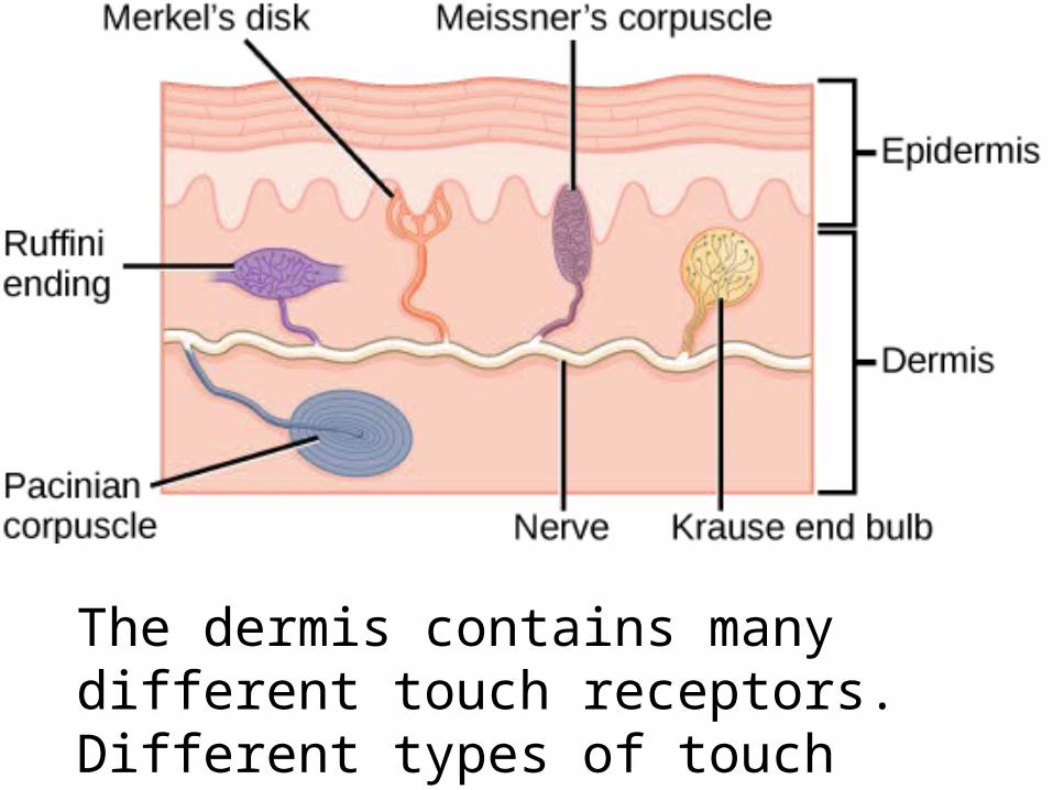
The dermis contains many different touch receptors. Different types of touch have different receptors.
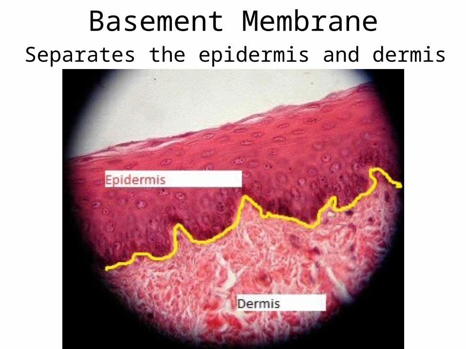
Basement MembraneSeparates the epidermis and dermis
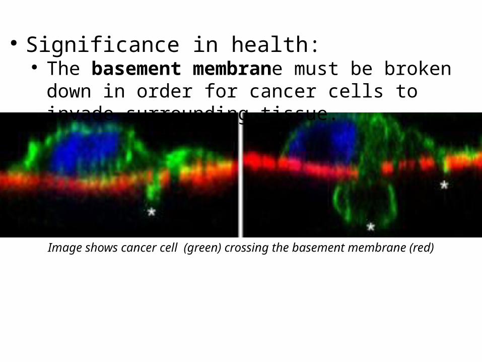
● Significance in health:● The basement membrane must be broken down in
order for cancer cells to invade surrounding tissue.
Image shows cancer cell (green) crossing the basement membrane (red)
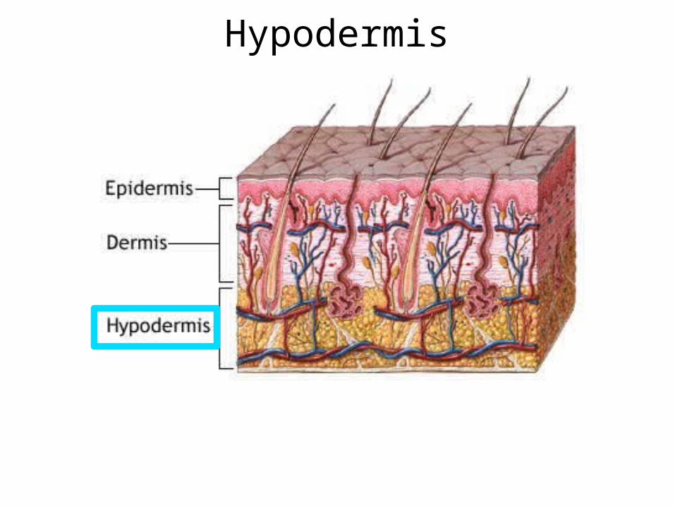
Hypodermis
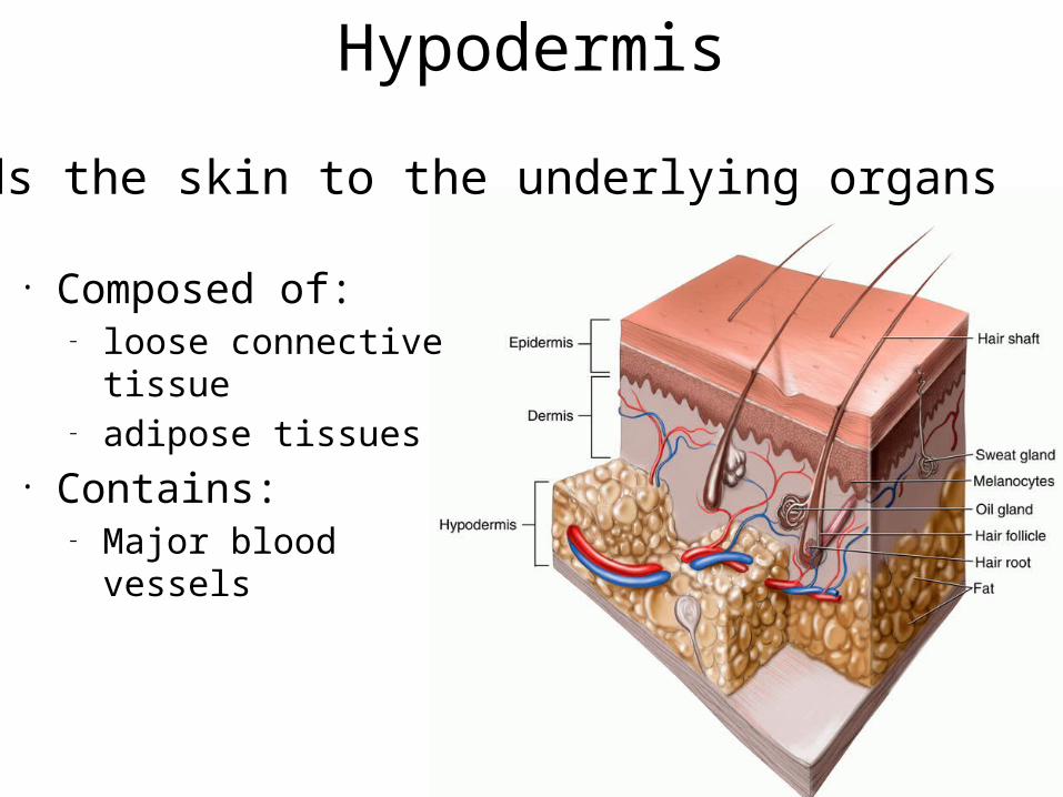
Hypodermis
• Composed of:– loose connective tissue– adipose tissues
• Contains:– Major blood vessels
•Binds the skin to the underlying organs
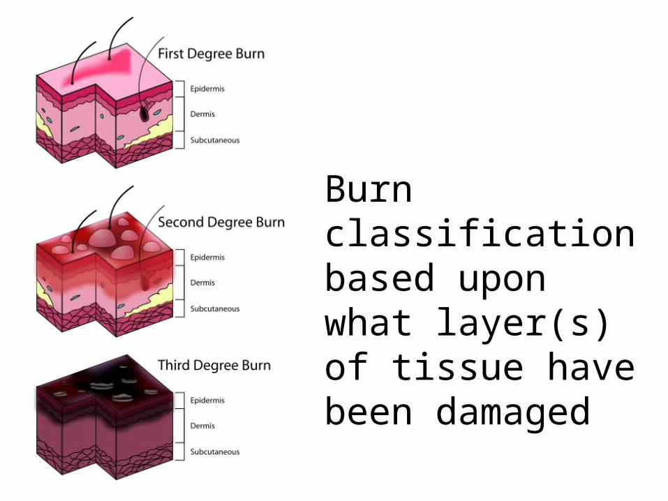
Burn classification based upon what layer(s) of tissue have been damaged
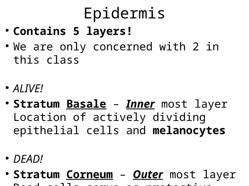
Epidermis● Contains 5 layers!● We are only concerned with 2 in this class
● ALIVE!● Stratum Basale – Inner most layer
Location of actively dividing epithelial cells and melanocytes
● DEAD!● Stratum Corneum – Outer most layer
Dead cells serve as protective barrier
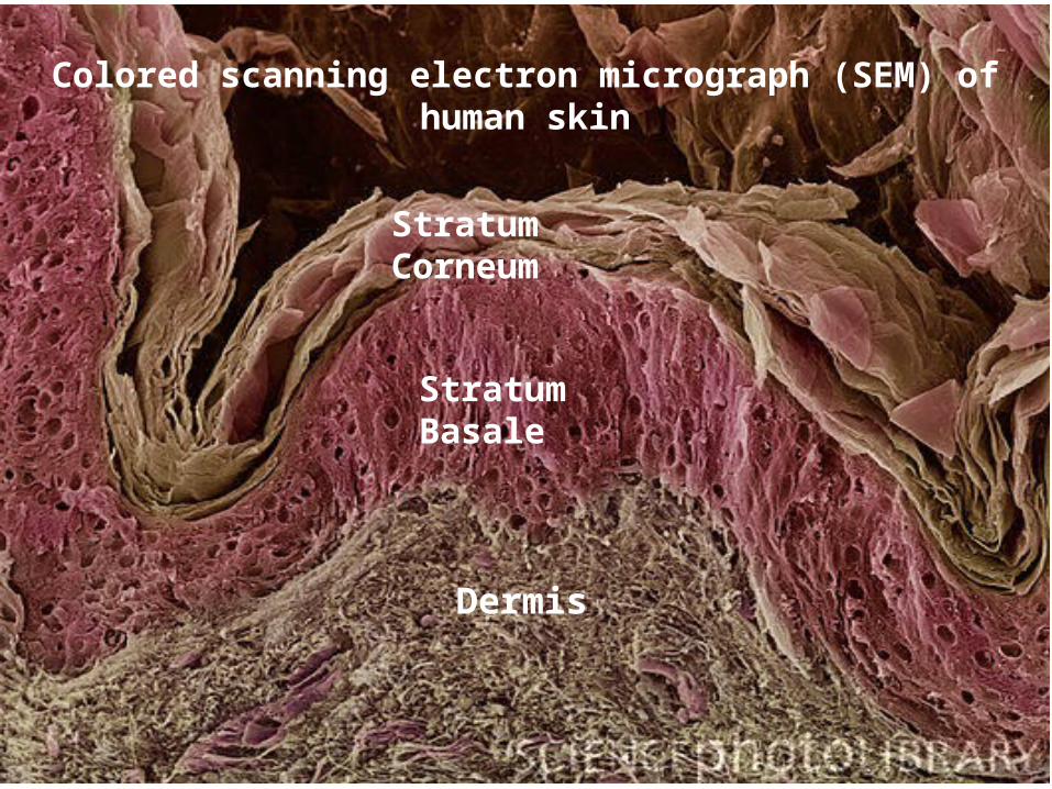
Stratum Corneum
Stratum Basale
Dermis
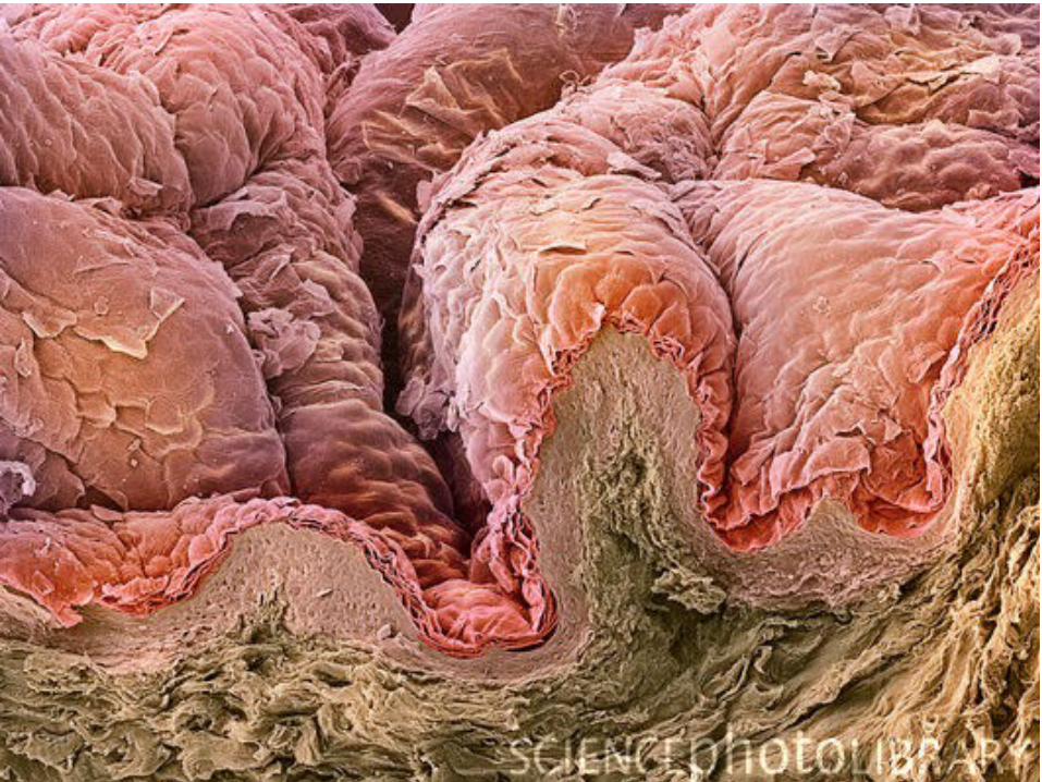
Colored scanning electron micrograph (SEM) of human skin
Keratinization● Cells divide in Stratum Basale and push
older cells up to surface. ● Cells starve and die as pushed away from
dermis.● Keratinization is the hardening process
these cells undergo as they die.
Keratin is a type of fibrous structural proteins.
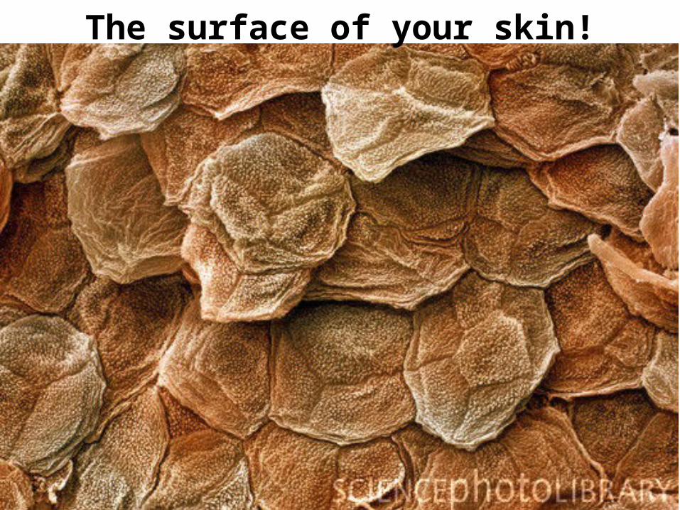
The surface of your skin!
Keratinization● Keratinization- the cementing of keratin
fibers(cytoskeleton protein) in the dead cells.
● Constant shedding and replacement grows new EPIdermis every 25-45 days
● Calluses- Thickening of the Stratum Corneum due to rubbing and pressure
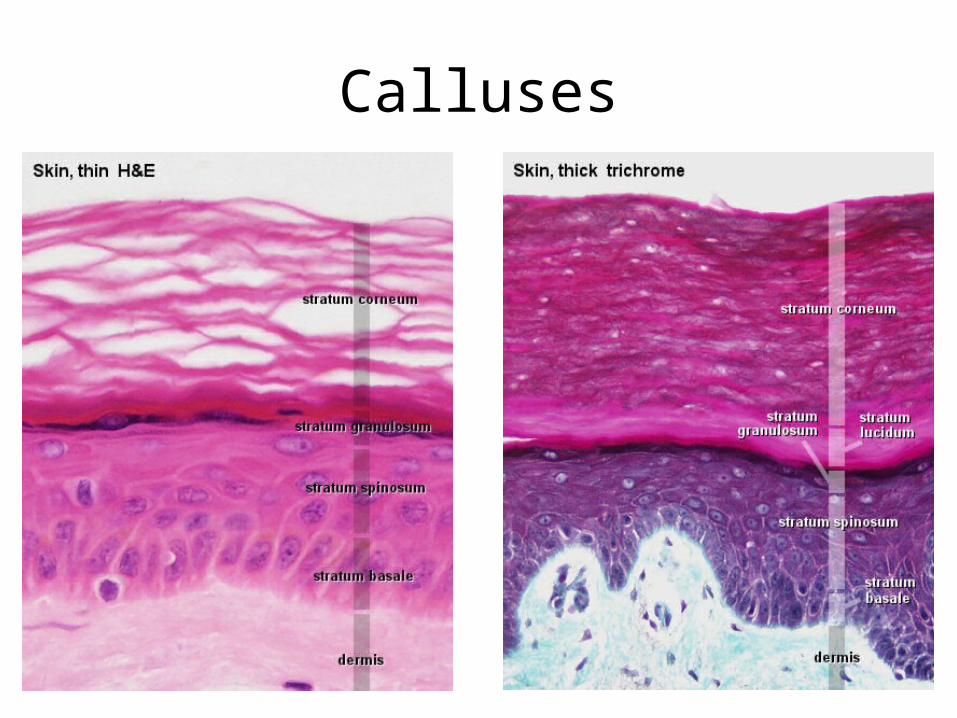
Calluses
Accessory Structures of the Skin
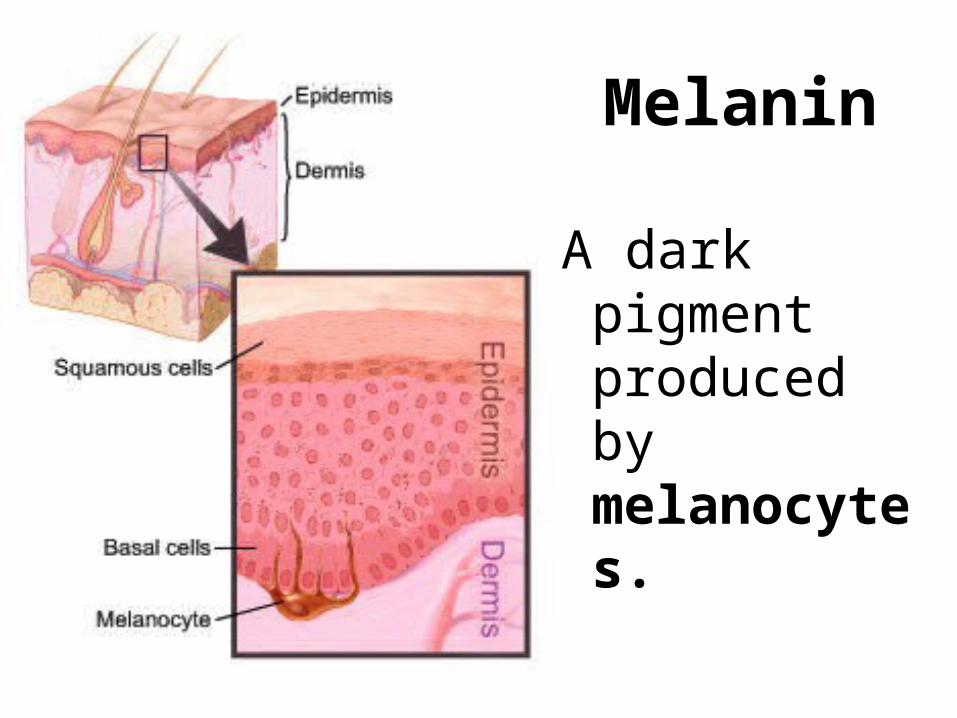
A dark pigment produced by melanocytes.
Melanin
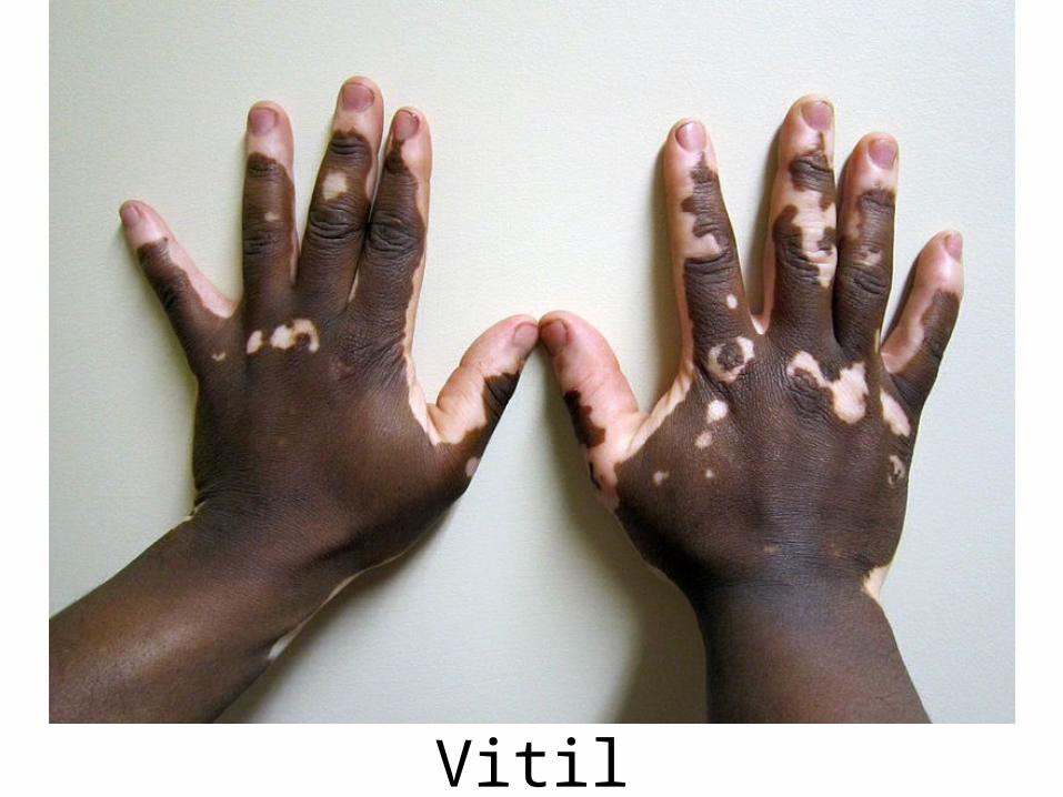
Vitiligo
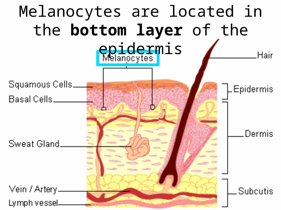
Melanocytes are located in the bottom layer of the epidermis
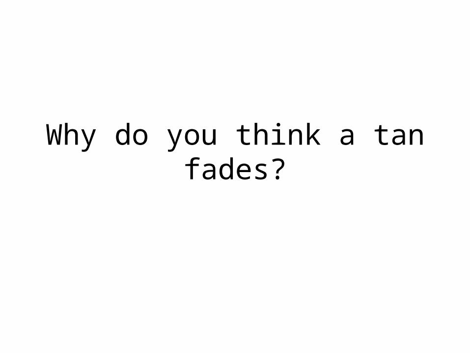
Why do you think a tan fades?
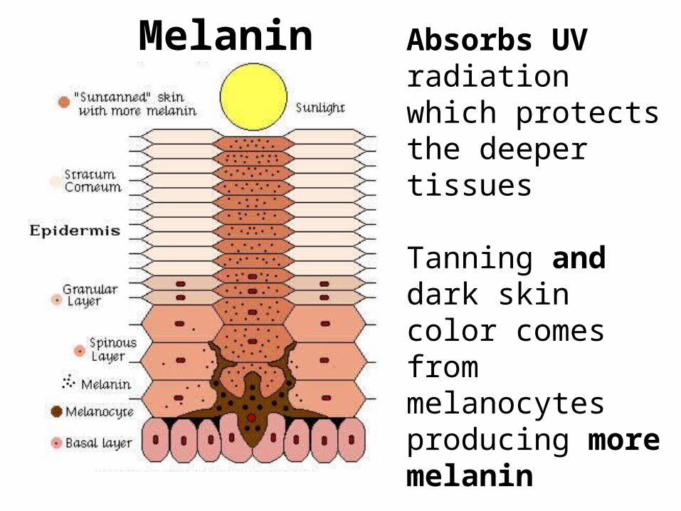
Absorbs UV radiation which protects the deeper tissues
Tanning and dark skin color comes from melanocytes producing more melanin
NOT due to more melanocytes
Melanin
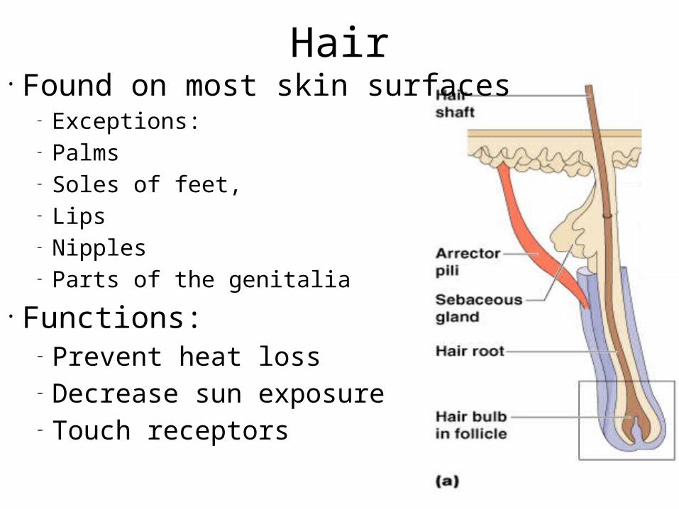
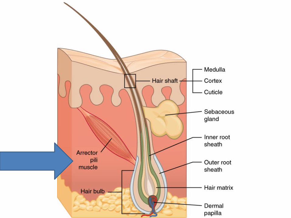
Hair• Found on most skin surfaces
– Exceptions: – Palms – Soles of feet, – Lips– Nipples– Parts of the genitalia
• Functions:– Prevent heat loss– Decrease sun exposure– Touch receptors
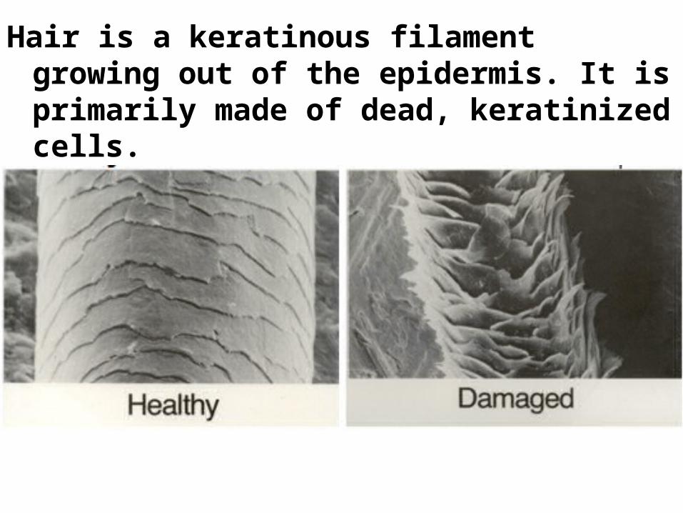
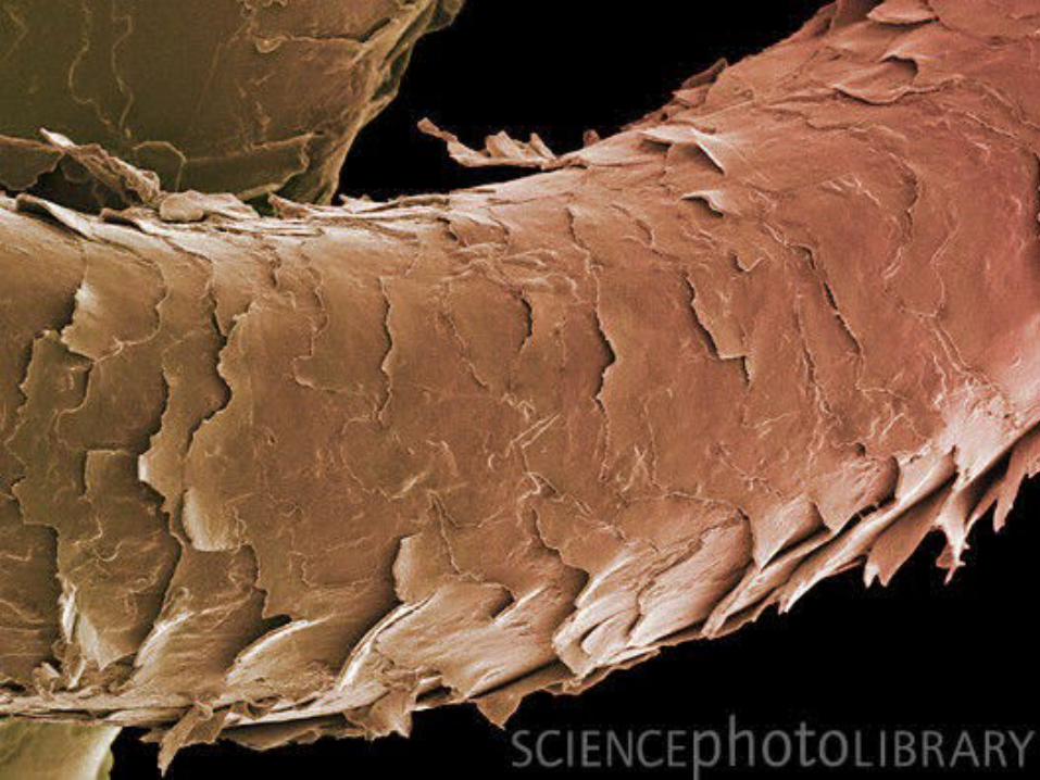
Hair is a keratinous filament growing out of the epidermis. It is primarily made of dead, keratinized cells.
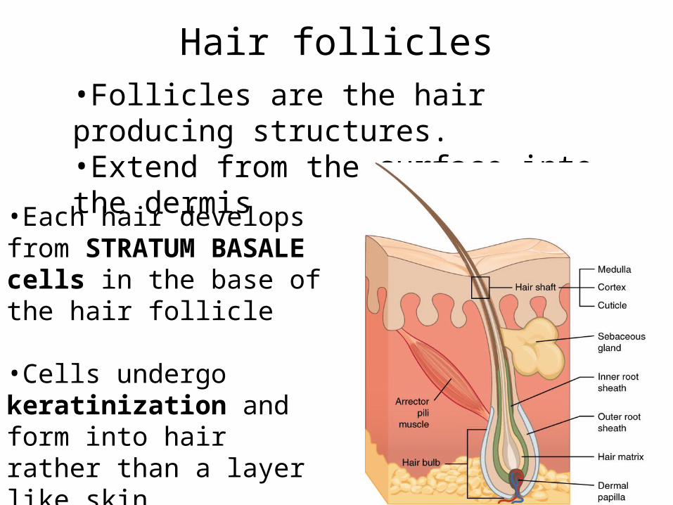
Hair follicles•Follicles are the hair producing structures.•Extend from the surface into the dermis
•Each hair develops from STRATUM BASALE cells in the base of the hair follicle
•Cells undergo keratinization and form into hair rather than a layer like skin
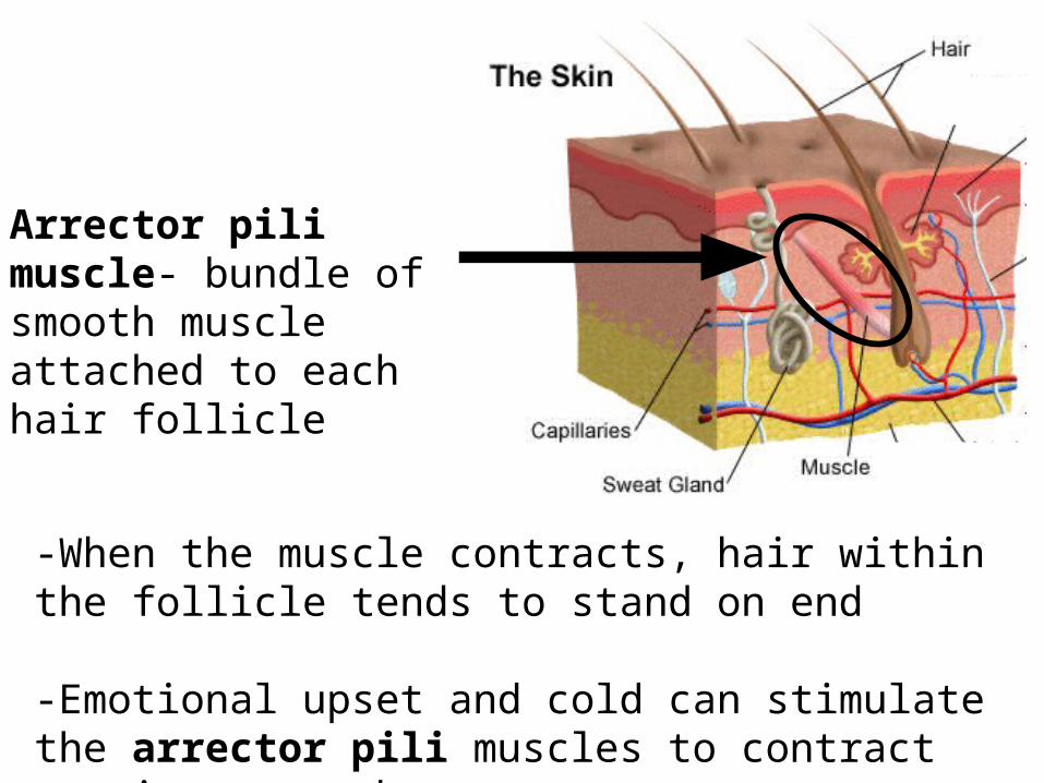
Arrector pili muscle- bundle of smooth muscle attached to each hair follicle
-When the muscle contracts, hair within the follicle tends to stand on end
-Emotional upset and cold can stimulate the arrector pili muscles to contract causing goose bumps
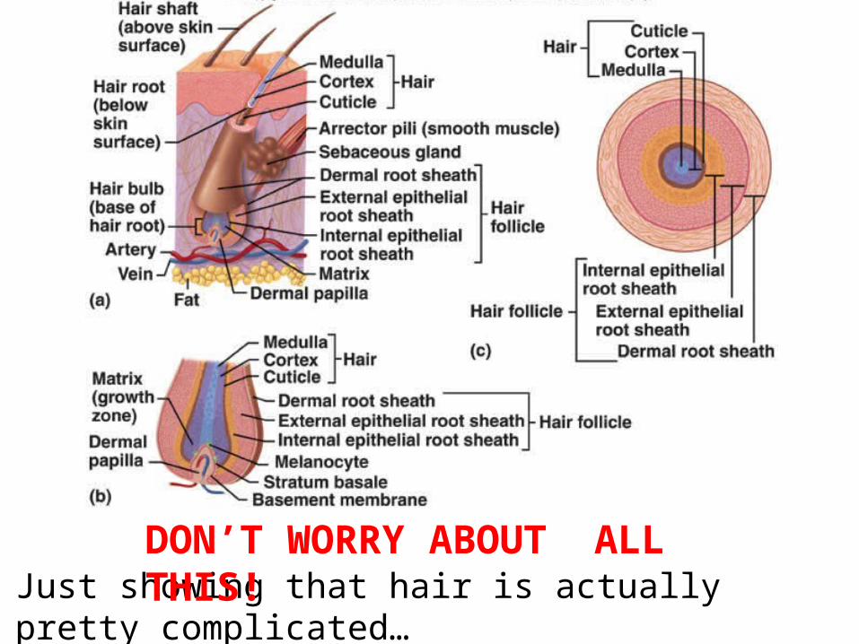
Just showing that hair is actually pretty complicated…DON’T WORRY ABOUT ALL THIS!
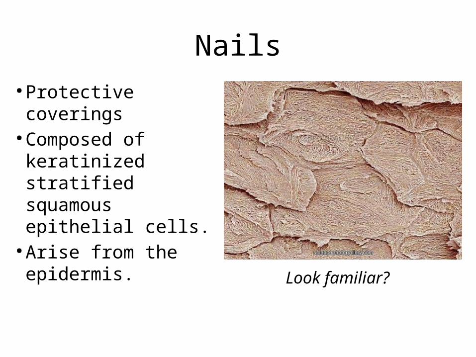
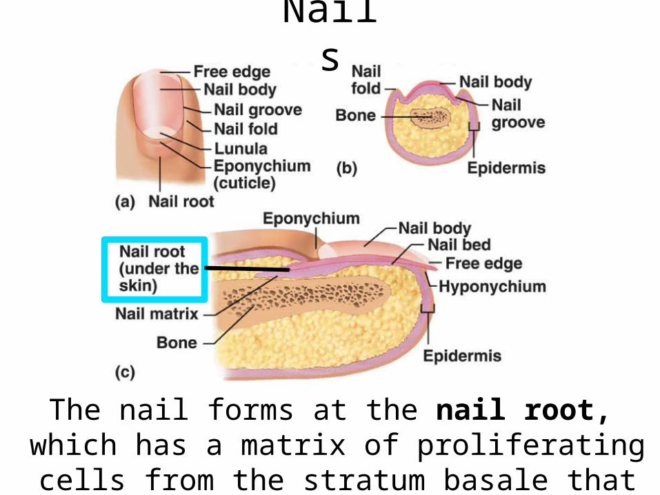
Nails● Protective coverings
● Composed of keratinized stratified squamous epithelial cells.
● Arise from the epidermis. Look familiar?
Nails
The nail forms at the nail root, which has a matrix of proliferating cells from the stratum basale that
enables the nail to grow continuously
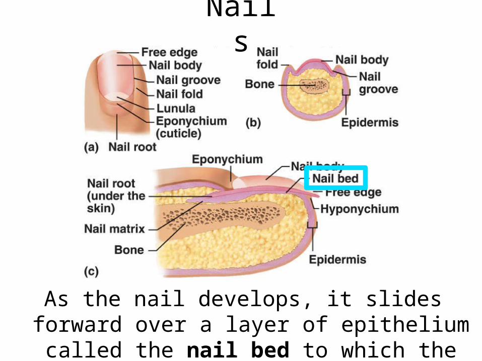
As the nail develops, it slides forward over a layer of epithelium called the nail bed to which the nail
remains attached
Nails
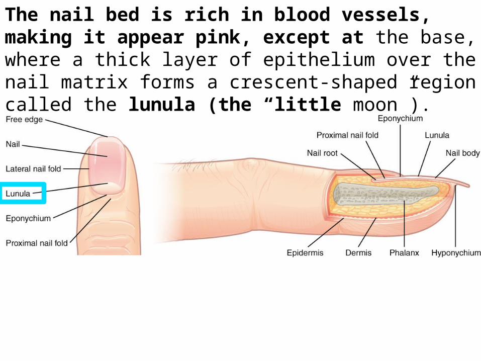
The nail bed is rich in blood vessels, making it appear pink, except at the base, where a thick layer of epithelium over the nail matrix forms a crescent-shaped region called the lunula (the “little moon”).
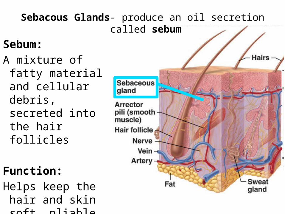
Sebum:
A mixture of fatty material and cellular debris, secreted into the hair follicles
Function:
Helps keep the hair and skin soft, pliable and relatively waterproof.
Sebacous Glands- produce an oil secretion called sebum
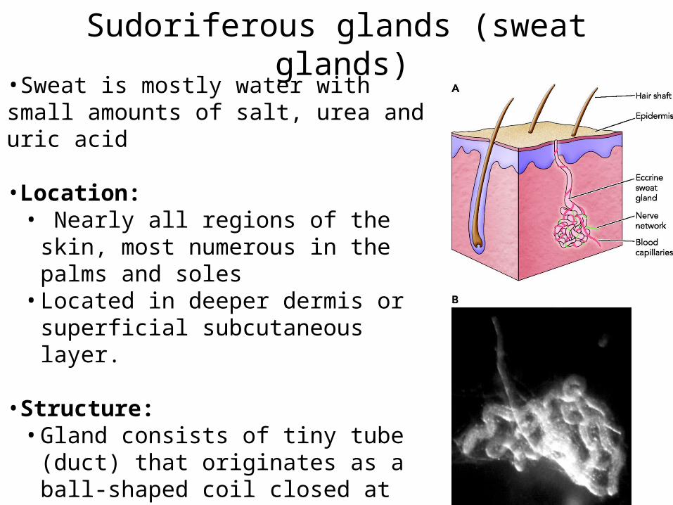
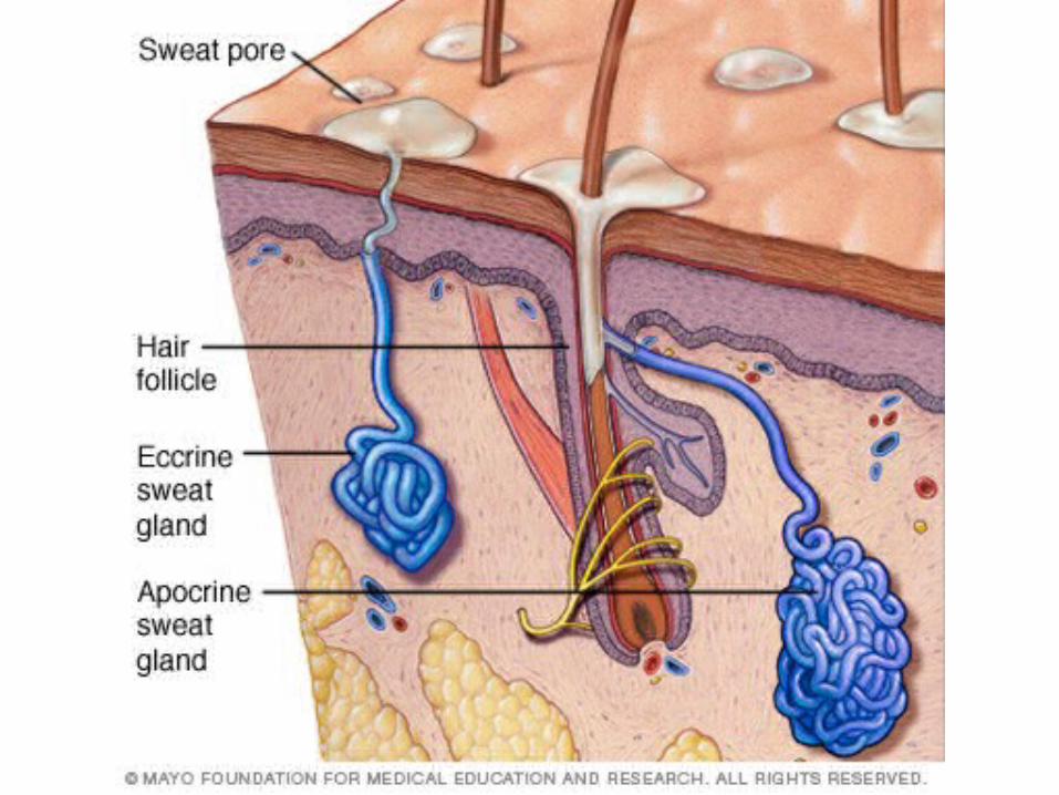
Sudoriferous glands (sweat glands)•Sweat is mostly water with small amounts of salt, urea and uric acid
•Location:• Nearly all regions of the skin, most
numerous in the palms and soles• Located in deeper dermis or superficial
subcutaneous layer.
•Structure:• Gland consists of tiny tube (duct) that
originates as a ball-shaped coil closed at its deepest end
• Coiled duct lined with sweat secreting epithelial cells
• Duct opens at the surface as a pore
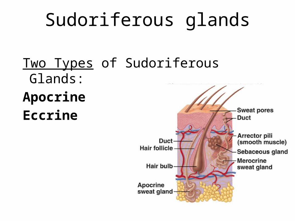
Two Types of Sudoriferous Glands:
Apocrine
Eccrine
Sudoriferous glands
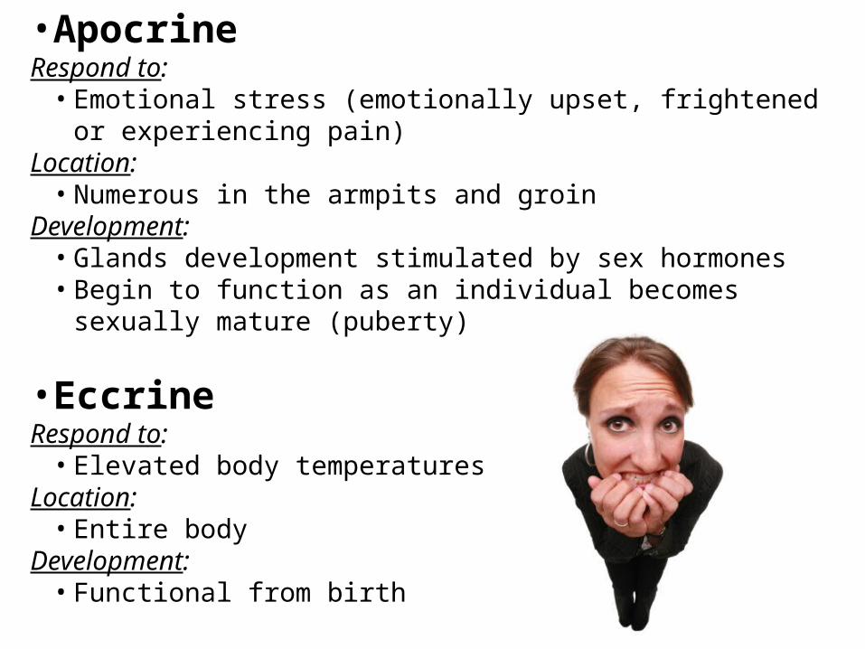
•ApocrineRespond to:
• Emotional stress (emotionally upset, frightened or experiencing pain)
Location:• Numerous in the armpits and groin
Development:• Glands development stimulated by sex hormones• Begin to function as an individual becomes sexually mature
(puberty)
•EccrineRespond to:
• Elevated body temperaturesLocation:
• Entire bodyDevelopment:
• Functional from birth
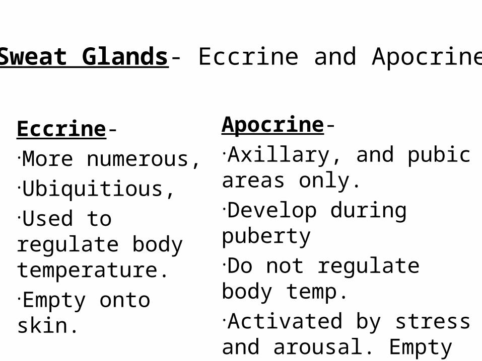
Eccrine- •More numerous, •Ubiquitious, •Used to regulate body temperature. •Empty onto skin.
Apocrine- •Axillary, and pubic areas only. •Develop during puberty •Do not regulate body temp. •Activated by stress and arousal. Empty onto hair follicle.
Sweat Glands- Eccrine and Apocrine

Diseases, Disorders, and Injuries of the Integumentary System
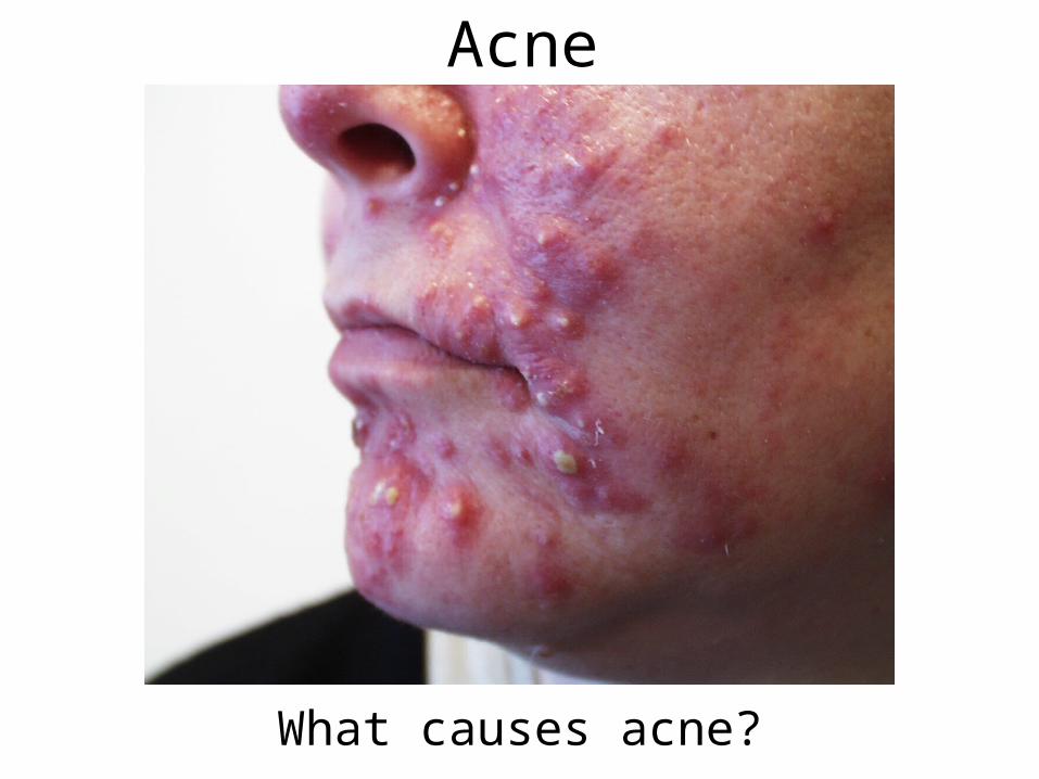
Acne
What causes acne?
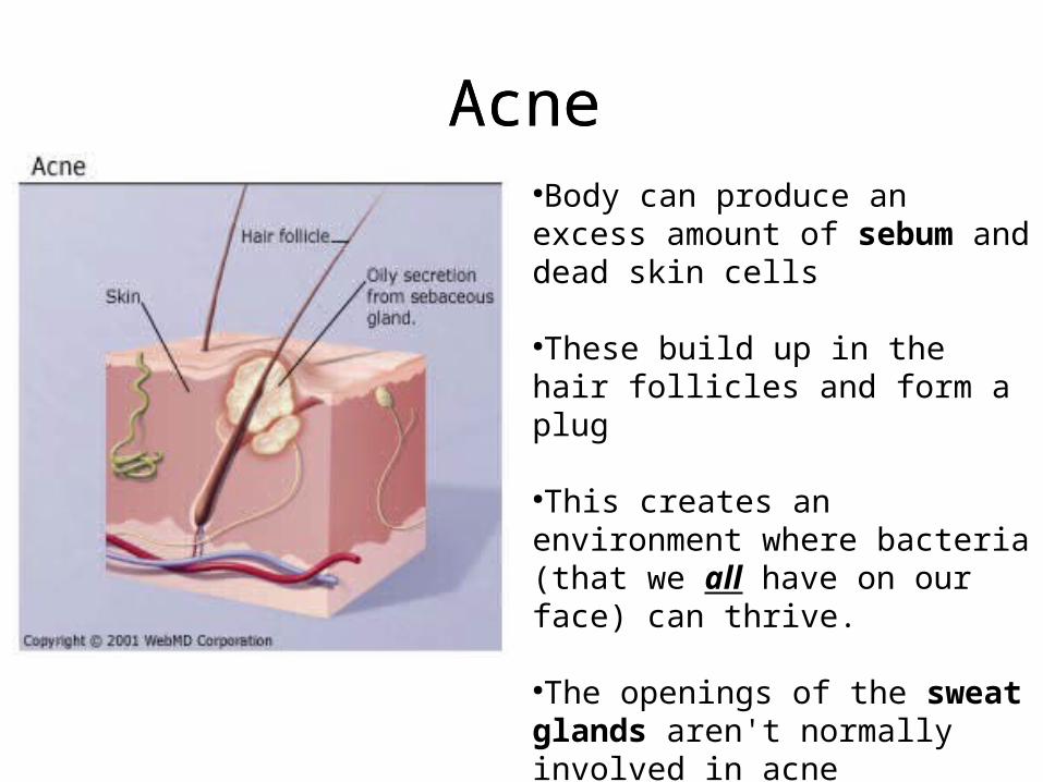
Acne●Body can produce an excess amount of sebum and dead skin cells
●These build up in the hair follicles and form a plug
●This creates an environment where bacteria (that we all have on our face) can thrive.
●The openings of the sweat glands aren't normally involved in acne
Acne
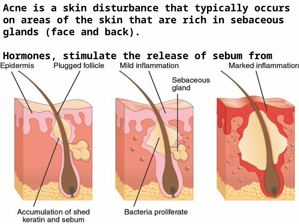
Acne is a skin disturbance that typically occurs on areas of the skin that are rich in sebaceous glands (face and back).
Hormones, stimulate the release of sebum from sebaceous glands.
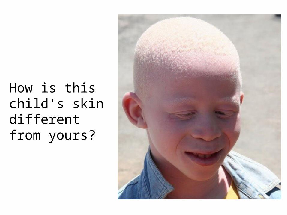
How is this child's skin different from yours?
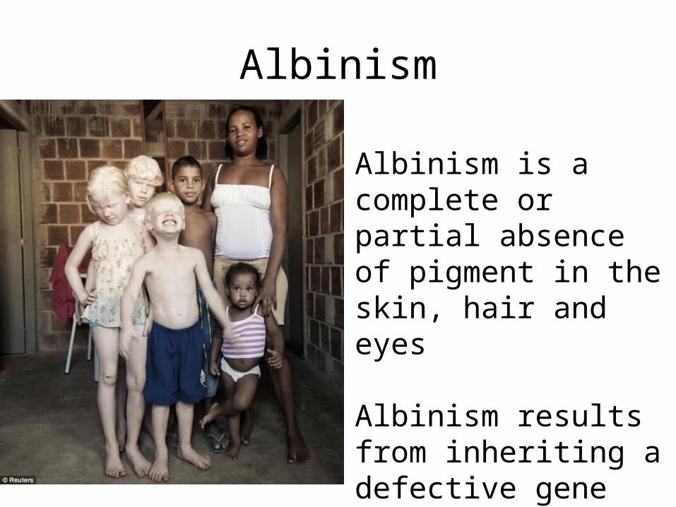
Albinism
Albinism is a complete or partial absence of pigment in the skin, hair and eyes
Albinism results from inheriting a defective gene involved in the production of melanin.
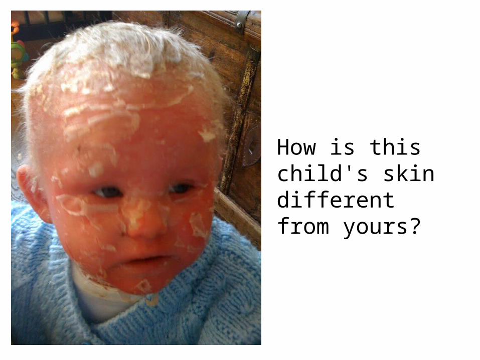
How is this child's skin different from yours?

Netherton Syndrome
Defect in keritinization causes over shedding of cells
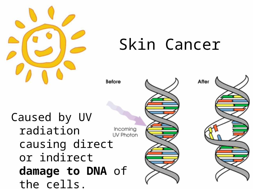
Skin Cancer
Caused by UV radiation causing direct or indirect damage to DNA of the cells.
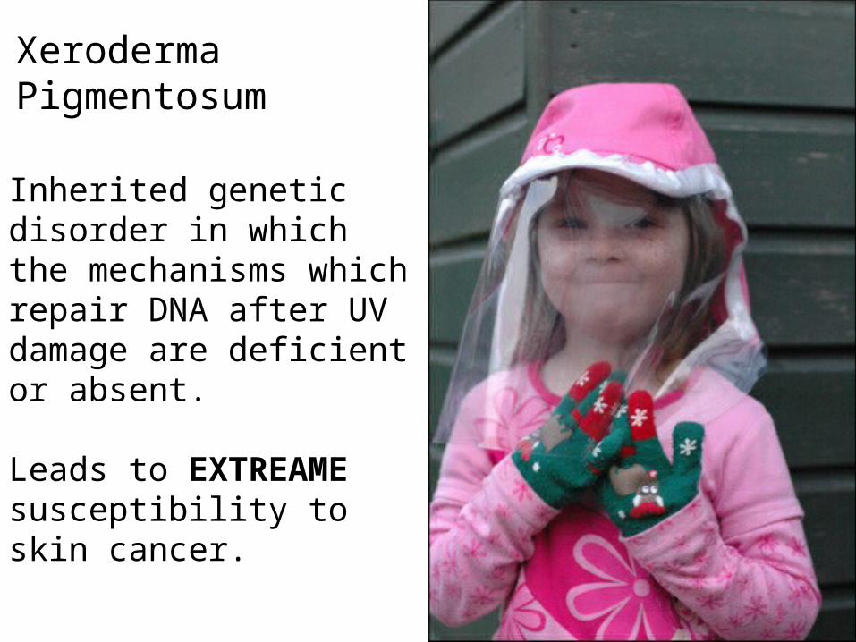
Xeroderma Pigmentosum
Inherited genetic disorder in which the mechanisms which repair DNA after UV damage are deficient or absent.
Leads to EXTREAME susceptibility to skin cancer.

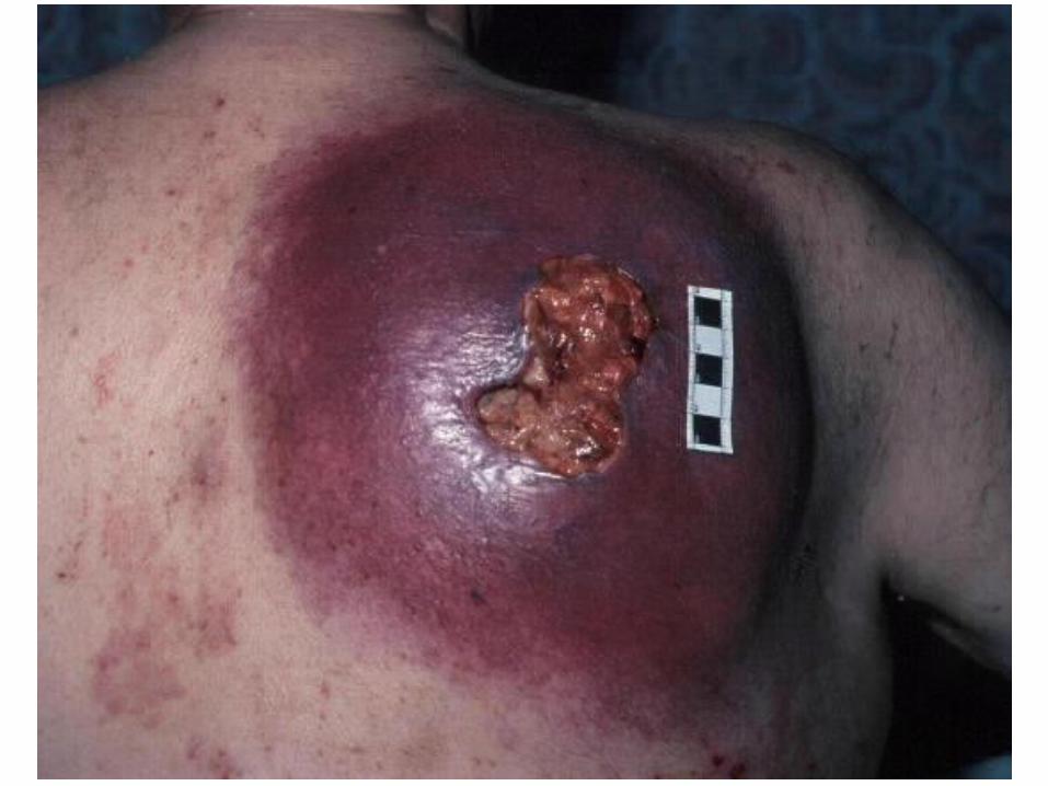
WARNING!GRAPHIC CONTENT!