Embed Size (px)
Citation preview
Da
ID
a
ARRAA
KEFES
1
tewp[fiai
ecioibo
U
h2c
CASE REPORT – OPEN ACCESSInternational Journal of Surgery Case Reports 17 (2015) 55–57
Contents lists available at ScienceDirect
International Journal of Surgery Case Reports
journa l h omepage: www.caserepor ts .com
ouble esophageal perforation by ingested foreign body: Endoscopicnd surgical approach. A case report.
. Ugenti, R. Digennaro, G. Martines ∗, O. Caputi Iambrenghiepartment of Emergency and Organ Trasplantation, University of Bari, Italy
r t i c l e i n f o
rticle history:eceived 16 July 2015eceived in revised form 22 October 2015ccepted 25 October 2015vailable online 30 October 2015
eywords:sophageal perforationoreign bodyndoscopic treatmenturgical treatment
a b s t r a c t
INTRODUCTION: Esophageal perforation in adults is most frequently caused by ingested foreign bodies.They can migrate through the esophageal wall, damaging the nearby organs such as the aorta or thetrachea, with fatal outcome. After the diagnosis, the viable treatments for extracting the foreign bodyand repairing the perforation are several. The appropriate treatment, may be endoscopic, surgical orcombined, depending on the level of the perforation, on the co-morbidities of the patient and on theavailable resources.PRESENTATION OF CASE: This paper describes a case of a 68 years old patient with a double EP caused bya meat-bone that perforated the thoracic esophageal wall, approaching the aorta on the left side and theazygos vein on the right side.DISCUSSION: Because of the double transfixion and the position near the aorta and the azygos, it wasnot possible to remove safely the bone during the endoscopy. The management required a combinedendoscopic and surgical approach. This way it was possible to detect easily the location of the perforation,
to remove safely the foreign body, to repair the perforation both from the outside and from the inside,and to place the nasogastric tube under direct vision.CONCLUSION: Even when the type of esophageal perforation requires surgical treatment, the simultane-ous use of endoscopy proved to be an advantage in order to extract the foreign body safely, to perform aratioPublihe CC
double repair of the perfo© 2015 The Authors.
access article under t
. Introduction
Esophageal perforation (EP) is a rare and potentially life-hreatening event with an 20% survival rate [1]. The first mention ofsophageal perforation was made by Hermann Boerhaave in 1724,ho reported a spontaneous rupture after repeated vomiting inost-mortem evidence in the Grand Admiral of the Dutch fleet2]. Two hundred years later, in 1947, Barrett and Olson made therst attempts at surgical repair of a EP [3–4]. Since then, variouspproaches have been discussed, but its treatment is still challeng-ng as there is still no agreed management algorithm.
Esophageal perforation can happen due to a number of differ-nt causes. It may be iatrogenic (most frequently), spontaneous oraused by trauma, tumors or foreign bodies. Ingested foreign bod-es are responsible for 80% of cervical perforations1 and for 9–35%f all esophageal perforations [5]. In adult patients, the foreign bod-
es most commonly involved are dental prostheses and meat or fishones. Usually, the foreign body hits the esophageal wall at the levelf the narrow portion of the esophagus, Prolonged contact can lead∗ Corresponding author at: Department of Emergency and Organ Trasplantation,niversity of Bari, Piazza G. Cesare, 11 70124 Bari, Italy.
E-mail address: [email protected] (G. Martines).
ttp://dx.doi.org/10.1016/j.ijscr.2015.10.033210-2612/© 2015 The Authors. Published by Elsevier Ltd. on behalf of IJS Publishing Greativecommons.org/licenses/by-nc-nd/4.0/).
n and to place the nasogastric tube under direct vision.shed by Elsevier Ltd. on behalf of IJS Publishing Group Ltd. This is an open
BY-NC-ND license (http://creativecommons.org/licenses/by-nc-nd/4.0/).
to extraluminal migration of the foreign body which can perforatenearby organs, such as the trachea or the aorta, often with fatalconsequences [6] or it can cause infection which spreads easily asthe esophagus is surrounded by loose stromal connective tissue.
Making a diagnosis of thoracic EP is difficult because the symp-toms may simulate other disorders, like myocardial infarction,aortic dissection, spontaneous pneumothorax or pulmonary dis-ease. The most common early symptoms and signs are: vomiting(84%), thoracic pain (79%), dyspnea (53%), epigastric pain (47%) anddysphagia (21%). This atypical clinical presentation explains why,in 60% of cases, diagnosis takes longer than 24 h [7].
Usually diagnosis requires radiologic and endoscopic exami-nation. Conventional radiology and contrast radiology can showpneumothorax, pneumoperitoneum, pneumomediastinum, subcu-taneous emphysema and occasionally may also highlight a foreignbody. A CT scan can help confirm the diagnosis. For the detectionof EP, the sensitivity of the endoscopy is nearly 100%, and its speci-ficity is 83%, but it is not always recommended because the airinsufflation caused can enlarge the perforation [8]. Nevertheless,endoscopy is the only exam that can be diagnostic and therapeuticat the same time, enabling the extraction of the foreign body.
Once the foreign body has been indentified, there are sev-eral possible therapeutic strategies for its extraction, ranging fromendoscopy to surgery including traditional thoracotomy and video-
roup Ltd. This is an open access article under the CC BY-NC-ND license (http://
CASE REPORT – OPEN ACCESS56 I. Ugenti et al. / International Journal of Surgery Case Reports 17 (2015) 55–57
apcTfo
1
rai
setto1
iwas
t
ae
msg
Asie
es
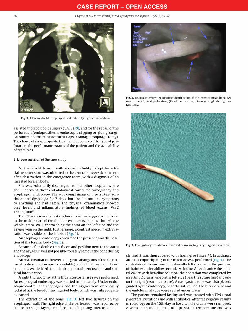
Fig. 2. Endoscopic view: endoscopic identification of the ingested meat–bone. (A)meat bone; (B) right perforation; (C) left perforation; (D) outside light during tho-racotomy.
Fig. 1. CT scan: double esophageal perforation by ingested meat–bone.
ssisted thoracoscopic surgery (VATS) [9], and for the repair of theerforation (endoprosthesis, endoscopic clipping or gluing, surgi-al suture and/or reinforcement flaps, drainage, esophagectomy).he choice of an appropriate treatment depends on the type of per-oration, the performance status of the patient and the availabilityf resources.
.1. Presentation of the case study
A 68-year-old female, with no co-morbidity except for arte-ial hypertension, was admitted to the general surgery departmentfter observation in the emergency room, with a diagnosis of anngested foreign body.
She was voluntarily discharged from another hospital, wherehe underwent chest and abdominal computed tomography andsophageal endoscopy. She was complaining of a persistent sorehroat and dysphagia for 7 days, but she did not link symptomso anything she had eaten. The physical examination showednly fever, and inflammatory findings of blood exams: WBC4,090/mm3.
The CT scan revealed a 4 cm linear shadow suggestive of bonen the middle part of the thoracic esophagus, passing through the
hole lateral wall, approaching the aorta on the left side and thezygos vein on the right. Furthermore, a contrast medium extrava-ation was visible on the left side (Fig. 1).
An esophageal endoscopy confirmed the presence and the posi-ion of the foreign body (Fig. 2).
Because of its double transfixion and position next to the aortand the azygos, it was not possible to safely remove the bone duringndoscopy.
After a consultation between the general surgeons of the depart-ent (where endoscopy is available) and the throat and heart
urgeons, we decided for a double approach, endoscopic and sur-ical intervention.
A right thoracotomy at the fifth intercostal area was performed.n esophageal endoscopy was started immediately. Under endo-copic control, the esophagus and the azygos vein were easilysolated at the level of the ingested body, which was subsequently
xtracted.The extraction of the bone (Fig. 3) left two fissures on thesophageal wall. The right edge of the perforation was repaired byuture in a single layer, a reinforcement flap using intercostal mus-
Fig. 3. Foreign body: meat–bone removed from esophagus by surgical extraction.
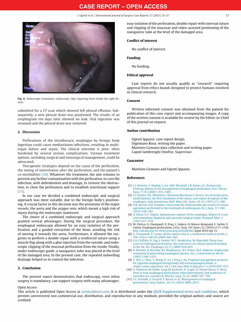
cle, and it was then covered with fibrin glue (Tiseel®). In addition,an endoscopic clipping of the mucosae was performed (Fig. 4). Thecontralateral fissure was intentionally left open with the purposeof draining and enabling secondary closing. After cleaning the pleu-ral cavity with betadine solution, the operation was completed byinserting 2 drains: one on the left side (near the suture line) and oneon the right (near the fissure). A nasogastric tube was also placed,guided by the endoscopy, near the suture line. The three drains andthe endoluminal tube were sealed under water.
The patient remained fasting and was treated with TPN (totalparenteral nutrition) and with antibiotics. After the negative resultsin radiology on the 15th day in hospital, the drains were removed.A week later, the patient had a persistent temperature and was
CASE REPORT – OI. Ugenti et al. / International Journal of Su
Fs
sser
2
ioboa
tcpit[
aivi
gefogmsuod
3
s
OTpc
ig. 4. Endoscopic treatment: endoscopic clips repairing from inside the right fis-ure.
ubmitted for a CT scan which showed left pleural effusion. Sub-equently, a new pleural drain was positioned. The results of ansophogram ten days later showed no leak. Oral ingestion wasesumed and the pleural drain was removed.
. Discussion
Perforations of the intrathoracic esophagus by foreign bodyngestion could cause mediastinum infections, resulting in multi-rgan failure and sepsis. The clinical outcome is poor, oftenurdened by several serious complications. Various treatmentptions, including surgical and nonsurgical management, could bedvocated.
Therapeutic strategies depend on the cause of the perforation,he timing of intervention after the perforation, and the patient’so-morbidities [10]. Whatever the treatment, the aim remains torevent any further contamination with the perforation, to cure the
nfection, with debridement and drainage, to remove the obstruc-ion, to close the perforation and to establish nutritional support11].
In our case we decided a combined endoscopic and surgicalpproach was most suitable, due to the foreign body’s position-ng. A crucial factor in this decision was the proximity of the majoressels, the aorta and the azygos vein, which meant a high risk ofnjury during the endoscopic maneuver.
The choice of a combined endoscopic and surgical approachranted several advantages. During the surgical procedure, thesophageal endoscopy allowed for an easy isolation of the per-oration and a guided extraction of the bone, avoiding the riskf moving it towards the aorta. Furthermore, it allowed the sur-eons to perform a double repair with a reinforced suture using auscle flap along with a glue injection from the outside, and endo-
copic clipping of the mucosal perforation from the inside. Finally,nder endoscopic guide, a nasogastric tube was placed at the levelf the damaged area. In the present case, the repeated indwellingrainage helped us to control the infection.
. Conclusion
The present report demonstrates that endoscopy, even whenurgery is mandatory, can support surgery with many advantages:
[
[
pen Accesshis article is published Open Access at sciencedirect.com. It is distribermits unrestricted non commercial use, distribution, and reproductredited.
PEN ACCESSrgery Case Reports 17 (2015) 55–57 57
easy isolation of the perforation, double repair with external sutureand clipping of the mucosae and video-assisted positioning of thenasogastric tube at the level of the damaged area.
Conflict of interest
No conflict of interest.
Funding
No funding.
Ethical approval
Case reports do not usually qualify as “research” requiringapproval from ethics boards designed to protect humans involvedin clinical research.
Consent
Written informed consent was obtained from the patient forpublication of this case report and accompanying images. A copyof the written consent is available for review by the Editor-in-Chiefof this journal on request.
Author contribution
Ugenti Ippazio: case report design.Digennaro Rosa: writing the paper.Martines Gennaro data collection and writing paper.Caputi Iambrenghi Onofrio: Supervisor.
Guarantor
Martines Gennaro and Ugenti Ippazio.
References
[1] C.J. Brinster, S. Singhal, L. Lee, M.B. Marshall, L.R. Kaiser, J.C. Kucharczuk,Evolving options in the management of esophageal perforation, Ann. Thorac.Surg. 77 (4) (2004) 1475–1483.
[2] V.J. Derbes, R.E. Mitchell Jr., Hermann Boerhaave’s Atrocis, nec descripti prius,morbi historia, the first translation of the classic case report of rupture of theesophagus, with annotations, Bull. Med. Libr. Assoc. 43 (2) (1955) 217–240.
[3] N.R. Barrett, R.H. Franklin, Concerning the unfavourable late results of certainoperations performed in the treatment of cardiospasm, Br. J. Surg. 37 (146)(2015) 194–202.
[4] A. Olson, O.T. Clagett, Spontaneous rupture of the esophagus. Report of a casewith immediate diagnosis and succesful surgical repair, Postgrad. Med. 2(1947) 417–419.
[5] M. Chirica, A. Champault, X. Dray, L. Sulpice, N. Munoz-Bongrand, E. Sarfati, P.Cattan, Esophageal perforations, J.Visc. Surg. 147 (June (3)) (2010) e117–e128,http://dx.doi.org/10.1016/j.jviscsurg.2010.08.003, Epub 2010 Sep 15.
[6] A. Champault, P. Cattan, Aortic rupture due to a swallowed medical needle, J.Chir. (Paris) 146 (4) (2009) 442–443.
[7] E.A. Griffiths, N. Yap, J. Poulter, M.T. Hendrickse, M. Khurshid, Thirty-fourcases of esophageal perforation: the experience of a district general hospitalin the UK, Dis. Esophagus 22 (7) (2009) 616–625.
[8] B. Horwitz, B. Krevsky, R.F. Buckman Jr., R.S. Fisher, M.A. Dabezies, Endoscopicevaluation of penetrating esophageal injuries, Am. J. Gastroenterol. 88 (8)(1993) 1249–1253.
[9] Y. Wei, L. Chen, Y. Wang, D. Yu, J. Peng, J. Xu, Proposed management protocolfor ingested esophageal foreign body and aortoesophageal fistula: asingle-center experience, Int. J. Clin. Exp. Med. 8 (January (1)) (2015) 607–615.
10] A. Udelnow, M. Huber-Lang, M. Juchems, K. Trager, D. Henne-Bruns, P. Wurl,How to treat esophageal perforations when determinants and predictors ofmortality are considered, World J. Surg. 33 (4) (2009) 787–796.
11] S.C. Schmidt, S. Strauch, T. Rosch, et al., Management of esophagealperforations, Surg. Endosc. 24 (11) (2010) 2809–2813.
uted under the IJSCR Supplemental terms and conditions, whichion in any medium, provided the original authors and source are