Embed Size (px)
Citation preview
719ISSN 1755-530210.2217/ICA.10.61 © 2010 Future Medicine Ltd Interv. Cardiol. (2010) 2(5), 719–733
Intravascular ultrasound guidance for percutaneous coronary intervention in the current practice era
Review
Intravascular ultrasound (IVUS) is an invasive imaging modality used to visualize coronary cross-sectional anatomy. IVUS technology has been proven to be superior to coronary angio graphy for assessment of vessel size, plaque composition, cal-cium content and lesion severity [1–3]. The role of IVUS guidance after stent implantation has been previously explored; however, limited informa-tion is available on how the preprocedural use of IVUS might impact the intervention strategy and clinical outcome, parti cularly when approaching complex coronary lesions. We believe that IVUS provides additional information beyond angio-graphy, often leading to more optimal results and improving the outcome after percutaneous coro-nary intervention (PCI). Given the continuous expansion of PCI for treating sicker patients and more complex coronary lesions, we believe that IVUS plays a central role in the current practice era. In this article, we examine the role of IVUS guidance for PCI in the current practice era in view of the latest clinical evidence.
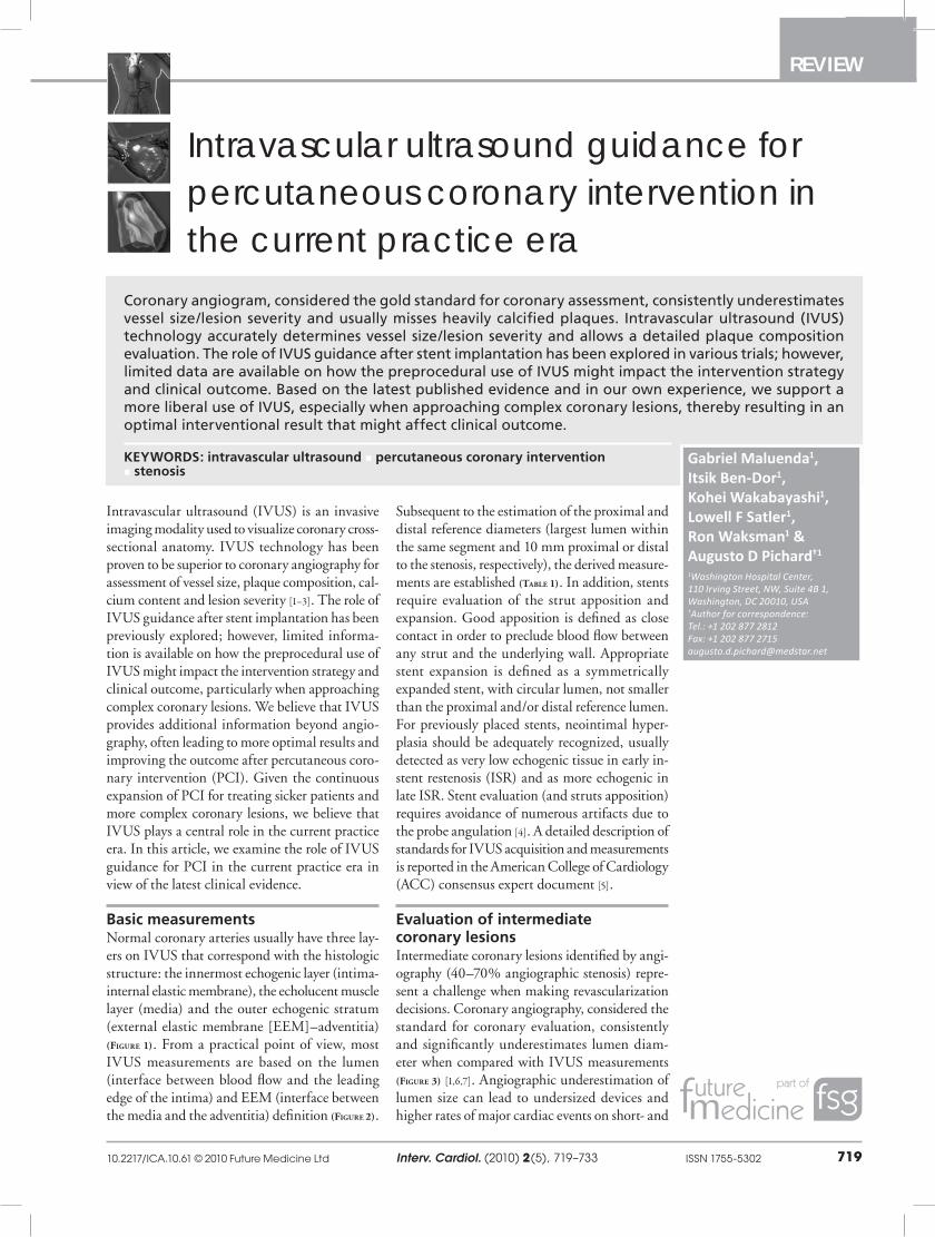
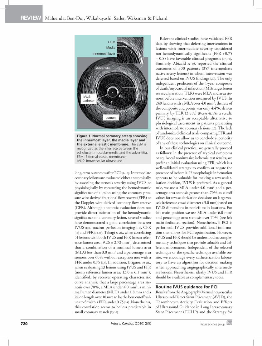
Basic measurementsNormal coronary arteries usually have three lay-ers on IVUS that correspond with the histologic structure: the innermost echogenic layer (intima-internal elastic membrane), the echolucent muscle layer (media) and the outer echogenic stratum (external elastic membrane [EEM]–adventitia) (Figure 1). From a practical point of view, most IVUS measurements are based on the lumen (interface between blood flow and the leading edge of the intima) and EEM (interface between the media and the adventitia) definition (Figure 2).
Subsequent to the estimation of the proximal and distal reference diameters (largest lumen within the same segment and 10 mm proximal or distal to the stenosis, respectively), the derived measure-ments are established (Table 1). In addition, stents require evaluation of the strut apposition and expansion. Good apposition is defined as close contact in order to preclude blood flow between any strut and the underlying wall. Appropriate stent expansion is defined as a symmetrically expanded stent, with circular lumen, not smaller than the proximal and/or distal reference lumen. For previously placed stents, neointimal hyper-plasia should be adequately recognized, usually detected as very low echogenic tissue in early in-stent restenosis (ISR) and as more echogenic in late ISR. Stent evaluation (and struts apposition) requires avoidance of numerous artifacts due to the probe angulation [4]. A detailed description of standards for IVUS acquisition and measurements is reported in the American College of Cardiology (ACC) c onsensus expert document [5].
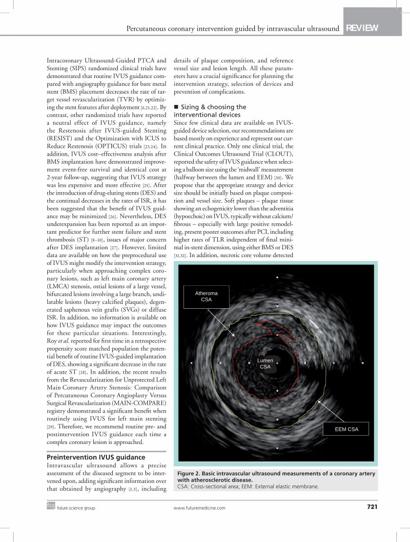
Evaluation of intermediate coronary lesionsIntermediate coronary lesions identified by angi-ography (40–70% angiographic stenosis) repre-sent a challenge when making revascularization decisions. Coronary angiography, considered the standard for coronary evaluation, consistently and significantly underestimates lumen diam-eter when compared with IVUS measurements (Figure 3) [1,6,7]. Angiographic underestimation of lumen size can lead to undersized devices and higher rates of major cardiac events on short- and
Coronary angiogram, considered the gold standard for coronary assessment, consistently underestimates vessel size/lesion severity and usually misses heavily calcified plaques. Intravascular ultrasound (IVUS) technology accurately determines vessel size/lesion severity and allows a detailed plaque composition evaluation. The role of IVUS guidance after stent implantation has been explored in various trials; however, limited data are available on how the preprocedural use of IVUS might impact the intervention strategy and clinical outcome. Based on the latest published evidence and in our own experience, we support a more liberal use of IVUS, especially when approaching complex coronary lesions, thereby resulting in an optimal interventional result that might affect clinical outcome.
KEYWORDS: intravascular ultrasound n percutaneous coronary intervention n stenosis
Gabriel Maluenda1, Itsik Ben-Dor1, Kohei Wakabayashi1, Lowell F Satler1, Ron Waksman1 & Augusto D Pichard†1
1Washington Hospital Center, 110 Irving Street, NW, Suite 4B 1, Washington, DC 20010, USA †Author for correspondence: Tel.: +1 202 877 2812 Fax: +1 202 877 2715 [email protected]
Interv. Cardiol. (2010) 2(5)720 future science group
Review Maluenda, Ben-Dor, Wakabayashi, Satler, Waksman & Pichard
long-term outcomes after PCI [8–10]. Intermediate coronary lesions are evaluated either anatomically by assessing the stenosis severity using IVUS or physiologically by measuring the hemodynamic significance of a lesion using the coronary pres-sure wire-derived fractional flow reserve (FFR) or the Doppler wire-derived coronary flow reserve (CFR). Although anatomic evaluation does not provide direct estimation of the hemodynamic significance of a coronary lesion, several studies have demonstrated a good correlation between IVUS and nuclear perfusion imaging [11], CFR [12] and FFR [13,14]. Takagi et al., when correlating 51 lesions with both IVUS and FFR (mean refer-ence lumen area: 9.26 ± 2.72 mm2) determined that a combination of a minimal lumen area (MLA) less than 3.0 mm2 and a percentage area stenosis over 60% without exception met with a FFR under 0.75 [13]. In addition, Briguori et al., when evaluating 53 lesions using IVUS and FFR (mean reference lumen area: 13.0 ± 6.1 mm2), identified, by receiver operating characteristic curve analysis, that a large percentage area ste-nosis over 70%, a MLA under 4.0 mm2, a mini-mal lumen diameter (MLD) under 1.8 mm and a lesion length over 10 mm to be the best cutoff val-ues to fit with a FFR under 0.75 [14]. Nonetheless, this correlation seems to be less predictable in small coronary vessels [15,16].
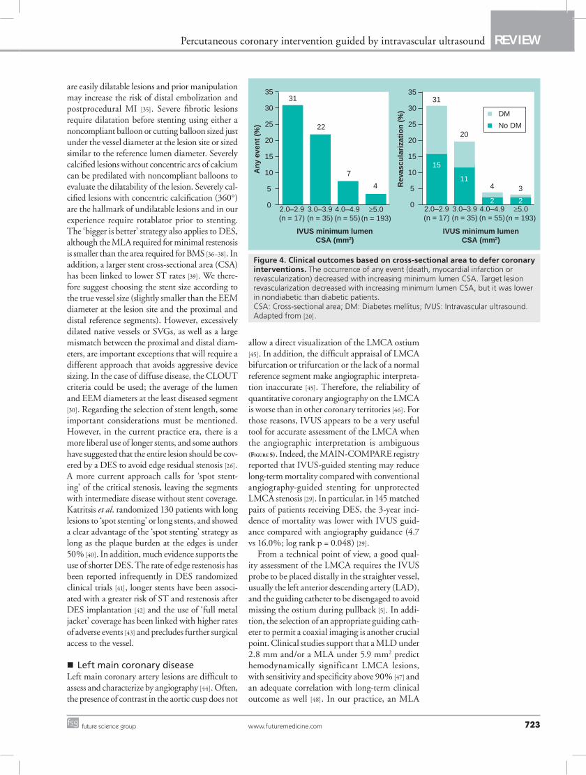
Relevant clinical studies have validated FFR data by showing that deferring interventions in lesions with intermediate severity considered not hemodynamically significant (FFR >0.75 – 0.8) have favorable clinical prognosis [17–19]. Similarly, Abizaid et al. reported the clinical outcomes of 300 patients (357 intermediate native artery lesions) in whom intervention was deferred based on IVUS findings [20]. The only independent predictors of the 1-year composite of death/myocardial infarction (MI)/target lesion revascularization (TLR) were MLA and area ste-nosis before intervention measured by IVUS. In 248 lesions with a MLA over 4.0 mm2, the rate of the composite end points was only 4.4%, driven primary by TLR (2.8%) (Figure 4). As a result, IVUS imaging is an acceptable alternative to physiological assessment in patients presenting with intermediate coronary lesions [20]. The lack of randomized clinical trials comparing FFR and IVUS does not allow us to conclude superiority of any of these technologies on clinical outcome.
In our clinical practice, we generally proceed as follows: in the presence of atypical symptoms or equivocal noninvasive ischemia test results, we prefer an initial evaluation using FFR, which is a well-validated strategy to confirm or negate the presence of ischemia. If morphologic information appears to be valuable for making a revascular-ization decision, IVUS is preferred. As a general rule, we use a MLA under 4.0 mm2 and a per-centage area stenosis greater than 70% as cutoff values for revascularization decisions on large ves-sels (reference vessel diameter >3.0 mm) based on IVUS dimensions in nonleft main locations. For left main position we use MLA under 6.0 mm2 and percentage area stenosis over 70% (see left main-dedicated section). Nonetheless, if PCI is performed, IVUS provides additional informa-tion that allows for PCI optimization. However, IVUS and FFR should be understood as comple-mentary techniques that provide valuable and dif-ferent information. Independent of the selected technique or the specific technique available on-site, we encourage every catheterization labora-tory to have an algorithm for decision making when approaching angiographically intermedi-ate lesions. Nevertheless, ideally IVUS and FFR should be available as complementary tools.
Routine IVUS guidance for PCIResults from the Angiography Versus Intravascular Ultrasound-Direct Stent Placement (AVID), the Thrombocyte Activity Evaluation and Effects of Ultrasound Guidance in Long Intracoronary Stent Placement (TULIP) and the Strategy for
IVUScatheter
Lumen
Adventitia
EEM
Media
Innermost layer
Figure 1. Normal coronary artery showing the innermost layer, the media layer and the external elastic membrane. The EEM is recognized as the interface between the echolucent muscular-media and the adventitia. EEM: External elastic membrane; IVUS: Intravascular ultrasound.
www.futuremedicine.com 721future science group
Percutaneous coronary intervention guided by intravascular ultrasound Review
Intracoronary Ultrasound-Guided PTCA and Stenting (SIPS) randomized clinical trials have demonstrated that routine IVUS guidance com-pared with angiography guidance for bare metal stent (BMS) placement decreases the rate of tar-get vessel revascularization (TVR) by optimiz-ing the stent features after deployment [6,21,22]. By contrast, other randomized trials have reported a neutral effect of IVUS guidance, namely the Restenosis after IVUS-guided Stenting (RESIST) and the Optimization with ICUS to Reduce Restenosis (OPTICUS) trials [23,24]. In addition, IVUS cost–effectiveness a nalysis after BMS implantation have demonstrated improve-ment event-free survival and identical cost at 2-year follow-up, suggesting that IVUS strategy was less expensive and more effective [25]. After the introduction of drug-eluting stents (DES) and the continual decreases in the rates of ISR, it has been suggested that the benefit of IVUS guid-ance may be minimized [26]. Nevertheless, DES underexpansion has been reported as an impor-tant predictor for further stent failure and stent thrombosis (ST) [8–10], issues of major concern after DES implantation [27]. However, limited data are available on how the preprocedural use of IVUS might modify the intervention strategy, particularly when approaching complex coro-nary lesions, such as left main coronary artery (LMCA) stenosis, ostial lesions of a large vessel, bifurcated lesions involving a large branch, undi-latable lesions (heavy calcified plaques), degen-erated saphenous vein grafts (SVGs) or diffuse ISR. In addition, no information is available on how IVUS guidance may impact the outcomes for these particular situations. Interestingly, Roy et al. reported for first time in a retrospective propensity score matched population the poten-tial benefit of routine IVUS-guided implantation of DES, showing a significant decrease in the rate of acute ST [28]. In addition, the recent results from the Revascularization for Unprotected Left Main Coronary Artery Stenosis: Comparison of Percutaneous Coronary Angioplasty Versus Surgical Revascularization (MAIN-COMPARE) registry demonstrated a significant benefit when routinely using IVUS for left main stenting [29]. Therefore, we recommend routine pre- and postintervention IVUS guidance each time a c omplex coronary lesion is approached.
Preintervention IVUS guidanceIntravascular ultrasound allows a precise a ssessment of the diseased segment to be inter-vened upon, adding significant information over that obtained by angiography [1,3], including
details of plaque composition, and reference vessel size and lesion length. All these param-eters have a crucial significance for planning the intervention strategy, selection of devices and p revention of complications.
n Sizing & choosing the interventional devicesSince few clinical data are available on IVUS-guided device selection, our recommendations are based mostly on experience and represent our cur-rent clinical practice. Only one clinical trial, the Clinical Outcomes Ultrasound Trial (CLOUT), reported the safety of IVUS guidance when select-ing a balloon size using the ‘midwall’ measurement (halfway between the lumen and EEM) [30]. We propose that the appropriate strategy and device size should be initially based on plaque composi-tion and vessel size. Soft plaques – plaque tissue showing an echogenicity lower than the adventitia (hypoechoic) on IVUS, typically without calcium/fibrous – especially with large positive remodel-ing, present poorer outcomes after PCI, including higher rates of TLR independent of final mini-mal in-stent dimension, using either BMS or DES [31,32]. In addition, necrotic core volume detected
EEM CSA
AtheromaCSA
LumenCSA
Figure 2. Basic intravascular ultrasound measurements of a coronary artery with atherosclerotic disease.CSA: Cross-sectional area; EEM: External elastic membrane.
Interv. Cardiol. (2010) 2(5)722 future science group
Review Maluenda, Ben-Dor, Wakabayashi, Satler, Waksman & Pichard
by using virtual histology (VH) IVUS correlates with the risk prediction of peri-interventional myocardial injury and presumably distal embo-lization after primary stent deployment in acute
MI and stable patients [33,34]. It is our opinion that soft plaques, especially those with a large plaque burden or necrotic core, should be stented directly without prior balloon dilatation since they
MLD: 1.82 mmMLA: 2.51 mm2
Figure 3. Angiographic underestimated lesion severity compared with intravascular ultrasound and computed tomography angiogram. (A) Multiplanar cardiac CT reconstruction showing the presence of a moderate stenosis caused by a noncalcified plaque (full arrow) and a more proximal severe calcified lesion (dashed arrow) involving the proximal right coronary artery. (B) Angiography of the same vessel showing the presence of a 30% stenotic lesion in the proximal RCA. (C) Cross-sectional intravascular ultrasound view of the pointed lesion (full arrows in [A, B & D]) showing the presence of a mixed plaque that determines a severe stenotic lesion. (D) Long intravascular ultrasound run view of the RCA. MLA: Minimal lumen area; MLD: Minimal lumen diameter.
Table 1. Standard derived intracoronary ultrasound measurements.
Measurement Definition
Lumen CSA Area bounded by the luminal border
Minimum diameter Shortest diameter through the center point of the lumen
Lumen area stenosis (Reference lumen CSA – minimum lumen CSA)/reference lumen CSA
Plaque + media (or atheroma) CSA EEM CSA – lumen CSA
Plaque (or atheroma) burden (Plaque + media CSA)/EEM CSA
Stent CSA Area bounded by the stent borderCSA: Cross-sectional area; EEM: External elastic membrane.
www.futuremedicine.com 723future science group
Percutaneous coronary intervention guided by intravascular ultrasound Review
are easily dilatable lesions and prior manipulation may increase the risk of distal embolization and postprocedural MI [35]. Severe fibrotic lesions require dilatation before stenting using either a noncompliant balloon or cutting balloon sized just under the vessel diameter at the lesion site or sized similar to the reference lumen diameter. Severely calcified lesions without concentric arcs of calcium can be predilated with noncompliant balloons to evaluate the dilatability of the lesion. Severely cal-cified lesions with concentric calcification (360°) are the hallmark of undilatable lesions and in our experience require rotablator prior to stenting. The ‘bigger is better’ strategy also applies to DES, although the MLA required for minimal restenosis is smaller than the area required for BMS [36–38]. In addition, a larger stent cross-sectional area (CSA) has been linked to lower ST rates [39]. We there-fore suggest choosing the stent size according to the true vessel size (slightly smaller than the EEM diameter at the lesion site and the proximal and distal reference segments). However, excessively dilated native vessels or SVGs, as well as a large mismatch between the proximal and distal diam-eters, are important exceptions that will require a different approach that avoids aggressive device sizing. In the case of diffuse disease, the CLOUT criteria could be used; the average of the lumen and EEM diameters at the least diseased segment [30]. Regarding the selection of stent length, some important considerations must be mentioned. However, in the current practice era, there is a more liberal use of longer stents, and some authors have suggested that the entire lesion should be cov-ered by a DES to avoid edge residual stenosis [26]. A more current approach calls for ‘spot stent-ing’ of the critical stenosis, leaving the segments with intermediate disease without stent coverage. Katritsis et al. randomized 130 patients with long lesions to ‘spot stenting’ or long stents, and showed a clear advantage of the ‘spot stenting’ strategy as long as the plaque burden at the edges is under 50% [40]. In addition, much evidence supports the use of shorter DES. The rate of edge restenosis has been reported infrequently in DES randomized clinical trials [41], longer stents have been associ-ated with a greater risk of ST and restenosis after DES implantation [42] and the use of ‘full metal jacket’ coverage has been linked with higher rates of adverse events [43] and precludes further surgical access to the vessel.
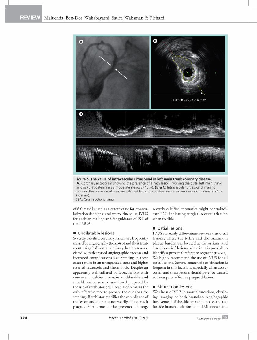
n Left main coronary diseaseLeft main coronary artery lesions are difficult to assess and characterize by angiography [44]. Often, the presence of contrast in the aortic cusp does not
allow a direct visualization of the LMCA ostium [45]. In addition, the difficult appraisal of LMCA bifurcation or trifurcation or the lack of a normal reference segment make angiographic interpreta-tion inaccurate [45]. Therefore, the reliability of quantitative coronary angiography on the LMCA is worse than in other coronary territories [46]. For those reasons, IVUS appears to be a very useful tool for accurate assessment of the LMCA when the angiographic interpretation is ambiguous (Figure 5). Indeed, the MAIN-COMPARE registry reported that IVUS-guided stenting may reduce long-term mortality compared with conventional angiography-guided stenting for unprotected LMCA stenosis [29]. In particular, in 145 matched pairs of patients receiving DES, the 3-year inci-dence of mortality was lower with IVUS guid-ance compared with angiography guidance (4.7 vs 16.0%; log rank p = 0.048) [29].
From a technical point of view, a good qual-ity assessment of the LMCA requires the IVUS probe to be placed distally in the straighter vessel, usually the left anterior descending artery (LAD), and the guiding catheter to be disengaged to avoid missing the ostium during pullback [5]. In addi-tion, the selection of an appropriate guiding cath-eter to permit a coaxial imaging is another crucial point. Clinical studies support that a MLD under 2.8 mm and/or a MLA under 5.9 mm2 predict hemodynamically significant LMCA lesions, with sensitivity and specificity above 90% [47] and an adequate correlation with long-term clinical outcome as well [48]. In our practice, an MLA
Any
eve
nt
(%)
Rev
ascu
lari
zati
on
(%
)
IVUS minimum lumenCSA (mm2)
IVUS minimum lumenCSA (mm2)
35
30
25
20
15
10
5
0
35
30
25
20
15
10
5
02.0–2.9(n = 17)
3.0–3.9(n = 35)
4.0–4.9(n = 55)
≥5.0(n = 193)
31
22
7
4
31
20
4 3
15
11
2 22.0–2.9(n = 17)
3.0–3.9(n = 35)
4.0–4.9(n = 55)
≥5.0(n = 193)
DM
No DM
Figure 4. Clinical outcomes based on cross-sectional area to defer coronary interventions. The occurrence of any event (death, myocardial infarction or revascularization) decreased with increasing minimum lumen CSA. Target lesion revascularization decreased with increasing minimum lumen CSA, but it was lower in nondiabetic than diabetic patients. CSA: Cross-sectional area; DM: Diabetes mellitus; IVUS: Intravascular ultrasound. Adapted from [20].
Interv. Cardiol. (2010) 2(5)724 future science group
Review Maluenda, Ben-Dor, Wakabayashi, Satler, Waksman & Pichard
of 6.0 mm2 is used as a cutoff value for revascu-larization decisions, and we routinely use IVUS for decision making and for guidance of PCI of the LMCA.
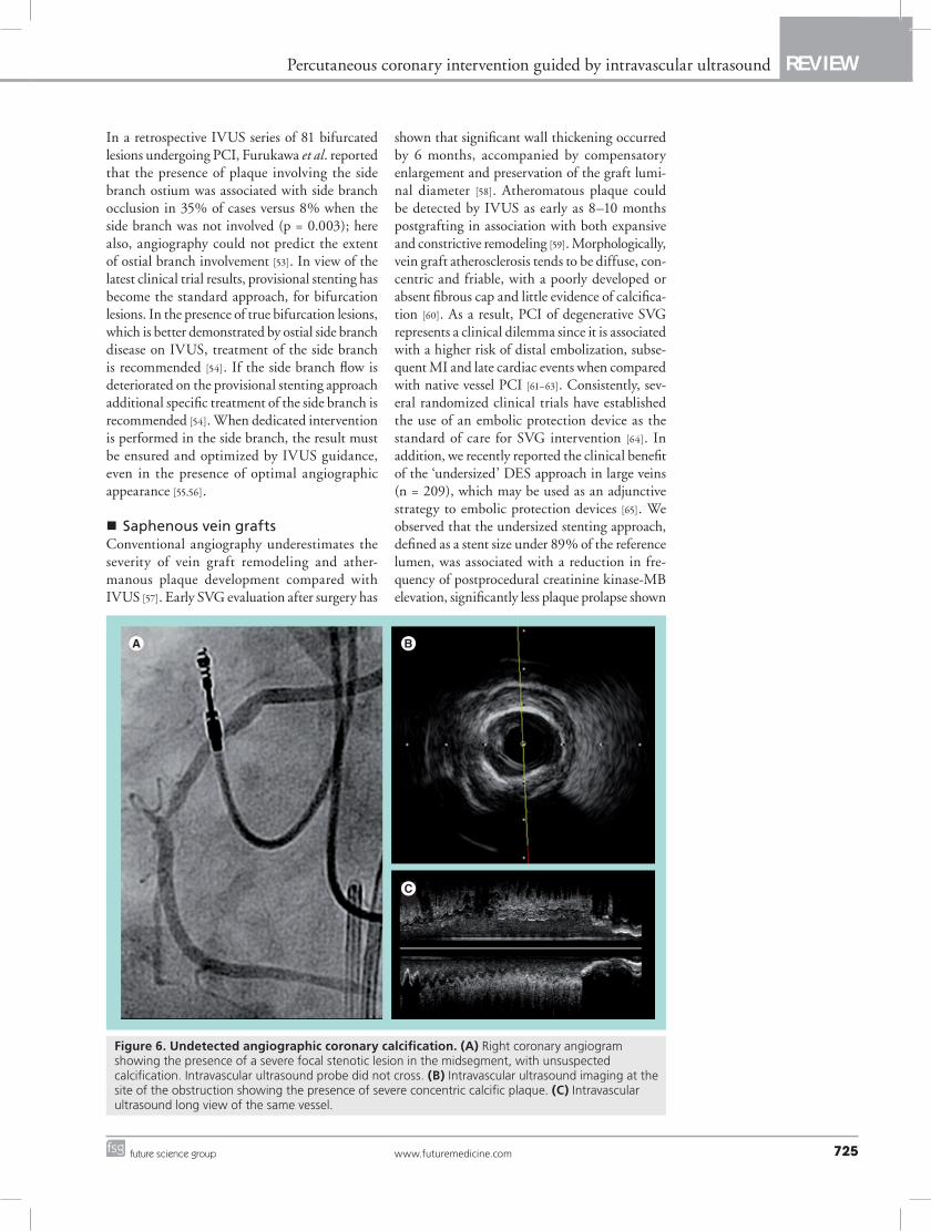
n Undilatable lesionsSeverely calcified coronary lesions are frequently missed by angiography (Figure 6) [3] and their treat-ment using balloon angioplasty has been asso-ciated with decreased angiographic success and increased complications [49]. Stenting in these cases results in an unexpanded stent and higher rates of restenosis and thrombosis. Despite an apparently well-inflated balloon, lesions with concentric calcium remain undilatable and should not be stented until well prepared by the use of rotablator [50]. Rotablator remains the only effective tool to prepare these lesions for stenting. Rotablator modifies the compliance of the lesion and does not necessarily ablate much plaque. Furthermore, the presence of long,
severely calcified coronaries might contraindi-cate PCI, i ndicating surgical revascularization when feasible.
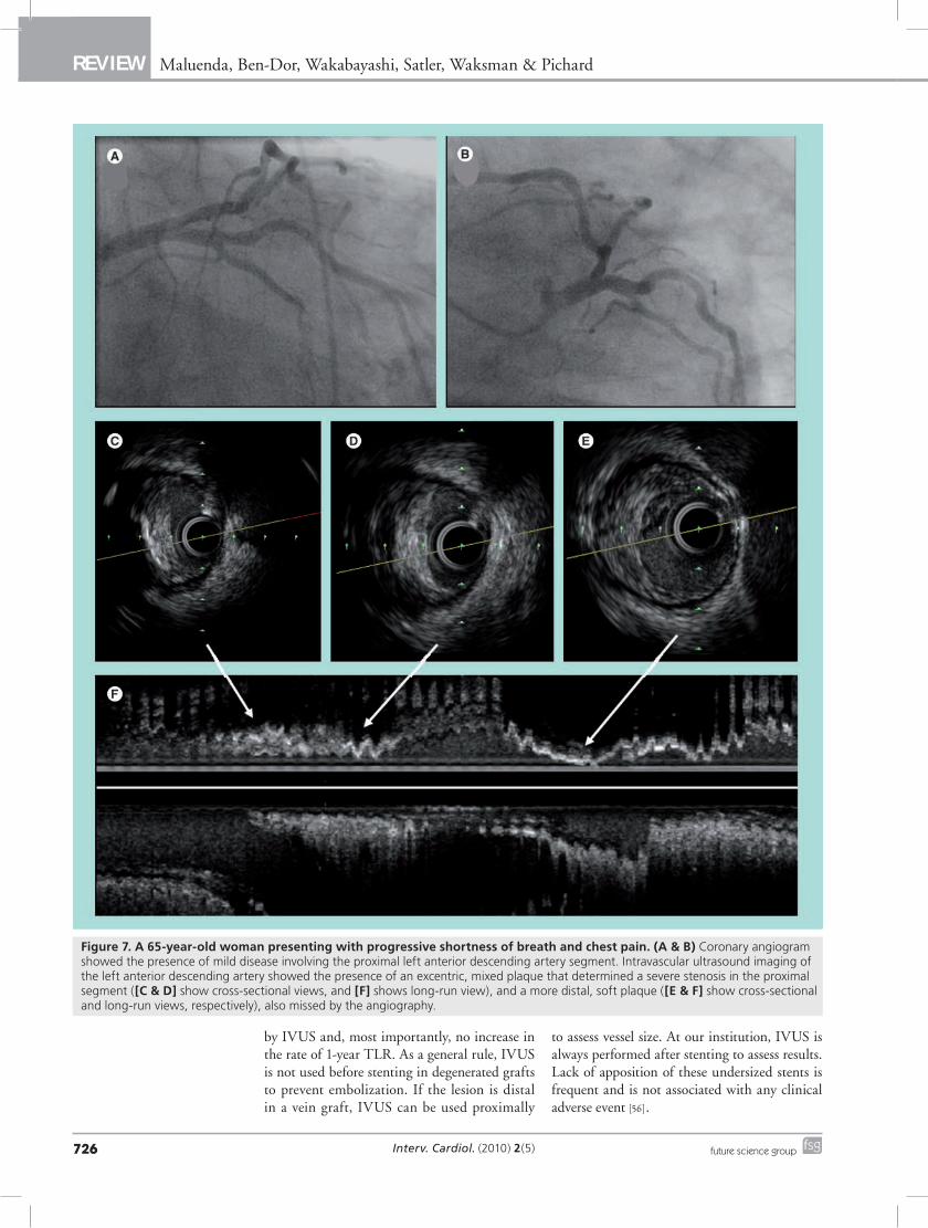
n Ostial lesionsIVUS can easily differentiate between true ostial lesions, where the MLA and the maximum plaque burden are located at the ostium, and ‘pseudo-ostial’ lesions, wherein it is possible to identify a proximal reference segment (Figure 7). We highly recommend the use of IVUS for all ostial lesions. Severe, concentric calcification is frequent in this location, especially when aorto-ostial, and these lesions should never be stented without prior effective plaque dilation.
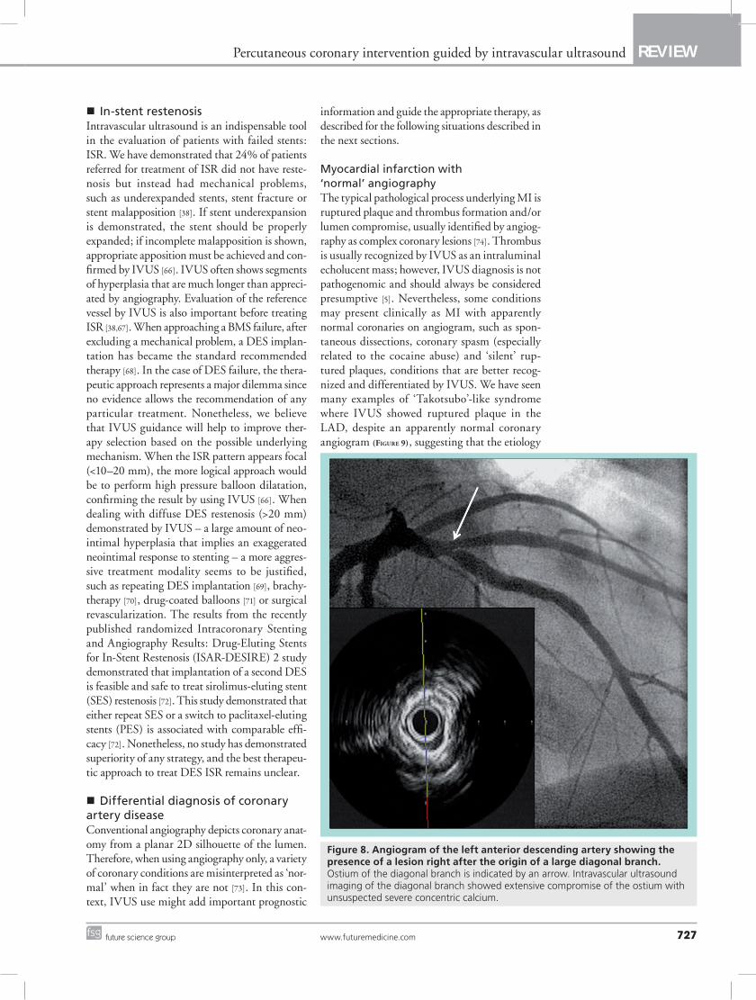
n Bifurcation lesionsWe also use IVUS in most bifurcations, obtain-ing imaging of both branches. Angiographic involvement of the side branch increases the risk for side-branch occlusion [51] and MI (Figure 8) [52].
Lumen CSA = 3.6 mm2
Figure 5. The value of intravascular ultrasound in left main trunk coronary disease. (A) Coronary angiogram showing the presence of a hazy lesion involving the distal left main trunk (arrows) that determines a moderate stenosis (40%). (B & C) Intravascular ultrasound imaging showing the presence of a severe calcified lesion that determines a severe stenosis (minimal CSA of 3.6 mm2). CSA: Cross-sectional area.
www.futuremedicine.com 725future science group
Percutaneous coronary intervention guided by intravascular ultrasound Review
In a retrospective IVUS series of 81 bifurcated lesions undergoing PCI, Furukawa et al. reported that the presence of plaque involving the side branch ostium was associated with side branch occlusion in 35% of cases versus 8% when the side branch was not involved (p = 0.003); here also, angiography could not predict the extent of ostial branch involvement [53]. In view of the latest clinical trial results, provisional stenting has become the standard approach, for bifurcation lesions. In the presence of true bifurcation lesions, which is better demonstrated by ostial side branch disease on IVUS, treatment of the side branch is recommended [54]. If the side branch flow is deteriorated on the provisional stenting approach additional specific treatment of the side branch is recommended [54]. When dedicated intervention is performed in the side branch, the result must be ensured and optimized by IVUS guidance, even in the p resence of o ptimal angiographic appearance [55,56].
n Saphenous vein graftsConventional angiography underestimates the severity of vein graft remodeling and ather-manous plaque development compared with IVUS [57]. Early SVG evaluation after surgery has
shown that significant wall thickening occurred by 6 months, accompanied by compensatory enlargement and preservation of the graft lumi-nal diameter [58]. Atheromatous plaque could be detected by IVUS as early as 8–10 months postgrafting in association with both expansive and constrictive remodeling [59]. Morphologically, vein graft atherosclerosis tends to be diffuse, con-centric and friable, with a poorly developed or absent fibrous cap and little evidence of calcifica-tion [60]. As a result, PCI of degenerative SVG represents a clinical dilemma since it is associated with a higher risk of distal embolization, subse-quent MI and late cardiac events when compared with native vessel PCI [61–63]. Consistently, sev-eral randomized clinical trials have established the use of an embolic protection device as the standard of care for SVG intervention [64]. In addition, we recently reported the clinical benefit of the ‘undersized’ DES approach in large veins (n = 209), which may be used as an adjunctive strategy to embolic protection devices [65]. We observed that the undersized stenting approach, defined as a stent size under 89% of the reference lumen, was associated with a reduction in fre-quency of postprocedural creatinine kinase-MB elevation, significantly less plaque prolapse shown
Figure 6. Undetected angiographic coronary calcification. (A) Right coronary angiogram showing the presence of a severe focal stenotic lesion in the midsegment, with unsuspected calcification. Intravascular ultrasound probe did not cross. (B) Intravascular ultrasound imaging at the site of the obstruction showing the presence of severe concentric calcific plaque. (C) Intravascular ultrasound long view of the same vessel.
Interv. Cardiol. (2010) 2(5)726 future science group
Review Maluenda, Ben-Dor, Wakabayashi, Satler, Waksman & Pichard
by IVUS and, most importantly, no increase in the rate of 1-year TLR. As a general rule, IVUS is not used before stenting in degenerated grafts to prevent embolization. If the lesion is distal in a vein graft, IVUS can be used proximally
to assess vessel size. At our institution, IVUS is always performed after stenting to assess results. Lack of apposition of these undersized stents is frequent and is not associated with any clinical adverse event [56].
Figure 7. A 65-year-old woman presenting with progressive shortness of breath and chest pain. (A & B) Coronary angiogram showed the presence of mild disease involving the proximal left anterior descending artery segment. Intravascular ultrasound imaging of the left anterior descending artery showed the presence of an excentric, mixed plaque that determined a severe stenosis in the proximal segment ([C & D] show cross-sectional views, and [F] shows long-run view), and a more distal, soft plaque ([E & F] show cross-sectional and long-run views, respectively), also missed by the angiography.
www.futuremedicine.com 727future science group
Percutaneous coronary intervention guided by intravascular ultrasound Review
n In-stent restenosisIntravascular ultrasound is an indispensable tool in the evaluation of patients with failed stents: ISR. We have demonstrated that 24% of patients referred for treatment of ISR did not have reste-nosis but instead had mechanical problems, such as underexpanded stents, stent fracture or stent malapposition [38]. If stent underexpansion is demonstrated, the stent should be properly expanded; if incomplete malapposition is shown, appropriate apposition must be achieved and con-firmed by IVUS [66]. IVUS often shows segments of hyperplasia that are much longer than appreci-ated by angiography. Evaluation of the reference vessel by IVUS is also important before treating ISR [38,67]. When approaching a BMS failure, after excluding a mechanical problem, a DES implan-tation has became the standard recommended therapy [68]. In the case of DES failure, the thera-peutic approach represents a major dilemma since no evidence allows the recommendation of any particular treatment. Nonetheless, we believe that IVUS guidance will help to improve ther-apy selection based on the possible underlying mechanism. When the ISR pattern appears focal (<10–20 mm), the more logical approach would be to perform high pressure balloon dilatation, confirming the result by using IVUS [66]. When dealing with diffuse DES resteno sis (>20 mm) demonstrated by IVUS – a large amount of neo-intimal hyperplasia that implies an exaggerated neointimal response to stenting – a more aggres-sive treatment modality seems to be justified, such as repeating DES implantation [69], brachy-therapy [70], drug-coated balloons [71] or surgical revascularization. The results from the recently published randomized Intracoronary Stenting and Angiography Results: Drug-Eluting Stents for In-Stent Restenosis (ISAR-DESIRE) 2 study demonstrated that implantation of a second DES is feasible and safe to treat sirolimus-eluting stent (SES) restenosis [72]. This study demonstrated that either repeat SES or a switch to paclitaxel-eluting stents (PES) is associated with comparable effi-cacy [72]. Nonetheless, no study has demonstrated superiority of any strategy, and the best therapeu-tic approach to treat DES ISR remains unclear.
n Differential diagnosis of coronary artery diseaseConventional angiography depicts coronary anat-omy from a planar 2D silhouette of the lumen. Therefore, when using angiography only, a variety of coronary conditions are misinterpreted as ‘nor-mal’ when in fact they are not [73]. In this con-text, IVUS use might add important prognostic
information and guide the appropriate therapy, as described for the following situations described in the next sections.
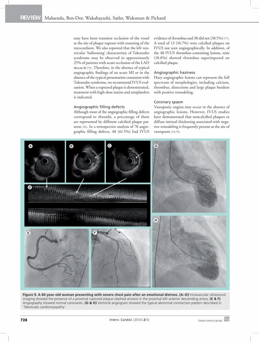
Myocardial infarction with ‘normal’ angiographyThe typical pathological process underlying MI is ruptured plaque and thrombus formation and/or lumen compromise, usually identified by angiog-raphy as complex coronary lesions [74]. Thrombus is usually recognized by IVUS as an intraluminal echolucent mass; however, IVUS diagnosis is not pathogenomic and should always be considered presumptive [5]. Nevertheless, some conditions may present clinically as MI with apparently normal coronaries on angiogram, such as spon-taneous dissections, coronary spasm (especially related to the cocaine abuse) and ‘silent’ rup-tured plaques, conditions that are better recog-nized and differentiated by IVUS. We have seen many examples of ‘Takotsubo’-like syndrome where IVUS showed ruptured plaque in the LAD, despite an apparently normal coronary angiogram (Figure 9), suggesting that the etiology
Figure 8. Angiogram of the left anterior descending artery showing the presence of a lesion right after the origin of a large diagonal branch. Ostium of the diagonal branch is indicated by an arrow. Intravascular ultrasound imaging of the diagonal branch showed extensive compromise of the ostium with unsuspected severe concentric calcium.
Interv. Cardiol. (2010) 2(5)728 future science group
Review Maluenda, Ben-Dor, Wakabayashi, Satler, Waksman & Pichard
may have been transient occlusion of the vessel at the site of plaque rupture with stunning of the myocardium. We also reported that the left ven-tricular ‘ballooning’ characteristic of Takotsubo syndrome may be observed in approximately 25% of patients with acute occlusion of the LAD (Figure 9) [75]. Therefore, in the absence of typical angiographic findings of an acute MI or in the absence of the typical presentation consistent with Takotsubo syndrome, we recommend IVUS eval-uation. When a ruptured plaque is demonstrated, treatment with high-dose statins and antiplatelets is indicated.
Angiographic filling defectsAlthough most of the angiographic filling defects correspond to thrombi, a percentage of them are represented by different calcified plaque pat-terns [76]. In a retrospective analysis of 78 angio-graphic filling defects, 48 (61.5%) had IVUS
evidence of thrombus and 30 did not (38.5%) [77]. A total of 13 (16.7%) were calcified plaques on IVUS not seen angiographically. In addition, of the 48 IVUS thrombus-containing lesions, nine (18.8%) showed thrombus superimposed on c alcified plaque.
Angiographic hazinessHazy angiographic lesions can represent the full spectrum of morphologies, including calcium, thrombus, dissections and large plaque burdens with positive remodeling.
Coronary spasmVasospastic angina may occur in the absence of angiographic lesions. However, IVUS studies have demonstrated that noncalcified plaques or diffuse intimal thickening associated with nega-tive remodeling is frequently present at the site of vasospasm [78,79].
Figure 9. A 60-year-old woman presenting with severe chest pain after an emotional distress. (A–D) Intravascular ultrasound imaging showed the presence of a proximal ruptured plaque (dashed arrows) in the proximal left anterior descending artery. (E & F) Angiography showed normal coronaries. (G & H) Ventricle angiogram showed the typical abnormal contraction pattern described in ‘Takotsubo cardiomyopathy’.
www.futuremedicine.com 729future science group
Percutaneous coronary intervention guided by intravascular ultrasound Review
Therefore, we propose a more liberal use of IVUS for patients presenting with ambiguous angiographic coronary lesions and/or u ndergoing potentially complex interventions to improve diagnostic accuracy and a ppropriate guidance.
Postprocedural IVUS guidanceThe role of IVUS in the optimization of stent implantation has been established when IVUS observations revealed that incomplete stent apposition significantly contributes to early ST occurrence [80]. These observations led to the widespread adoption of high-pressure bal-loon postdilatation after stent deployment [81]. As earlier described, the results of the AVID, TULIP, and SIPS support the routine use of IVUS to ensure good stent expansion and appo-sition when using BMS [6,21,22,82]. However, with the emergence of DES and significant late loss decreases, different authors suggest that routine IVUS guidance after DES deployment is not required. Nevertheless, incomplete stent
expansion and smaller minimum stent area after DES implantation measured by IVUS are reported to correlate with restenosis [8,83] and ST [9,10]. In addition, using the traditional criteria for inadequate stent expansion, defined per the Multicenter Ultrasound Stenting in Coronaries (MUSIC) study (final stent CSA >80% of the reference CSA, or >90% if reference CSA area was <9 mm2) [84], we reported significantly higher rates of stent underexpansion with SES and PES at conventional delivery pressures [85,86]. In view of the exposed evidence, we support a more liberal use of IVUS to ensure an appropriate result after stent deployment, especially when concerned with approaching complex coronary lesions or patients who are theoretically at higher risk of ST.
Postintervention complicationsThe rate of persistent angiographic haziness proximal or distal to the stent is approximately 15% after high-pressure stent deployment. Stent edge dissection is the most common reason;
Executive summary
Utility of intravascular ultrasound in intermediate coronary lesions � Cutoff values for minimal lumen area under 4.0 mm2 and percentage area of stenosis over 60–70% correlate appropriately with
fractional flow reserve value under 0.75 and clinical outcomes in large none left main vessel (>3.0 mm reference diameter). � Intravascular ultrasound (IVUS) and fractional flow reserve should be used as complementary tools.
Clinical evidence for routine use of IVUS guiding percutaneous coronary intervention � The results of the Angiography Versus Intravascular Ultrasound-Direct Stent Placement (AVID), the Thrombocyte Activity Evaluation and
Effects of Ultrasound Guidance in Long Intracoronary Stent Placement (TULIP) and the Strategy for Intracoronary Ultrasound-Guided PTCA and Stenting (SIPS) randomized trials support the routine use of IVUS to ensure good stent expansion and apposition when using bare-metal stents.
� Incomplete stent expansion and smaller minimum stent area after drug-eluting stent implantation correlate with restenosis and stent thrombosis.
� Retrospective data suggest that routine intravascular ultrasound-guided percutaneous coronary intervention using drug-eluting stent might decrease the risk of acute stent thrombosis and improve the outcomes in unprotected left main trunk percutaneous coronary intervention.
Role of IVUS sizing & choosing the interventional device � A precisely sized device and interventional strategy based on IVUS findings is recommended.
Role of IVUS in complex coronary interventions � Routine IVUS guidance is recommended for complex coronary intervention including:
– Protected and unprotected left main trunk disease
– Undilatable lesions
– Ostial lesions
– Bifurcations
– Saphenous vein graft
– In-stent restenosis
IVUS value in the differential diagnosis of coronary artery disease � IVUS use might add important prognostic information and guide the appropriate therapy in the following situations:
– Myocardial infarction with ‘normal’ angiography – Angiographic filling defects – Angiographic haziness – Coronary spasm
IVUS assessment of complications after percutaneous coronary intervention � Stent edge dissection is frequent as detected by IVUS, usually a benign phenomenon that does not require additional stent implantation
if the area stenosis is less than 60% at the site of dissection.
Interv. Cardiol. (2010) 2(5)730 future science group
Review Maluenda, Ben-Dor, Wakabayashi, Satler, Waksman & Pichard
however, other conditions, such as thrombus, calcification, intramural hematoma or material prolapse could be distinguished by IVUS and further treated if necessary. Stent edge dissection is a frequent phenomenon detected by IVUS and does not necessarily proscribe an adverse progno-sis [87]. Indeed, Nishida et al. reported the results of 124 consecutive native coronary lesions with angiographic non-obstructive residual dissec-tion in 97 patients compared with 124 lesions in 100 matched patients without residual dissection [88]. They observed that most nonflow-limiting residual dissections occurring after successful PCI have a good long-term prognosis and do not need additional stenting. More importantly, IVUS examination identifies an area stenosis over 60% at the site of dissection to be the best threshold for distinguishing patients who had in-hospital major adverse cardiac events. Therefore, we encourage IVUS guidance for complications occurring after PCI, especially to prevent the unnecessary deployment of a dditional stents.
ConclusionAs a result of the described evidence and our clinical experience, we provided a comprehensive approach for a practical use of IVUS in a mod-ern catheterization laboratory. We believe that routine use of IVUS, especially when approach-ing complex coronary lesions, allows for better definition of the nature of the disease, thereby leading to a more tailored and focused therapeu-tic strategy resulting in an optimal interventional
outcome. As multiple new therapies become available, where efficacy is the main objective, but safety is the major concern, we support a more liberal use of IVUS.
Future perspectiveIVUS imaging has played a key role in the u nderstanding and development of currently avail-able coronary intervention technologies. Multiple datasets support the concept that r outine use of IVUS impacts positively p rocedural results and long-term outcomes. Nonetheless, few data are available on how the preprocedural use of IVUS might impact intervention s trategy and clinical outcome, and limited studies have specifically addressed this issue in the DES era. As percuta-neous intervention is expanding into more com-plex and high-risk lesion subsets, and higher-risk patients, IVUS guidance may become more important for improved short- and long-term out-come; p rospective, r andomized clinical designs would be ideal to prove this concept.
Financial & competing interest disclosureThe authors have no relevant affiliations or financial involvement with any organization or entity with a fi nancial interest in or financial conflict with the subject matter or materials discussed in the manuscript. This includes employment, consultancies, honoraria, stock o wnership or options, expert testimony, grants or patents received or pending, or royalties.
No writing assistance was utilized in the production of this manuscript.
BibliographyPapers of special note have been highlighted as:n of interest
1 Mintz GS, Painter JA, Pichard AD et al.: Atherosclerosis in angiographically ‘normal’ coronary artery reference segments: an intravascular ultrasound study with clinical correlations. J. Am. Coll. Cardiol. 25, 1479–1485 (1995).
2 Mintz GS, Popma JJ, Pichard AD et al.: Limitations of angiography in the assessment of plaque distribution in coronary artery disease: a systematic study of target lesion eccentricity in 1446 lesions. Circulation 93, 924–931 (1996).
3 Baptista J, di Mario C, Escaned J et al.: Intracoronary two-dimensional ultrasound imaging in the assessment of plaque morphologic features and the planning of coronary interventions. Am. Heart J. 129, 177–187 (1995).
4 Finet G, Cachard C, Delachartre P, Maurincomme E, Beaune J: Artifacts in intravascular ultrasound imaging during
coronary artery stent implantation. Ultrasound Med. Biol. 24, 793–802 (1998).
5 Mintz GS, Nissen SE, Anderson WD et al.: American College of Cardiology Clinical Expert Consensus Document on Standards for Acquisition, Measurement and Reporting of Intravascular Ultrasound Studies (IVUS). A report of the American College of Cardiology Task Force on Clinical Expert Consensus Documents. J. Am. Coll. Cardiol. 37, 1478–1492 (2001).
6 Russo RJ, Silva PD, Teirstein PS et al.: A randomized controlled trial of angiography versus intravascular ultrasound-directed bare-metal coronary stent placement (the AVID Trial). Circ. Cardiovasc. Interv. 2, 113–123 (2009).
n Randomized study emphasizes that stent underexpansion is a frequent problem and expansion optimized by intravascular ultrasound (IVUS) reduces the target lesion revascularization rate.
7 Maehara A, Mintz GS, Bui AB et al.: Determinants of angiographically silent stenoses in patients with coronary artery disease. Am. J. Cardiol. 91, 1335–1338 (2003).
8 Fujii K, Mintz GS, Kobayashi Y et al.: Contribution of stent underexpansion to recurrence after sirolimus-eluting stent implantation for in-stent restenosis. Circulation 109, 1085–1088 (2004).
n Highlights the importance of stent underexpansion as a cause of failure after sirolimus-eluting stents (SES) implantation treatment of in-stent restenosis.
9 Fujii K, Carlier SG, Mintz GS et al.: Stent underexpansion and residual reference segment stenosis are related to stent thrombosis after sirolimus-eluting stent implantation: an intravascular ultrasound study. J. Am. Coll. Cardiol. 45, 995–998 (2005).
n Demonstrates the association between stent underexpansion and residual reference segment stenosis with stent thrombosis after SES implantation.
www.futuremedicine.com 731future science group
Percutaneous coronary intervention guided by intravascular ultrasound Review
10 Okabe T, Mintz GS, Buch AN et al.: Intravascular ultrasound parameters associated with stent thrombosis after drug-eluting stent deployment. Am. J. Cardiol. 100, 615–620 (2007).
11 Nishioka T, Amanullah AM, Luo H et al.: Clinical validation of intravascular ultrasound imaging for assessment of coronary stenosis severity: comparison with stress myocardial perfusion imaging. J. Am. Coll. Cardiol. 33, 1870–1878 (1999).
12 Abizaid A, Mintz GS, Pichard AD et al.: Clinical, intravascular ultrasound, and quantitative angiographic determinants of the coronary flow reserve before and after percutaneous transluminal coronary angioplasty. Am. J. Cardiol. 82, 423–428 (1998).
13 Takagi A, Tsurumi Y, Ishii Y, Suzuki K, Kawana M, Kasanuki H: Clinical potential of intravascular ultrasound for physiological assessment of coronary stenosis: relationship between quantitative ultrasound tomography and pressure-derived fractional flow reserve. Circulation 100, 250–255 (1999).
14 Briguori C, Anzuini A, Airoldi F et al.: Intravascular ultrasound criteria for the assessment of the functional significance of intermediate coronary artery stenoses and comparison with fractional flow reserve. Am. J. Cardiol. 87, 136–141 (2001).
15 Costa MA, Sabate M, Staico R et al.: Anatomical and physiologic assessments in patients with small coronary artery disease: final results of the Physiologic and Anatomical Evaluation Prior to and After Stent Implantation in Small Coronary Vessels (PHANTOM) trial. Am. Heart J. 153, 296 E1–E7 (2007).
16 Lee CH, Tai BC, Soon CY et al.: New set of intravascular ultrasound-derived anatomic criteria for defining functionally significant stenoses in small coronary arteries (results from Intravascular Ultrasound Diagnostic Evaluation of Atherosclerosis in Singapore [IDEAS] study). Am. J. Cardiol. 105, 1378–1384 (2010).
17 Bech GJ, De Bruyne B, Bonnier HJ et al.: Long-term follow-up after deferral of percutaneous transluminal coronary angioplasty of intermediate stenosis on the basis of coronary pressure measurement. J. Am. Coll. Cardiol. 31, 841–847 (1998).
18 Meuwissen M, Chamuleau SA, Siebes M et al.: The prognostic value of combined intracoronary pressure and blood flow velocity measurements after deferral of percutaneous coronary intervention. Catheter Cardiovasc. Interv. 71, 291–297 (2008).
19 Tonino PA, De Bruyne B, Pijls NH et al.: Fractional flow reserve versus angiography for guiding percutaneous coronary intervention. N. Engl. J. Med. 360, 213–224 (2009).
20 Abizaid AS, Mintz GS, Mehran R et al.: Long-term follow-up after percutaneous transluminal coronary angioplasty was not performed based on intravascular ultrasound findings: importance of lumen dimensions. Circulation 100, 256–261 (1999).
n Landmark study demonstrating the prognostic value of deferring intervention based on IVUS lumen dimension.
21 Oemrawsingh PV, Mintz GS, Schalij MJ, Zwinderman AH, Jukema JW, van der Wall EE: Intravascular ultrasound guidance improves angiographic and clinical outcome of stent implantation for long coronary artery stenoses: final results of a randomized comparison with angiographic guidance (TULIP Study). Circulation 107, 62–67 (2003).
22 Frey AW, Hodgson JM, Muller C, Bestehorn HP, Roskamm H: Ultrasound-guided strategy for provisional stenting with focal balloon combination catheter: results from the randomized Strategy for Intracoronary Ultrasound-guided PTCA and Stenting (SIPS) trial. Circulation 102, 2497–2502 (2000).
23 Schiele F, Meneveau N, Vuillemenot A et al.: Impact of intravascular ultrasound guidance in stent deployment on 6-month restenosis rate: a multicenter, randomized study comparing two strategies – with and without intravascular ultrasound guidance. RESIST Study Group. REStenosis after Ivus guided STenting. J. Am. Coll. Cardiol. 32, 320–328 (1998).
24 Mudra H, di Mario C, de Jaegere P et al.: Randomized comparison of coronary stent implantation under ultrasound or angiographic guidance to reduce stent restenosis (OPTICUS Study). Circulation 104, 1343–1349 (2001).
25 Mueller C, Hodgson JM, Schindler C, Perruchoud AP, Roskamm H, Buettner HJ: Cost-effectiveness of intracoronary ultrasound for percutaneous coronary interventions. Am. J. Cardiol. 91, 143–147 (2003).
26 Orford JL, Lerman A, Holmes DR: Routine intravascular ultrasound guidance of percutaneous coronary intervention: a critical reappraisal. J. Am. Coll. Cardiol. 43, 1335–1342 (2004).
27 Maluenda G, Lemesle G, Waksman R: A critical appraisal of the safety and efficacy of drug-eluting stents. Clin. Pharmacol. Ther. 85, 474–480 (2009).
28 Roy P, Steinberg DH, Sushinsky SJ et al.: The potential clinical utility of intravascular ultrasound guidance in patients undergoing percutaneous coronary intervention with drug-eluting stents. Eur. Heart J. 29, 1851–1857 (2008).
n First clinical experience suggesting a benefit of IVUS-guided drug-eluting stent implantation by reducing the rate of acute stent thrombosis.
29 Park SJ, Kim YH, Park DW et al.: Impact of intravascular ultrasound guidance on long-term mortality in stenting for unprotected left main coronary artery stenosis. Circ. Cardiovasc. Interv. 2, 167–177 (2009).
n Current registry suggesting a mortality benefit of IVUS-guided percutaneous coronary intervention for unprotected left main disease.
30 Stone GW, Hodgson JM, St Goar FG et al.: Improved procedural results of coronary angioplasty with intravascular ultrasound-guided balloon sizing: the CLOUT Pilot Trial. Clinical Outcomes With Ultrasound Trial (CLOUT) Investigators. Circulation 95, 2044–2052 (1997).
31 Sahara M, Kirigaya H, Oikawa Y et al.: Soft plaque detected on intravascular ultrasound is the strongest predictor of in-stent restenosis: an intravascular ultrasound study. Eur. Heart J. 25, 2026–2033 (2004).
32 Okura H, Morino Y, Oshima A et al.: Preintervention arterial remodeling affects clinical outcome following stenting: an intravascular ultrasound study. J. Am. Coll. Cardiol. 37, 1031–1035 (2001).
33 Kawaguchi R, Oshima S, Jingu M et al.: Usefulness of virtual histology intravascular ultrasound to predict distal embolization for ST-segment elevation myocardial infarction. J. Am. Coll. Cardiol. 50, 1641–1646 (2007).
34 Bose D, von Birgelen C, Zhou XY et al.: Impact of atherosclerotic plaque composition on coronary microembolization during percutaneous coronary interventions. Basic Res. Cardiol. 103, 587–597 (2008).
35 Piscione F, Piccolo R, Cassese S et al.: Is direct stenting superior to stenting with predilation in patients treated with percutaneous coronary intervention? Results from a meta-analysis of 24 randomised controlled trials. Heart 96, 588–594 (2010).
36 Sonoda S, Morino Y, Ako J et al.: Impact of final stent dimensions on long-term results following sirolimus-eluting stent implantation: serial intravascular ultrasound analysis from the sirius trial. J. Am. Coll. Cardiol. 43, 1959–1963 (2004).
37 Doi H, Maehara A, Mintz GS et al.: Impact of in-stent minimal lumen area at 9 months poststent implantation on 3-year target lesion revascularization-free survival: a serial intravascular ultrasound analysis from the TAXUS IV, V, and VI trials. Circ. Cardiovasc. Interv. 1, 111–118 (2008).
38 Castagna MT, Mintz GS, Leiboff BO et al.: The contribution of ‘mechanical’ problems to in-stent restenosis: an intravascular ultrasonographic analysis of 1090 consecutive in-stent restenosis lesions. Am. Heart J. 142, 970–974 (2001).
Interv. Cardiol. (2010) 2(5)732 future science group
Review Maluenda, Ben-Dor, Wakabayashi, Satler, Waksman & Pichard
39 Roy P, Torguson R, Okabe T et al.: Angiographic and procedural correlates of stent thrombosis after intracoronary implantation of drug-eluting stents. J. Interv. Cardiol. 20, 307–313 (2007).
40 Katritsis DG, Korovesis S, Tzanalaridou E, Giazitzoglou E, Voridis E, Meier B: Comparison of long versus short (‘spot’) drug-eluting stenting for long coronary stenoses. Am. J. Cardiol. 104, 786–790 (2009).
41 Dawkins KD, Grube E, Guagliumi G et al.: Clinical efficacy of polymer-based paclitaxel-eluting stents in the treatment of complex, long coronary artery lesions from a multicenter, randomized trial: support for the use of drug-eluting stents in contemporary clinical practice. Circulation 112, 3306–3313 (2005).
42 Wiviott SD, Braunwald E, McCabe CH et al.: Intensive oral antiplatelet therapy for reduction of ischaemic events including stent thrombosis in patients with acute coronary syndromes treated with percutaneous coronary intervention and stenting in the TRITON-TIMI 38 trial: a subanalysis of a randomised trial. Lancet 371, 1353–1363 (2008).
43 Sharp AS, Latib A, Ielasi A et al.: Long-term follow-up on a large cohort of ‘full-metal jacket’ percutaneous coronary intervention procedures. Circ. Cardiovasc. Interv. 2, 416–422 (2009).
44 Isner JM, Kishel J, Kent KM, Ronan JA Jr, Ross AM, Roberts WC: Accuracy of angiographic determination of left main coronary arterial narrowing. Angiographic – histologic correlative analysis in 28 patients. Circulation 63, 1056–1064 (1981).
45 Sano K, Mintz GS, Carlier SG et al.: Assessing intermediate left main coronary lesions using intravascular ultrasound. Am. Heart J. 154, 983–988 (2007).
46 Fisher LD, Judkins MP, Lesperance J et al.: Reproducibility of coronary arteriographic reading in the coronary artery surgery study (CASS). Cathet. Cardiovasc. Diagn. 8, 565–575 (1982).
47 Jasti V, Ivan E, Yalamanchili V, Wongpraparut N, Leesar MA: Correlations between fractional flow reserve and intravascular ultrasound in patients with an ambiguous left main coronary artery stenosis. Circulation 110, 2831–2836 (2004).
48 Abizaid AS, Mintz GS, Abizaid A et al.: One-year follow-up after intravascular ultrasound assessment of moderate left main coronary artery disease in patients with ambiguous angiograms. J. Am. Coll. Cardiol. 34, 707–715 (1999).
49 Ellis SG, Roubin GS, King SB III et al.: Angiographic and clinical predictors of acute closure after native vessel coronary angioplasty. Circulation 77, 372–379 (1988).
50 Cavusoglu E, Kini AS, Marmur JD, Sharma SK: Current status of rotational atherectomy. Catheter Cardiovasc. Interv. 62, 485–498 (2004).
51 Meier B, Gruentzig AR, King SB III et al.: Risk of side branch occlusion during coronary angioplasty. Am. J. Cardiol. 53, 10–14 (1984).
52 Al Suwaidi J, Yeh W, Cohen HA, Detre KM, Williams DO, Holmes DR Jr. Immediate and one-year outcome in patients with coronary bifurcation lesions in the modern era (NHLBI dynamic registry). Am. J. Cardiol. 87, 1139–1144 (2001).
53 Furukawa E, Hibi K, Kosuge M et al.: Intravascular ultrasound predictors of side branch occlusion in bifurcation lesions after percutaneous coronary intervention. Circ. J. 69, 325–330 (2005).
54 Latib A, Colombo A: Bifurcation disease: what do we know, what should we do? JACC Cardiovasc. Interv. 1, 218–226 (2008).
55 Robinson NM, Balcon R, Layton CA, Mills PG, Timmis AD, Rothman MT: Intravascular ultrasound assessment of culotte stent deployment for the treatment of stenoses at major coronary bifurcations. Int. J. Cardiovasc. Intervent. 4, 21–27 (2001).
56 Costa RA, Mintz GS, Carlier SG et al.: Bifurcation coronary lesions treated with the ‘crush’ technique: an intravascular ultrasound analysis. J. Am. Coll. Cardiol. 46, 599–605 (2005).
57 Nase-Hueppmeier S, Uebis R, Doerr R, Hanrath P: Intravascular ultrasound to assess aortocoronary venous bypass grafts in vivo. Am. J. Cardiol. 70, 455–458 (1992).
58 Higuchi Y, Hirayama A, Shimizu M, Sakakibara T, Kodama K: Postoperative changes in angiographically normal saphenous vein coronary bypass grafts using intravascular ultrasound. Heart Vessels 17, 57–60 (2002).
59 Hong MK, Mintz GS, Hong MK et al.: Intravascular ultrasound assessment of the presence of vascular remodeling in diseased human saphenous vein bypass grafts. Am. J. Cardiol. 84, 992–998 (1999).
60 Kalan JM, Roberts WC: Morphologic findings in saphenous veins used as coronary arterial bypass conduits for longer than 1 year: necropsy analysis of 53 patients, 123 saphenous veins, and 1865 five-millimeter segments of veins. Am. Heart J. 119, 1164–1184 (1990).
61 Hong MK, Mehran R, Dangas G et al.: Creatine kinase-MB enzyme elevation following successful saphenous vein graft intervention is associated with late mortality. Circulation 100, 2400–2405 (1999).
62 Bhargava B, Kornowski R, Mehran R et al.: Procedural results and intermediate clinical outcomes after multiple saphenous vein graft stenting. J. Am. Coll. Cardiol. 35, 389–397 (2000).
63 Hong MK, Mehran R, Dangas G et al.: Are we making progress with percutaneous saphenous vein graft treatment? A comparison of 1990 to 1994 and 1995 to 1998 results. J. Am. Coll. Cardiol. 38, 150–154 (2001).
64 Moris C, Lozano I, Martin M, Rondan J, Avanzas P: Embolic protection devices in saphenous percutaneous intervention. EuroIntervention 5(Suppl. D), D45–D50 (2009).
65 Hong YJ, Pichard DA, Mintz GS et al.: Outcome of undersized drug-eluting stents for percutaneous coronary intervention of saphenous vein graft lesions. Am. J. Cardiol. 105, 179–185 (2010).
66 Aminian A, Kabir T, Eeckhout E: Treatment of drug-eluting stent restenosis: an emerging challenge. Catheter Cardiovasc. Interv. 74, 108–116 (2009).
67 Cheneau E, Pichard AD, Satler LF, Suddath WO, Weissman NJ, Waksman R: Intravascular ultrasound stent area of sirolimus-eluting stents and its impact on late outcome. Am. J. Cardiol. 95, 1240–1242 (2005).
68 King SB III, Smith SC Jr, Hirshfeld JW Jr et al.: 2007 Focused Update of the ACC/AHA/SCAI 2005 Guideline Update for Percutaneous Coronary Intervention: a report of the American College of Cardiology/American Heart Association Task Force on Practice Guidelines: 2007 Writing Group to Review New Evidence and Update the ACC/AHA/SCAI 2005 Guideline Update for Percutaneous Coronary Intervention, Writing on Behalf of the 2005 Writing Committee. Circulation 117, 261–295 (2008).
69 Garg S, Smith K, Torguson R et al.: Treatment of drug-eluting stent restenosis with the same versus different drug-eluting stent. Catheter Cardiovasc. Interv. 70, 9–14 (2007).
70 Torguson R, Sabate M, Deible R et al.: Intravascular brachytherapy versus drug-eluting stents for the treatment of patients with drug-eluting stent restenosis. Am. J. Cardiol. 98, 1340–1344 (2006).
www.futuremedicine.com 733future science group
Percutaneous coronary intervention guided by intravascular ultrasound Review
71 Unverdorben M, Vallbracht C, Cremers B et al.: Paclitaxel-coated balloon catheter versus paclitaxel-coated stent for the treatment of coronary in-stent restenosis. Circulation 119, 2986–2994 (2009).
72 Mehilli J, Byrne RA, Tiroch K et al.: Randomized trial of paclitaxel- versus sirolimus-eluting stents for treatment of coronary restenosis in sirolimus-eluting stents: the ISAR-DESIRE 2 (Intracoronary Stenting and Angiographic Results: Drug Eluting Stents for In-Stent Restenosis 2) study. J. Am. Coll. Cardiol. 55, 2710–2716 (2010).
73 Topol EJ, Nissen SE: Our preoccupation with coronary luminology. The dissociation between clinical and angiographic findings in ischemic heart disease. Circulation 92, 2333–2342 (1995).
74 Fujii K, Kobayashi Y, Mintz GS et al.: Intravascular ultrasound assessment of ulcerated ruptured plaques: a comparison of culprit and nonculprit lesions of patients with acute coronary syndromes and lesions in patients without acute coronary syndromes. Circulation 108, 2473–2478 (2003).
75 Chao T, Lindsay J, Collins S et al.: Can acute occlusion of the left anterior descending coronary artery produce a typical ‘Takotsubo’ left ventricular contraction pattern? Am. J. Cardiol. 104, 202–204 (2009).
76 Duissaillant GR, Mintz GS, Pichard AD et al.: Intravascular ultrasound identification of calcified intraluminal lesions misdiagnosed as thrombi by coronary angiography. Am. Heart J. 132, 687–689 (1996).
77 Kotani J, Mintz GS, Rai PB et al.: Intravascular ultrasound assessment of angiographic filling defects in native coronary arteries: do they always contain thrombi? J. Am. Coll. Cardiol. 44, 2087–2089 (2004).
78 Hong MK, Park SW, Lee CW et al.: Intravascular ultrasound findings of negative arterial remodeling at sites of focal coronary spasm in patients with vasospastic angina. Am. Heart J. 140, 395–401 (2000).
79 Miyao Y, Kugiyama K, Kawano H et al.: Diffuse intimal thickening of coronary arteries in patients with coronary spastic angina. J. Am. Coll. Cardiol. 36, 432–437 (2000).
80 Nakamura S, Colombo A, Gaglione A et al.: Intracoronary ultrasound observations during stent implantation. Circulation 89, 2026–2034 (1994).
81 Colombo A, Hall P, Nakamura S et al.: Intracoronary stenting without anticoagulation accomplished with intravascular ultrasound guidance. Circulation 91, 1676–1688 (1995).
82 Fitzgerald PJ, Oshima A, Hayase M et al.: Final results of the Can Routine Ultrasound Influence Stent Expansion (CRUISE) study. Circulation 102, 523–530 (2000).
83 Hong MK, Mintz GS, Lee CW et al.: Intravascular ultrasound predictors of angiographic restenosis after sirolimus-eluting stent implantation. Eur. Heart J. 27, 1305–1310 (2006).
84 de Jaegere P, Mudra H, Figulla H et al.: Intravascular ultrasound-guided optimized stent deployment. Immediate and 6 months clinical and angiographic results from the Multicenter Ultrasound Stenting in Coronaries Study (MUSIC Study). Eur. Heart J. 19, 1214–1223 (1998).
85 Cheneau E, Satler LF, Escolar E et al.: Underexpansion of sirolimus-eluting stents: incidence and relationship to delivery pressure. Catheter Cardiovasc. Interv. 65, 222–226 (2005).
86 Javaid A, Chu WW, Cheneau E et al.: Comparison of paclitaxel-eluting stent and sirolimus-eluting stent expansion at incremental delivery pressures. Cardiovasc. Revasc. Med. 7, 208–211 (2006).
87 Sheris SJ, Canos MR, Weissman NJ: Natural history of intravascular ultrasound-detected edge dissections from coronary stent deployment. Am. Heart J. 139, 59–63 (2000).
88 Nishida T, Colombo A, Briguori C et al.: Outcome of nonobstructive residual dissections detected by intravascular ultrasound following percutaneous coronary intervention. Am. J. Cardiol. 89, 1257–1262 (2002).
n Study demonstrating that most of nonflow-limiting residual dissections that occur after successful percutaneous coronary intervention have a good long-term prognosis and do not need additional stenting.