Embed Size (px)
Citation preview
Spectral Analysis Reviews, 2013, 1, 1-9 http://dx.doi.org/10.4236/sar.2013.11001 Published Online July 2013 (http://www.scirp.org/journal/sar)
1
Investigation of Interaction of Some Chalcones and Cyclic Chalcone Analogues with Outer Mitochondrial Membrane by UV-VIS and Fluorescence Spectroscopy
Vladimíra Tomečková1, Miroslava Štefanišinová1, Beáta Veliká1, Krisztina Fodor2, Pal Perjési2, Marek Stupák1, Juraj Guzy1, Štefan Tóth Jr.1, Tímea Pekárová1
1Department of Medical Chemistry, Biochemistry and Clinical Biochemistry, Faculty of Medicine, Pavol Jozef Šafárik University in Košice, Košice, Slovakia; 2Institute of Pharmaceutical Chemistry, Faculty of Medicine, University of Pécs, Pécs, Hungary. Email: [email protected] Received June 1st, 2013; revised July 11th, 2013; accepted July 21st, 2013 Copyright © 2013 Vladimíra Tomečková et al. This is an open access article distributed under the Creative Commons Attribution License, which permits unrestricted use, distribution, and reproduction in any medium, provided the original work is properly cited.
ABSTRACT
Interaction of the synthetic chalcones (1b,1c) and their cyclic analogues (2b,2c) with bovine (BSA) and human serum albumin (HSA) as well as with rat liver mitochondria (RLM) was studied by fluorescence spectroscopy. The maxima of emission fluorescence spectra were changed only in the case of 2b and 2c during interaction with BSA, HSA as well as mitochondrial outer membrane showing a slight hypsochromic shift and decrease of fluorescence. Interaction of the methoxy-(1b,2b) and the dimethylamino-substituted (1c,2c) compounds with outer mitochondrial membrane were studied by fluorescence polarization. Fluorescence polarization of 1b in the presence of the two proteins and mitochon- dria was found to be unchanged. Under similar conditions (2b,1c,2c) showed continuously increasing fluorescence po- larization signal during the 30 minute period of investigations. Since fluorescence polarization supposes that as a result of binding these substances to proteins and lipids. Compound 2c displayed a continuous increase of fluorescence po- larization signal in the presence of proteins (BSA, HSA), yeast cytoplasm (YC) and mitochondria (YM and RLM). This compound displayed a significant cytotoxic effect. This pattern of interaction with proteins might be one of the contrib- uting vectors of the observed cytotoxicity against several human carcinoma cell lines. Keywords: Chalcones; Yeast Cytoplasm; Yeast Mitochondria; Rat Liver Mitochondria; Fluorescence Polarization
Fluorescence Synchronous Fingerprint
1. Introduction
Flavanones, flavonols, chalcones and dihydrochalcones are biochemically related compounds sometimes being present in food at a dietary significant concentration 1. Flavonoids, chalcones and chalcone derivatives have been reported to display among others e.g. cytotoxic, antitumor, antiinflammatory, immunosuppressive, an- tiplasmodial and antioxidant properties [2-4]. Among the naturally occurring chalcones and their synthetic ana- logues several compounds displayed antineoplastic activ- ity [4]. Recently we have investigated in vitro antineo- plastic activity of several synthetic chalcones and cyclic chalcone analogues [5-7]. Among the compounds inves- tigated E-2-(4’-methoxybenzylidene)-1-benzosuberone (2b) and E-2-(4’-dimethylaminobenzylidene)-1-benzo- suberone (2c) (Figure 1) had the greatest tumour cyto-
toxicity [5]. Earlier results suggested that the chalcone derivatives
exert their biological activities through noncovalent in- teractions with cellular macromolecules [5-7]. UV-VIS study of methoxy and dimethylamino substituted chal- cones and cyclic chalcone analogues with bovine (BSA) and human serum albumin (HSA) resulted in a slight hypsochromic shift of 2b and 2c indicating changing the polar environment to a less polar one [8]. Such an obser- vation is in accord with interaction of the molecules with the hydrophobic binding site(s) of the two proteins.
While investigating the mechanism of cytotoxicity of the compounds, effect on mitochondrial outer membrane of some methyl- and methoxy-substituted E-2-arylme- thylene-1-tetralones and E-2-arylmethylene-1-benzo- suberones were investigated by fluorescence spectros- copy [9]. It was found that the most cytotoxic benzo-
Copyright © 2013 SciRes. SAR
Investigation of Interaction of Some Chalcones and Cyclic Chalcone Analogues with Outer Mitochondrial Membrane by UV-VIS and Fluorescence Spectroscopy
2
O
X
O
X 1
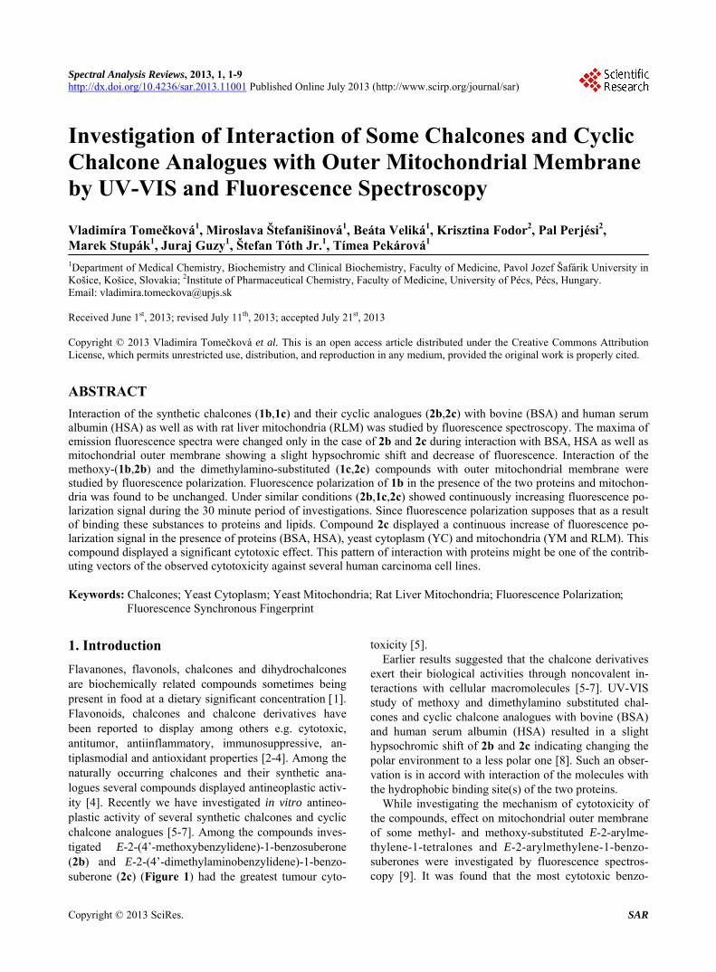
a) X = H b) X = OCH3 c) X = N(CH3)2
Figure 1. Structure of chalcones (1) and E-2-arylidene- benzosuberones (2). suberone derivative (2b) displayed a continuous increase of fluorescence polarization in the presence of rat liver mitochondria [9].
As a continuation of our work aims at gaining a better insight of interaction of the compounds with biological macromolecules, we investigated interaction of methoxy and dimethylamino-substituted chalcones (1b,c) and (E)-2-arylmethylene-1-benzosuberones (2b,c) with BSA, HSA, yeast cytoplasm (YC) as well as with yeast (YM) and rat liver mitochondria (RLM) by fluorescence spec- troscopic methods. Since FP is most readily applicable to the analysis of the binding interaction between small- molecular-mass compound and a receptor molecule, we considered applying this technique to the analysis of chalcones (1b,c) and (E)-2-arylmethylene-1-benzo- suberones (2b,c) by investigating their interaction with BSA, HSA, yeast cytoplasm, yeast mitochondria and rat liver mitochondria. In practice, low-molecular-weight fluorophores are very flexible and rotate rapidly in solu- tion, depolarizing the plane-polarized light. On the other hand, large fluorescently labelled molecules tumble more slowly; thus, the polarization of the light remains rela- tively constant between excitation and emission states. Therefore, low-molecular-mass compounds have low po- larization values while high-molecular-weight compounds show greater polarization values.
2. Materials and Methods
Compounds 1b,c and 2b,c Figure 1 were synthesized, and their structures were characterized as described be- fore [5,10]. Their structures were characterized by IR and 1H NMR spectroscopy. Their purity was checked by TLC (thin layer chromatography) and GC (gas chromatogra- phy) methods [5,10]. Other chemicals used were of the analytical grade available and, if not otherwise specified, purchased from Sigma-Aldrich (Hungary, Budapest) or Serva (Heidelberg, Germany). Compounds 1 and 2 were dissolved in DMSO immediately before use. The respira-
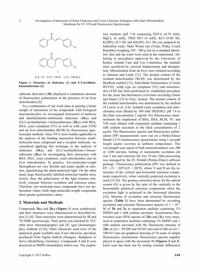
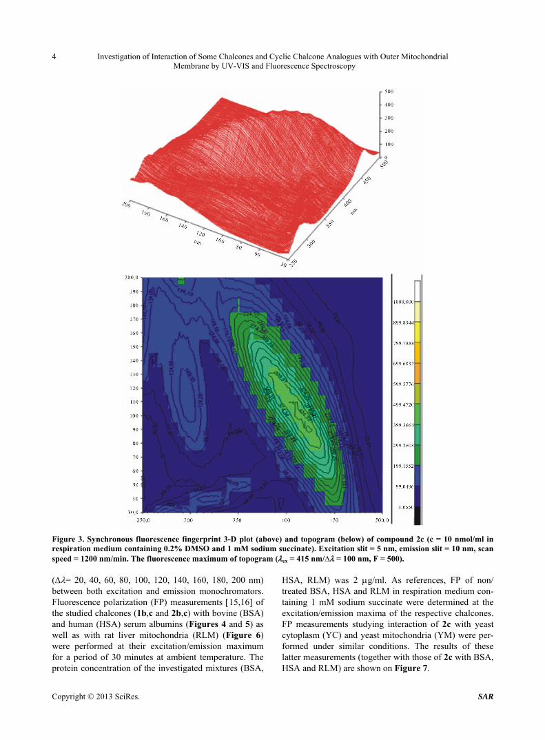
tion medium (pH 7.4) containing EDTA (0.78 mM), MgCl2 (6 mM), TRIS HCl (4 mM), KCl (0.08 M), K2HPO4 (0.3 M) and KH2PO4 (0.3 M) was prepared by bidistilled water. Male Wistar rats (Velaz, Praha, Czech Republic) weighing 250 - 300 g fed on a standard labora- tory diet and tap water were used in the experiment. Ad- hering to procedures approved by the University of Košice Animal Care and Use Committee, the animals were sacrificed by cervical displacement and decapita- tion. Mitochondria from rat liver were isolated according to Johnson and Lardy [11]. The protein content of the isolated mitochondria (RLM) was determined by the Bradford method [12]. Subcellular fractionation of strain BY4741, wilde type on cytoplasm (YC) and mitochon- dria (YM) has been performed by established procedure for the yeast Saccharomyces cerevisiae according Zinser and Daum [13] in Graz, Austria. The protein content of the isolated mitochondria was determined by the method of Lowry et al. [14]. Isolated yeast cytoplasm and mito- chondria were diluted by 100 mM TRIS/HCl, pH 7.4 to the final concentration 2 mg/ml. For fluorescence meas- urements the suspension of BSA, HSA, RLM, YC and YM were diluted with respiration medium containing 1 mM sodium succinate to the final concentration of 2 g/ml. The fluorescence spectra and fluorescence polari- zation (FP) measurements were run on a Perkin-Elmer Model LS 55 luminescence spectrometer using 1 cm path length quartz cuvettes at ambient temperature. The wavelength scan speed of both monochromators was 200 or 1200 nm/min. Setting of instruments excitation slit was 5 nm and emission slit was 10 nm. Data processing was managed by the FL Winlab (Perkin-Elmer) software package. Fluorescence polarization (FP) was defined as FP = (V − H)*G/(V + H)*G, where V and H are the in- tensities of the vertical and horizontal emission compo- nents respectively, when vertically polarized excitation is used [15,16]. The grating correction factor for the optical system (G) is given by the ratio of the vertically to the horizontally polarized emission components when the excitation light is polarized in the horizontal direction [16]. Maxima of excitation and emission fluorescence spectra (Table 1) have been determined by recording excitation and emission fluorescence spectra of 1 × 10⁻⁵ M of 2b and 2c in respiration medium containing 1% DMSO and 1 mM sodium succinate. Synchronous fluo- rescence scan (SFS) spectra of (2b) and (2c) were meas- ured in respiration medium containing 1% DMSO and 1 mM sodium succinate with the fluorescent maxima of (2b) at ( = 30/280 and 50/365 nm) and of (2c) at ( = 100/415 nm) are graphical drawing of 10 scans of simple fluorescence synchronous fluorescence spectra together placed in space with the increment 20 (Figures 2 and 3). Each scan has been run by setting constant differences
Copyright © 2013 SciRes. SAR
Investigation of Interaction of Some Chalcones and Cyclic Chalcone Analogues with Outer Mitochondrial Membrane by UV-VIS and Fluorescence Spectroscopy
Copyright © 2013 SciRes. SAR
3
Table 1. Characteristic fluorescence parameters of compounds 1b, 1c, 2b, and 2c determined in respiration medium contain-ing 1% DMSO and 1 mM sodium succinate.
Compound Concentration
(nmol/ml) Excitation maximum
ex (nm) Fluorescence intensity
Fex Emission maximum
em (nm) Fluorescence intensity
Fem Fluorescence polarization
FP
1b 10 363 583 485 586 0.398
2b 10 365 112 417 114 0.563
1c 10 477 708 542 719 0.04
2c 10 415 580 516 590 0.090
Figure 2. Synchronous fluorescence fingerprint 3-D plot (above) and topogram (below) of compound 2b (c = 10 nmol/ml in respiration medium containing 0.2% DMSO and 1 mM sodium succinate). Excitation slit = 5 nm, emission slit = 10 nm, scan speed = 200 nm/min. The fluorescence maxima of topogram (ex = 365 nm/∆ = 50 nm, F = 116; ex = 280 nm/∆ = 30 nm, F = 118).
Investigation of Interaction of Some Chalcones and Cyclic Chalcone Analogues with Outer Mitochondrial Membrane by UV-VIS and Fluorescence Spectroscopy
4
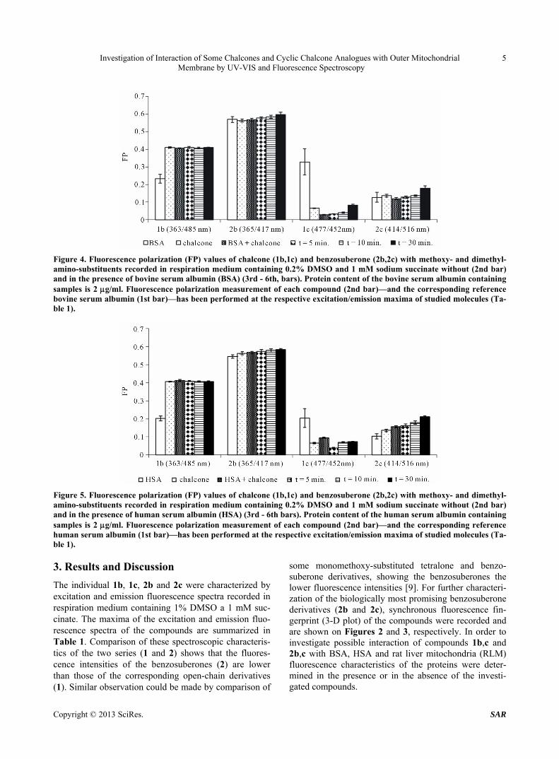
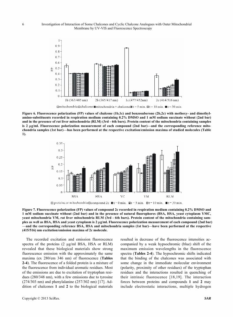
Figure 3. Synchronous fluorescence fingerprint 3-D plot (above) and topogram (below) of compound 2c (c = 10 nmol/ml in respiration medium containing 0.2% DMSO and 1 mM sodium succinate). Excitation slit = 5 nm, emission slit = 10 nm, scan speed = 1200 nm/min. The fluorescence maximum of topogram (ex = 415 nm/∆ = 100 nm, F = 500). (= 20, 40, 60, 80, 100, 120, 140, 160, 180, 200 nm) between both excitation and emission monochromators. Fluorescence polarization (FP) measurements 15,16 of the studied chalcones (1b,c and 2b,c) with bovine (BSA) and human (HSA) serum albumins (Figures 4 and 5) as well as with rat liver mitochondria (RLM) (Figure 6) were performed at their excitation/emission maximum for a period of 30 minutes at ambient temperature. The protein concentration of the investigated mixtures (BSA,
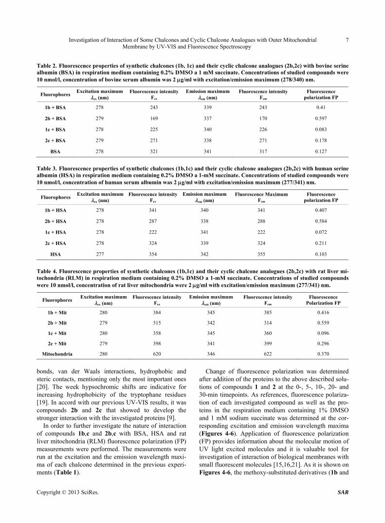
HSA, RLM) was 2 g/ml. As references, FP of non/ treated BSA, HSA and RLM in respiration medium con- taining 1 mM sodium succinate were determined at the excitation/emission maxima of the respective chalcones. FP measurements studying interaction of 2c with yeast cytoplasm (YC) and yeast mitochondria (YM) were per- formed under similar conditions. The results of these latter measurements (together with those of 2c with BSA,
SA and RLM) are shown on Figure 7. H
Copyright © 2013 SciRes. SAR
Investigation of Interaction of Some Chalcones and Cyclic Chalcone Analogues with Outer Mitochondrial Membrane by UV-VIS and Fluorescence Spectroscopy
5
Figure 4. Fluorescence polarization (FP) values of chalcone (1b,1c) and benzosuberone (2b,2c) with methoxy- and dimethyl-amino-substituents recorded in respiration medium containing 0.2% DMSO and 1 mM sodium succinate without (2nd bar) and in the presence of bovine serum albumin (BSA) (3rd - 6th, bars). Protein content of the bovine serum albumin containing samples is 2 g/ml. Fluorescence polarization measurement of each compound (2nd bar)—and the corresponding reference bovine serum albumin (1st bar)—has been performed at the respective excitation/emission maxima of studied molecules (Ta-ble 1).
Figure 5. Fluorescence polarization (FP) values of chalcone (1b,1c) and benzosuberone (2b,2c) with methoxy- and dimethyl-amino-substituents recorded in respiration medium containing 0.2% DMSO and 1 mM sodium succinate without (2nd bar) and in the presence of human serum albumin (HSA) (3rd - 6th bars). Protein content of the human serum albumin containing samples is 2 g/ml. Fluorescence polarization measurement of each compound (2nd bar)—and the corresponding reference human serum albumin (1st bar)—has been performed at the respective excitation/emission maxima of studied molecules (Ta-ble 1). 3. Results and Discussion
The individual 1b, 1c, 2b and 2c were characterized by excitation and emission fluorescence spectra recorded in respiration medium containing 1% DMSO a 1 mM suc- cinate. The maxima of the excitation and emission fluo- rescence spectra of the compounds are summarized in Table 1. Comparison of these spectroscopic characteris- tics of the two series (1 and 2) shows that the fluores- cence intensities of the benzosuberones (2) are lower than those of the corresponding open-chain derivatives (1). Similar observation could be made by comparison of
some monomethoxy-substituted tetralone and benzo- suberone derivatives, showing the benzosuberones the lower fluorescence intensities [9]. For further characteri- zation of the biologically most promising benzosuberone derivatives (2b and 2c), synchronous fluorescence fin- gerprint (3-D plot) of the compounds were recorded and are shown on Figures 2 and 3, respectively. In order to investigate possible interaction of compounds 1b,c and 2b,c with BSA, HSA and rat liver mitochondria (RLM) fluorescence characteristics of the proteins were deter- mined in the presence or in the absence of the investi- gated compounds.
Copyright © 2013 SciRes. SAR
Investigation of Interaction of Some Chalcones and Cyclic Chalcone Analogues with Outer Mitochondrial Membrane by UV-VIS and Fluorescence Spectroscopy
6
Figure 6. Fluorescence polarization (FP) values of chalcone (1b,1c) and benzosuberone (2b,2c) with methoxy- and dimethyl-amino-substituents recorded in respiration medium containing 0.2% DMSO and 1 mM sodium succinate without (2nd bar) and in the presence of rat liver mitochondria (RLM) (3rd - 6th bars). Protein content of the mitochondria containing samples is 2 g/ml. Fluorescence polarization measurement of each compound (2nd bar)—and the corresponding reference mito-chondria samples (1st bar)—has been performed at the respective excitation/emission maxima of studied molecules (Table 1).
Figure 7. Fluorescence polarization (FP) values of compound 2c recorded in respiration medium containing 0.2% DMSO and 1 mM sodium succinate without (2nd bar) and in the presence of natural fluorophores (BSA, HSA, yeast cytoplasm YMC, yeast mitochondria YM, rat liver mitochondria RLM (3rd - 6th bars). Protein content of the mitochondria containing sam-ples as well as BSA, HSA and yeast cytoplasm is 2 g/ml. Fluorescence polarization measurement of each compound (2nd bar) —and the corresponding reference BSA, HSA and mitochondria samples (1st bar)—have been performed at the respective (415/516) nm excitation/emission maxima of 2c molecule.
The recorded excitation and emission fluorescence spectra of the proteins (2 µg/ml BSA, HSA or RLM) revealed that these biological materials show strong fluorescence emission with the approximately the same maxima (ex 280/em 346 nm) of fluorescence (Tables 2-4). The fluorescence of a folded protein is a mixture of the fluorescence from individual aromatic residues. Most of the emissions are due to excitation of tryptophan resi- dues (280/348 nm), with a few emissions due to tyrosine (274/303 nm) and phenylalanine (257/302 nm) [17]. Ad- dition of chalcones 1 and 2 to the biological materials
resulted in decrease of the fluorescence intensities ac- companied by a weak hypsochromic (blue) shift of the maximum emission wavelengths in the fluorescence spectra (Tables 2-4). The hypsochromic shifts indicated that the binding of the chalcones was associated with some change in the immediate molecular environment (polarity, proximity of other residues) of the tryptophan residues and the interactions resulted in quenching of their intrinsic fluorescence [18,19]. The interaction forces between proteins and compounds 1 and 2 may include electrostatic interactions, multiple hydrogen
Copyright © 2013 SciRes. SAR
Investigation of Interaction of Some Chalcones and Cyclic Chalcone Analogues with Outer Mitochondrial Membrane by UV-VIS and Fluorescence Spectroscopy
7
Table 2. Fluorescence properties of synthetic chalcones (1b, 1c) and their cyclic chalcone analogues (2b,2c) with bovine serine albumin (BSA) in respiration medium containing 0.2% DMSO a 1 mM succinate. Concentrations of studied compounds were 10 nmol/l, concentration of bovine serum albumin was 2 g/ml with excitation/emission maximum (278/340) nm.
Fluorophores Excitation maximum
ex (nm) Fluorescence intensity
Fex Emission maximum
em (nm) Fluorescence intensity
Fem Fluorescence
polarization FP
1b + BSA 278 243 339 243 0.41
2b + BSA 279 169 337 170 0.597
1c + BSA 278 225 340 226 0.083
2c + BSA 279 271 338 271 0.178
BSA 278 321 341 317 0.127
Table 3. Fluorescence properties of synthetic chalcones (1b,1c) and their cyclic chalcone analogues (2b,2c) with human serine albumin (HSA) in respiration medium containing 0.2% DMSO a 1-mM succinate. Concentrations of studied compounds were 10 nmol/l, concentration of human serum albumin was 2 g/ml with excitation/emission maximum (277/341) nm.
Fluorophores Excitation maximum
ex (nm) Fluorescence intensity
Fex Emission maximum
em (nm) Fluorescence Maximum
Fem Fluorescence
polarization FP
1b + HSA 278 341 340 341 0.407
2b + HSA 278 287 338 288 0.584
1c + HSA 278 222 341 222 0.072
2c + HSA 278 324 339 324 0.211
HSA 277 354 342 355 0.103
Table 4. Fluorescence properties of synthetic chalcones (1b,1c) and their cyclic chalcone analogues (2b,2c) with rat liver mi-tochondria (RLM) in respiration medium containing 0.2% DMSO a 1-mM succinate. Concentrations of studied compounds were 10 nmol/l, concentration of rat liver mitochondria were 2 g/ml with excitation/emission maximum (277/341) nm.
Fluorophores Excitation maximum
ex (nm) Fluorescence intensity
Fex Emission maximum
em (nm) Fluorescence intensity
Fem Fluorescence
Polarization FP
1b + Mit 280 384 345 385 0.416
2b + Mit 279 315 342 314 0.559
1c + Mit 280 358 345 360 0.096
2c + Mit 279 398 341 399 0.296
Mitochondria 280 620 346 622 0.370
bonds, van der Waals interactions, hydrophobic and steric contacts, mentioning only the most important ones [20]. The week hypsochromic shifts are indicative for increasing hydrophobicity of the tryptophane residues [19]. In accord with our previous UV-VIS results, it was compounds 2b and 2c that showed to develop the stronger interaction with the investigated proteins [9].
In order to further investigate the nature of interaction of compounds 1b,c and 2b,c with BSA, HSA and rat liver mitochondria (RLM) fluorescence polarization (FP) measurements were performed. The measurements were run at the excitation and the emission wavelength maxi- ma of each chalcone determined in the previous experi- ments (Table 1).
Change of fluorescence polarization was determined after addition of the proteins to the above described solu- tions of compounds 1 and 2 at the 0-, 5-, 10-, 20- and 30-min timepoints. As references, fluorescence polariza- tion of each investigated compound as well as the pro- teins in the respiration medium containing 1% DMSO and 1 mM sodium succinate was determined at the cor- responding excitation and emission wavelength maxima (Figures 4-6). Application of fluorescence polarization (FP) provides information about the molecular motion of UV light excited molecules and it is valuable tool for investigation of interaction of biological membranes with small fluorescent molecules [15,16,21]. As it is shown on Figures 4-6, the methoxy-substituted derivatives (1b and
Copyright © 2013 SciRes. SAR
Investigation of Interaction of Some Chalcones and Cyclic Chalcone Analogues with Outer Mitochondrial Membrane by UV-VIS and Fluorescence Spectroscopy
8
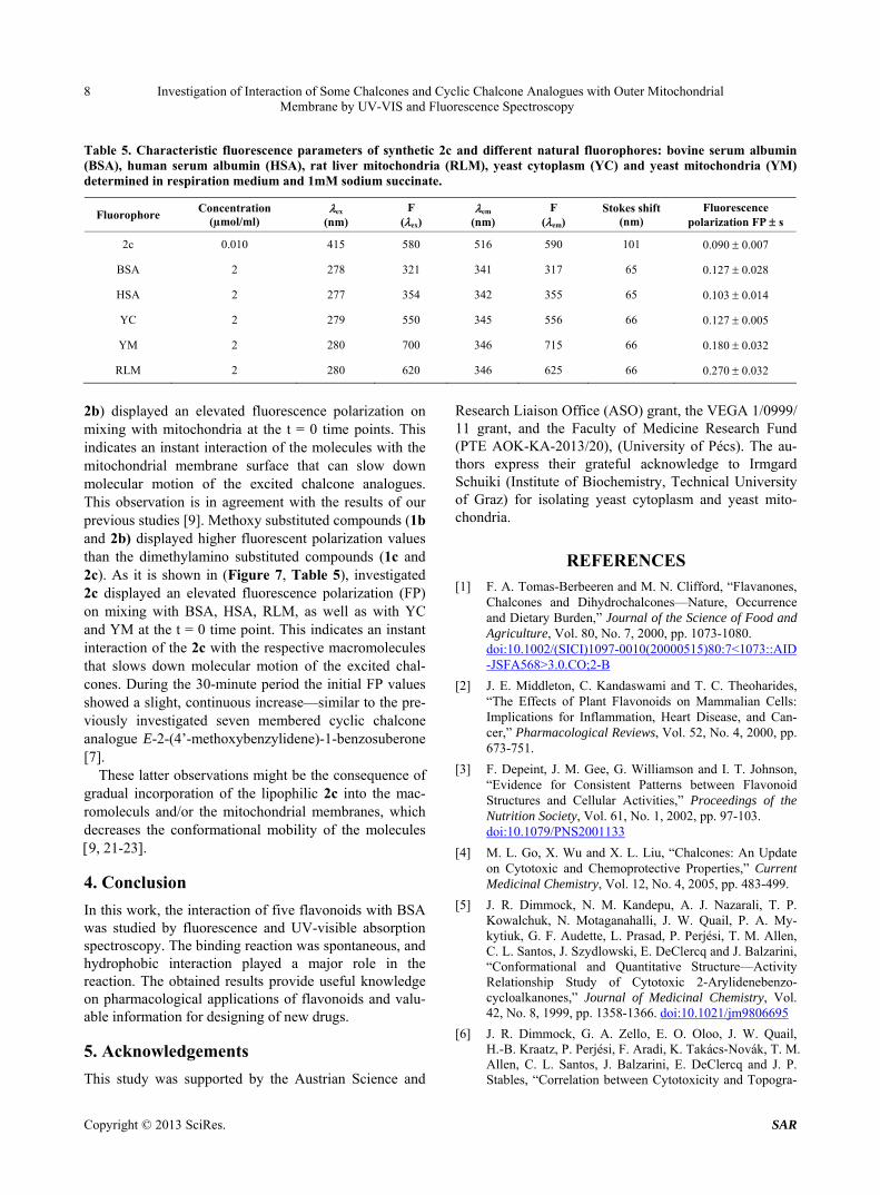
Table 5. Characteristic fluorescence parameters of synthetic 2c and different natural fluorophores: bovine serum albumin (BSA), human serum albumin (HSA), rat liver mitochondria (RLM), yeast cytoplasm (YC) and yeast mitochondria (YM) determined in respiration medium and 1mM sodium succinate.
Fluorophore Concentration
(µmol/ml) ex
(nm) F
(ex) em
(nm) F
(em) Stokes shift
(nm) Fluorescence
polarization FP s
2c 0.010 415 580 516 590 101 0.090 0.007
BSA 2 278 321 341 317 65 0.127 0.028
HSA 2 277 354 342 355 65 0.103 0.014
YC 2 279 550 345 556 66 0.127 0.005
YM 2 280 700 346 715 66 0.180 0.032
RLM 2 280 620 346 625 66 0.270 0.032
2b) displayed an elevated fluorescence polarization on mixing with mitochondria at the t = 0 time points. This indicates an instant interaction of the molecules with the mitochondrial membrane surface that can slow down molecular motion of the excited chalcone analogues. This observation is in agreement with the results of our previous studies [9]. Methoxy substituted compounds (1b and 2b) displayed higher fluorescent polarization values than the dimethylamino substituted compounds (1c and 2c). As it is shown in (Figure 7, Table 5), investigated 2c displayed an elevated fluorescence polarization (FP) on mixing with BSA, HSA, RLM, as well as with YC and YM at the t = 0 time point. This indicates an instant interaction of the 2c with the respective macromolecules that slows down molecular motion of the excited chal- cones. During the 30-minute period the initial FP values showed a slight, continuous increase—similar to the pre- viously investigated seven membered cyclic chalcone analogue E-2-(4’-methoxybenzylidene)-1-benzosuberone [7].
These latter observations might be the consequence of gradual incorporation of the lipophilic 2c into the mac- romoleculs and/or the mitochondrial membranes, which decreases the conformational mobility of the molecules 9, 21-23.
4. Conclusion
In this work, the interaction of five flavonoids with BSA was studied by fluorescence and UV-visible absorption spectroscopy. The binding reaction was spontaneous, and hydrophobic interaction played a major role in the reaction. The obtained results provide useful knowledge on pharmacological applications of flavonoids and valu- able information for designing of new drugs.
5. Acknowledgements
This study was supported by the Austrian Science and
Research Liaison Office (ASO) grant, the VEGA 1/0999/ 11 grant, and the Faculty of Medicine Research Fund (PTE AOK-KA-2013/20), (University of Pécs). The au- thors express their grateful acknowledge to Irmgard Schuiki (Institute of Biochemistry, Technical University of Graz) for isolating yeast cytoplasm and yeast mito- chondria.
REFERENCES [1] F. A. Tomas-Berbeeren and M. N. Clifford, “Flavanones,
Chalcones and Dihydrochalcones—Nature, Occurrence and Dietary Burden,” Journal of the Science of Food and Agriculture, Vol. 80, No. 7, 2000, pp. 1073-1080. doi:10.1002/(SICI)1097-0010(20000515)80:7<1073::AID-JSFA568>3.0.CO;2-B
[2] J. E. Middleton, C. Kandaswami and T. C. Theoharides, “The Effects of Plant Flavonoids on Mammalian Cells: Implications for Inflammation, Heart Disease, and Can- cer,” Pharmacological Reviews, Vol. 52, No. 4, 2000, pp. 673-751.
[3] F. Depeint, J. M. Gee, G. Williamson and I. T. Johnson, “Evidence for Consistent Patterns between Flavonoid Structures and Cellular Activities,” Proceedings of the Nutrition Society, Vol. 61, No. 1, 2002, pp. 97-103. doi:10.1079/PNS2001133
[4] M. L. Go, X. Wu and X. L. Liu, “Chalcones: An Update on Cytotoxic and Chemoprotective Properties,” Current Medicinal Chemistry, Vol. 12, No. 4, 2005, pp. 483-499.
[5] J. R. Dimmock, N. M. Kandepu, A. J. Nazarali, T. P. Kowalchuk, N. Motaganahalli, J. W. Quail, P. A. My- kytiuk, G. F. Audette, L. Prasad, P. Perjési, T. M. Allen, C. L. Santos, J. Szydlowski, E. DeClercq and J. Balzarini, “Conformational and Quantitative Structure—Activity Relationship Study of Cytotoxic 2-Arylidenebenzo- cycloalkanones,” Journal of Medicinal Chemistry, Vol. 42, No. 8, 1999, pp. 1358-1366. doi:10.1021/jm9806695
[6] J. R. Dimmock, G. A. Zello, E. O. Oloo, J. W. Quail, H.-B. Kraatz, P. Perjési, F. Aradi, K. Takács-Novák, T. M. Allen, C. L. Santos, J. Balzarini, E. DeClercq and J. P. Stables, “Correlation between Cytotoxicity and Topogra-
Copyright © 2013 SciRes. SAR
Investigation of Interaction of Some Chalcones and Cyclic Chalcone Analogues with Outer Mitochondrial Membrane by UV-VIS and Fluorescence Spectroscopy
9
phy of Some 2-Arylidenebenzocyclanones Determined by X-Ray Crystallography,” Journal of Medicinal Chemistry, Vol. 45, No. 14, 2002, pp. 3103-3111. doi:10.1021/jm010559p
[7] P. Perjési, U. Das, E. De Clercq, J. Balzarini, M. Kawase, H. Sakagami, J. P. Stables, T. Loránd, Z. Rozmer and J. R. Dimmock, “Design, Synthesis and Antiproliferative Ac- tivity of Some 3-Benzylidene-2,3-dihydro-1-ben-zopy- ran-4-ones Which Display Selective Toxicity for Malig- nant Cells,” European Journal of Medicinal Chemistry, Vol. 43, No. 4, 2008, pp. 839-845. doi:10.1016/j.ejmech.2007.06.017
[8] K. Fodor, V. Tomečková, T. Koszegi, I. Kron and P. Per- jési, “(E)-2-Benzylidenecyclanones: Part VI. Solvent Ef- fect on the UV and Fluorescence Properties of Some Chalcones and Their Cyclic Analogues. Interaction of 4-Dimethylaminochalcones with Bovine and Human Se- rum Albumin: A UV-VIS Study,” Monatshefte für Che- mie, Vol. 142, No. 5, 2011, pp. 463-468. doi:10.1007/s00706-011-0463-0
[9] V. Tomečková, P. Perjési, J. Guzy, J. Kusnir, Z. Cho- vanová, Z. Chavková and M. Mareková, “Comparison of Effect of Selected Synthetic Chalcone Analogues on Mi- tochondrial Outer Membrane Determined by Fluores- cence Spectroscopy,” Journal of Biochemical and Bio- physical Methods, Vol. 61, No. 1-2, 2004, pp. 135-141. doi:10.1016/j.jbbm.2004.04.010
[10] P. Perjési, T. Nusser, G. Tarczay and P. Sohar, “E-2-Ben- zylidenebenzocycloalkanones. Stereostructure and NMR Spectroscopic Investigation,” Journal of Molecular Struc- ture, Vol. 479, No. 1, 1999, pp. 13-19. doi:10.1016/S0022-2860(98)00805-9
[11] D. Johnson and H. Lardy, “Isolation of Rat Liver and Kidney Mitochondria,” Methods in Enzymology, Vol. 10, 1967, pp. 94-96. doi:10.1016/0076-6879(67)10018-9
[12] M. M. Bradford, “A Rapid and Sensitive for the Quanti- fication of Microgram Quantities of Protein Utilizing the Principle of Protein-Dye Binding,” Analytical Bioche- mistry, Vol. 72, No. 1-2, 1976, pp. 248-254. doi:10.1016/0003-2697(76)90527-3
[13] E. Zinser and G. Daum, “Isolation and Biochemical Characterisation of Organelles Form the Yeast, Saccha- romyces cerevisiae,” Yeast, Vol. 11, No. 6, 1995, pp. 493-
536. doi:10.1002/yea.320110602
[14] O. H. Lowry, N. J. Rosebrough, A. L. Farr and R. Ram- dall, “Protein Measurement with the Folin Phenol Re- agent,” Journal of Biological Chemistry, Vol. 193, No. 1, 1951, pp. 265-275.
[15] W. J. Checovich, R. E. Bolger and T. Burke, “Fluores- cence Polarization—A New Tool for Cell and Molecular Biology,” Nature, Vol. 375, 1995, pp. 253-256. doi:10.1038/375254a0
[16] B. J. Litman and Y. Barenholz, “Fluorescence Probe: Difenylhexatriene,” Methods in Enzymology, Vol. 81, 1982, pp. 678-685. doi:10.1016/S0076-6879(82)81093-8
[17] J. R. Lakowicz, “Principles of Fluorescence Spectros- copy,” 2nd Edition, Kluwer Academic/Plenum Publishers, New York, 1999. doi:10.1007/978-1-4757-3061-6
[18] J. T. Vivian and P. R. Callis, “Mechanisms of Tryptophan Fluorescence Shifts in Proteins,” Biophysical Journal, Vol. 80, No. 5, 2001, pp. 2093-2109. doi:10.1016/S0006-3495(01)76183-8
[19] S. Gorinstein, I. Goshev, S. Moncheva, M. Zemser, M. Weisz, A. Caspi, I. Libman, H. T. Lerner, S. Trakhten- berg and O. Martín-Belloso, “Intrinsic Tryptophan Fluo- rescence of Human Serum Proteins and Related Confor- mational Changes,” Journal of Protein Chemistry, Vol. 19, No. 8, 2000, pp. 637-642. doi:10.1023/A:1007192017291
[20] D. Leckband, “Measuring the Forces that Control Protein Interactions,” Annual Review of Biophysics and Biomo- lecular Structure, Vol. 29, 2000, pp. 1-26. doi:10.1146/annurev.biophys.29.1.1
[21] L. A. Sklar, “Chapter 3. Fluorescence Polarization Stud-ies of Membrane Fluidity: Where Do We Go from Here?” In: Biomembranes, Plenum Press, New York, 1984, pp. 99-131.
[22] M. Shinitzky and Y. Berenholz, “Fluidity Parameters of Lipid Regions Determined by Fluorescence Polarization,” Biochimica et Biophysica Acta, Vol. 515, No. 4, 1978, pp. 367-394. doi:10.1016/0304-4157(78)90010-2
[23] B. H. Havsteen, “The Biochemistry and Medical Signifi- cance of the Flavonoids,” Pharmacology and Therapeu- tics, Vol. 96, No. 2-3, 2002, pp. 67-202. doi:10.1016/S0163-7258(02)00298-X
Copyright © 2013 SciRes. SAR


![Preparation and Antifungal Properties of Chalcone and ... · antimicrobial activities [9-11]. Notably, the 4, 6-diphenyl-2-thiopyrimidine and epoxide derivatives of chalcones had](https://img.pdfslide.net/doc/110x75/6086b07b4b8005680c667375/preparation-and-antifungal-properties-of-chalcone-and-antimicrobial-activities.jpg)