Embed Size (px)
Citation preview
1
Involvement of Host Cell Integrin α2 in Cryptosporidium parvum Infection 1 2 Haili Zhang1, Fengguang Guo1, and Guan Zhu1,2,* 3 4 1 Department of Veterinary Pathobiology, College of Veterinary Medicine & Biomedical 5 Sciences, Texas A&M University, College Station, Texas 77843-4467, USA 6 2 Faculty of Genetics Program, Texas A&M University, College Station, Texas 77843-4467, 7 USA 8 9 Running Title: Host Cell ITGA2 in Cryptosporidium Infection 10 11 Address corresponding to: 12 Guan Zhu, PhD 13 Department of Veterinary Pathobiology 14 College of Veterinary Medicine & Biomedical Sciences 15 4467 TAMU 16 College Station, Texas 77843-4467, USA. 17 Tel: +1 (979) 845-6981 18 Fax: +1 (979) 845-9972 19 Email: [email protected] 20 21 22
Copyright © 2012, American Society for Microbiology. All Rights Reserved.Infect. Immun. doi:10.1128/IAI.05862-11 IAI Accepts, published online ahead of print on 21 February 2012
on February 12, 2018 by guest
http://iai.asm.org/
Dow
nloaded from
2
ABSTRACT 23 Cryptosporidium parvum is an opportunistic pathogen in AIDS patients. It is an intracellular 24
but extracytoplasmic parasite residing in a host cell-derived parasitophorous vacuole. It is still 25 poorly understood how this parasite interacts with host cells. We observed that expression of the 26 integrin α2 (ITGA2) gene in host cells was significantly up-regulated upon C. parvum infection, 27 and a higher level of ITGA2 protein was present in the parasite infection sites. The infection 28 could be reduced by the treatment of antibodies against ITGA2 and integrin β1 (ITGB1) 29 subunits, as well as by type I collagen (an integrin α2β1 ligand). We also generated stable 30 knockdown of ITGA2 gene expression in HCT-8 cells, and observed consistent reduction of 31 parasite infection in these knockdown cells. Collectively, our evidence indicates that host cell 32 ITGA2 might be involved in interacting with Cryptosporidium during infection, probably acting 33 as part of the regulatory elements upstream to the reported recruiting and reorganization of F-34 actin at the infection sites. 35 36
37 Keywords: Cryptosporidium parvum; Apicomplexa; Infection; Integrin α2 (ITGA2); HCT-8 38 cells; Host-parasite interactions39
on February 12, 2018 by guest
http://iai.asm.org/
Dow
nloaded from
3
INTRODUCTION 40 Cryptosporidium parvum is an apicomplexan parasite infecting both humans and animals 41
(30, 31, 36). It is also an AIDS opportunistic pathogen, for which treatment is currently 42 unavailable (9). Like other apicomplexans, Cryptosporidium possesses a complex life cycle that 43 starts with the ingestion of oocysts, followed by excystation in the intestine to release sporozoites 44 that invade host gastric or intestinal epithelial cells. During intracellular development, 45 Cryptosporidium resides within a host cell-derived membrane parasitophorous vacuole 46 membrane (PVM). The PVM-contained parasite is connected to the host cell cytosol only by an 47 electron-dense juncture, rather than residing within host cell cytoplasm. Therefore, 48 Cryptosporidium is considered an intracellular, but extracytoplasmic parasite, which differs from 49 the majority of other intracytoplasmic apicomplexans. 50
Although morphology at the parasite-infection site has been extensively studied, very 51 limited knowledge has accumulated on how the parasite interacts with host cell molecules. 52 Several C. parvum membrane proteins and antigens that might be involved in the interaction 53 with host cells have been reported. These include various mucin-like proteins, thrombospondin-54 related adhesive proteins (TRAPs), and circumsporozoite-like antigen/ligand (CSL) (5, 26, 32). 55 It is known that host cell F-actin is reorganized and accumulates underneath the electron-dense 56 membrane structure (13, 14). Using biliary epithelial cells as a model of cryptosporidial 57 infection, several host cell factors and pathways have been shown to possibly be involved in the 58 remodeling of F-actin, which include c-Src-dependent tyrosine phosphorylation for the 59 accumulation of cortactin, and phosphatidylinositol-3-kinase (PI3K) and frabin-mediated 60 activation of CDC42 for the recruiting of Neural Wiskott-Alrich Symdrome protein (WASP) (7, 61 8, 10). However, the upstream elements within the pathways have yet to be defined, as well as 62
on February 12, 2018 by guest
http://iai.asm.org/
Dow
nloaded from
4
the host cell membrane proteins that may interact directly with the parasite during invasion and 63 development. 64
Integrins (ITGs) are a family of surface receptors associated with extracellular matrix 65 (ECM) complexes. These receptors consist of α- and β-subunits. Each subunit has several 66 isoforms that form up to 24 prototypes of αβ heterodimers in higher vertebrates (2, 3, 17-20). 67 ITGs in the cytoplasmic membrane are involved in the transduction of both outside-in and 68 inside-out signals to regulate cell polarity, migration, growth, survival and differentiation (11, 69 20, 22, 28). One important function of ITGs is to regulate the dynamics and reorganization of 70 actins at the sites of adhesion via a matrix of proteins and pathways, such as FAK/Src, PI3K, 71 ILK, Rho, Rac and the Cdc42-WASP-Arp2/3 pathway (23). 72
In the present study, we observed that the expression of ITGA2 in human cells was up-73 regulated upon infection by C. parvum and ITGA2 protein was recruited to the sites of infection. 74 We have confirmed that infection could be reduced by the knockdown of ITGA2 expression in 75 host cells, and by treatment using antibodies specific to ITGA2 and the ligand collagen-I. These 76 observations indicate that host cell ITGA2 may be involved in the interaction with 77 cryptosporidial infection. 78 79 MATERIALS AND METHODS 80
Parasite and in vitro cultivation. Fresh oocysts of C. parvum (Iowa strain) were 81 purchased from Bunch Grass Farm (Deary, Idaho). Oocysts were further purified by a Percoll-82 based gradient centrifugation method and surface-sterilized with 10% bleach for 7 min on ice, 83 followed by washes with PBS for as described (1, 6, 25). A human ileocecal epithelial cell line 84 (HCT-8, ATCC# CCL-244) was used to assay cryptosporidial infection and to generate stable 85 ITGA2-knockdown (KD) cells. HCT-8 cells were maintained as described (6). In a typical 86
on February 12, 2018 by guest
http://iai.asm.org/
Dow
nloaded from
5
infection assay, HCT-8 and ITGA2-KD cells were seeded into 24-well plates and allowed to 87 grow overnight until they reached ~80% confluence. Oocysts (less than 3 months old) were 88 suspended in the same culture medium and added to plates with specified parasite:host cell ratios 89 as described below. Cells receiving no infection or sham-infection with heat-killed oocysts by 90 pre-treating oocysts at 65 °C for 30 min were included as controls. Uninfected controls received 91 the same treatments as experimental groups. After incubation for 3 h to allow parasite 92 excystation and invasion, uninfected parasites were removed by a medium exchange. Cells at 93 this stage might be harvested at 3 h post-infection to determine the level of parasite invasion or 94 allowed to grow further for a total 8 to 18 h to determine the effects of described conditions on 95 early intracellular development of the parasite. The selection of these time points was mainly to 96 restrict the parasite growth within the first cell cycle (i.e., first merogony development) before 97 the release of merozoites that might occur ~20 - 24 h post-infection. The release of merozoites 98 would damage and trigger apoptosis in host cells, and the unsynchronized second parasite cell 99 cycle by the released merozoites (i.e., second merogony) in a mixture of damaged and healthy 100 cells would also complicate the study of host cell-parasite molecular interactions. It was also 101 noticed that ITGA2-KD cells might gradually increase the expression of ITGA2 upon parasite 102 infection, particularly after >8 h of infection (see below for details). 103 104
Effects of integrin antibodies and ligand collagen-I on parasite infection. Monoclonal 105 antibody (mAb) against ITGA2 (clone C-9) was purchased from Santa Cruz Biotechnology 106 (Santa Cruz, CA). Additional mAbs against β1 (6S6), α6 (NKI-GoH3), β4 mAb (ASC-3), α3 107 (P1B5), β3 (B3A) and αvβ3 (23C6) subunits were purchased from Millipore (Billerica, MA). 108 Integrin α2β1 ligand collagen-I was purchased from Sigma-Aldrich (St. Louis, MO). HCT-8 cell 109 monolayers in 24-well plates were pre-incubated with mAbs or collagen-I for 1 h prior to the 110
on February 12, 2018 by guest
http://iai.asm.org/
Dow
nloaded from
6
addition of C. parvum oocysts. The final concentration of each antibody and ligand was 10 111 μg/well. Host cells were infected with live or heat-killed sham parasite with an oocyst:host cell 112 ratio at 1:5. Cells were washed with RNase-free PBS 3 times and harvested at 3 h and 18 h after 113 infection for the isolation of total RNA and detection of parasite load by qRT-PCR as described 114 below. 115 116
Generation of stable integrin α2-knockdown (ITGA2-KD) cells. Stable ITGA2-KD 117 cells were generated by small-hairpin RNA (shRNA) based methods. SureSilencing shRNA 118 vector for human ITGA2 (Cat. # KH00625N) and a negative control vector containing only 119 scrambled artificial sequence without matching any human genes were purchased from 120 SABiosciences (now part of Qiagen Inc., Frederick, MD). HCT-8 cells with ~80% confluence in 121 24-well plates were transfected with shRNA plasmids (0.8 μg/well) using a lipofectamine 122 protocol as recommended by the manufacturer. After 24 to 48 h post-transfection, cells were re-123 plated to a low density at <10% confluence in culture medium containing 580 μg/ml neomycin 124 that was exchanged every 2 to 3 days. Cells were re-plated every week for two weeks. 125 “Monoclonal” populations were obtained by diluting cells to ~1 cell/400 μl and plating them into 126 96-well plates at 200 μl/well. Cells were allowed to form large colonies, and wells containing 127 single colonies were selected and maintained in medium containing 300 μg/ml neomycin. 128 129
Western blot analysis of ITGA2 protein in WT and KD cells. Cultured WT and ITGA2-130 KD cells were removed from plates with a cell scraper and lysed in radio-immunoprecipitation 131 assay (RIPA) buffer containing a protease inhibitor cocktail for mammals (Sigma-Aldrich). Total 132 protein concentrations were determined by the Bradford method. Protein lysates (20 µg/lane) 133 were separated by 7% SDS-PAGE and transferred onto nitrocellulose membranes. The blots 134
on February 12, 2018 by guest
http://iai.asm.org/
Dow
nloaded from
7
were treated with 5% BSA in 10 mM Tris-HCl (pH 7.5) containing 166 mM NaCl and 0.05% 135 Tween-20 (5% BSA-TBST), incubated with anti-ITGA2 mAb (1:200 dilution in 5% BSA-136 TBST), and then horseradish peroxidase (HRP)-conjugated goat-anti-mouse IgG antibody 137 (1:5000 dilution). Each incubation step was carried out for 1 h at room temperature, followed by 138 3 washes in TBST. Human glyceraldehyde-3-phosphate dehydrogenase (GAPDH) was detected 139 with mAb (Millipore, 1:1000 dilution) as a loading control. Antibody-labeled blots were 140 visualized using an enhanced chemiluminescent (ECL) reagent (Sigma-Aldrich). 141 142
Detection of parasite growth in vitro and gene expressions by qRT-PCR. Total RNA 143 was isolated from HCT-8 or ITGA2-KD cells uninfected or infected with C. parvum for various 144 times using an RNeasy mini kit (Qiagen Inc., Valencia, CA). Cell monolayers were gently 145 washed 3 times with nuclease-free PBS, and lysed in 350 μL of lysis buffer by incubating cells at 146 37 °C for 20 min, followed by gentle pipetting at least 20 times through 200 μl pipetting tips. 147 Samples were treated with RNase-free DNase (Qiagen Inc., Valecia, CA) to remove DNA 148 according to the manufacturer’s protocol. The quality and purity of RNA was determined using a 149 NanoDrop ND-1000 spectrophotometer at 260/280 nm (NanoDrop Technologies, Wilmington, 150 DE). A Qiagen one-step RT-PCR QuantiTect SYBR-Green RT-PCR kit was employed to 151 evaluate both gene expression levels and parasite growth as described below. Each 25 μl reaction 152 mixture contained 2 ng total RNA, 500 nM each primer, 10 nM FITC, 0.25 μl RT-master mix 153 and 1X QuantiTect SYBR-Green. The mixtures were incubated at 50 °C for 30 min to synthesize 154 cDNA, heated at 95 °C for 15 min to inactivate the reverse transcriptase, and then subjected to 155 40 thermal cycles of PCR amplification (95 °C for 20 s, 58 °C for 30 s, and 72 °C for 30 s) with 156 an iCycler iQ real-time PCR detection system (Bio-Rad Laboratories, Hercules, CA). At least 2 157
on February 12, 2018 by guest
http://iai.asm.org/
Dow
nloaded from
8
replicate qRT-PCR reactions were performed for each sample. All reagents for the qRT-PCR 158 were loaded manually. 159
Parasite growth in HCT-8 and ITGA2-KD cells was assayed after infecting host cells for 3 160 and 8 h (oocyst:host cell ratio 1:20) by detecting parasite 18S rRNA levels using primers Cp18S-161 1011F [5'-TTGTTCCTTACTCCTTCAGCAC-3'] and Cp18S-1185R [5'-162 TCCTTCCTATGTCTGGACCTG-3’] as described (6). Human 18S rRNA levels were detected 163 using primers Hs18S-1F [5'-GGCGCCCCCTCGATGCTCTTA-3'] and Hs18S-1R [5'-164 CCCCCGGCCGTCCCTCTTA-3'] for evaluating host cell integrity and for normalization (6). 165 The relative levels of human ITG subunit transcripts in WT and ITGA2-KD cells were 166 determined by qRT-PCR using the following primer pairs, which were designed using the 167 Primer3Plus server (http://www.bioinformatics.nl/cgi-bin/primer3plus/primer3plus.cgi) (33), and 168 validated by BLAST searches against the NCBI nucleotide databases 169 (http://blast.ncbi.nlm.nih.gov/Blast.cgi): 5’-AGGACGGACTTTGCATTTCTGAT-3’ and 5’-170 CCACCTGGCATGTTACTTCTGT-3’ for α2 (ITGA2); 5’-GTGCAAATCCCACAACACTG-3’ 171 and 5’-TTTCAATAGTCCAGGAAGAAAAGG-3’ for β1; 5’-AAGGCTCCTGTTTTGCACAG-172 3’ and 5’-ATGTAAGTCAGCCACGCCA-3’ for α6; 5’-CCCGCCACGTCCCACTAG-3’ and 173 5’-TTTTTTTAGCAGTAGCAAAACCA-3’ for β4. All experiments were performed at least in 174 triplicate for each experimental condition. Relative levels of gene expression were calculated by 175 a ΔΔCT method with a general formula of 2-ΔΔCT, in which ΔCT values between specified gene 176 transcripts and human 18S rRNA were first determined by ΔCT = CT[gene] - CT[18S], followed by 177 the calculation of ΔΔCT between KD and WT cells or between treated and untreated samples. 178 Statistical significance on the relative levels of gene expression and on the parasite infections 179 was examined by the Student’s t-test. 180
on February 12, 2018 by guest
http://iai.asm.org/
Dow
nloaded from
9
181 Immunofluorescent microscopy of ITGA2 protein in infected cells. The same quantity 182
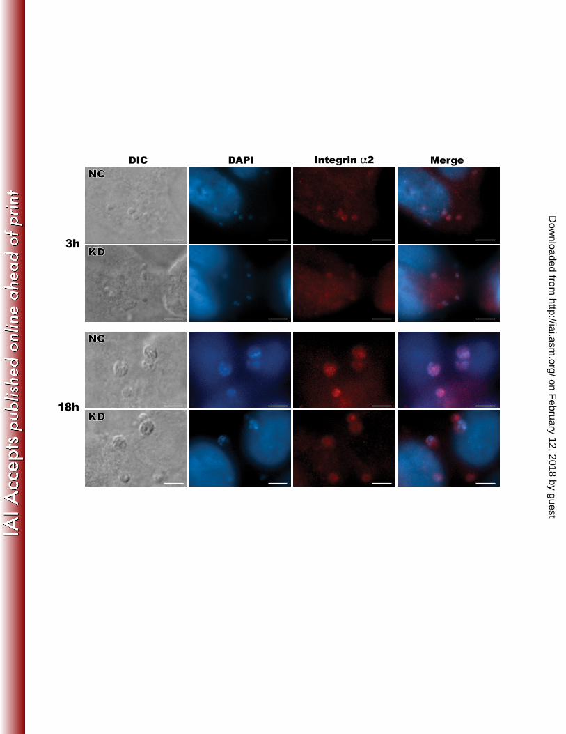
of ITGA2-KD (KD137-2-3) and vector control containing a scrambled sequence (NC1-2) cells 183 were seeded into 24-well plates containing coverslips and allowed to grow to ~80% confluence. 184 Cells were infected with C. parvum (oocyst:host cell ratio 2:1) for 3 h and 18 h, respectively, and 185 then fixed in PBS-buffered paraformaldehyde (3.7%) for 30 min. After 3 washes in PBS, excess 186 paraformaldehyde was quenched with 50 mM NH4Cl for 15 min. Fixed cells on coverslips were 187 washed and permeabilized with 0.1% Triton X-100 and 0.05% SDS in PBS for 5 min, washed in 188 5% FBS-PBS 3 times (5 min each), and then incubated with anti-ITGA2 mAb (4 μg/ml in 5% 189 FBS-PBS) for 1 h. After 3 washes in PBS, cells were incubated with Alexa 594-labeled goat 190 anti-mouse IgG antibody diluted 1:1000 in PBS for 45 min, washed 3 times with PBS, and then 191 mounted on glass microscopy slides with a SlowFade kit containing 4',6-diamidino-2-192 phenylindole (DAPI) for counter-staining of nuclei (Molecular Probes/Invitrogen). Cells labeled 193 with fluorescent molecules were examined with an Olympus BX51 research microscope 194 equipped with appropriate filter sets (i.e., excitation at 510-550 nm with a 590 nm barrier filter 195 for Alexa-594, and excitation at 330-385 nm with a 420 nm barrier filter for DAPI). Images 196 captured under the same exposure parameters with a Retiga SRV CCD Digital Camera 197 (QImaging) were used for comparison of fluorescence intensity among different samples. 198 199 RESULTS 200
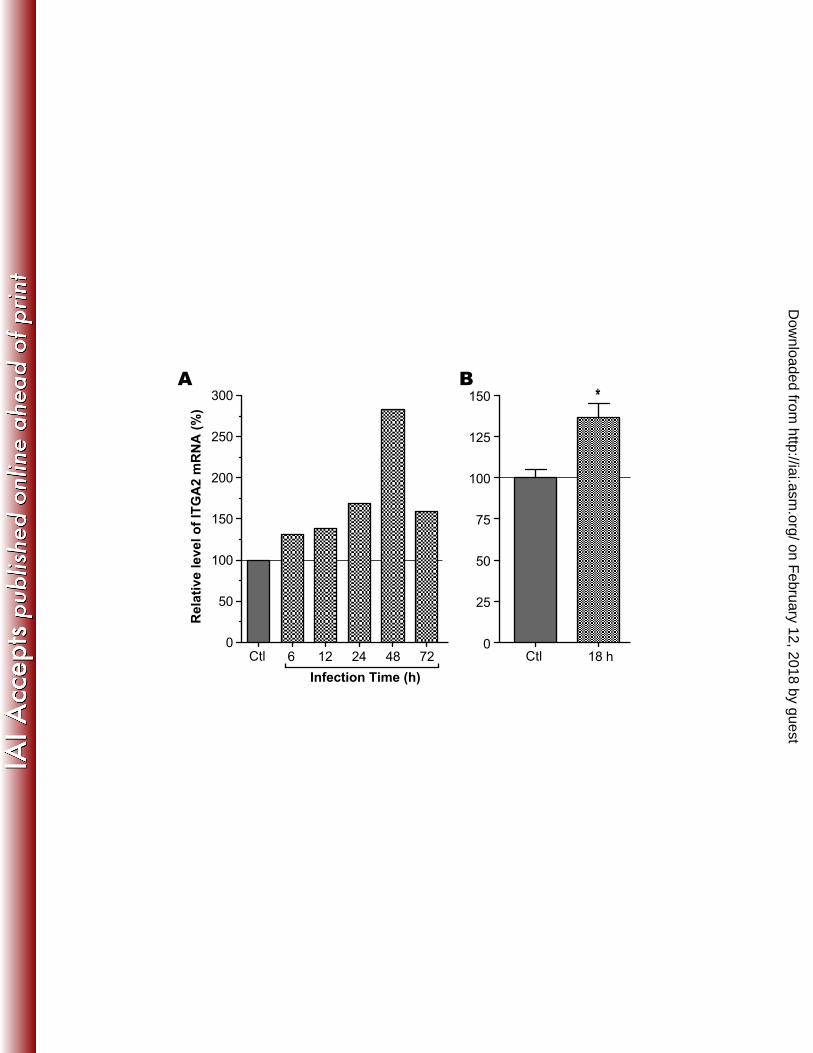
The expression of host cell ITGA2 gene was up-regulated upon C. parvum infection. 201 We observed that ITGA2 gene expressed in HCT-8 cells was consistently up-regulated upon C. 202 parvum infection at various time points from published microarray data (Figure 1A) (12). Similar 203 results were also obtained in Caco-2 cells infected with C. parvum for 18 h in our unpublished 204
on February 12, 2018 by guest
http://iai.asm.org/
Dow
nloaded from
10
analysis using Affymetrix U133A microarrays (i.e., 40% increase, p = 0.00296). These 205 observations were further validated by qRT-PCR in HCT-8 cells inoculated with C. parvum 206 oocysts for 18 h, in which the levels of ITGA2 transcripts were increased by 37% (Figure 1B). 207 208
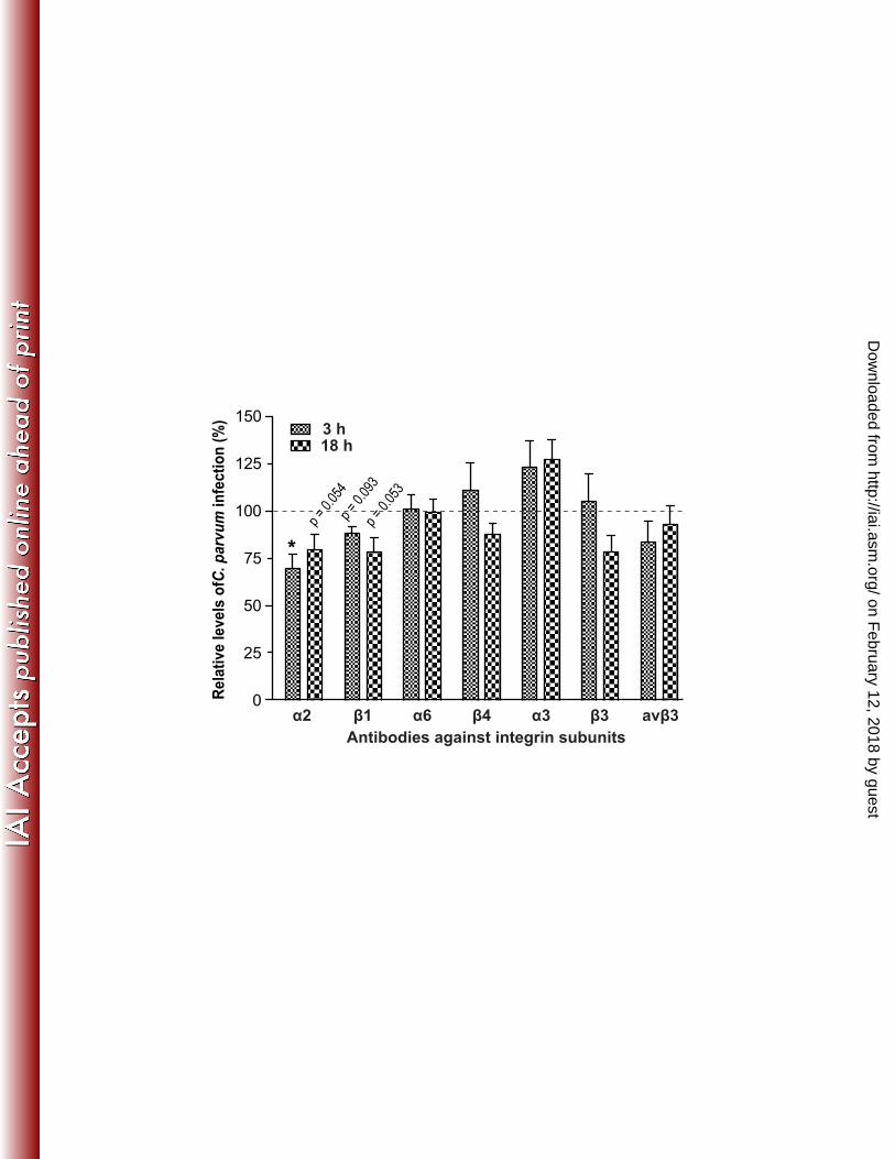
Antibody specific to ITGA2 and its ligand collagen-I could reduce C. parvum infection. 209 To determine whether ITGA2 was involved in C. parvum infection, we treated HCT-8 cells with 210 mAbs against ITGA2 and 6 other subunits (i.e., β1, α6, β4, α3, β3 and αvβ3) to test their effects 211 on the parasite invasion and growth in vitro. Among them, antibodies to α2 and β1 consistently 212 inhibited the parasite growth by ~20% - 30% in both 3 h and 18 h infection assays with p-values 213 below (3 h α2) or near 0.05% (other three groups), whereas all other antibodies except for the α3 214 antibody either had no or little effect on parasite infection (Figure 2). On the other hand, 215 antibody against α3 promoted both invasion and intracellular development by 24% and 28%, 216 respectively. 217
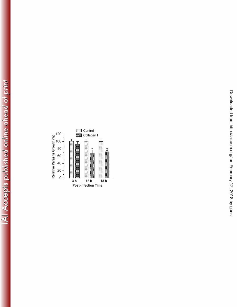
Because collagen-I is a known ECM ligand for the ITG α2β1 complex (3, 29), we also 218 examined the effect of this ligand on C. parvum infection in HCT-8 cells. When soluble 219 collagen-I was added to the culture, only a slight reduction of parasite invasion (6%) was 220 observed in the 3 h infection assay. However, a significant reduction of intracellular 221 development (32% and 28%, respectively) was detected in the 12 h and 18 h infection assays 222 (Figure 3). These data are suggestive that collagen-I might play a more important role for the 223 intracellular development of the parasite rather than for invasion, probably via acting as a critical 224 ECM component connecting ITGA2 and parasite at the host cell-parasite juncture. 225 226
on February 12, 2018 by guest
http://iai.asm.org/
Dow
nloaded from
11
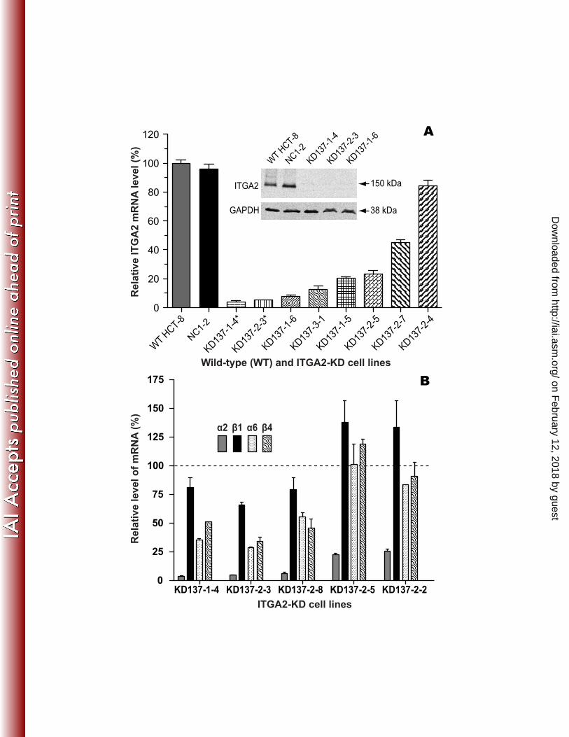
Stably transfected HCT-8 cells could be generated to silence ITGA2 gene expression 227 at varied levels. We developed stable transfection of a serial cloned ITGA2-KD cell lines 228 derived from HCT-8 cells to test the effect of reduced ITGA2 expression on C. parvum infection. 229 In comparison with WT cells, the levels of ITGA2 mRNA in various ITGA2-KD lines were 230 reduced from 16% to 96%, whereas no or little changes were observed in the negative control 231 cells (NC1-2) as determined by qRT-PCR (Figure 4A). The gene silencing was further validated 232 by western blot analysis, in which ITGA2 protein was clearly detectable in the WT and NC1-2 233 controls, but undetectable in 3 ITGA2-KD lines that displayed the highest reduction in ITGA2 234 expression (i.e., KD137-1-4, KD137-2-3 and KD137-1-6) (Figure 4A, inset). 235
We also examined the effect of ITGA2 knockdown on the expression of 3 other integrin 236 subunits (i.e., β1, α6 and β4) among select ITGA2-KD cell lines and observed a general pattern. 237 In comparison with NC1-2 controls, the expression of β1, α6 and β4 integrins was significantly 238 down-regulated in ITGA2-KD cells that had the lowest ITGA2 expressions (i.e., in KD137-1-4, 239 KD137-2-3 and KD137-2-8 cells with >95% reduction in ITGA2 expression), but either up-240 regulated or only slightly down-regulated in cells with relatively "higher levels" of ITGA2 241 expression (i.e., in KD137-2-5 and KD137-2-2 cells with ~75% reduction) (Figure 4B). 242
In comparison with the control cells, ITGA2-KD cells did not show much difference in cell 243 adhesion and growth during the first 6-12 h of growth after seeding. However, the morphological 244 difference between the negative control and ITGA2-KD cells was more obvious from 12 h to 48 245 h after seeding. The KD cells generally displayed more round or clustered shapes than vector 246 control cells (Figure S1), which may indicate a migration defect in knockdown cells. 247 Surprisingly however, at 24 - 48 h after seeding, ITGA2-KD cells showed ~25% higher growth 248 rate than negative control cells (Figure S2). Flow cytometric analysis of cells double-stained with 249 propidium iodide and Annexin V-FITC indicated no statistically significant differences in the 250
on February 12, 2018 by guest
http://iai.asm.org/
Dow
nloaded from
12
ratios of dead or apoptotic cells between WT, NT-21, and KD137-1-4, KD137-2-3 cell lines 251 (Figure S3), suggesting that apoptosis had no or little effect on the subsequent parasite infection 252 assays. 253 254
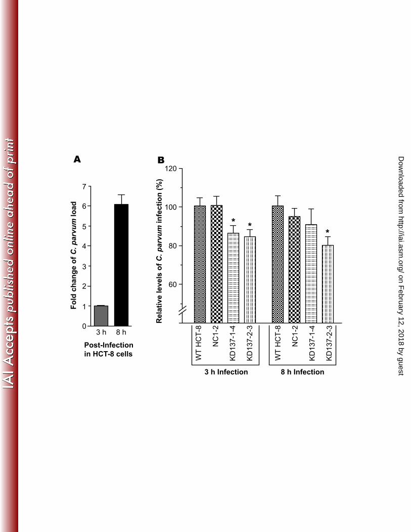
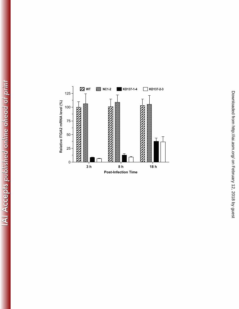
Cryptosporidium early infection was reduced in ITGA2-KD cells. Two ITGA2-KD cell 255 lines with the lowest level of ITGA2 expression (KD137-1-4 and KD137-2-3) were used to test 256 the effect of knockdown on parasite infection by an 18S rRNA-based qRT-PCR method. C. 257 parvum was allowed to infect WT and KD cells for 3 and 8 h to represent parasite invasion and 258 early stages of intracellular development. There was an approximate 6-fold increase in the level 259 of C. parvum 18S rRNA (Cp18S) from 3 h to 8 h infection in WT cells (Figure 5A), suggesting a 260 rapid parasite growth in the early stage of intracellular development. In comparison with WT 261 cells, both parasite invasion (3 h infection) and early intracellular development (8 h infection) 262 were moderately, but consistently reduced by ~15% - 20% in the 2 ITGA2-KD cell lines (Figure 263 5B). On the other hand, no changes or only a slight reduction were observed in negative control 264 NC1-2 cells (Figure 5B). Data on the parasite growth for 18 h in KD cells were inconclusive as 265 they varied greatly between experiments, although there was a general tendency on the reduction 266 of parasite growth in KD137-2-3 cells, but not in KD137-1-4 cells (data not shown). This is 267 probably due to the significant rebound of ITGA2 mRNA levels from ~5% back to ~40% in both 268 KD cell lines (in comparison with those in WT and NC1-2 controls) upon parasite infection for 269 18 h (Figure 6). 270 271
ITGA2 protein was recruited to the C. parvum infection sites at the parasite 272 intracellular development stage. In addition to the increased level of ITGA2 transcripts of host 273 cells in response to C. parvum infection, we also observed that ITGA2 was recruited to the 274
on February 12, 2018 by guest
http://iai.asm.org/
Dow
nloaded from
13
infection sites during parasite intracellular development (Figure 7). Immunofluorescence 275 microscopy using anti-ITGA2 mAb indicated that this protein was more concentrated at the 276 infection sites in both 3 h and 18 h assays. To our surprise, however, ITGA2 proteins were also 277 detectable at the infection sites in ITGA2-KD cells with fluorescence signal intensity comparable 278 to that in the control cells (Figure 7, marked as KD), indicating the presence of a low, but 279 adequate amount of ITGA2 protein in these knockdown cells. Nonetheless, the presence of 280 ITGA2 protein in KD cells was consistent with the rebound of ITGA2 gene expression in KD 281 cells upon infection for 18 h as described above (Figure 6). 282 283 DISCUSSION 284
It is known that a number of intracellular microbial pathogens could directly or indirectly 285 explore ITGs and associated pathways to breach the host cell surface barrier (4, 15, 16, 21). 286 Some pathogens might bind to the host cell ITGs with their special outer membrane proteins 287 (21), while others may interact with host ITGs via ECM proteins, which serve as receptors for 288 pathogen adherence and internalization (4). More recently, the ITG α2β1 complex was also 289 reported to mediate the entry of Bacillus anthracis spores into epithelial cells (34). Our 290 observations provide evidence to also support the involvement of ITGA2 in C. parvum infection 291 based on the following: 1) host cell ITGA2 gene expression was significantly up-regulated upon 292 infection in both WT and KD cells; 2) ITGA2 was recruited to the parasite infection sites in 293 cultured host cells; 3) parasite invasion and intracellular development were reduced in ITGA2-294 KD cells, and by its specific mAbs and its ligand type I collagen. 295
Although the observed effects in parasite infections by antibody and ligand treatments, as 296 well as by gene knockdown were at most moderate (Figures 2, 3 and 5), the reductions were 297 consistent with p-values smaller or near 0.05%. These moderate effects imply that ITGA2 is 298
on February 12, 2018 by guest
http://iai.asm.org/
Dow
nloaded from
14
involved in parasite infection, but its role might be non-critical. Or it might be critical, but the 299 effect could not be fully assessed by a gene knockdown approach as it is unable to completely 300 eliminate the expression of ITGA2. We observed the presence of ITGA2 at the infection sites in 301 ITGA2-KD cells (Figure 6), despite that its protein level was undetectable by western blot 302 analysis (Figure 4A). These observations suggest that although the ITGA2 mRNA level in these 303 KD cells was reduced by >95%, and ITGA2 protein level was below the sensitivity of western 304 blot detection, a very small amount of ITGA2 protein was still present and could be recruited to 305 the infection sites. Additionally, the apparent rebound of ITGA2 mRNA levels in KD cells upon 306 relatively long parasite infection as shown in figure 6 also attributes to the synthesis of even 307 more ITGA2 proteins. Therefore, a complete deletion/knockout of the ITGA2 gene in HCT-8 308 cells may be needed to ultimately test the necessity of ITGA2 in Cryptosporidium infection. 309 Additionally, it is possible that other ITG subunits might also interact with the parasite during its 310 infection and intracellular development, thus compensating the loss of ITGA2 in the ITGA2-KD 311 cells. On the other hand, the observed rebound of ITGA2 expression further supports the notion 312 that ITGA2 expression is up-regulated upon C. parvum infection. 313
ITGA2 typically forms a heterodimer with the β1 subunit (α2β1). Our current data show 314 that antibody against the β1 subunit could also inhibit parasite infection (Figure 2), which is well 315 correlated with the reduction of parasite infection in ITGA2-KD cells and by anti-ITGA2 316 antibody treatment. It is known that ITGs are not only capable of directly anchoring actin 317 filaments on the cytosolic side of the cytoplasmic membrane, but are also able to regulate F-actin 318 remodeling via FAK/Src/CDC42 associated signal transduction pathway (24, 27, 29). On the 319 other hand, it has been reported that host cell c-Src dependent tyrosine phosphorylation and 320 PI3K-mediated activation of CDC42 are involved in interactions with parasite infection via 321 regulating the recruiting of F-actin at the host-parasite interface (7, 8, 10). Collectively, albeit for 322
on February 12, 2018 by guest
http://iai.asm.org/
Dow
nloaded from
15
further investigations to make firm conclusions, we speculate that ITGs may act at the upstream 323 level to regulate the F-actin remodeling via FAK/Src/CDC42 pathway. 324
The HCT-8 cell line is commonly used to study cryptosporidial infection and test drug 325 efficacies in vitro (6). However, except for a transient gene expression knockdown with 326 microRNA (35), stable knockdown of gene expression was unreported. This is the first time a 327 stable gene knockdown has been obtained for HCT-8 cells, showing that shRNA-based gene 328 knockdown could be applied to this cell line to study the roles of other genes of interest on 329 infection by C. parvum or other pathogens. 330 331 Acknowledgements 332
The authors thank Dr. Beiyan Zhou and Dr. Guoqing Zhuang for their technical assistance 333 in the flow cytometric analysis, and Dr. Jason M. Fritzler for his critical reading of the 334 manuscript. This study was supported by a grant from the National Institutes of Health (NIH), 335 National Institute of Allergic and Infectious Diseases (NIAID) [R21 AI055278 to G.Z.] 336 337 338
on February 12, 2018 by guest
http://iai.asm.org/
Dow
nloaded from
16
REFERENCES 339 340 1. Arrowood, M. J., and C. R. Sterling. 1987. Isolation of Cryptosporidium oocysts and 341
sporozoites using discontinuous sucrose and isopycnic Percoll gradients. J Parasitol 73:314-342 319. 343
2. Banno, A., and M. H. Ginsberg. 2008. Integrin activation. Biochem Soc Trans 36:229-234. 344 3. Barczyk, M., S. Carracedo, and D. Gullberg. 2010. Integrins. Cell Tissue Res 339:269-345
280. 346 4. Bergmann, S., A. Lang, M. Rohde, V. Agarwal, C. Rennemeier, C. Grashoff, K. T. 347
Preissner, and S. Hammerschmidt. 2009. Integrin-linked kinase is required for 348 vitronectin-mediated internalization of Streptococcus pneumoniae by host cells. J Cell Sci 349 122:256-267. 350
5. Borad, A., and H. Ward. 2010. Human immune responses in cryptosporidiosis. Future 351 Microbiol 5:507-519. 352
6. Cai, X., K. M. Woods, S. J. Upton, and G. Zhu. 2005. Application of quantitative real-353 time reverse transcription-PCR in assessing drug efficacy against the intracellular pathogen 354 Cryptosporidium parvum in vitro. Antimicrob Agents Chemother 49:4437-4442. 355
7. Chen, X. M., B. Q. Huang, P. L. Splinter, H. Cao, G. Zhu, M. A. McNiven, and N. F. 356 LaRusso. 2003. Cryptosporidium parvum invasion of biliary epithelia requires host cell 357 tyrosine phosphorylation of cortactin via c-Src. Gastroenterology 125:216-228. 358
8. Chen, X. M., B. Q. Huang, P. L. Splinter, J. D. Orth, D. D. Billadeau, M. A. McNiven, 359 and N. F. LaRusso. 2004. Cdc42 and the actin-related protein/neural Wiskott-Aldrich 360 syndrome protein network mediate cellular invasion by Cryptosporidium parvum. Infect 361 Immun 72:3011-3021. 362
on February 12, 2018 by guest
http://iai.asm.org/
Dow
nloaded from
17
9. Chen, X. M., J. S. Keithly, C. V. Paya, and N. F. LaRusso. 2002. Cryptosporidiosis. N 363 Engl J Med 346:1723-1731. 364
10. Chen, X. M., P. L. Splinter, P. S. Tietz, B. Q. Huang, D. D. Billadeau, and N. F. 365 LaRusso. 2004. Phosphatidylinositol 3-kinase and frabin mediate Cryptosporidium parvum 366 cellular invasion via activation of Cdc42. J Biol Chem 279:31671-31678. 367
11. Damsky, C. H., and D. Ilic. 2002. Integrin signaling: it's where the action is. Curr Opin Cell 368 Biol 14:594-602. 369
12. Deng, M., C. A. Lancto, and M. S. Abrahamsen. 2004. Cryptosporidium parvum 370 regulation of human epithelial cell gene expression. Int J Parasitol 34:73-82. 371
13. Elliott, D. A., and D. P. Clark. 2000. Cryptosporidium parvum induces host cell actin 372 accumulation at the host-parasite interface. Infect Immun 68:2315-2322. 373
14. Elliott, D. A., D. J. Coleman, M. A. Lane, R. C. May, L. M. Machesky, and D. P. Clark. 374 2001. Cryptosporidium parvum infection requires host cell actin polymerization. Infect 375 Immun 69:5940-5942. 376
15. Eto, D. S., T. A. Jones, J. L. Sundsbak, and M. A. Mulvey. 2007. Integrin-mediated host 377 cell invasion by type 1-piliated uropathogenic Escherichia coli. PLoS Pathog 3:e100. 378
16. Garrigues, H. J., Y. E. Rubinchikova, C. M. Dipersio, and T. M. Rose. 2008. Integrin 379 alphaVbeta3 Binds to the RGD motif of glycoprotein B of Kaposi's sarcoma-associated 380 herpesvirus and functions as an RGD-dependent entry receptor. J Virol 82:1570-1580. 381
17. Gilcrease, M. Z. 2007. Integrin signaling in epithelial cells. Cancer Lett 247:1-25. 382 18. Harburger, D. S., and D. A. Calderwood. 2009. Integrin signalling at a glance. J Cell Sci 383
122:159-163. 384 19. Hynes, R. O. 2004. The emergence of integrins: a personal and historical perspective. 385
Matrix Biol 23:333-340. 386
on February 12, 2018 by guest
http://iai.asm.org/
Dow
nloaded from
18
20. Hynes, R. O. 2002. Integrins: bidirectional, allosteric signaling machines. Cell 110:673-687. 387 21. Isberg, R. R., and J. M. Leong. 1990. Multiple beta 1 chain integrins are receptors for 388
invasin, a protein that promotes bacterial penetration into mammalian cells. Cell 60:861-389 871. 390
22. Kumar, C. C. 1998. Signaling by integrin receptors. Oncogene 17:1365-1373. 391 23. Martin, K. H., J. K. Slack, S. A. Boerner, C. C. Martin, and J. T. Parsons. 2002. 392
Integrin connections map: to infinity and beyond. Science 296:1652-1653. 393 24. Mitra, S. K., and D. D. Schlaepfer. 2006. Integrin-regulated FAK-Src signaling in normal 394
and cancer cells. Curr Opin Cell Biol 18:516-523. 395 25. Nesterenko, M. V., and S. J. Upton. 1996. A rapid microcentrifuge procedure for 396
purification of Cryptosporidium sporozoites. J Microbiol Meth 25:87-89. 397 26. Petersen, C., D. A. Barnes, and L. Gousset. 1997. Cryptosporidium parvum GP900, a 398
unique invasion protein. J Eukaryot Microbiol 44:89S-90S. 399 27. Sanz-Moreno, V., and C. J. Marshall. 2010. The plasticity of cytoskeletal dynamics 400
underlying neoplastic cell migration. Curr Opin Cell Biol 22:690-696. 401 28. Takada, Y., X. Ye, and S. Simon. 2007. The integrins. Genome Biol 8:215. 402 29. Takagi, J. 2007. Structural basis for ligand recognition by integrins. Curr Opin Cell Biol 403
19:557-564. 404 30. Templeton, T. J., S. Enomoto, W. J. Chen, C. G. Huang, C. A. Lancto, M. S. 405
Abrahamsen, and G. Zhu. 2010. A genome-sequence survey for Ascogregarina 406 taiwanensis supports evolutionary affiliation but metabolic diversity between a Gregarine 407 and Cryptosporidium. Mol Biol Evol 27:235-248. 408
31. Thompson, R. C., M. E. Olson, G. Zhu, S. Enomoto, M. S. Abrahamsen, and N. S. 409 Hijjawi. 2005. Cryptosporidium and cryptosporidiosis. Adv Parasitol 59:77-158. 410
on February 12, 2018 by guest
http://iai.asm.org/
Dow
nloaded from
19
32. Tzipori, S., and H. Ward. 2002. Cryptosporidiosis: biology, pathogenesis and disease. 411 Microbes Infect 4:1047-1058. 412
33. Untergasser, A., H. Nijveen, X. Rao, T. Bisseling, R. Geurts, and J. A. Leunissen. 2007. 413 Primer3Plus, an enhanced web interface to Primer3. Nucleic Acids Res 35:W71-74. 414
34. Xue, Q., C. Gu, J. Rivera, M. Hook, X. Chen, A. Pozzi, and Y. Xu. 2011. Entry of 415 Bacillus anthracis spores into epithelial cells is mediated by the spore surface protein BclA, 416 integrin alpha2beta1 and complement component C1q. Cell Microbiol 13:620-634. 417
35. Yan, R., X. Zu, J. Ma, Z. Liu, M. Adeyanju, and D. Cao. 2007. Aldo-keto reductase 418 family 1 B10 gene silencing results in growth inhibition of colorectal cancer cells: 419 Implication for cancer intervention. Int J Cancer 121:2301-2306. 420
36. Zhu, G., J. S. Keithly, and H. Philippe. 2000. What is the phylogenetic position of 421 Cryptosporidium? Int J Syst Evol Microbiol 50 Pt 4:1673-1681. 422
423 424 on February 12, 2018 by guest
http://iai.asm.org/
Dow
nloaded from
20
Figure 1. Up-regulated human ITGA2 gene expression in HCT-8 cells upon infection with C. 425 parvum. A) Relative levels of ITGA2 transcripts based on data extracted from an earlier 426 microarray analysis at NCBI Gene Express Omnibus Datasets for GSE2077 427 (http://www.ncbi.nlm.nih.gov/geo/query/acc.cgi?acc=GSE2077) [Deng et al., 2004]; B) Relative 428 levels of ITGA2 transcripts in uninfected control cells and those infected with C. parvum for 18 429 h as determined by qRT-PCR in this study. The transcript levels were first normalized with those 430 of host cell 18S rRNA and then displayed in relative to those in uninfected cells. An asterisk (*) 431 indicates p < 0.05 by Student’s t test versus uninfected control. 432 433 Figure 2. Effects of treatment by antibodies against various integrin subunits on the infection of 434 C. parvum in HCT-8 cells. Significant reduction in infection was observed in the group treated 435 with anti-integrin α2 antibodies in 3 h infection assays. Bars represent standard-error-of-the-436 means derived from triplicate treatments. An asterisk (*) indicates p < 0.05 by Student’s t test 437 versus untreated control cells. 438 439 Figure 3. Effects of collagen-I on C. parvum infection. Treatment of HCT-8 cells by collagen-I 440 significantly reduced the infection by C. parvum as determined by qRT-PCR detection of 441 parasite 18S rRNA levels in the 12 h and 18 h infection assays. Levels of infection were 442 expressed relative to the untreated control. Bars represent standard-error-of-the-means derived 443 from triplicate treatments. An asterisk (*) indicates p < 0.05 by Student’s t test versus untreated 444 control cells. 445 446 447
on February 12, 2018 by guest
http://iai.asm.org/
Dow
nloaded from
21
Figure 4. Gene expression profiles in ITGA2-KD cells. A) Relative expression levels of host 448 ITGA2 mRNA and protein (inset) in various control and ITGA2-KD cells as determined by 449 qRT-PCR and western blot analysis. GAPDH was used as a protein load control. B) Levels of 450 gene expression of 4 different integrin subunits in select ITGA2-KD lines as determined by qRT-451 PCR relative to those in WT HCT-8 cells. The ITGA2-KD lines marked with asterisks (*) were 452 used in subsequent infection assays. Bars represent standard-error-of-the-means derived from 453 biological triplicates. WT: wild-type HCT-8 cell line, NC1-2: negative vector control with a 454 scrambled sequence. 455 456 Figure 5. The levels of C. parvum infection in control and ITGA2-KD cell lines. A) The rapid 457 growth of C. parvum in the early stage of infection in HCT-8 cells as determined by qRT-PCR. 458 A 6-fold increase of parasite 18S rRNA levels was observed during the period of 3 h to 8 h post-459 infection. B) The infection of C. parvum was significantly decreased in two ITGA2-KD cell lines 460 (KD137-1-4 and KD137-2-3) in comparison to those in WT and negative control (NC1-2) cells 461 in both 3 h and 8 h infection assays. Bars represent standard-error-of-the-means derived from 462 triplicate treatments. An asterisk (*) indicates p<0.05 by Student’s t test versus C. parvum 463 infected WT control. 464 465 Figure 6. Significant rebound of ITGA2 gene expression in knockdown (KD) cell lines upon 466 infection by C. parvum as determined by qRT-PCR. 467 468 469
on February 12, 2018 by guest
http://iai.asm.org/
Dow
nloaded from
22
Figure 7. ITGA2 protein was recruited to the infection sites in vector control NC1-2 (NC) and 470 KD137-1-4 (KD) cells infected with C. parvum for 3 h and 18 h, respectively, as determined by 471 immunofluorescence microscopy. DIC: differential inference contrast; DAPI: 4',6-diamidino-2-472 phenylindole for counter-staining nuclei; Integrin α2: Alexa 594-labeled integrin α2 protein; 473 Merge: DAPI-stained nuclei + Alexa 594-labeled α2 protein. Bar = 5 μm. 474
on February 12, 2018 by guest
http://iai.asm.org/
Dow
nloaded from

![REVIEW Open Access Mechanism of regulation of stem cell ......MSC myogenic differentiation induced by substrates with medium stiffness [12], while α2 integrin modulates osteogenesis](https://img.pdfslide.net/doc/110x75/609d7071995e0c33fd1fef73/review-open-access-mechanism-of-regulation-of-stem-cell-msc-myogenic-differentiation.jpg)