Embed Size (px)
Citation preview
IQGAP1-Dependent Signaling Pathway RegulatesEndothelial Cell Proliferation and AngiogenesisRosana D. Meyer1, David B. Sacks2, Nader Rahimi1*
1 Departments of Pathology and Ophthalmology, School of Medicine, Boston University, Boston, Massachusetts, United States of America, 2 Department of Pathology,
Brigham and Women’s Hospital and Harvard Medical School, Boston, Massachusetts, United States of America
Abstract
Background: Vascular endothelial growth factor receptor-2 (VEGFR-2) signaling is an obligate requirement for normaldevelopment and pathological angiogenesis such as cancer and age-related macular degeneration. Although autopho-sphorylation of tyrosine 1173 (Y1173) of VEGFR-2 is considered a focal point for its angiogenic signal relay, however, themechanism of phosphorylation of Y1173, signaling proteins that are recruited to this residue and their role in angiogenesisis not fully understood.
Methodology/Principal Findings: In this study we demonstrate that c-Src kinase directly through its Src homology 2 (SH2)domain and indirectly via c-Cbl binds to phospho-Y1057 of VEGFR-2. Activation of c-Src kinase by a positive feedbackmechanism phosphorylates VEGFR-2 at multi-docking site, Y1173. c-Src also catalyzes tyrosine phosphorylation of IQGAP1and acts as an adaptor to bridge IQGAP1 to VEGFR-2. In turn, IQGAP1 activates b-Raf and mediates proliferation ofendothelial cells. Silencing expression of IQGAP1 and b-Raf revealed that their activity is essential for VEGF to stimulateangiogenesis in an in vivo angiogenesis model of chicken chorioallantoic membrane (CAM).
Conclusions/Significance: Angiogenesis contributes to the pathology of numerous human diseases ranging from cancer toage-related macular degeneration. Determining molecular mechanism of tyrosine phosphorylation of VEGFR-2 andidentification of molecules that are relaying its angiogenic signaling may identify novel targets for therapeutic interventionagainst angiogenesis-associated diseases. Our study shows that recruitment and activation of c-Src by VEGFR-2 plays apivotal role in relaying angiogenic signaling of VEGFR-2; it phosphorylates VEGFR-2 at Y1173, facilitates association andactivation of IQGAP1 and other signaling proteins to VEGFR-2. IQGAP1-dependent signaling, in part, is critically required forendothelial cell proliferation, a key step in angiogenesis. Thus, Y1057 of VEGFR-2 serves to regulate VEGFR-2 function in acombinatorial manner by supporting both diversity of recruitment of angiogenic signaling proteins to VEGFR-2, and itsability to promote angiogenesis.
Citation: Meyer RD, Sacks DB, Rahimi N (2008) IQGAP1-Dependent Signaling Pathway Regulates Endothelial Cell Proliferation and Angiogenesis. PLoS ONE 3(12):e3848. doi:10.1371/journal.pone.0003848
Editor: Nick Gay, University of Cambridge, United Kingdom
Received May 20, 2008; Accepted November 10, 2008; Published December 3, 2008
Copyright: � 2008 Meyer et al. This is an open-access article distributed under the terms of the Creative Commons Attribution License, which permitsunrestricted use, distribution, and reproduction in any medium, provided the original author and source are credited.
Funding: This study was supported in part by grants from the National Institutes of Health (NR and DBS) and Massachusetts Lions Foundation grant to theDepartment of Ophthalmology.
Competing Interests: The authors have declared that no competing interests exist.
* E-mail: [email protected]
Introduction
Activation of vascular endothelial growth factor receptor-2
(VEGFR-2 also called FLK-1/KDR) plays a pivotal role in
mediating growth of blood vessels, angiogenesis [1]. VEGF
stimulation of VEGFR-2 provokes pleiotropic responses in
endothelial cells including endothelial cell growth, survival,
differentiation, migration, and tube formation [2]. Autopho-
sphorylation of tyrosine 1173 (Y1173) of VEGFR-2 is a focal
point for activation of angiogenic signal relay of VEGFR-2, as it
has emerged as a multi-functional docking site involved in the
recruitment of multiple signaling proteins including the p85
regulatory subunit of phosphatidylinositol 3-kinase (p85/PI3-K)
[3,4], phospholipase Cc1 (PLCc1) [5,6], Shb [7], and the Shc-
related adaptor protein, Sck [8–10] and the transmission of a
range of biological signals to coordinate endothelial cell function
and angiogenesis. The critical and direct role of Y1173 in enabling
VEGFR-2 to promote angiogenesis was recently revealed by a
gene targeting study where mice homozygous for the mutant
VEGFR-2Y1173F knock-in allele died between E8.5 and E9.5
without any organized blood vessels [11] similar to the VEGFR-2
null mice [12]. More recent studies indicate that in addition to
Y1173, the kinase domain tyrosine, Y1057 also takes part in
VEGFR-2 function and is involved in VEGFR-2-mediated cell
proliferation [13]. Y1057 along with Y1173 also is engaged in the
recruitment and activation of the ubiquitin E3 ligase, c-Cbl.
Phospho-Y1057 contributes to the recruitment of c-Cbl to
VEGFR-2 by directly associating with c-Cbl, while phospho-
Y1173 indirectly via PLCc1 participates in its recruitment to
VEGFR-2 [14], resulting in the ubiquitination of PLCc1 and
inhibition of angiogenesis in vitro [14].
The recent study indicates that IQGAP1 associates with
VEGFR-2 and its activity is required for proliferation of
endothelial cells in vitro [26]. It remains, however, unclear how
IQGAP1 interacts with VEGFR-2 since it lacks phospho-tyrosine
binding domains such as SH2 and PTB and how its activity is
PLoS ONE | www.plosone.org 1 December 2008 | Volume 3 | Issue 12 | e3848
regulated by VEGFR-2. IQGAP1 contains multiple protein-
interaction domains including calponin homology domain (CHD),
poly-proline-binding domain (WW), calmodulin-binding domain
(IQ) and rasGTPase-activating protein (GAP)-related domain
(GRD) and tyrosine and serine/threonine phosphorylation sites
[33] and is involved in multiple cellular functions including
calcium/calmodulin signaling, MAPK signaling and regulation of
cytoskeletal structure, cell adhesion and cell motility [27,31,33].
In this study, we uncovered a surprising correlation between
phosphorylation of Y1057 and Y1173 and that phosphorylation of
Y1057 plays a multitasking role in mediating VEGFR-2-directed
angiogenic signaling events in endothelial cells. Phospho-Y1057
recruits c-Src kinase to VEGFR-2, and in part catalyzes
phosphorylation of Y1173 via Src kinases. c-Src also bridges
IQGAP1 to VEGFR-2 and directly phosphorylates IQGAP1 and
promotes endothelial cell proliferation, a key step in angiogenesis.
Results
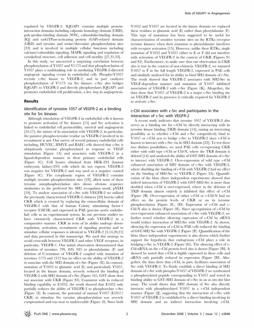
Identification of tyrosine 1057 of VEGFR-2 as a bindingsite for Src kinases
Although stimulation of VEGFR-2 in endothelial cells is known
to promote activation of Src kinases [15] and Src activation is
linked to endothelial cell permeability, survival and angiogenesis
[16,17], the nature of its association with VEGFR-2, in particular,
the putative phospho-tyrosine residue on VEGFR-2 involved in its
recruitment is not. Our survey of several primary endothelial cells
including, HUVEC, HMVE and BAEC cells showed that c-Src is
ubiquitously tyrosine phosphorylated in response to VEGF
stimulation (Figure 1A) and associates with VEGFR-2 in a
ligand-dependent manner in these primary endothelial cells
(Figure 1C). Cell lysates obtained from HEK-293 (human
embryonic kidney-293) cells which do not express VEGFR-2
was negative for VEGFR-2 and was used as a negative control
(Figure 1C). The cytoplasmic region of VEGFR-2 contains
multiple tyrosine phosphorylation sites [18], albeit none of these
tyrosine autophosphorylation sites shows obvious sequence
similarities to the preferred Src SH2 recognition motif, pYEEI
[19]. To analyze association of c-Src with VEGFR-2 we utilized
the previously characterized VEGFR-2 chimera [20] herein called
CKR which is created by replacing the extracellular domain of
VEGFR-2 with that of human Colony stimulating factor-1
receptor (CSF1R) and expressed in PAE (porcine aortic endothe-
lial) cells as an experimental system. In our previous studies we
have extensively characterized CKR with VEGFR-2 in a
comparative manner. CKR in term of its ability undergo down-
regulation, activation, recruitment of signaling proteins and to
stimulate cellular responses is identical to VEGFR-2 [3,14,20,21]
and data presented in this manuscript. We used this strategy to
avoid cross-talk between VEGFR-2 and other VEGF receptors, in
particular, VEGFR-1. Our initial observation demonstrated that
mutation of tyrosines 799, 820, 925 to phenylalanine (F) and
deletion of C-terminus of VEGFR-2 coupled with mutation of
tyrosines 1173 and 1212 has no effect on the ability of VEGFR-2
to associate with the SH2 domain of c-Src (Figure 1E). In contrast,
mutation of Y1052 to glutamic acid (E) and particularly Y1057,
located in the kinase domain, severely reduced the binding of
VEGFR-2 with SH2 domain of c-Src (Figure 1G). GST alone does
not associate with CKR (Figure S3). Consistent with its reduced
binding capability to E1052, the result showed that E1052 only
partially reduces the ability of VEGFR-2 to phosphorylate c-Src
(Figure 1I). In contrast, the potential of mutant Y1057 (E1057/
CKR) to stimulate Src tyrosine phosphorylation was severely
compromised and was near to undetectable (Figure 1I). Since both
Y1052 and Y1057 are located in the kinase domain we replaced
these residues to glutamic acid (E) rather than phenylalanine (F).
This type of mutations has been suggested to be useful for
characterization of sensitive tyrosine sites on the RTKs (receptor
tyrosine kinases) when their mutation to phenylalanine interferes
with receptor activation [15]. However, unlike these RTKs, single
mutation of Y1052 and Y1057 either to E or F did not interfere
with activation of VEGFR-2 in the context of CKR (Figures S1
and S2). Furthermore, to make sure that our observation in CKR
also is true in the context of non-chimeric VEGFR-2, we mutated
Y1057 to F in the full length VEGFR-2, expressed in PAE cells
and similarly analyzed for its ability to bind SH2 domain of c-Src.
The result showed that VEGFR-2 associates with SH2-Src in
VEGF-dependent manner and mutation of Y1057 inhibited
association of VEGFR-2 with c-Src (Figure 1K). Altogether, the
data show that Y1057 of VEGFR-2 is a major c-Src binding site
on VEGFR-2 and its presence is critically required for VEGFR-2
to activate c-Src.
c-Cbl associates with c-Src and participates in theinteraction of c-Src with VEGFR-2
A recent study indicates that tyrosine 1057 of VEGFR-2 also
serves as a binding site for c-Cbl by directly interacting with its
tyrosine kinase binding (TKB) domain [14], raising an interesting
possibility as to whether c-Cbl and c-Src competitively bind to
Y1057 or c-Cbl acts to bridge c-Src to VEGFR-2 since c-Cbl is
known to interact with c-Src via its SH3 domain [22]. To test these
two distinct possibilities, we used PAE cells co-expressing CKR
either with wild type c-Cbl or Cbl-N, where the TKB domain is
deleted [14] and analyzed the ability of GST-SH2 domain of c-Src
to interact with VEGFR-2. Over-expression of wild type c-Cbl
enhanced association of SH2 domain of c-Src with VEGFR-2,
where disabling the binding of c-Cbl with VEGFR-2 had no effect
on the binding of SH2-Src to VEGFR-2 (Figure 2A). Quantifi-
cation of the blots (three independent experiments) showed that
indeed interaction of VEGFR-2 with GST-SH2-Src is more than
doubled when c-Cbl is over-expressed, where as the deletion of
TKB domain almost entirely is inhibited this effect of c-Cbl
(Figure 2B). Over-expression of either c-Cbl or c-Cbl-N had no
effect on the protein levels of CKR or on its tyrosine
phosphorylation (Figure 2C, 2D). Expression of c-Cbl and c-
Cbl-N also is shown (Figure 2E). Since up-regulation of c-Cbl by
over-expression enhanced association of c-Src with VEGFR-2, we
further tested whether silencing expression of c-Cbl by siRNA
would reduce interaction of SH2-Src with VEGFR-2. As shown,
silencing the expression of c-Cbl in PAE cells reduced the binding
of GST-SH2 Src with VEGFR-2 (Figure 2F). Quantification of the
blots (three independent experiments) is also shown which further
support the hypothesis that endogenous c-Cbl plays a role in
bridging c-Src to VEGFR-2 (Figure 2G). The silencing effect of c-
Cbl-siRNA on the c-Cbl protein level also is shown (Figure 2H). It
showed be noted that c-Cbl is highly expressed in these cells and
siRNA only partially reduced its expression (Figure 2H). Alto-
gether, the data show that c-Cbl, in part, facilitates association of
Src with VEGFR-2. To firmly establish a direct binding of SH2
domain of c-Src with phospho-Y1057 of VEGFR-2 we synthesized
a phosphorylated peptide corresponding to Y1057 and tested its
binding ability to GST-SH2 domain of c-Src in an in vitro dot blot
assay. The result shows that SH2 domain of Src also directly
interacts with phosphorylated Y1057 in a c-Cbl independent
manner (Figure 2J), suggesting that the interaction of c-Src with
Y1057 of VEGFR-2 is established by a direct binding involving its
SH2 domain and an indirect interaction involving c-Cbl.
Role of IQGAP1 in Angiogenesis
PLoS ONE | www.plosone.org 2 December 2008 | Volume 3 | Issue 12 | e3848
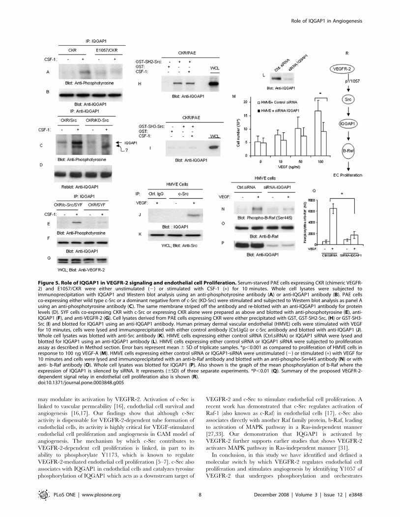
Figure 1. Tyrosine 1057 mediates recruitment of c-Src to and its activation by VEGFR-2. HUVEC, HMVE and BAE cells were stimulated withVEGF-A (100 gg/ml) for indicated times. Whole cell lysates (WCL) were immunoblotted with an anti-phospho-Src (pY416) antibody (A) or an anti-Srcantibody (B). HUVEC, HMVE and HEK-293 cells were either unstimulated (2) or stimulated with VEGF (+) for 10 minutes, lysed, immunoprecipitatedwith c-Src antibody and immunoblotted with an anti-VEGFR-2 antibody (C). Whole cell lysates (WCL) from the same group was immunoblotted withan anti-VEGFR-2 as a control (D). PAE cells expressing wild type chimeric VEGFR-2 (CKR) and tyrosine mutant CKRs, F799/CKR, F820/CKR, F925/CKRand carboxyl terminus deleted CKR coupled with mutation of Y1173 and Y1212 denoted as DCKR/F1173/F1212 was stimulated with CSF-1 for10 minutes. Cells were lysed, and whole cell lysates were incubated with purified GST-SH2-Src protein. The GST-SH2-Src bound proteins weresubjected to Western blot analysis using an anti-VEGFR-2 antibody (E). Whole cell lysates from the same groups was blotted with an anti-VEGFR-2antibody (F). PAE cells expressing CKR, E1052/CKR, and E1057/CKR were either unstimulated or stimulated with CSF-1 for 10 minutes and cells werelysed and cell lysates was subjected to GST-SH2-Src pull-down assay (G). An immunoblot of whole cell lysates also were probed with an anti-VEGFR-2antibody (F,H), an anti-phospho-Src (pY416) antibody (I) or an anti-Src antibody (J). PAE cells expressing VEGFR-2 or F1057/VEGFR-2 were preparedand subjected to pull-down analysis as panel E (K) or whole cell lysates was blotted with an anti-VEGFR-2 antibody (L). The results shown in A–L arerepresentative of three separate experiments.doi:10.1371/journal.pone.0003848.g001
Role of IQGAP1 in Angiogenesis
PLoS ONE | www.plosone.org 3 December 2008 | Volume 3 | Issue 12 | e3848
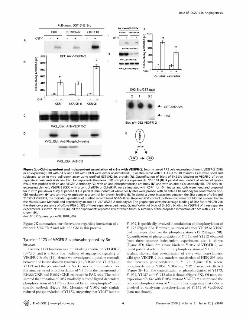
Figure 2K summarizes our observation regarding interaction of c-
Src with VEGFR-2 and role of c-Cbl in this process.
Tyrosine 1173 of VEGFR-2 is phosphorylated by Srckinases
Tyrosine 1173 functions as a multitasking residue on VEGFR-2
[3–7,10] and is a bona fide mediator of angiogenic signaling of
VEGFR-2 in vivo [11]. Hence we investigated a possible crosstalk
between the kinase domain tyrosines (i.e., Y1052 and Y1057) and
Y1173 and the potential role of Src kinases in this crosstalk. For
this aim, we tested phosphorylation of Y1173 in the background of
E1052/CKR and E1057/CKR expressed in PAE cells. The result
showed that mutation of 1057 markedly reduced ligand-dependent
phosphorylation of Y1173 as detected by an anti-phospho-Y1173
specific antibody (Figure 3A). Mutation of Y1052 only slightly
reduced phosphorylation of Y1173, suggesting that Y1057 but not
Y1052, is specifically involved in modulation of phosphorylation of
Y1173 (Figure 3A). However, mutation of either Y1052 or Y1057
had no major effect on the phosphorylation Y1212 (Figure 3B).
Quantification of phosphorylation of Y1173 and Y1212 obtained
from three separate independent experiments also is shown
(Figure 3D). Since Src kinase binds to Y1057 of VEGFR-2, we
tested potential role of Src in the phosphorylation of Y1173. Our
analysis showed that co-expression of v-Src with non-chimeric
wild-type VEGFR-2 in a transient transfection of HEK-293 cells
also increases phosphorylation of Y1173 (Figure 3D), where
phosphorylation of Y1052, Y1057 and Y1212 were not effected
(Figure 3F–H). The quantification of phosphorylation of Y1173,
Y1052, Y1057 and Y1212 also is shown (Figure 3K). Of note, co-
expression of v-Src with E1057 mutant VEGFR-2 also rescued the
reduced phosphorylation of Y1173 further suggesting that c-Src is
involved in catalyzing phosphorylation of Y1173 of VEGFR-2
(data not shown).
Figure 2. c-Cbl-dependent and independent association of c-Src with VEGFR-2. Serum-starved PAE cells expressing chimeric VEGFR-2 (CKR)or co-expressing CKR with c-Cbl and CKR with Cbl-N were either unstimulated (2) or stimulated with CSF-1 (+) for 10 minutes. Cells were lysed andsubjected to an in vitro pull-down assay using purified GST-SH2-Src protein (A). Quantification of blots of SH2-Src binding to VEGFR-2 of threeseparate experiments is shown. Each bar represents the mean 6SD of triplicate experiments. *P,0.01 (B). A parallel immunoblot of whole cell lysates(WCL) was probed with an anti-VEGFR-2 antibody (C), with an anti-phosphotyrosine antibody (D) and with an anti-c-Cbl antibody (E). PAE cells co-expressing chimeric VEGFR-2 (CKR) with a control siRNA or Cbl-siRNA were stimulated with CSF-1 for 10 minutes and cells were lysed and preparedfor in vitro pull-down assay as panel A (F). A parallel immunoblot of whole cell lysates were probed with an anti-c-Cbl antibody for confirmation of c-Cbl knockdown (H) and anti-Hsp70 antibody as a control for protein loading (I). To detect a direct interaction between the SH2 domain of c-Src andY1057 of VEGFR-2, the indicated quantities of purified recombinant GST-SH2-Src (top) and GST control (bottom row) were dot blotted as described inthe Materials and Methods and detected by an anti-pY1057 VEGFR-2 antibody (J). The graph represents the average binding of SH2-Src to VEGFR-2 inthe absence or presence of c-Cbl-siRNA (6SD) of three separate experiments. Quantification of blots of SH2-Src binding to VEGFR-2 of three separateexperiments is shown. *P,0.01 (G). All the experiments repeated at least three times. A summary of the proposed interaction of c-Src with VEGFR-2 isshown (K).doi:10.1371/journal.pone.0003848.g002
Role of IQGAP1 in Angiogenesis
PLoS ONE | www.plosone.org 4 December 2008 | Volume 3 | Issue 12 | e3848
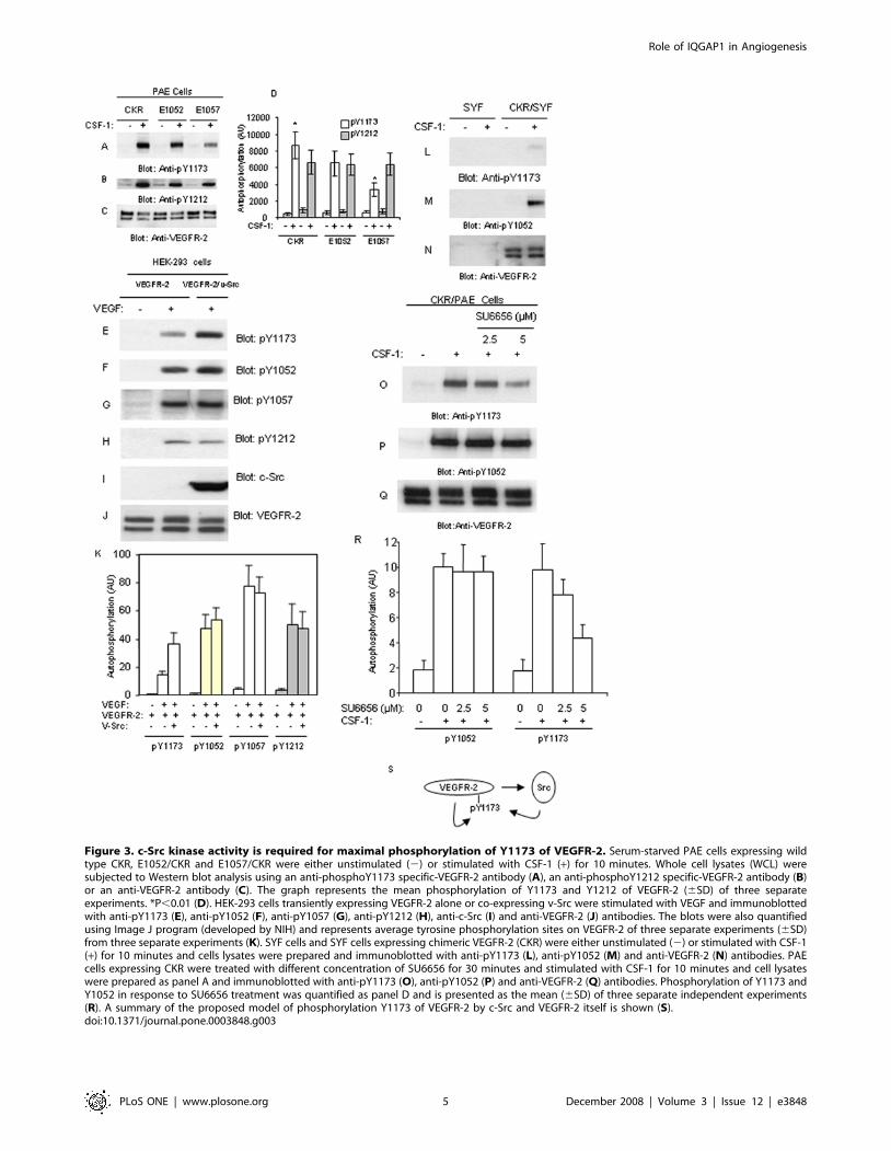
Figure 3. c-Src kinase activity is required for maximal phosphorylation of Y1173 of VEGFR-2. Serum-starved PAE cells expressing wildtype CKR, E1052/CKR and E1057/CKR were either unstimulated (2) or stimulated with CSF-1 (+) for 10 minutes. Whole cell lysates (WCL) weresubjected to Western blot analysis using an anti-phosphoY1173 specific-VEGFR-2 antibody (A), an anti-phosphoY1212 specific-VEGFR-2 antibody (B)or an anti-VEGFR-2 antibody (C). The graph represents the mean phosphorylation of Y1173 and Y1212 of VEGFR-2 (6SD) of three separateexperiments. *P,0.01 (D). HEK-293 cells transiently expressing VEGFR-2 alone or co-expressing v-Src were stimulated with VEGF and immunoblottedwith anti-pY1173 (E), anti-pY1052 (F), anti-pY1057 (G), anti-pY1212 (H), anti-c-Src (I) and anti-VEGFR-2 (J) antibodies. The blots were also quantifiedusing Image J program (developed by NIH) and represents average tyrosine phosphorylation sites on VEGFR-2 of three separate experiments (6SD)from three separate experiments (K). SYF cells and SYF cells expressing chimeric VEGFR-2 (CKR) were either unstimulated (2) or stimulated with CSF-1(+) for 10 minutes and cells lysates were prepared and immunoblotted with anti-pY1173 (L), anti-pY1052 (M) and anti-VEGFR-2 (N) antibodies. PAEcells expressing CKR were treated with different concentration of SU6656 for 30 minutes and stimulated with CSF-1 for 10 minutes and cell lysateswere prepared as panel A and immunoblotted with anti-pY1173 (O), anti-pY1052 (P) and anti-VEGFR-2 (Q) antibodies. Phosphorylation of Y1173 andY1052 in response to SU6656 treatment was quantified as panel D and is presented as the mean (6SD) of three separate independent experiments(R). A summary of the proposed model of phosphorylation Y1173 of VEGFR-2 by c-Src and VEGFR-2 itself is shown (S).doi:10.1371/journal.pone.0003848.g003
Role of IQGAP1 in Angiogenesis
PLoS ONE | www.plosone.org 5 December 2008 | Volume 3 | Issue 12 | e3848
To further link Src kinase activity to phosphorylation of Y1173
of VEGFR-2, we tested the phosphorylation of Y1173 of VEGFR-
2 in the triple knockout SYF (c-Src, c-Yes and c-Fyn) cells [24].
The data showed that stimulation of SYF cells ectopically
expressing chimeric VEGFR-2 (CKR) with ligand increases
phosphorylation of Y1052. In contrast, phosphorylation of
VEGFR-2 at Y1173 was almost undetectable (Figure 3L and
3M). Only a long exposure of the film detected a weak
phosphorylation of Y1173 (data not shown), suggesting that in
addition to Src kinases, VEGFR-2 itself also catalyzes phosphor-
ylation of Y1173 of VEGFR-2. Since in the recent years various
Src inhibitors were used for therapeutic purposes in cancer and
anti-angiogenesis treatments [25], we tested the effect of Src-
specific inhibitor, SU6656 to inhibit phosphorylation of VEGFR-2
at Y1173. The result showed that SU6656 selectively inhibits
phosphorylation of VEGFR-2 at Y1173 in a dose-dependent
manner but had no effect on the phosphorylation of Y1052
(Figure 3O, 3P). The quantification of inhibition of phosphory-
lation of Y1173 of VEGFR-2 by SU6656 also is shown (Figure 3R).
Taken together, the data demonstrate that Src kinases upon
activation by VEGFR-2 phosphorylate Y1173 of VEGFR-2
(Figure 3S).
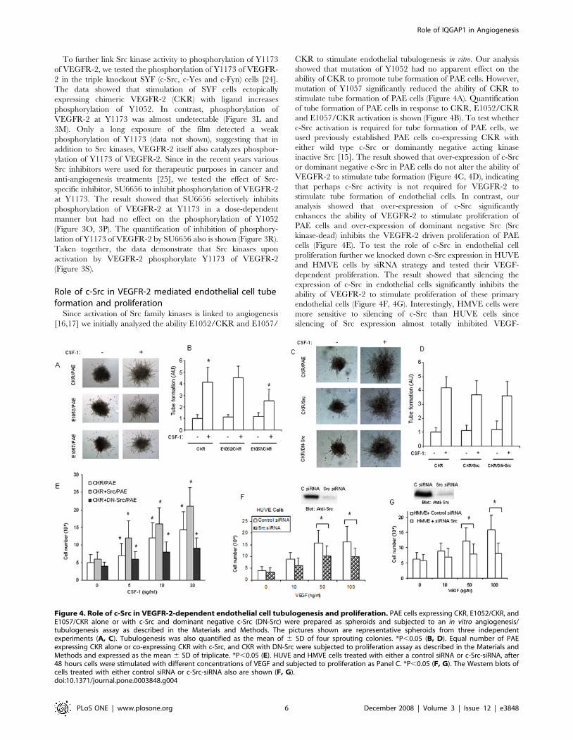
Role of c-Src in VEGFR-2 mediated endothelial cell tubeformation and proliferation
Since activation of Src family kinases is linked to angiogenesis
[16,17] we initially analyzed the ability E1052/CKR and E1057/
CKR to stimulate endothelial tubulogenesis in vitro. Our analysis
showed that mutation of Y1052 had no apparent effect on the
ability of CKR to promote tube formation of PAE cells. However,
mutation of Y1057 significantly reduced the ability of CKR to
stimulate tube formation of PAE cells (Figure 4A). Quantification
of tube formation of PAE cells in response to CKR, E1052/CKR
and E1057/CKR activation is shown (Figure 4B). To test whether
c-Src activation is required for tube formation of PAE cells, we
used previously established PAE cells co-expressing CKR with
either wild type c-Src or dominantly negative acting kinase
inactive Src [15]. The result showed that over-expression of c-Src
or dominant negative c-Src in PAE cells do not alter the ability of
VEGFR-2 to stimulate tube formation (Figure 4C, 4D), indicating
that perhaps c-Src activity is not required for VEGFR-2 to
stimulate tube formation of endothelial cells. In contrast, our
analysis showed that over-expression of c-Src significantly
enhances the ability of VEGFR-2 to stimulate proliferation of
PAE cells and over-expression of dominant negative Src (Src
kinase-dead) inhibits the VEGFR-2 driven proliferation of PAE
cells (Figure 4E). To test the role of c-Src in endothelial cell
proliferation further we knocked down c-Src expression in HUVE
and HMVE cells by siRNA strategy and tested their VEGF-
dependent proliferation. The result showed that silencing the
expression of c-Src in endothelial cells significantly inhibits the
ability of VEGFR-2 to stimulate proliferation of these primary
endothelial cells (Figure 4F, 4G). Interestingly, HMVE cells were
more sensitive to silencing of c-Src than HUVE cells since
silencing of Src expression almost totally inhibited VEGF-
Figure 4. Role of c-Src in VEGFR-2-dependent endothelial cell tubulogenesis and proliferation. PAE cells expressing CKR, E1052/CKR, andE1057/CKR alone or with c-Src and dominant negative c-Src (DN-Src) were prepared as spheroids and subjected to an in vitro angiogenesis/tubulogenesis assay as described in the Materials and Methods. The pictures shown are representative spheroids from three independentexperiments (A, C). Tubulogenesis was also quantified as the mean of 6 SD of four sprouting colonies. *P,0.05 (B, D). Equal number of PAEexpressing CKR alone or co-expressing CKR with c-Src, and CKR with DN-Src were subjected to proliferation assay as described in the Materials andMethods and expressed as the mean 6 SD of triplicate. *P,0.05 (E). HUVE and HMVE cells treated with either a control siRNA or c-Src-siRNA, after48 hours cells were stimulated with different concentrations of VEGF and subjected to proliferation as Panel C. *P,0.05 (F, G). The Western blots ofcells treated with either control siRNA or c-Src-siRNA also are shown (F, G).doi:10.1371/journal.pone.0003848.g004
Role of IQGAP1 in Angiogenesis
PLoS ONE | www.plosone.org 6 December 2008 | Volume 3 | Issue 12 | e3848
stimulated proliferation (Figure 4G). Taken together, our results
identify c-Src as a key component of mitogenic signaling of
VEGFR-2 in endothelial cells.
Identification of IQGAP1 as a novel Src kinase substrateIQGAP1 is a scaffold protein and participates in signaling
cascades mediated by a diverse group of cell surface receptors [33],
including VEGFR-2 [26]. In addition, we recently have identified
IQGAP1 as a binding partner of VEGFR-2 by a proteomic
approach (our unpublished data), raising the likelihood for the
involvement of Y1057 in the recruitment and tyrosine phosphor-
ylation of IQGAP1 by VEGFR-2. Our initial observation showed
that IQGAP1 is tyrosine phosphorylated in PAE cells by VEGFR-
2 and mutation of Y1057 reduces the ability of VEGFR-2 to
stimulate its tyrosine phosphorylation (Figure 5A). IQGAP1 also is
tyrosine phosphorylated by other RTKs including, ErbB1/EGFR-
1 and PDGFRb ectopically expressed in PAE cells (our
unpublished data), suggesting that IQGAP1 serves as a common
substrate for RTKs. In addition, our data show that over-
expression of c-Src in PAE cells highly enhanced tyrosine
phosphorylation of IQGAP1. In a sharp contrast, over-expression
of a dominant negatively acting c-Src inhibited tyrosine phos-
phorylation of IQGAP1 (Figure 5C).
To further establish role of c-Src in phosphorylation of IQGAP1
we show that in SYF knockout cells where Src family kinases (Yes,
Fyn and Src) are absent, IQGAP1 is not tyrosine phosphorylated
in response to activation of VEGFR-2, where re-introduction of c-
Src rescued phosphorylation of IQGAP1 by VEGFR-2 (Figure 5E).
Our further studies showed that in an in vitro kinase assay
recombinant c-Src protein phosphorylates GST-fusion IQGAP1
(data not shown). Taken together, the data support the hypothesis
that IQGAP1 directly associates with c-Src and undergoes c-Src-
dependent tyrosine phosphorylation. Hence to further establish as
to whether c-Src interacts with IQGAP1 we tested the ability of
SH2 and SH3 domains of c-Src to associate with IQGAP1 in an in
vitro pull-down assay. The result showed that SH2 domain of c-Src
interacts with IQGAP1 independent of its tyrosine phosphoryla-
tion since SH2 domain of c-Src forms a complex with IQGAP1
prior to stimulation of cells with ligand (Figure 5H). Additional
analysis using PAE cells null for chimeric VEGFR-2 revealed that
indeed association of SH2 domain of Src with IQGAP1 is
independent of VEGFR-2 (data not shown). In contrast to the
SH2 domain, the SH3 domain of c-Src failed to interact with
IQGAP1 with or without stimulation of VEGFR-2 (Figure 5I).
The data suggest that SH2 domain mediates the interaction of c-
Src with IQGAP1 by a novel mechanism that is independent of
tyrosine phosphorylation of IQGAP1.
IQGAP1 signaling pathway is required for VEGF-inducedangiogenesis
To establish the biological importance of IQGAP1 in VEGFR-2
signaling, we initially examined its role in proliferation of
endothelial cells. To this end, we employed siRNA-mediated
knockdown strategy and analyzed proliferation of endothelial cells
in response to VEGF stimulation. IQGAP1 siRNA significantly
reduced expression of IQGAP1 in HMVE cells (Figure 5L). We
further analyzed these cells for their ability to undergo prolifer-
ation in response to VEGF. Silencing the expression of IQGAP1
in primary endothelial cells, but not control siRNA, significantly
inhibited VEGF-dependent cell proliferation (Figure 5M), sug-
gesting that VEGFR-2/c-Src orchestrated tyrosine phosphoryla-
tion of IQGAP1 serves an important role in endothelial cell
proliferation. Our recent study indicates that IQGAP1 is capable
of modulating activity of b-Raf [27], to test whether IQGAP1
activity is also required for b-Raf phosphorylation in endothelial
cells in response to VEGFR-2 activation we tested phosphoryla-
tion of b-Raf where expression of IQGAP1 is knocked down by
siRNA. The data show that stimulation of endothelial cells with
VEGF promotes phosphorylation of b-Raf at Ser 455 and
silencing expression of IQGAP1 notably reduced its phosphory-
lation (Figure 5N). Quantification of phosphorylation of Ser 455 of
b-Raf based on the three independent experiments also is shown
(Figure 5Q). The reduced phosphorylation of b-Raf in IQGAP1
siRNA treated cells was not due to the differential amount of
protein since relatively equal amount of b-Raf protein is present in
each group (Figure 5O). The expression of IQGAP1 in cells
treated with control siRNA or IQGAP1 siRNA also is shown
(Figure 5P). In short, the data indicate that VEGFR-2-dependent
endothelial cell proliferation, in part, is established by Src,
IQGAP1and b-Raf axis (Figure 5R).
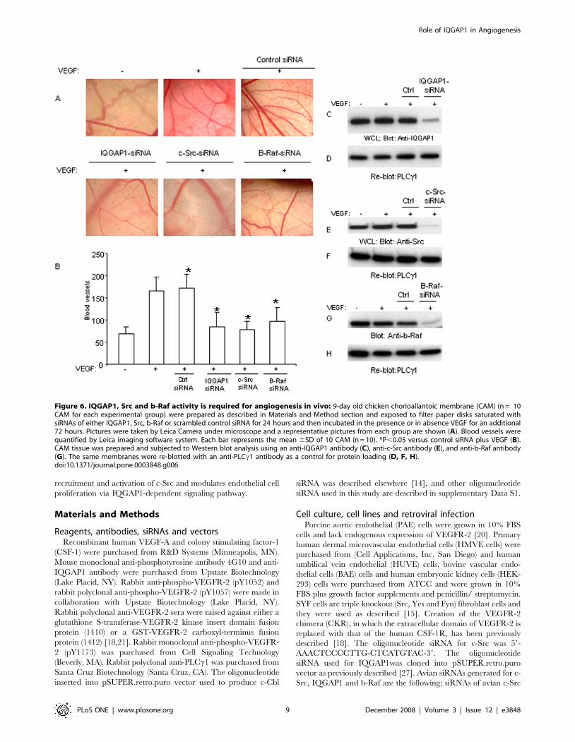
To further address the physiological importance of c-Src,
IQGAP1and b-Raf in angiogenesis in an in vivo setting, we used
chicken chorioallantoic membrane (CAM) angiogenesis assay
where their expression were knocked down by siRNA strategy.
The result showed that targeting IQGAP1, c-Src and b-Raf
individually by siRNA suppresses the ability of VEGF to stimulate
angiogenesis (Figure 6A), where control siRNA had no negative
effect on angiogenesis (Figure 6A). Furthermore, quantification of
CAM assay showed that VEGF treatment of CAM induced robust
angiogenic response where IQGAP1-siRNA, c-Src-siRNA and b-
Raf-siRNA each decreased VEGF-induced angiogenesis
(Figure 6B). As noted the siRNA-mediated inhibition of VEGF-
induced angiogenesis was near to baseline angiogenesis (Figure 6B)
underscoring the importance of this pathway for VEGF-induced
angiogenesis. The ability of these specific siRNAs to knockdown
expression of c-Src, IQGAP1 and b-Raf also are analyzed
(Figure 6C, 6E, 6G). The same membranes were re-probed for
PLCc1 as a loading control (Figure 6D, 6F, 6H). In sum, our data
demonstrate that IQGAP1, c-Src and b-Raf pathway plays a
critical role both in vitro and in vivo in VEGF-mediated
angiogenesis.
Discussion
In this report, we have identified a novel angiogenic pathway
emanating from phospho-Y1057 of VEGFR-2. Phosphorylation of
Y1057 plays a combinatorial role in VEGFR-2 signaling; it
recruits c-Src kinase to VEGFR-2, regulates phosphorylation of
multi-docking Y1173 site, mediates VEGFR-2-dependent prolif-
eration of endothelial cells and angiogenesis through IQGAP1 and
b-Raf. One of the most interesting aspects of this study is the
discovery of an unexpected role of Src family kinases in the
phosphorylation of Y1173 of VEGFR-2. Y1173 of VEGFR-2 is
considered to be a bona fide mediator of angiogenic signaling of
VEGFR-2 in vivo since its mutation abrogates its ability to
stimulate angiogenesis during mouse embryonic development
[6,11] and Y1173 functions as a multi-docking residue on
VEGFR-2 that recruits p85/PI3kinase, PLCc1, Shb, and Sck
[3–7,10]. Thus, phosphorylation of Y1173 by Src kinases
represents one pathway by which the intensity and extent of
Y1173-dependent signaling of VEGFR-2 is controlled. Converse-
ly, inhibition of Src kinases could skew the binding of these
signaling proteins to VEGFR-2 and their subsequent activation by
VEGFR-2.
A recent study indicates that c-Cbl directly interacts with Y1057
of VEGFR-2 [14]. Interestingly, not only does c-Src bind directly
to Y1057, but c-Cbl provides an additional mechanism for c-Src to
interact with VEGFR-2 by serving as an ‘‘adaptor’’ which in turn
Role of IQGAP1 in Angiogenesis
PLoS ONE | www.plosone.org 7 December 2008 | Volume 3 | Issue 12 | e3848
may modulate its activation by VEGFR-2. Activation of c-Src is
linked to vascular permeability [16], endothelial cell survival and
angiogenesis [16,17]. Our findings show that although c-Src
activity is dispensable for VEGFR-2-dependent tube formation of
endothelial cells, its activity is highly critical for VEGF-stimulated
endothelial cell proliferation and angiogenesis in CAM model of
angiogenesis. The mechanism by which c-Src contributes to
VEGFR-2-dependent cell proliferation is linked, in part to its
ability to phosphorylate Y1173, which is known to regulate
VEGFR-2-mediated endothelial cell proliferation [5–7]. c-Src also
associates with IQGAP1 in endothelial cells and catalyzes tyrosine
phosphorylation of IQGAP1 which acts as a downstream target of
VEGFR-2 and c-Src to stimulate endothelial cell proliferation. A
recent work has demonstrated that c-Src regulates activation of
Raf-1 (also known as c-Raf) in endothelial cells [17]. c-Src also
associates directly with another Raf family protein, b-Raf, leading
to activation of MAPK pathway in a Ras-independent manner
[27,33]. Our demonstration that IQGAP1 is activated by
VEGFR-2 further supports earlier studies that shows VEGFR-2
activates MAPK pathway in Ras-independent manner [31].
In conclusion, in this study we have identified and defined a
molecular switch by which VEGFR-2 regulates endothelial cell
proliferation and stimulates angiogenesis by identifying Y1057 of
VEGFR-2 that undergoes phosphorylation and orchestrates
Figure 5. Role of IQGAP1 in VEGFR-2 signaling and endothelial cell Proliferation. Serum-starved PAE cells expressing CKR (chimeric VEGFR-2) and E1057/CKR were either unstimulated (2) or stimulated with CSF-1 (+) for 10 minutes. Whole cell lysates were subjected toimmunoprecipitation with IQGAP1 and Western blot analysis using an anti-phosphotyrosine antibody (A) or anti-IQGAP1 antibody (B). PAE cellsco-expressing either wild type c-Src or a dominant negative form of c-Src (KD-Src) were stimulated and subjected to Western blot analysis as panel Ausing an anti-phosphotyrosine antibody (C). The same membrane striped off the antibody and re-blotted with an anti-IQGAP1 antibody for proteinlevels (D). SYF cells co-expressing CKR with c-Src or expressing CKR alone were prepared as above and blotted with anti-phosphotyrosine (E), anti-IQGAP1 (F), and anti-VEGFR-2 (G). Cell lysates derived from PAE cells expressing CKR were either precipitated with GST, GST-SH2-Src, (H) or GST-SH3-Src (I) and blotted for IQGAP1 using an anti-IQGAP1 antibody. Human primary dermal vascular endothelial (HMVE) cells were stimulated with VEGFfor 10 minutes, cells were lysed and immunoprecipitated with either control antibody (Ctrl.IgG) or c-Src antibody and blotted with anti-IQGAP1 (J).Whole cell lysates was blotted with anti-Src antibody (K). HMVE cells expressing either control siRNA (Ctrl.siRNA) or IQGAP1 siRNA were lysed andblotted for IQGAP1 using an anti-IQGAP1 antibody (L). HMVE cells expressing either control siRNA or IQGAP1 siRNA were subjected to proliferationassay as described in Method section. Error bars represent mean 6 SD of triplicate samples. *p,0.001 as compared to proliferation of HMVE cells inresponse to 100 gg VEGF-A (M). HMVE cells expressing either control siRNA or IQGAP1-siRNA were unstimulated (2) or stimulated (+) with VEGF for10 minutes and cells were lysed and immunoprecipitated with an anti-b-Raf antibody and blotted with an anti-phospho-Ser445 antibody (N) or withanti- b-Raf antibody (O). Whole cell lysates was blotted for IQGAP1 (P). Also shown is the graph of the mean phosphorylation of b-Raf where theexpression of IQGAP1 is silenced by siRNA. It represents (6SD) of three separate experiments. *P,0.01 (Q). Summary of the proposed VEGFR-2-dependent signal relay in endothelial cell proliferation also is shown (R).doi:10.1371/journal.pone.0003848.g005
Role of IQGAP1 in Angiogenesis
PLoS ONE | www.plosone.org 8 December 2008 | Volume 3 | Issue 12 | e3848
recruitment and activation of c-Src and modulates endothelial cell
proliferation via IQGAP1-dependent signaling pathway.
Materials and Methods
Reagents, antibodies, siRNAs and vectorsRecombinant human VEGF-A and colony stimulating factor-1
(CSF-1) were purchased from R&D Systems (Minneapolis, MN).
Mouse monoclonal anti-phosphotyrosine antibody 4G10 and anti-
IQGAP1 antibody were purchased from Upstate Biotechnology
(Lake Placid, NY). Rabbit anti-phospho-VEGFR-2 (pY1052) and
rabbit polyclonal anti-phospho-VEGFR-2 (pY1057) were made in
collaboration with Upstate Biotechnology (Lake Placid, NY).
Rabbit polyclonal anti-VEGFR-2 sera were raised against either a
glutathione S-transferase-VEGFR-2 kinase insert domain fusion
protein (1410) or a GST-VEGFR-2 carboxyl-terminus fusion
protein (1412) [18,21]. Rabbit monoclonal anti-phospho-VEGFR-
2 (pY1173) was purchased from Cell Signaling Technology
(Beverly, MA). Rabbit polyclonal anti-PLCc1 was purchased from
Santa Cruz Biotechnology (Santa Cruz, CA). The oligonucleotide
inserted into pSUPER.retro.puro vector used to produce c-Cbl
siRNA was described elsewhere [14], and other oligonucleotide
siRNA used in this study are described in supplementary Data S1.
Cell culture, cell lines and retroviral infectionPorcine aortic endothelial (PAE) cells were grown in 10% FBS
cells and lack endogenous expression of VEGFR-2 [20]. Primary
human dermal microvascular endothelial cells (HMVE cells) were
purchased from (Cell Applications, Inc. San Diego) and human
umbilical vein endothelial (HUVE) cells, bovine vascular endo-
thelial cells (BAE) cells and human embryonic kidney cells (HEK-
293) cells were purchased from ATCC and were grown in 10%
FBS plus growth factor supplements and penicillin/ streptomycin.
SYF cells are triple knockout (Src, Yes and Fyn) fibroblast cells and
they were used as described [15]. Creation of the VEGFR-2
chimera (CKR), in which the extracellular domain of VEGFR-2 is
replaced with that of the human CSF-1R, has been previously
described [18]. The oligonucleotide siRNA for c-Src was 59-
AAACTCCCCTTG-CTCATGTAC-39. The oligonucleotide
siRNA used for IQGAP1was cloned into pSUPER.retro.puro
vector as previously described [27]. Avian siRNAs generated for c-
Src, IQGAP1 and b-Raf are the following; siRNAs of avian c-Src
Figure 6. IQGAP1, Src and b-Raf activity is required for angiogenesis in vivo: 9-day old chicken chorioallantoic membrane (CAM) (n = 10CAM for each experimental group) were prepared as described in Materials and Method section and exposed to filter paper disks saturated withsiRNAs of either IQGAP1, Src, b-Raf or scrambled control siRNA for 24 hours and then incubated in the presence or in absence VEGF for an additional72 hours. Pictures were taken by Leica Camera under microscope and a representative pictures from each group are shown (A). Blood vessels werequantified by Leica imaging software system. Each bar represents the mean 6SD of 10 CAM (n = 10). *P,0.05 versus control siRNA plus VEGF (B).CAM tissue was prepared and subjected to Western blot analysis using an anti-IQGAP1 antibody (C), anti-c-Src antibody (E), and anti-b-Raf antibody(G). The same membranes were re-blotted with an anti-PLCc1 antibody as a control for protein loading (D, F, H).doi:10.1371/journal.pone.0003848.g006
Role of IQGAP1 in Angiogenesis
PLoS ONE | www.plosone.org 9 December 2008 | Volume 3 | Issue 12 | e3848
were; CAUUGCCAAGGUCAGCGAU UU and AUGACGC-
CAC AGCGCAGGC UU, siRNA for avian b-raf were;
UUGGCUGGGACACUGACAU UU and AGGAUAGGAU-
CUGGAUCAU UU; siRNA for avian IQGAP1 was; ACUCUG-
CAAGCCUUACA GAUU. Creation of mutant VEGFR-2s and
other related methods are described in supplementary Data S1.
In vitro angiogenesis/tubulogenesis assayAngiogenesis assay was performed essentially as described
[14,28]. Briefly, PAE cells were suspended in DMEM containing
1% fetal bovine serum and 0.24% (w/v) carboxymethylcellulose
(4000 centipoise) in non-adherent round-bottom 96-well plates.
After 24 h, all cells formed one single spheroid per well. Spheroids
were cultured for additional 48 h before using them in the in vitro
angiogenesis assay in the manner previously described [14,28].
Sprouting and tubulogenesis was observed under an inverted
phase-contrast microscope and pictures were taken using a Leica
digitial camera system. For statistical analysis purposes four
sprouting colonies were used for each experimental condition
and their sprouting was quantified using image J program (NIH).
Immunoprecipitation, immunoblotting and in vitro pull-down assay
Cells were prepared for immunoprecipitation as described [14].
Briefly, cells were grown in 10-cm culture dishes until 80–90%
confluent and after serum starvation, cells were left either resting
or stimulated with appropriate ligands as indicated in figure
legends. Cells were lysed and normalized whole cell lysates were
subject to immunoprecipitation by incubating with appropriate
antibodies. Immunocomplexes were captured by incubating with
either Protein-A Sepharose or Protein-G-Agarose beads and
immunoprecipitated protein were subjected to immunoblotting
analysis. Additional experimental details are provided in Supple-
mentary Data S1. In some occasions the blots were scanned and
they were subsequently quantified using Image J program (NIH).
Statistical AnalysisFor statistical purposes the blots of three independent
experiments were scanned and blots were quantified using NIH
Image J program. Comparison of the different parameters for the
blots was determined by repeated measures analysis of variance
(ANOVA). Significant differences were assigned using Kruskal-
Wallis post hoc test. The criterion for significance for all tests was
set at p , 0.05. Specific software was used to assist in the data
analysis (GraphPadPrism v4.0b, GraphPad Software, San Diego,
USA).
GST-Pull down assayIn vitro GST fusion protein binding experiments were performed
as described [30]. To this end, equal numbers of endothelial cells
were grown to 90% confluency prior to serum starvation for
overnight. Unstimulated or ligand-stimulated cells were lysed in
ice-cold lysis buffer supplemented with 2 mM Na3VO4 and a
protease inhibitor cocktail. Equal amounts of the appropriate
immobilized GST fusion proteins were incubated with normalized
whole cell lysates by rocking for 3 h at 4uC. The beads were
washed four times in the presence of protease inhibitors and
Na3VO4, and proteins were eluted and analyzed in Western blot
analysis using appropriate antibody.
Dot blot assayPurified recombinant GST, GST-SH2-c-Src and GST-SH3-c-
Src were eluted from glutathione-Sepharose beads and subjected
to dot blot. Further information regarding this assay is provided in
Supplementary Data S1.
Cell proliferation assayCell proliferation was evaluated by direct cell counting.
Endothelial cells were seeded at a density of 26104 cells/well in
24-well plates and cultured overnight; the cells were then
incubated in serum-free medium for 12 hours. Cells were
stimulated with recombinant human CSF-1 or VEGF-A at
different concentrations as indicated in figure legends, and after
48 hours they were washed with PBS, harvested by mild
trypsinization, and counted with a hematocytometer. Experiments
were performed in triplicate and 3 separate experiments were
performed and where appropriate, values were presented as means
of 6 SD.
Chicken chorioallantoic membrane (CAM) angiogenesisassay
Pathogen free grade fertilized chick embryos (purchased from
SPAFAS, Preston, CT) were incubated for 10 days (37uC with
70% humidity). A hole was made with a drill over the air sac at the
end of the egg as described [16]. A small window was cut in the
egg shell over the dropped CAM, in order to place growth factor
and siRNA on the CAM [16,17]. Filter disks were soaked in
cortisone acetate, VEGF (200 gg), siRNA and plus transfection
agent and the disk filters were applied to the CAM of 9 day chick
embryos and incubated at 37uC for 72 hours. 10 CAM were used
per each group. Pictures were taken by Leica Camera under
microscope from three different area randomly and these areas
were further used to quantify blood vessels using Leica imaging
software system. Statistical Analysis: Data represent mean 6SD of
10 CAM which includes three different fields from each CAM was
used unless otherwise stated. Statistical significance was calculated
by ANOVA soft ware program (prism v4.0b), considering p,0.05
as statistically significant. Tissue preparation for Western blotting:
CAM tissue was also harvested for Western blotting. For this
purpose CAM tissue was snap frozen in liquid nitrogen and
samples were homogenized in a modified RIPA buffer. Five
CAMs from each experimental group were homogenized in 1 ml
of the RIPA buffer. Equal amounts of protein were subjected to
Western blot analysis using desired antibody as described in the
figure legends.
Supporting Information
Data S1
Found at: doi:10.1371/journal.pone.0003848.s001 (0.03 MB
DOC)
Figure S1 Effect of mutation of tyrosines 1052 and 1057 to
phenylalanine: Equal number of serum-starved PAE cells
expressing wild type CKR, F1052/CKR, F1057/CKR and
F1052/F1057/CKR were either unstimulated (2) or stimulated
with CSF-1 (+) for 10 minutes. Total cell lysates were subjected to
Western blot analysis using an anti-phosphoY1052 specific-
VEGFR-2 antibody (A), an anti-phosphoY1057 specific anti-
VEGFR-2 antibody (B), an anti-phosphotyrosine antibody (C),
and an anti-VEGFR-2 antibody (D). Equal number of serum-
starved PAE cells expressing wild type CKR, F1052/CKR,
F1057/CKR and F1052/F1057/CKR without stimulation were
lysed and immunoprecipitated with an anti-VEGFR-2 antibody.
The immunoprecipitated proteins were subjected to an in vitro
kinase assay with increasing concentrations of ATP as indicated
and described in Materials and Methods and immunoblotted with
Role of IQGAP1 in Angiogenesis
PLoS ONE | www.plosone.org 10 December 2008 | Volume 3 | Issue 12 | e3848
an anti-phosphotyrosine antibody (E). The same membranes were
re-blotted with an anti-VEGFR-2 antibody for proteins levels (F).
Found at: doi:10.1371/journal.pone.0003848.s002 (0.22 MB TIF)
Figure S2 Effect of mutation of Tyrosines 1052 and 1057 to
glutamic acid. Partial amino acid sequence of mouse VEGFR-2
containing Y1052 and Y1057 and the schematic presentation of
VEGFR-2 are shown (A). Equal number of serum-starved PAE
cells expressing wild type CKR, E1052/CKR, E1057/CKR and
E1052/E1057/CKR were either unstimulated (2) or stimulated
with CSF-1 (+) for 10 minutes. Total cell lysates were subjected to
Western blot analysis using an anti-phosphoY1052 specific-
VEGFR-2 antibody (B), an anti-phosphoY1057 specific anti-
VEGFR-2 antibody (C), an anti-phosphotyrosine antibody (D),
and an anti-VEGFR-2 antibody (E). Equal number of serum-
starved PAE cells expressing wild type CKR, E1052/CKR,
E1057/CKR and E1052/E1057/CKR without stimulation were
lysed and immunoprecipitated with an anti-VEGFR-2 antibody.
The immunoprecipitated proteins were subjected to an in vitro
kinase assay with increasing concentrations of ATP as indicated
and described in Materials and Methods and immunoblotted with
an anti-phosphotyrosine antibody (F). The same membranes were
re-blotted with an anti-VEGFR-2 antibody for proteins levels (G).
The image analysis program was used to quantify the data. The
tyrosine phosphorylation values were normalized over total
protein (H). Arbitrary unit (AU).
Found at: doi:10.1371/journal.pone.0003848.s003 (0.24 MB TIF)
Figure S3 GST-Src but not GST binds to VEGFR-2: Serum-
starved PAE cells expressing CKR were either unstimulated (2) or
stimulated (+) with CSF-1 for 10 minutes, lysed, and whole cell
lysates were incubated with purified GST alone or GST-SH2-Src
protein. The GST-SH2-Src bound proteins were subjected to
Western blot analysis using an anti-VEGFR-2 antibody (A). Whole
cell lysates were blotted with anti-VEGFR-2 antibody as a control
(B).
Found at: doi:10.1371/journal.pone.0003848.s004 (0.09 MB TIF)
Author Contributions
Conceived and designed the experiments: RDM NR. Performed the
experiments: RDM NR. Analyzed the data: RDM NR. Contributed
reagents/materials/analysis tools: DBS. Wrote the paper: NR.
References
1. Carmeliet P (2005) Angiogenesis in life, disease and medicine. Nature 438:
932–936.
2. Olsson AK, Dimberg A, Kreuger J, Claesson-Welsh L (2006) VEGF receptorsignalling-in control of vascular function. Nat Rev Mol Cell Biol 7: 359–371.
3. Dayanir V, Meyer RD, Lashkari K, Rahimi N (2001) Identification of tyrosineresidues in vascular endothelial growth factor receptor-2/FLK-1 involved in
activation of phosphatidylinositol 3-kinase and cell proliferation. J Biol Chem
276: 17686–17692.4. Tzima E, Irani-Tehrani M, Kiosses WB, Dejana E, Schultz DA, et al. (2005) A
mechanosensory complex that mediates the endothelial cell response to fluidshear stress. Nature 437: 426–431.
5. Takahashi T, Yamaguchi S, Chida K, Shibuya M (2001) A single autopho-
sphorylation site on KDR/Flk-1 is essential for VEGF-A-dependent activation ofPLC-gamma and DNA synthesis in vascular endothelial cells. EMBO J 20:
2768–2778.6. Meyer RD, Latz C, Rahimi N (2003) Recruitment and activation of
phospholipase Cgamma1 by vascular endothelial growth factor receptor-2 arerequired for tubulogenesis and differentiation of endothelial cells. J Biol Chem
278: 16347–16355.
7. Holmqvist K, Cross MJ, Rolny C, Hagerkvist R, Rahimi N, et al. (2004) Theadaptor protein shb binds to tyrosine 1175 in vascular endothelial growth factor
(VEGF) receptor-2 and regulates VEGF-dependent cellular migration. J BiolChem 279: 22267–22275.
8. Igarashi K, Shigeta K, Isohara T, Yamano T, Uno I (1998) Sck interacts with
KDR and Flt-1 via its SH2 domain. Biochem Biophys Res Commun 251:77–82.
9. Warner AJ, Lopez-Dee J, Knight EL, Feramisco JR, Prigent SA (2000) The Shc-related adaptor protein, Sck, forms a complex with the vascular-endothelial-
growth-factor receptor KDR in transfected cells. Biochem J 347: 501–509.10. Ratcliffe KE, Tao Q, Yavuz B, Stoletov KV, Spring SC, et al. (2002) Sck is
expressed in endothelial cells and participates in vascular endothelial growth
factor-induced signaling. Oncogene 21: 6307–6316.11. Sakurai Y, Ohgimoto K, Kataoka Y, Yoshida N, Shibuya M (2005) Essential
role of Flk-1 (VEGF receptor 2) tyrosine residue 1173 in vasculogenesis in mice.Proc Natl Acad Sci U S A 102: 1076–1081.
12. Shalaby F, Rossant J, Yamaguchi TP, Gertsenstein M, Wu XF, et al. (1995)
Failure of blood-island formation and vasculogenesis in Flk-1-deficient mice.Nature 376: 62–66.
13. Zeng H, Sanyal S, Mukhopadhyay D (2001) Tyrosine residues 951 and 1059 ofvascular endothelial growth factor receptor-2 (KDR) are essential for vascular
permeability factor/vascular endothelial growth factor-induced endotheliummigration and proliferation, respectively. J Biol Chem 276: 32714–32719.
14. Singh AJ, Meyer RD, Navruzbekov G, Shelke R, Duan L, et al. (2007) A critical
role for the E3-ligase activity of c-Cbl in VEGFR-2-mediated PLCgamma1activation and angiogenesis. Proc Natl Acad Sci U S A 104: 5413–5418.
15. Meyer RD, Dayanir V, Majnoun F, Rahimi N (2002) The presence of a singletyrosine residue at the carboxyl domain of vascular endothelial growth factor
receptor-2/FLK-1 regulates its autophosphorylation and activation of signaling
molecules. J Biol Chem 277: 27081–27087.
16. Eliceiri BP, Paul R, Schwartzberg PL, Hood JD, Leng J, et al. (1999) Selective
requirement for Src kinases during VEGF-induced angiogenesis and vascular
permeability. Mol Cell 4: 915–924.
17. Alavi A, Hood JD, Frausto R, Stupack DG, Cheresh DA (2003) Role of Raf in
vascular protection from distinct apoptotic stimuli. Science 301: 94–96.
18. Rahimi N (2006) VEGFR-1 and VEGFR-2: two non-identical twins with a
unique physiognomy. Front Biosci 11: 818–829.
19. Marengere LE, Songyang Z, Gish GD, Schaller MD, Parsons JT, et al. (1994)
SH2 domain specificity and activity modified by a single residue. Nature 369:
502–505.
20. Rahimi N, Dayanir V, Lashkari K (2000) Receptor chimeras indicate that the
vascular endothelial growth factor receptor-1 (VEGFR-1) modulates mitogenic
activity of VEGFR-2 in endothelial cells. J Biol Chem 275: 16986–16992.
21. Singh AJ, Meyer RD, Band H, Rahimi N (2005) The carboxyl terminus of
VEGFR-2 is required for PKC-mediated down-regulation. Mol Biol Cell 16:
2106–2118.
22. Sanjay A, Miyazaki T, Itzstein C, Purev E, Horne WC, et al. (2006)
Identification and functional characterization of an Src homology domain 3
domain-binding site on Cbl. FEBS J 273: 5442–5456.
23. Nolen B, Taylor S, Ghosh G (2004) Regulation of protein kinases; controlling
activity through activation segment conformation. Mol Cell 15: 661–675.
24. Klinghoffer RA, Sachsenmaier C, Cooper JA, Soriano P (1999) Src family
kinases are required for integrin but not PDGFR signal transduction. EMBO J
18: 2459–2471.
25. Summy JM, Gallick GE (2003) Src family kinases in tumor progression and
metastasis. Cancer Metastasis Rev 22: 337–358.
26. Yamaoka-Tojo M, Ushio-Fukai M, Hilenski L, Dikalov SI, Chen YE, et al.
(2004) IQGAP1, a novel vascular endothelial growth factor receptor binding
protein, is involved in reactive oxygen species-dependent endothelial migration
and proliferation. Circ Res 95: 276–283.
27. Ren JG, Li Z, Sacks DB (2007) IQGAP1 modulates activation of B-Raf. Proc
Natl Acad Sci U S A. 104: 10465–10469.
28. Tanaka S, Amling M, Neff L, Peyman A, Uhlmann E, et al. (1996) c-Cbl is
downstream of c-Src in a signalling pathway necessary for bone resorption.
Nature 383: 528–531.
29. Bao J, Gur G, Yarden Y (2003) Src promotes destruction of c-Cbl: implications
for oncogenic synergy between Src and growth factor receptors. Proc Natl Acad
Sci U S A 100: 2438–2443.
30. Meyer RD, Singh A, Majnoun F, Latz C, Lashkari K, et al. (2004) Substitution
of C-terminus of VEGFR-2 with VEGFR-1 promotes VEGFR-1 activation and
endothelial cell proliferation. Oncogene 23: 5523–5531.
31. Roy M, Li Z, Sacks DB (2004) IQGAP1 binds ERK2 and modulates its activity.
J Biol Chem 279: 17329–17337.
32. Takahashi T, Ueno H, Shibuya M (1999) VEGF activates protein kinase C-
dependent, but Ras-independent Raf-MEK-MAP kinase pathway for DNA
synthesis in primary endothelial cells. Oncogene 18: 2221–2230.
33. Briggs MW, Sacks DB (2003) IQGAP proteins are integral components of
cytoskeletal regulation. EMBO Rep 4: 571–574.
Role of IQGAP1 in Angiogenesis
PLoS ONE | www.plosone.org 11 December 2008 | Volume 3 | Issue 12 | e3848


![2008 3723-3736 3723 The Brain, the Penis and Steroid ... Brain... · 2) regulates Nitric Oxide(NO)synthase(NOS) in the hypothalamus [8] and modulates vascular endothelial growth permeability](https://img.pdfslide.net/doc/110x75/5edd15f3ad6a402d66680b2e/2008-3723-3736-3723-the-brain-the-penis-and-steroid-brain-2-regulates.jpg)