Embed Size (px)
Citation preview
An Isoform-specific Mutation in the Protein 4.1 Gene Results in HereditaryElliptocytosis and Complete Deficiencyof Protein 4.1 in Erythrocytes but not in Nonerythroid CellsJohn G. Conboy, Joel Anne Chasis, Ricky Winardi, Gil Tchernia, Yuet Wai Kan, and Narla MohandasCell and Molecular Biology Division, Lawrence Berkeley Laboratory, University of California, Berkeley, California 94720; Departmentof Laboratory Medicine and Howard Hughes Medical Institute, University of California, San Francisco, California 94143;and H6pital Bicetre, 94270 Kremlin Bicetre, Paris, France
Abstract
Multiple protein 4.1 isoforms are expressed in a variety of tis-sues through complex alternative pre-mRNA splicing events,one function of which is to regulate use of two alternative trans-lation initiation signals. Late erythroid cells express mainly thedownstream initiation site for synthesis of prototypical 80-kDisoforms; nonerythroid cells in addition use an upstream site toencode higher molecular mass isoform(s). In this study, weexamined the effects of a 5' gene rearrangement in a family withhereditary elliptocytosis and complete deficiency of erythro-cyte 4.1 protein on 4.1 isoform expression in erythroid vs. non-erythroid cells. Patient 4.1 mRNAsfrom reticulocytes, fibro-blasts, and B lymphocytes were amplified by reverse transcrip-tase / polymerase chain reaction techniques and shown toexhibit a 318-nucleotide deletion that encompasses the down-stream AUG, but leaves intact the upstream AUG. Immuno-blot analysis revealed a total deficiency of 4.1 in patient redcells and a selective deficiency of 80-kD isoform(s) but nothigh molecular weight 4.1 in patient nonerythroid cells. Thus,the 4.1 gene mutation in this family produces an isoform-speci-fic deficiency that is manifested clinically in tissue-specific fash-ion, such that red cells are affected but other cell types areunaffected because of tissue-specific differences in RNAsplic-ing and translation initiation. (J. Clin. Invest. 1993. 91:77-82.)Key words: erythrocyte - alternative splicing * protein 4.1translation initiation * hereditary elliptocytosis
Introduction
Studies on red cells from patients with abnormal red cell mor-phology and clinically significant hemolytic anemia have fo-cused on the structure and function of the "membrane skele-ton,' a highly organized network of proteins supporting theinner surface of the cell membrane. Several red cell disorders,including hereditary spherocytosis and hereditary elliptocyto-sis (HE),' can result from disruption of membrane function by
Address correspondence to John Conboy, Cell and Molecular Biology,University of California, Lawrence Berkeley Laboratory, 1 CyclotronRoad, Building 74-157, Berkeley, CA 94720.
Receivedfor publication 28 April 1992 and in revised form 2 July1992.
1. Abbreviations uised in this paper: HE, hereditary elliptocytosis;PBGD, porophobilinogen deaminase; PCR, polymerase chain reac-tion; RFLPs, restriction fragment polymorphisms.
mutation of the gene(s) for major membrane skeletal proteinsincluding protein 4.1, a- and f-spectrin, ankyrin, and band3 (1).
Although most of these skeletal proteins were originallythought to be restricted to erythroid cells, it is now known thatclosely homologous proteins are also found in most nonery-throid cell types. Interestingly, the nonerythroid homologs of-ten vary with respect to size, sequence, or subcellular locationcompared to the red cell forms. These different but homolo-gous forms (isoforms) of membrane proteins may arise as prod-ucts of distinct genes, or by differences in expression of a singlegene in erythroid versus nonerythroid cells. One well-knownmechanism for differential gene expression is alternative pre-mRNAsplicing, a process by which a single gene can generatedifferent isoforms in different tissues.
Protein 4.1 is a key membrane skeletal protein which playsan important role in determining red cell shape and integritythrough specific interactions with other skeletal and membranecomponents. The major form of protein 4.1 in red cells is apolypeptide of - 80 kD, while nonerythroid cells contain aheterogeneous array of 4.1 polypeptides that range in size from30 to 210 kD (2). Despite the size and structural variationsamong these proteins, all appear to be encoded by a single largegene (> 100 kb) (3). Isoform variation is generated by alterna-tive splicing of the 4.1 pre-mRNA transcribed from this gene, acomplex process in which discrete exons are either spliced in(included) or spliced out (deleted) to produce an array of ma-ture 4.1 messenger RNAs that can be translated into differentprotein isoforms (Fig. 1 A) (4-7). In addition, either of twopotential translation initiation sites (denoted by the trinucleo-tide AUG) can be used depending on whether the upstream(more 5') or downstream sites are included in the maturemRNA(5, 7) (Fig. 1 B). Reticulocyte 4.1 mRNAspossess onlythe downstream AUGand can synthesize only the 80-kD redcell isoform, while nucleated erythroid progenitors and nonery-throid cells also contain the upstream initiation sites and syn-thesize larger protein 4.1 isoforms having an elongated NH2-ter-minal sequences (Fig. 1 B).
Mutations in the protein 4.1 gene result in hereditary ellip-tocytosis. To date, mutations that result in elongated or short-ened 4.1 polypeptides (8-12) or deficiency of red cell protein4.1 (10, 13-17) have been described. Wehave previously re-ported that in an Algerian family, several siblings suffer fromHE and severe hemolytic anemia caused by the complete ab-sence of erythroid protein 4. 1. These patients, the offspring of aconsanguinous marriage, were homozygous for a structural re-arrangement of the protein 4.1 gene located around the down-stream AUGcodon (3). Surprisingly, although the same geneencodes protein 4.1 isoforms in erythroid and nonerythroidtissue, no clinical manifestations other than the hemolytic ane-
Isoform-specific Deficiency Protein of Protein 4.1 in Hereditary Elliptocytosis 77
J. Clin. Invest.©3 The American Society for Clinical Investigation, Inc.0021-9738/93/01/077/06 $2.00Volume 91, January 1993, 77-82
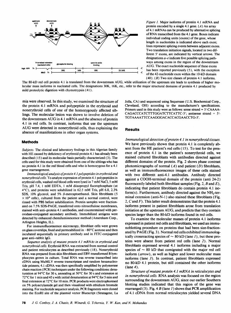
upGream downGream Figure 1. Major isoforms of protein 4.1 mRNAand1 t t U protein encoded by a single 4.1 gene. (A) An array
213 129 //0\ 81of 4.1 mRNAscan be produced by alternative splicingof RNAtranscribed from the 4.1 gene. Boxes indicateindividual coding units (exons) of the gene, whoselength in nucleotides is indicated above each exon;lines represent splicing events between adjacent exons.Two translation initiation signals, located in two dif-ferent 5' exons, are indicated by vertical arrows. Thedesignations a-e indicate five possible splicing path-
N-terminal extensbon tycophorin bindint spectrin-actin ways among exons in the region of the downstream(nucteated cetls) 9 binding AUG. The exact nucleotide sequence of these exonshigh MW N. I1i IIC
30K 16K 10K 22-24K has been reported previously (5), with the exceptionSOkD N. I I I -c of the 42-nucleotide exon within the 10-kD domain(40). (B) Two size classes of protein 4.1 isoforms.
The 80-kD red cell protein 4.1 is translated from the downstream AUG, while utilization of the upstream site leads to synthesis of higher mo-lecular mass isoforms in nucleated cells. The designations 30K, 16K, etc., refer to the major structural domains of protein 4.1 produced bymild proteolytic digestion with chymotrypsin (41 ).
mia were observed. In this study, we examined the structure ofthe protein 4.1 mRNAand polypeptide in the erythroid andnonerythroid cells of one of the homozygously affected sib-lings. The molecular lesion was shown to involve deletion ofthe downstream AUGin 4.1 mRNAand the absence of protein4.1 in red cells. In contrast, isoforms that use the upstreamAUGwere detected in nonerythroid cells, thus explaining theabsence of manifestations in other organ systems.
Methods
Subjects. The clinical and laboratory findings in this Algerian familywith HEcaused by deficiency of erythroid protein 4.1 has already beendescribed ( 15) and its molecular basis partially characterized (3). Thecells used for this study were obtained from one of the siblings who hasno protein 4.1 in the red blood cells and who is homozygous for a 4.1gene rearrangement.
Immunological analysis ofprotein 4.1 polypeptides in erythroid andnonerythroid cells. 'To analyze expression of protein 4.1 polypeptides inerythroid cells, washed red blood cells were lysed in 7 mMNaCl, 5 mMTris, pH 7.4, 1 mMEDTA, 1 mMdiisopropyl fluorophosphate (at4°C), and proteins were solubilized in 62.5 mMTris, pH 6.8, 2.3%SDS, 10% glycerol, and 5% fl-mercaptoethanol. Skin fibroblasts (5x 10' cells), cultured from the patient and a normal control, wererinsed with PBS before solubilization. Protein samples were fraction-ated on 7.5% SDS-PAGE, transferred onto nitrocellulose membranes,stained with anti-protein 4.1 antibodies, and counterstained with per-oxidase-conjugated secondary antibody. Immobilized antigens weredetected by enhanced chemiluminescence method (Amersham Corp.,Arlington Heights, IL).
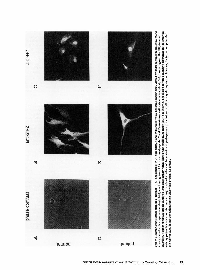
For immunofluorescence microscopy, fibroblast cells were grownon glass coverslips, fixed and permeabilized in -80°C acetone and thenincubated sequentially in primary antibody and in FITC-conjugatedgoat anti-rabbit IgG.
Sequence analysis of mutant protein 4.1 mRNAsin erythroid andnonerythroid cells. Erythroid RNAwas extracted from normal controland patient reticulocytes as described previously ( 18 ). NonerythroidRNAwas prepared from skin fibroblasts and EBV-transformed B lym-phocytes grown in culture. Total RNAwas reverse transcribed intocDNA using MuMLVreverse transcriptase and random hexanucleo-tide primers; 4.1 cDNAwas then specifically amplified by polymerasechain reaction (PCR) techniques under the following conditions: dena-turation at 94°C for 30 s, annealing at 50°C for 30 s and extension at72°C for 1 min and 45 s with initial denaturation at 94°C for 5 min andfinal extension at 720C for 10 min. PCRproducts were electrophoresedon 5%polyacrylamide gel and then visualized with ethidium bromidestaining. For nucleotide sequence analysis, PCRfragments were clonedinto the EcoRI site of the plasmid vector Bluescript (Stratagene, La
Jolla, CA) and sequenced using Sequenase (U.S. Biochemical Corp.,Cleveland, OH) according to the manufacturer's specifications.Primers used in this study were as follows: sense strand = 5'-CAAAA-CAGACCCATCTTTGGATCTTCATTC-3';antisense strand = 5'-TGTAAAATTCCAAGGGACACCACGAACCTG-3'.
Results
Immunological detection ofprotein 4.1 in nonerythroid tissues.Wehave previously shown that protein 4.1 is completely ab-sent from the HEpatient's red cells ( 15 ). To test for the pres-ence of protein 4.1 in the patient's nonerythroid cells, westained cultured fibroblasts with antibodies directed againstdifferent domains of the protein. Fig. 2 shows phase contrastphotomicrographs of normal (A) and patient (D) fibroblasts,as well as immunofluorescence images of these cells stainedwith two different anti-4. 1 antibodies. Antibody directedagainst a COOH-terminal domain of the protein (anti 24-2)fluorescently labeled both fibroblast samples (Fig. 2, B and E),indicating that patient fibroblasts do contain protein 4.1 iso-form(s). Furthermore, antibody directed against the NH2-ter-minal extension (anti-N- 1) also stained these fibroblasts (Fig.2, Cand F). This latter result demonstrates that the protein 4.1isoforms present in patient fibroblasts arose from translationinitiation at the upstream AUG, and predicts that they containspecies larger than the 80-kD isoforms found in red cells.
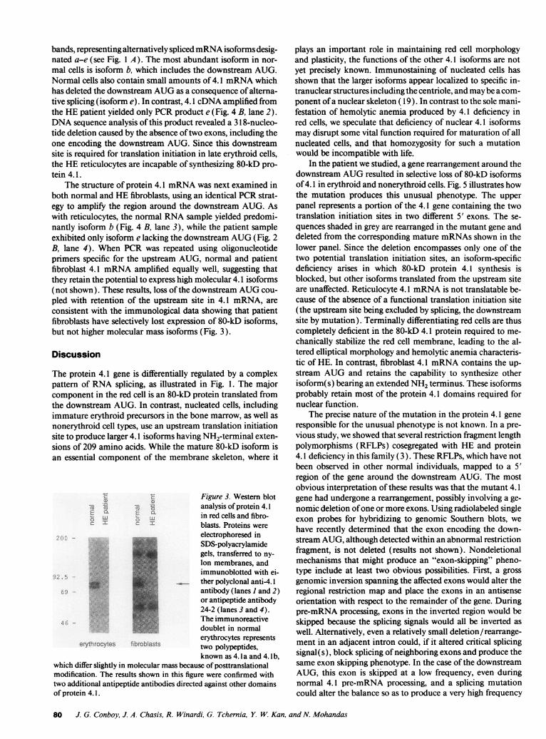
To examine the molecular masses of protein 4.1 isoformsexpressed in patient red cells and fibroblasts, we used an immu-noblotting procedure on proteins that had been size-fraction-ated by PAGE(Fig. 3). Normal red cells exhibited immunolog-ically crossreacting species of 80 kD (lane 1), but these pro-teins were absent from patient red cells (lane 2). Normalfibroblasts expressed several 4.1 isoforms including a majorspecies of - 80 kD that comigrated with the major red cellisoform (arrow), as well as higher and lower molecular massisoforms (lane 3). In contrast, patient fibroblasts expressedno 80-kD 4.1 protein, but still contained the other isoforms(lane 4).
Structure of mutant protein 4.1 mRNAin reticulocytes andin nonerythroid cells. RNAanalysis was focused on the regionsurrounding the downstream AUG, since our earlier Southernblotting studies indicated that this region of the gene wasrearranged (3). Fig. 4 B (lane 1) shows that PCRamplificationof 4.1 cDNA from normal reticulocytes yielded several DNA
78 J. G. Conboy, J. A. Chasis, R. Winardi, G. Tchernia, Y. W. Kan, and N. Mohandas
1:24
luealed
0'06
C)l
C13 . = >
CIS
.) 7 *- a
JD l =E
-
2C) C)O
>. .2_ X
Cu ) C)3
_ C
CIS
.a C
3 .z sO * Q * cS
2 < o Q
wD .- S .=
0.
Cu. - cj0. C) C)*
C.. e .- O0
o S. 2;
*- CuS 2> .a) -
EWo .N r
2 D Y -
= r
Isoform-specific Deficiency Protein of Protein 4.1 in Hereditary Elliptocytosis 79
C-)
CMICM
4-'
CI
P4:
cn
CucoL.
Cb0a,
CDCO-c0-
iewiou
cer
bands, representing alternatively spliced mRNAisoforms desig-nated a-e (see Fig. 1 A). The most abundant isoform in nor-mal cells is isoform b, which includes the downstream AUG.Normal cells also contain small amounts of 4.1 mRNAwhichhas deleted the downstream AUGas a consequence of alterna-tive splicing (isoform e). In contrast, 4.1 cDNAamplified fromthe HEpatient yielded only PCRproduct e (Fig. 4 B, lane 2).DNAsequence analysis of this product revealed a 31 8-nucleo-tide deletion caused by the absence of two exons, including theone encoding the downstream AUG. Since this downstreamsite is required for translation initiation in late erythroid cells,the HE reticulocytes are incapable of synthesizing 80-kD pro-tein 4.1.
The structure of protein 4.1 mRNAwas next examined inboth normal and HE fibroblasts, using an identical PCRstrat-egy to amplify the region around the downstream AUG. Aswith reticulocytes, the normal RNAsample yielded predomi-nantly isoform b (Fig. 4 B, lane 3), while the patient sampleexhibited only isoform e lacking the downstream AUG(Fig. 2B, lane 4). When PCRwas repeated using oligonucleotideprimers specific for the upstream AUG, normal and patientfibroblast 4.1 mRNAamplified equally well, suggesting thatthey retain the potential to express high molecular 4.1 isoforms(not shown). These results, loss of the downstream AUGcou-pled with retention of the upstream site in 4.1 mRNA, areconsistent with the immunological data showing that patientfibroblasts have selectively lost expression of 80-kD isoforms,but not higher molecular mass isoforms (Fig. 3).
Discussion
The protein 4.1 gene is differentially regulated by a complexpattern of RNAsplicing, as illustrated in Fig. 1. The majorcomponent in the red cell is an 80-kD protein translated fromthe downstream AUG. In contrast, nucleated cells, includingimmature erythroid precursors in the bone marrow, as well asnonerythroid cell types, use an upstream translation initiationsite to produce larger 4.1 isoforms having NH2-terminal exten-sions of 209 amino acids. While the mature 80-kD isoform isan essential component of the membrane skeleton, where it
t 'E Figure 3. Western blotCZ m,5 < 5analysis of protein 4.1E0
E L in red cells and fibro-cI CI blasts. Proteins were
electrophoresed inSDS-polyacrylamidegels, transferred to ny-lon membranes, andimmunoblotted with ei-ther polyclonal anti-4. 1antibody (lanes 1 and 2)or antipeptide antibody24-2 (lanes 3 and 4).The immunoreactivedoublet in normalerythrocytes represents
erythrocytes fibroblasts two polypeptides,
known as 4. la and 4. lb,which differ slightly in molecular mass because of posttranslationalmodification. The results shown in this figure were confirmed with
two additional antipeptide antibodies directed against other domainsof protein 4.1.
plays an important role in maintaining red cell morphologyand plasticity, the functions of the other 4.1 isoforms are notyet precisely known. Immunostaining of nucleated cells hasshown that the larger isoforms appear localized to specific in-tranuclear structures including the centriole, and maybe a com-ponent of a nuclear skeleton ( 19). In contrast to the sole mani-festation of hemolytic anemia produced by 4.1 deficiency inred cells, we speculate that deficiency of nuclear 4.1 isoformsmay disrupt some vital function required for maturation of allnucleated cells, and that homozygosity for such a mutationwould be incompatible with life.
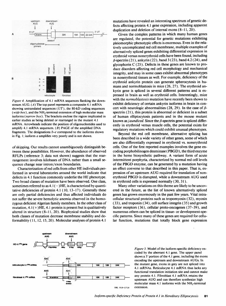
In the patient we studied, a gene rearrangement around thedownstream AUGresulted in selective loss of 80-kD isoformsof 4.1 in erythroid and nonerythroid cells. Fig. 5 illustrates howthe mutation produces this unusual phenotype. The upperpanel represents a portion of the 4.1 gene containing the twotranslation initiation sites in two different 5' exons. The se-quences shaded in grey are rearranged in the mutant gene anddeleted from the corresponding mature mRNAsshown in thelower panel. Since the deletion encompasses only one of thetwo potential translation initiation sites, an isoform-specificdeficiency arises in which 80-kD protein 4.1 synthesis isblocked, but other isoforms translated from the upstream siteare unaffected. Reticulocyte 4.1 mRNAis not translatable be-cause of the absence of a functional translation initiation site(the upstream site being excluded by splicing, the downstreamsite by mutation). Terminally differentiating red cells are thuscompletely deficient in the 80-kD 4.1 protein required to me-chanically stabilize the red cell membrane, leading to the al-tered elliptical morphology and hemolytic anemia characteris-tic of HE. In contrast, fibroblast 4.1 mRNAcontains the up-stream AUGand retains the capability to synthesize otherisoform(s) bearing an extended NH2terminus. These isoformsprobably retain most of the protein 4.1 domains required fornuclear function.
The precise nature of the mutation in the protein 4.1 generesponsible for the unusual phenotype is not known. In a pre-vious study, we showed that several restriction fragment lengthpolymorphisms (RFLPs) cosegregated with HE and protein4.1 deficiency in this family (3). These RFLPs, which have notbeen observed in other normal individuals, mapped to a 5'region of the gene around the downstream AUG. The mostobvious interpretation of these results was that the mutant 4.1gene had undergone a rearrangement, possibly involving a ge-nomic deletion of one or more exons. Using radiolabeled singleexon probes for hybridizing to genomic Southern blots, wehave recently determined that the exon encoding the down-stream AUG, although detected within an abnormal restrictionfragment, is not deleted (results not shown). Nondeletionalmechanisms that might produce an "exon-skipping" pheno-type include at least two obvious possibilities. First, a grossgenomic inversion spanning the affected exons would alter theregional restriction map and place the exons in an antisenseorientation with respect to the remainder of the gene. Duringpre-mRNA processing, exons in the inverted region would beskipped because the splicing signals would all be inverted aswell. Alternatively, even a relatively small deletion/ rearrange-ment in an adjacent intron could, if it altered critical splicingsignal(s), block splicing of neighboring exons and produce thesame exon skipping phenotype. In the case of the downstreamAUG, this exon is skipped at a low frequency, even duringnormal 4.1 pre-mRNA processing, and a splicing mutationcould alter the balance so as to produce a very high frequency
80 J. G. Conboy, J. A. Chasis, R. Winardi, G. Tchernia, Y W. Kan, and N. Mohandas
30K 16K110K22,2KL //-
oligonucleotide
pnmers
Efi E a
cI I
B bc -
d -
e -
retlculocyte tfiroblast
Figuire 4. Amplification of 4.1 mRNAsequences flanking the down-stream AUG. (A) The top panel represents a composite 4.1 mRNAshowing untranslated sequences (UT), the 80-kD coding sequences
(wide box), and the NH2-terminal extension of high molecular mass
isoforms (narrow box). The brackets enclose the region implicated inearlier studies as being deleted or rearranged in the mutant 4.1mRNA. Arrowheads indicate the position of oligonucleotides used toamplify 4.1 mRNAsequences. (B) PAGEof the amplified DNAfragments. The designations b-e correspond to the isoforms shownin Fig. 1; isoform a amplifies very poorly and is not shown.
of skipping. Our results cannot unambiguously distinguish be-tween these possibilities. However, the abundance of observedRFLPs (reference 3; data not shown) suggests that the rear-
rangement involves kilobases of DNArather than a small se-
quence change near intron/exon boundaries.Characterization of red cells from other HEindividuals per-
formed in several laboratories around the world indicate thatdefects in 4.1 function commonly underlie the HEphenotype.Two broad classes of mutation have been observed. One class,sometimes referred to as 4. 1 (- )HE, is characterized by quanti-tative deficiencies of protein 4.1 (10, 13-17). Generally theseare only partial deficiencies and thus affected individuals donot suffer the severe hemolytic anemia observed in the homo-zygous deficient Algerian family members. In the other class ofmutation, 4.1 (+ )HE, 4.1 protein is present but is qualitativelyaltered in structure (8-11, 20). Biophysical studies show thatboth classes of mutation decrease membrane stability and de-formability ( 1 1, 12, 15, 20). Molecular analyses of protein 4.1
upstream downstreamAUG AUG
HE4.1A.9gonen0b>4 21b 1
mutations have revealed an interesting spectrum of genetic de-fects affecting protein 4.1 gene expression, including apparentduplication and deletion of internal exons (8-11, 20).
Given the complex patterns in which many human genesare regulated, the potential for genetic mutations exhibitingpleiomorphic phenotypic effects is enormous. Even in the rela-tively uncomplicated red cell membrane, multiple examples ofalternatively spliced genes exhibiting differential expression inerythroid versus nonerythroid cells have been found, including,B-spectrin ( 21), ankyrin (22), band 3(23), band 4.2 (24), andglycophorin C (25). Defects in these genes are known to pro-duce disorders affecting red cell morphology and mechanicalintegrity, and may in some cases exhibit abnormal phenotypesin nonerythroid tissues as well. For example, deficiency of theerythroid ankyrin protein can generate spherocytosis in hu-mans and normoblastosis in mice (26, 27). The erythroid an-
kyrin gene is spliced in several different patterns and is ex-
pressed in brain as well as erythroid cells. Interestingly, micewith the normoblastosis mutation have recently been shown toexhibit deficiency of certain ankyrin isoforms in brain in con-
cert with neurologic abnormalities (28, 29). In the case of (3-
spectrin ( 21), this protein is abnormal or deficient in a subsetof human elliptocytosis patients and in the mouse mutantknown as jaundiced. Since the f-spectrin gene is spliced differ-ently in erythroid versus muscle cells, the potential exists forregulatory mutations which could exhibit unusual phenotypes.
Beyond the red cell membrane, alternative splicing hasbeen described in a wide variety of other genes, some of whichare also differentially expressed in erythroid vs. nonerythroidcells. One of the first reported examples involves the gene en-
coding porphobilinogen deaminase (PBGD), the third enzyme
in the heme biosynthetic pathway. A variant form of acuteintermittent porphyria, characterized by normal red cell levelsof the PBGDenzyme, can be generated by a mutation havingan effect converse to that described in this paper. That is, ex-
pression of an upstream AUGrequired for translation of non-
erythroid PBGDis disrupted, while a downstream AUGusedin erythroid cells is expressed normally (30, 31 ).
Many other variations on this theme are likely to be uncov-
ered in the future, as the list of known alternatively splicedgenes has grown enormously in the past few years. Vital intra-cellular structural proteins such as tropomyosin (32), myosin(33), and troponin (34), cell surface integrin (35) and growthfactor receptors (36), cellular proto-oncogenes (37-39), andnumerous others can be spliced in tissue- or development-spe-cific patterns. Since many of these genes are required for cellu-lar function, mutations that totally block gene expression
30K
AUG A ' .
I 'I
fibroblast 4.1AIg mRNA n _ 30K
L311bpdegted
rigure _. mnoGei oi -ne isoiorm-specinc aenciency en-coded by the aberrant 4.1 gene. The upper panelshows a 5' portion of the 4.1 gene, including the exons
encoding the upstream and downstream AUGs. In16K 10K 24K the mutant gene, exons in grey are not spliced into
4.1 mRNAs. Reticulocyte 4.1 mRNAthus lacks any
functional translation initiation site and cannot make
16K 10K 24K any protein 4.1. Fibroblast 4.1 mRNAretains theupstream AUGand can therefore synthesize highmolecular mass 4.1 isoforms with the NH2-terminal
XBL 918-1719 extension.
Isoform-specific Deficiency Protein of Protein 4.1 in Hereditary Elliptocytosis 81
upstream downstreamAUG AUG
I i5 UT
A N-term.
deletion?
reticulocyte 4.1AIg mRNA
It I1At
E-V... R
P;e"jr,o '. kAr%A,-i r%fths- AA4;g-;s-,nr--.y -r%-
might well be lethal. However, mutations in alternativelyspliced exons can have more subtle effects. At the molecularlevel, genetic alterations of this type might disrupt expressionof isoforms only in selected tissues; patients inheriting such amutation might well exhibit an unusual phenotype with tissue-specific manifestations.
Acknowledgments
The authors would like to acknowledge the invaluable assistance of Dr.C. Wallon and Dr. A. Boutron in establishing the B-cell and fibroblastcultures.
This work was supported by a grant from the National Institutes ofHealth (DK-32094) and by the Director, Office of Health and Environ-mental Research, Division of the U.S. Department of Energy, undercontract DE-AC03-76SF00098.
References
1. Davies, K. A., and S. E. Lux. 1989. Hereditary disorders of the red cellmembrane skeleton. Trends Genet. 5:222-227.
2. Anderson, R. A., I. Correas, C. Mazzucco, D. E. Castle, and V. T. Marchesi.1988. Tissue-specific analogues of erythrocyte protein 4.1 retain functional do-mains. J. Cell. Biochem. 37:269-284.
3. Conboy, J. G., N. Mohandas, G. Tchernia, and Y. W. Kan. 1986. Molecu-lar basis of hereditary elliptocytosis due to protein 4.1 deficiency. N. Engl. J. Med.315:680-685.
4. Conboy, J. G., J. Chan, N. Mohandas, and Y. W. Kan. 1988. Multipleprotein 4.1 isoforms produced by alternative splicing in human erythroid cells.Proc. Natl. Acad. Sci. USA. 85:9062-9065.
5. Conboy, J. G., J. Chan, J. A. Chasis, Y. W. Kan, and N. Mohandas. 1991.Tissue- and development-specific alternative RNAsplicing regulates expressionof multiple isoforms of erythroid membrane protein 4.1. J. Biol. Chem.266:8273-8280.
6. Tang, T. K., T. L. Leto, I. Correas, M. A. Alonso, V. T. Marchesi, and E. J.Benz. 1988. Selective expression of an erythroid-specific isoform of protein 4.1.Proc. Natl. Acad. Sci. USA. 85:3713-3717.
7. Tang, T. K., T. Leto, V. T. Marchesi, and E. J. Benz. 1990. Heterogeneity ofmRNAand protein products arising from the protein 4.1 gene in erythroid andnonerythroid tissues. J. Cell Biol. 110:617-624.
8. Alloisio, N., E. Dorleac, J. Delauney, et al. 1982. A shortened variant of redcell membrane protein 4.1. Blood. 60:265-267.
9. Morle, L., M. Garbarz, N. Alloisio, R. Girot, I. Chaveroche, P. Boivin, andJ. Delauney. 1985. The characterization of protein 4.1 Presles, a shortened vari-ant of RBCmembrane protein 4.1. Blood. 65:1511-1517.
10. McGuire, M., B. L. Smith, and P. Agre. 1988. Distinct variants of erythro-cyte protein 4.1 inherited in linkage with elliptocytosis and Rh type in three whitefamilies. Blood. 72:287-293.
11. Marchesi, S., J. Conboy, P. Agre, J. T. Letsinger, V. T. Marchesi, D. W.Speicher, and N. Mohandas. 1990. Molecular analysis of insertion/deletion mu-tations in protein 4.1 in elliptocytosis. I. Biochemical identification of rearrange-ments in the spectrin/actin binding domain and functional characterizations. J.Clin. Invest. 86:516-523.
12. Conboy, J., S. Marchesi, R. Kim, P. Agre, Y. W. Kan, and N. Mohandas.1990. Molecular analysis of insertion/deletion mutations in protein 4.1 in ellip-tocytosis. II. Determination of the molecular genetic origins of rearrangements. J.Clin. Invest. 86:524-530.
13. Alloisio, N., L. Morle, E. Dorleac, 0. Gentilhomme, D. Bachir, D. Gue-tarni, P. Colonna, B. M., Z. Zouaoui, L. Roda, D. Roussel, and J. Delauney.1985. The heterozygous form of 4.1 (-) hereditary elliptocytosis [the 4.1 (-)trait]. Blood. 65:46-51.
14. Mueller, T. J. and M. Morrison. 1990. Glycononnectin (PAS 2), a mem-brane attachment site for the human erythrocyte cytoskeleton. In ErythrocyteMembrane 2: Clinical and Experimental Advances. W. C. Kruckenberg, J. W.Eaton, and G. J. Brewer, editors. Alan R. Liss Inc., NewYork, p. 95-116.
15. Tchernia, G., N. Mohandas, and S. B. Shohet. 1981. Deficiency of skeletalmembrane protein 4.1 in homozygouis hereditary elliptocytosis. J. Clin. Invest.68:454-460.
16. Lambert, S., J. Conboy, and S. Zail. 1988. A molecular study of heterozy-gous protein 4.1 deficiency in hereditary elliptocytosis. Blood. 72:1926-1929.
17. Feddal, S., G. Brunet, L. Roda, S. Chabanis, N. Alloisio, L. Morle, M. T.Ducluzeau, J. Marechal, J. M. Robert, E. J. Benz, et al. 1991. Molecular analysisof hereditary elliptocytosis with reduced protein 4.1 in the French Northern Alps.Blood. 78:2113-2119.
18. Temple, G. F., J. C. Chang, and Y. W. Kan. 1977. Authentic 13-globinmRNAsequences in homozygous °3O thalassemia. Proc. Natl. Acad. Sci. USA.74:3047-3051.
19. Marchesi, V. T., S. Huang, T. K. Tang, and E. J. J. Benz. 1990. Intranu-clear localization of high molecular weight isoform of protein 4.1 generated byalternative mRNAsplicing. Blood. 76(Suppl.) 1:12a.
20. Conboy, J. G., R. Shitamoto, M. Parra, R. Winardi, A. Kabra, J. Smith,and N. Mohandas. 1991. Hereditary elliptocytosis due to both qualitative andquantitative defects in membrane skeletal protein 4.1. Blood. 78:2438-2843.
21. Winkelmann, J. C., F. F. Costa, B. L. Linzie, and B. G. Forget. 1990. Betaspectrin in human skeletal muscle. Tissue-specific differential processing of 3'beta spectrin pre-mRNA generates a beta spectrin isoform with a unique carboxylterminus. J. Biol. Chem. 265:20449-20454.
22. Otto, E., M. Kunimoto, T. McLaughlin, and V. Bennett. 1991. Isolationand characterization of cDNAsencoding human brain ankyrins reveal a family ofalternatively spliced genes. J. Cell Biol. 114:241-253.
23. Cox, J. V., and E. Lazarides. 1988. Alternative primary structures in thetransmembrane domain of the chicken erythroid anion transporter. Mol. Cell.Biol. 8:1327-1335.
24. Sung, L. A., S. Chien, L.-S. Chang, K. Lambert, S. A. Bliss, E. E. Bouhas-sira, R. L. Nagel, R. S. Schwartz, and A. C. Rybicki. 1990. Molecular cloning ofhuman protein 4.2: A major component of the erythrocyte membrane. Proc.Natl. Acad. Sci. USA. 87:955-959.
25. Le Van Kim, C., M. T. Mitjavila, M. Clerget, J. P. Cartron, and Y. Colin.1990. An ubiquitous isoform of glycophorin C is produced by alternative splicing.Nucleic Acids Res. 18:3076.
26. Lambert, S., H. Yu, J. T. Prchal, J. Lawler, P. Ruff, D. Speicher, M.-C.Cheung, Y. W. Kan, and J. Palek. 1990. cDNAsequence for human erythrocyteankyrin. Proc. Natl. Acad. Sci. USA. 87:1730-1734.
27. Lux, S. E., K. M. John, and V. Bennett. 1990. Analysis of cDNA forhuman erythrocyte ankyrin indicates a repeated structure with homology to tis-sue-differentiation and cell-cycle control proteins. Nature (Lond.). 344:36-42.
28. Peters, L. L., C. S. Birkenmeier, R. T. Bronson, R. A. White, S. E. Lux, E.Otto, V. Bennett, A. Higgins, and J. E. Barker. 1991. Purkinje cell degenerationassociated with erythroid ankyrin deficiency in nb/nb mice. J. Cell Biol.114:1233-1241.
29. Kordeli, E., and V. Bennett. 1991. Distinct ankyrin isoforms at neuroncell bodies and nodes of Ranvier resolved using erythrocyte ankyrin-deficientmice. J. Cell Biol. 114:1243-1259.
30. Grandchamp, B., V. H. De, C. Beaumont, S. Chretien, 0. Walter, and Y.Nordmann. 1987. Tissue-specific expression of porphobilinogen deaminase. Twoisoenzymes from a single gene. Eur. J. Biochem. 162:105-110.
31. Grandchamp, B., C. Picat, R. Kauppinen, V. Mignotte, L. Peltonen, P.Mustajoki, P. H. Romeo, M. Goossens, and Y. Nordmann. 1989. Molecularanalysis of acute intermittent porphyria in a Finnish family with normal erythro-cyte porphobilinogen deaminase. Eur. J. Clin. Invest. 19:415-418.
32. Helfman, D. M., R. F. Roscigno, G. J. Mulligan, L. A. Finn, and K. S.Weber. 1990. Identification of two distinct intron elements involved in alterna-tive splicing of b-tropomyosin pre-mRNA. Genes & Dev. 4:98-1 10.
33. Kronert, W. A., K. A. Edwards, E. S. Roche, L. Wells, and S. I. Bernstein.1991. Muscle-specific accumulation of Drosophila myosin heavy chains: a splic-ing mutation in an alternative exon results in an isoform substitution. EMBO(Eur. Mol. Biol. Organ.) J. 102479:88.
34. Breitbart, R. E., H. T. Nguyen, R. M. Medford, A. T. Destree, V. Mah-davi, and B. Nadal-Ginard. 1985. Intricate combinatorial patterns of exon splic-ing generate multiple regulated troponin T isoforms from a single gene. Cell.41:67-82.
35. Tamura, R. N., H. M. Cooper, G. Collo, and V. Quaranta. 1991. Celltype-specific integrin variants with alternative a chain cytoplasmic domains.Proc. Natl. Acad. Sci. USA. 88:10183-10187.
36. Werner, S., D. S. R. Duan, C. Devries, K. G. Peters, D. E. Johnson, andL. T. Williams. 1992. Differential splicing in the extracellular region of fibroblastgrowth factor receptor generates receptor variants with different ligand-bindingspecificities. Mol. Cell. Biol. 12:82-88.
37. Shen, 0. G., R. J. Skurla, J. D. Owens, and J. F. Mushinski. 1990. Alterna-tive splicing of RNAs transcribed from the human c-myb gene. Mol. Cell. Biol.10:27 15-2722.
38. Westin, E. H., K. M. Gorse, and M. F. Clarke. 1990. Alternative splicingof the human c-myb gene. Oncogene. 5:1117-1124.
39. Dasgupta, P., and E. P. Reddy. 1989. Identification of alternatively splicedtranscripts for human c-myb: molecular cloning and sequence analysis of humanc-myb exon 9A sequences. Oncogene. 4:1419-1423.
40. Benz, E. J., T. K. Tang, F. Baklouti, H. Huang, J. Cho, and V. T. Mar-chesi. 1991. Tissue specific selection of alternatively spliced exons of the protein4.1 gene generates multiple isoforms with altered spectrin actin binding domains.Blood. 78:365a. (Abstr.).
41. Leto, T. L., and V. T. Marchesi. 1984. A structural model of humanerythrocyte protein 4.1. J. Biol. Chem. 259:4603-4608.
82 J. G. Conboy, J. A. Chasis, R. Winardi, G. Tchernia, Y. W. Kan, and N. Mohandas