Embed Size (px)
Citation preview
Eur. J. Biochem. 243, 422-429 (1997) 0 FEBS 1997
Isolation and biochemical characterisation of monomeric and dimeric photosystem I1 complexes from spinach and their relevance to the organisation of photosystem I1 in vivo Ben HANKAMER’, Jon NlELD I, Daniella ZHELEVA’, Egbert BOEKEMA’, Stefan JANSSON ’ and James BARBER I
I Wolfson Laboratories, Department of Biochemistry, Imperial College of Science, Technology and Medicine, London, UK ’ Biofysische Chemie, Rijksuniversiteit, Groningen, The Netherlands ’ Department of Plant Physiology, University of Umei, Sweden
(Received 23 September 1996) - EJB 96 1410/6
Membranes enriched in photosystem TI were isolated from spinach and further solubilised using n-octyl 8-D-glucopyranoside (OctGlc) and n-dodecyl P-D-maltoside (DodClc,). The OctGlc preparation had high rates of oxygen evolution and when subjected to size-exclusion HPLC and sucrose density gradient centrifugation, in the presence of DodGlc,, separated into dimeric (430 kDa), monomeric (236 kDa) photosystem I1 cores and a fraction containing photosystem I1 light-harvesting complex (Lhcb) proteins. The dimeric core fraction was more stable, contained higher levels of chlorophyll, 8-carotene and plastoquinone per photosystem I1 reaction centre and had a higher oxygen-evolving activity than the monomeric cores. Their subunit composition was similar (CP43, CP47, DI , D2, cytochrome b 559 and several lower-molecular-mass components) except that the level of 33-kDa extrinsic protein was lower in the monomeric fraction. Direct solubilisation of photosystem-11-enriched membranes with DodGlc,, followed by sucrose density gradient centrifugation, yielded a super complex (700 kDa) containing the dimeric form of the photosystem I1 core and Lhcb proteins: Lhcbl, Lhcb2, Lhcb4 (CP29), and LhcbS (CP26). Like the dimeric and monomeric photosystem I1 core complexes, the photosystem 11-LHCII complex had lost the 23-kDa and 17-kDa extrinsic proteins, but maintained the 33-kDa protein and the ability to evolve oxygen. I t is suggested. with a proposed model, that the isolated photosystem 11-LHCII super complex represents an in vivn organisation that can sometimes form a lattice in granal membranes of the type detected by freeze-etch electron microscopy [Seibert, M., DeWit, M. & Staehelin, L. A. (1987) J . Cell B i d . 105, 2257-22651.
K K ~ V O K ~ S : dimer; photosynthesis; photosystem 11; spinach; structure.
Photosystem I1 (PSII) is a pigment-protein complex embed- ded in the thylakoid membrane of higher plants, algae, and cya- nobacteria. By utilizing sunlight, it catalyses the splitting of water into protons, electrons, and molecular oxygen. This is the most strongly oxidizing reaction known to occur in biology. The primary photochemical process driving this highly oxidizing re- action takes place in the reaction centre of PSII which, when isolated, consists of the D1 and D2 subunits, cytochrome b 559 (cyt b 559), and the psbl gene product (Nanba and Satoh, 1987; Barber et al., 1987). The reaction centre proteins are closely associated with two other chlorophyll-a-binding proteins (CP47 and CP43). as well as the oxygen-evolving complex (OEC) com- posed of a four-atom cluster of manganese and the 33-kDa PsbO
Corre.sponr1enc.c. to J . Barber. Biochemistry Department, Wolfson Laboratories, Imperial College of Science, Technology and Medicine, London, England SW7 2AY
Ahhreviutiorz.s. BBY. photosystein-11-enriched membranes isolated according to Berthold ct al. (19x1); Chl, chlorophyll; CP, chlorophyll protein; CP43, product of the p h C gene: CP47, product of the psbB gene: cyt b 559, cytochrome h 559, consisting of the (1- ( p b E gene product) and /1- ( p s b F gene product) subunits; D1, product of the psbA gene; D2. product of the psbU gene ; IhdClc,, n-dodecyl p-D-maltoside ; ESs. exoplasmic (lumenal) surface in a stacked membrane region; OEC, oxygen-evolving complex: OctGlc, ri-octyl /1-r)-glucopyranoside; PQ, plastoquinone; PS. photosystem: LHC, light-harvesting cotnplex ; Lhcb, light-harvesting complex of photosystem 11.
extrinsic protein (Ikeuchi et al., 1985). Unlike cyanobacteria, higher plants and algae have two additional extrinsic proteins associated with the OEC which have apparent molecular masses of 23 kDa and 17 kDa (PsbP and PsbQ proteins, respectively) (Murata and Miyao, 1985). Higher plants and green algae also have chlorophyll-ah-binding antenna systems that transfer exci- tation energy to the PSII reaction centre rather than phycobili- somes, which function in the same way in red algae and cyano- bacteria (Jansson, 1994). The chlorophyll-nlh-binding antenna consists of LHCII (Lhcbl, b2, and b3) and the minor LHCII proteins, CP29 (Lhcb4), CP26 (LhcbS), and CP24 (Lhcb6).
We recently presented electron micrograph images of nega- tively stained oxygen-evolving monomeric and dimeric PSII core complexes isolated from spinach consisting of CP47, CP43, D2, D1, cyt b 559 and the 33-kDa extrinsic subunit (Boekema et al.. 1995). These images, which were obtained by single particle ave;aging, yielded structural information at a resolution of about 25 A. The structure of a PSII-LHCII super complex isolated from spinach was characterised by the same procedure and found to have twofold symmetry with a dimeric core placed centrally. The dimeric PSII-LHCII super complex was isolated by gentle solubilisation of PSII-enriched membranes, while the highly purified dimeric and monomeric core complexes were obtained after a more rigorous detergent treatment. These results give credence to the theory that, within the granal regions, PSII
Hankamer et al. (Eur: J . Biochem. 243) 423
exists as a dimer in vivo (Ragner et al., 1996). Indeed, this postu- late previously emerged from electron microscopy analyses of thylakoid membranes (Seibert et al., 1987) and ordered PSII ar- rays (Bassi et al., 1989; Lyon et al., 1993; Santini et al., 1994), as well as from biochemical studies (Peter and Thornber, 1991 ; Santini et al., 1994). In this paper, we describe the isolation and biochemical characterisation of the monomeric and dimeric PSII core complexes, as well as the PSII-LHCII super complex, whose structures were reported by Boekema et al. (1995). The biochemical and structural characteristics of these complexes are compared and discussed in terms of their relevance to the organ- isation of PSII in the granal lamellae of thylakoid membranes.
MATERIALS AND METHODS
Isolation of PSII-enriched membranes from spinach. The isolation procedure used to obtain PSII-enriched membranes from market spinach leaves (Spinaceu oleruceu) was modified from that reported by Berthold et al. (1981). The chlorophyll concentration was measured using the method of Arnon (1949).
Isolation of oxygen-evolving PSII cores (OctGlc cores). To prepare OctGlc cores, PSII-enriched membranes (15 mg Chl) were resuspended in 20 ml core buffer A (0.5 M sucrose, 40 mM Mes, pH 6.0). The sample was then centrifuged at 48400 g for 15 min at 0°C. The washed PSII-enriched membrane pellet was carefully resuspended in 3 ml core buffer B (72 mM Mes, pH 6.0, 1.8 M sucrose, 72 mM MgCl,, 18 mM NaCI) and 2 ml 346 mM n-octyl P-D-glucopyranoside (OctGlc) purchased from Calbiochem. The sample was then thoroughly homogenised using a hand-held glass homogeniser and incubated in the dark, with constant stirring for 75 min at 4°C. The solubilised PSII core/LHCII mixture was diluted with 7.5 ml core buffer A and centrifuged at 48400 g for 10 min at 4°C to pellet any insoluble material. The resultant supernatant was then mixed with 15.5 ml 40 mM Mes, pH 6.0, and centrifuged at 150000 g at 4°C for 1 h to precipitate LHCII. The supernatant, which contained the oxygen-evolving PSII cores (OctGlc cores), was diluted with an equal volume of 40 mM Mes, pH 6.0, and centrifuged for 30 min at 150000 g at 4°C. The precipitated OctGlc cores were resuspended with 0.3 M sucrose, 25 mM Mes, pH 6.5, 10 mM NaCI, 5 mM CaCl,, 10 mM NaHCO,, supplemented with 2 mM n-dodecyl-P-D-maltoside (DodGlc,), before being flash frozen in liquid nitrogen and stored at -80°C.
Isolation of monomeric and dimeric oxygen-evolving PSI1 cores. To isolate PSII monomers and dimers, OctClc cores (150 pg Chl) were suspended in 25 mM Mes, pH 6.5, 10 mM NaCl, 5 mM CaCL, and 10 mM NaHCO,, supplemented with DodGlc, to give a detergent concentration of 25 mM in a final volume of 300 pl. The solubilised sample was homogenised and loaded onto a freshly prepared sucrose gradient. To prepare sucrose gradients, centrifuge tubes were filled with sucrose gra- dient mix solution (25 mM Mes, pH 6.5, 0.5 M sucrose, 10 mM NaCI, 5 mM CaCl,, and 0.03% DodGlc,) and frozen at -20°C. Slow thawing at 4°C resulted i n the formation of a sucrose den- sity gradient. The solubilised OctGlc cores were then loaded onto the gradients and centrifuged overnight (90000 g) at 4°C in a Beckman SW41 swing-out rotor. The chlorophyll-rich frac- tions were then removed from the sucrose gradients, frozen in liquid nitrogen and stored at -80°C.
Isolation of the PSII-LHCII super complex. The PSII- LHCII super complex was isolated by subjecting PSKenriched membranes (150 pg Chl at a final concentration of 1 mg m1-l
Chl) to mild solubilisation with 20 mM DodGlc, i n 25 mM Mes, pH 6.5, 10 mM NaCI, 5 mM CaC1, and 10 mM NaHCO,. The solution was homogenised five times before 1 SO pl were loaded
onto a sucrose gradient and centrifuged overnight at 90000 g in a Beckman SW41 swing-out rotor at 4°C. The PSII-LHCII super complex was located in the most dense band of the sucrose gra- dient.
SDSPAGE and western blotting. The polypeptide compo- sitions of the isolated PSII preparations were analysed by gradi- ent SDS/PAGE (10- 17 % polyacrylamide) containing 6 M urea, following the method of Laemmli (1970). The gels were stained with Coomassie brilliant blue R-250.
The protein profiles, resolved by SDSIPAGE, were transfer- red onto nitrocellulose (Marder et al.. 1987; Towbin et al., 1979) and immunolabelled with D2-, C-terminal D1-, and Lhcb1-6- specific antibodies. The D2-specific antibody was a gift from Dr Barbato and was raised according to Barbato et al. (1992). The C-terminal D1 (Dupont 304) antibody was a gift from Dr Nixon. It was raised against a synthetic oligopeptide of the D1 molecule corresponding to the region between residue 333 and the C-ter- minus. Lhcbl, 2, 3, and 5 proteins were detected using the spe- cific antibodies reported by Krol et al. (1995). Lhcb4 and Lhcb6 were detected using the specific antibodies reported by Falbel and Staehelin (1992) and Hgyer-Hansen et al. (1988). Biotin- ylated anti-rabbit IgG was used as the secondary antibody in all cases except for the Lhcb4 blots and was, in turn, labelled with ExtrAvidin-alkaline phosphatase conjugate (Sigma). The Lhcb4- specific antibody was raised in mice and on blotting was labelled with an anti-mouse IgG alkaline phosphatase conjugate. The blots were then incubated with the appropriate chromogenic sub- strates to facilitate band detection.
Oxygen evolution. Oxygen evolution measurements were made using a Clark-type oxygen electrode (Hansatech). All PSII complexes were suspended in 0.3 M sucrose, 25 mM Mes, pH 6.5, 10 mM NaCI, 5 mM CaCl,, 10 mM NaHCO, supple- mented with 1 mM 2.6-dichlorobenzoquinone and illuminated with white light. For oxygen evolution measurements, OctGlc cores, PSII monomer, and dimer samples were also supple- mented with 0.03 % DodGlc,.
HPLC size-exclusion analysis. HPLC size-exclusion analy- sis was carried out using a Zorbax GF-450 column 9.41250 mm (Jones Chromatography). The mobile phase consisted of 0.2 M Tris/HCl, pH 7.2, 0.05 % DodGlc, and was passed through the column at a rate of 0.5 ml min-I. The elution profiles were mon- itored at 418 nm.
Pigment analysis. To determine Chl u :Chl h $-carotene : plastoquinone-9 (PQ-9) pheophytin ratios of isolated spinach PSII complexes, their p gments were extracted with 80% ace- tone at 4°C under dim light conditions, before being vortexed for 30 s and centrifuged for 1 min. The pigments were resolved by passing the supernatant through an ODS-1 Spherisorb column (Anachem) in a 68% MeOH, 30% ethyl acetate, 2% water mo- bile phase with a flow rate of 1 ml min-l and detected at 453 nm and 663 nm. Chl a, Chl h, and P-carotene peaks were calibrated using absorption coefficients 76.79 mM-' cm-' at 663.6 nm in 80% acetone for Chl u (Porra et al., 1989), 47.04 mM-'cm-' at 646.6 nm in 80% acetone for Chl b (Porra et al., 1989), and 139 m M - ' cm-' at 452 nm i n 100% hexane for p-carotene (Zechmeister and Polgar, 1943). The pheophytin standard used for calibration was produced by acidifying Chl a standard with 2 mM HCI. PQ-9 (obtained from Sigma) was dissolved in 100% ethanol and its concentration determined using the absorption coefficient of 15.2 M- I cm I at 255 nm (Redfearn and Friend, 1962).
Single particle image averaging. Single particle image av- eraging was carried out using the procedures described in Boekema et al. (1995). Fig. 8A was produced using 1925 indi- vidual images of the F-type PSII-LHCII super complex. Fig. 8 E was produced by placing the images of the isolated PSII core
424 Hankamer et al. (Eur: J. Rioc~lzm?. 243)
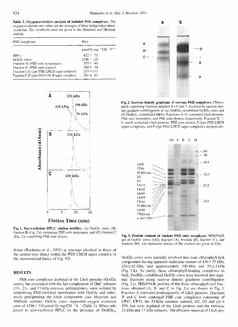
Table 1. Oxygen-evolution analysis of isolated PSII complexes. The oxygen-evolution rate values are the averages of three independent deter- minations. The conditions used are given in the Materials and Methods section.
PSII complexes Rate
pmol0,mg 'Chl ' h ' BBYs 4222 I1 OctClc cores 1246k 110 Fraction B (PSI1 core monomcrs) 3 5 5 2 60 Fraction C (PSII core dimers) 5 6 0 2 56 Fraction E (E-type PSIl-LHCII super complex) 215 i 113 Fraction F (F-type PSII-LHCII supei complex) 265 2 61
236 kDa !
A
/ .
236 kDa I
B
430 kDa I
C
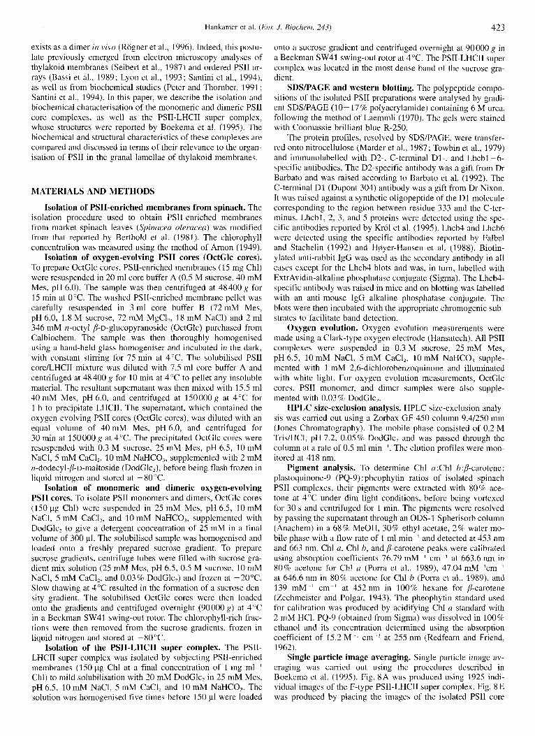
Elution Time (min) Fig. 1. Size-exclusion HPLC elution profiles. (A) OctGlc cores, (B) fraction B (Fig. 2a) containing PSI1 core monomers, and (C) fraction C (Fig. 2 a) containing PSII core dimers.
dimer (Boekema et al., 1995) at spacings identical to those of the central core diiner within the PSII-LHCII super complex i n the reconstructed lattice of' Fig. 8 D.
RESULTS
PSII core complexes depleted of the Lhcb proteins (OctGlc cores), but associated with the full complement of OEC subunits (33-, 23-, and 17-kDa extrinsic polypeptides), were isolated by soiubilising PSII-enriched membranes with OctGlc and selec- tively precipitating the Lhcb components (see Materials and Methods section). OctGlc cores supported oxygen evolution rates of 1246 -f 110 pmol O2 mg Chl ' h ~' (Table 1). When sub- jected to size-exclusion HPLC in the presence of DodGlc,,
a b
A-
B-
6-
Fig. 2. Sucrose density gradients of various PSII complexes. Chloro- phyll-containing fractions labelled A-F and *, resolved by sucrose den- sity gradient centrifugation of (a) DodGlc,-solubilised OctGlc cores and (b) DodClc2-solubilised BBYs. Fractions A-C contained Lhcb proteins, PSI1 core monomers, and PSI1 core dimers, respectively. Fraction D, *, E, and F contained Lhch proteins, PSII core dimers, E-type PSII-LHCII super complexes, and F-type PSII-LHCII super complexes, respectively.
O G A B C M
CP47
33 kDa ext. \ D2 D1
CP43 \
1
Lhcb3 23 kDa ext. Lhcb6 l7kDa ext. a cyt b-559
- 106 - 80
- 49.5
- 32.5
27.5
- 18.5
Fig. 3. Protein content of various PSI1 core complexes. SDS/PAGE gel of OctGlc cores (OG), fraction (A), fraction (B), fraction (C). and markers (M). The molecular masses of the markers are given in kDa.
OctGlc cores were partially resolved into four chlorophyll-rich components having apparent molecular masses of 430 t 37 kDa, 236 i 42 kDa, and approximately 100 kDa and 56 ? 3 kDa (Fig. 1 A). To purify these chlorophyll-binding complexes in bulk, DodGlc,-solubilised OctGlc cores were resolved into sepa- rate fractions using sucrose density gradients centrifugation (Fig. 2 a). SDSPACE profiles of the three chlorophyll-rich frac- tions obtained (A, B, and C in Fig. 2a) are shown in Fig. 3. Fraction A consisted predominantly of Lhcb proteins. Fractions B and C both contained PSII core complexes consisting of CP47, CP43, the 33-kDa extrinsic subunit, D2, D1 and cyt b 559, but were depleted of the Lhcb proteins and the extrinsic 23-kDa and 17-kDa subunits. The efficient removal of Lhcb pro-
Hankamer et al. (ELK J . Biochenz. 243) 425
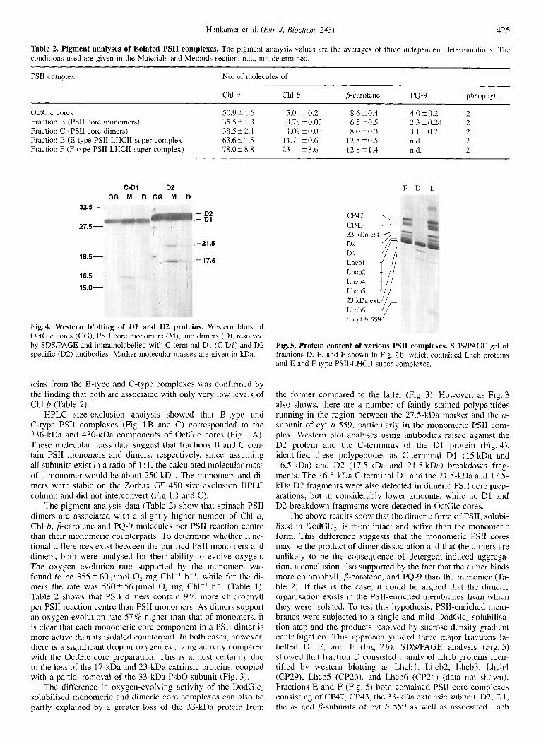
Table 2. Pigment analyses of isolated PSII complexes. The pigment analysis values are the averages of three independent determinations. The conditions used are given in the Materials and Methods section. ad., not determined.
PSII complex No. of molecules of
Chl a Chl b p-carotene PQ-9 pheophytin -~ -~ -
OctClc cores 50.9 i 1.6 5.0 20.2 8.6 2 0.4 4.0 2 0.2 2 Fraction B (PSII core monomers) 35.5 i 1.3 0.78 5 0.03 6.5 -+ 0.5 2.3 i 0.24 2 Fraction C (PSI1 core dimers) 38.5 2 2.1 1.09 i 0.03 8.0i 0.3 3.1 i 0 . 2 2 Fraction E (E-type PSII-LHCII super complex) 63.621.5 14.7 k0.6 12.5 k 0.5 n.d. 2 Fraction F (F-type PSII-LHCII super complex) 78.0 i 8.8 23 23.6 12.8 2 1.4 n.d. 2
C-DI D2 OG M D O G M D
32.5-
27.5-
- D2 - D1
-21.5
-17.5 18.5-
16.5-
15.0-
Fig.4. Western blotting of D1 and D2 proteins. Western blots of OctGlc cores (OG), PSI1 core monomers (M), and dimers (D), resolved by SDS/PAGE and immunolabelled with C-terminal D1 (C-D1) and D2 specific (D2) antibodies. Marker molecular masses are given in kDa.
teins from the B-type and C-type complexes was confirmed by the finding that both are associated with only very low levels of Chl b (Table 2).
HPLC size-exclusion analysis showed that B-type and C-type PSIl complexes (Fig. 1 B and C) corresponded to the 236-kDa and 430-kDa components of OctGlc cores (Fig. 1 A). These molecular mass data suggest that fractions B and C con- tain PSII monomers and dimers, respectively, since, assuming all subunits exist in a ratio of 1 : 1, the calculated molecular mass of a monomer would be about 250 kDa. The monomers and di- mers were stable on the Zorbax GF-450 size-exclusion HPLC column and did not interconvert (Fig.lB and C).
The pigment analysis data (Table 2) show that spinach PSII dimers are associated with a slightly higher number of Chl a, Chl b, p-carotene and PQ-9 molecules per PSII reaction centre than their monomeric counterparts. To determine whether func- tional differences exist between the purified PSII monomers and dimers, both were analysed for their ability to evolve oxygen. The oxygen evolution rate supported by the monomers was found to be 355 5 6 0 pmol 0, mg Chl-' h ~ - ' , while for the di- mers the rate was 5 6 0 2 5 6 pmol 0, mg C h l P h- ' (Table 1). Table 2 shows that PSII dimers contain 9% more chlorophyll per PSII reaction centre than PSII monomers. As dimers support an oxygen evolution rate 57% higher than that of monomers, it is clear that each monomeric core component in a PSII dimer is more active than its isolated counterpart. In both cases, however, there is a significant drop i n oxygen evolving activity compared with the OctGlc core preparation. This is almost certainly due to the loss of the 17-kDa and 23-kDa extrinsic proteins, coupled with a partial removal of the 33-kDa PsbO subunit (Fig. 3).
The difference in oxygen-evolving activity of the DodGlc, solubilised monomeric and dimeric core complexes can also be partly explained by a greater loss of the 33-kDa protein from
F D E
CP47 \ CP43 - D1
LhcbS 23 kDa ext. Lhcb6 (L cyt b-559
Fig. 5. Protein content of various PSII complexes. SDSPAGE gel of fractions D, E, and F shown in Fig. 2 b, which contained Lhcb proteins and E and F type PSIl-LHCII super complexes.
the former compared to the latter (Fig. 3). However, as Fig. 3 also shows, there are a number of faintly stained polypeptides running in the region between the 27.5-kDa marker and the a- subunit of cyt b 559, particularly in the monomeric PSII com- plex. Western blot analyses using antibodies raised against the D2 protein and the C-terminus of the D1 protein (Fig. 4), identified these polypeptides as C-terminal D1 (15 kDa and 16.5 kDa) and D2 (17.5 kDa and 21.5 kDa) breakdown frag- ments. The 16.5-kDa C-terminal D1 and the 21.5-kDa and 17.5- kDa D2 fragments were also detected in dimeric PSII core prep- arations, but in considerably lower amounts, while no D1 and D2 breakdown fragments were detected in OctGlc cores.
The above results show that the dimeric form of PSII, solubi- lised in DodGlc,, is more intact and active than the monomeric form. This difference suggests that the monomeric PSI1 cores may be the product of dimer dissociation and that the dimers are unlikely to be the consequence of detergent-induced aggrega- tion, a conclusion also supported by the fact that the dimer binds more chlorophyll, p-carotene, and PQ-9 than the monomer (Ta- ble 2). If this is the case, it could be argued that the dimeric organisation exists in the PSII-enriched membranes from which they were isolated. To test this hypothesis, PSII-enriched mem- branes were subjected to a single and mild DodGlc, solubilisa- tion step and the products resolved by sucrose density gradient centrifugation. This approach yielded three major fractions la- belled D, E, and F (Fig. 2b). SDS/PAGE analysis (Fig. 5) showed that fraction D consisted mainly of Lhcb proteins iden- tified by western blotting as Lhcbl, Lhcb2, Lhcb3, Lhcb4 (CP29), Lhcb5 (CP26), and Lhcb6 (CP24) (data not shown). Fractions E and F (Fig. 5) both contained PSII core complexes consisting of CP47, CP43, the 33-kDa extrinsic subunit, D2, D1, the n- and p-subunits of cyt b 559 as well as associated Lhcb
426 Hankamer et al. (ELK J. Biochem. 243)
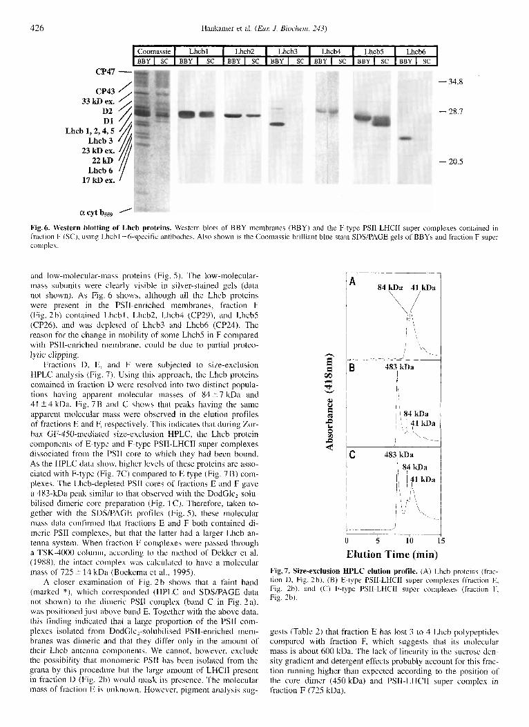
Coomassie I Lhcbl I Lhcb2 I Lhcb3 I Lhcb4 I Lhcb5 I Lhcb6 BBY I SC I BBY I SC I BBY I SC I BBY I SC I BBY I SC I BBY I SC I BBY I SC
CP47 - 33 kD ex.
22 kD Lhcb 6
17 kD ex.
- 34.8
- 28.7
- 20.5
acytb559 ' Fig. 6. Western blotting of Lhcb proteins. Western blots of BBY membranes (BBY) and the F-type PSlI-LHCII super complexes contained in fraction F (SC), using Lhcbl -6-specific antibodies. Also shown is the Coomassie brilliant blue stain SDYPAGE gels of BBYs and fraction F super complex.
and low-molecular-mass proteins (Fig. 5) . The low-molecular- mass subunits were clearly visible in silver-stained gels (data not shown). As Fig. 6 shows, although all the Lhcb proteins were present in the PSII-enriched membranes, fraction F (Fig. 2b) contained Lhcbl, Lhcb2, Lhcb4 (CP29), and LhcbS (CP26), and was depleted of Lhcb3 and Lhcb6 (CP24). The reason for the change in mobility of some Lhcb5 in F compared with PSI1-enriched meinbrane, could be due to partial proteo- lytic clipping.
Fractions D. E, and F were subjected to size-exclusion HPLC analysis (Fig. 7). Using this approach, the Lhcb proteins contained in fraction D were resolved into two distinct popula- tions having apparent molecular masses of 84 2 7 kDa and 41 24 kDa. Fig. 7 B and C shows that peaks having the same apparent molecular mass were observed in the elution profiles of fractions E and F, respectively. This indicates that during Zor- bax GF-450-mediated size-exclusion HPLC, the Lhcb protein components of E-type and F-type PSII-LHCJI super complexes dissociated from the PSII core to which they had been bound. As the HPLC data show, higher levels of these proteins are asso- ciated with F-type (Fig. 7C) compared to E type (Fig. 7 B) com- plexes. The Lhcb-depleted PSII cores of fractions E and F gave a 483-kDa peak similar to that observed with the DodClc,-solu- bilised dimeric core preparation (Fig. 1 C). Therefore, taken to- gether with the SDS/PAGE profiles (Fig. S ) , these molecular mass data confirmed that fractions E and F both contained di- meric PSII complexes, but that the latter had a larger Lhcb an- tenna system. When fraction F complexes were passed through a TSK-4000 column, according to the method of Dekker et al. (1988), the intact complex was calculated to have a molecular mass of 725 5 1 4 kDa (Boekema et al., 1995).
A closer examination of Fig. 2 b shows that a faint band (marked d'), which corresponded (HPLC and SDSPAGE data not shown) to the dimeric PSII complex (band C in Fig. 2a), was positioned just above band E. Together with the above data, this finding indicated that a large proportion of the PSII com- plexes isolated from DodGlcz-solubilised PSII-enriched mem- branes was dimeric and that they differ only in the amount of their Lhcb antenna components. We cannot, however, exclude the possibility that monomeric PSII has been isolated from the grana by this procedure but the large amount of LHCII present in fraction D (Fig. 2b) would mask its presence. The molecular mass of fraction E is unknown. However, pigment analysis sug-
483 kDa
' I I
B
/ I I \ 84 kDa ~ j y a I
5 10 5
Elution Time (min) Fig. 7. Size-exclusion HPLC elution profile. (A) Lhcb proteins (frac- tion D, Fig. 2b), (B) E-type PSII-LHCII super complexes (fraction E. Fig. 2b), and (C) F-type PSII-LHCII super complexes (fraction F, Fig. 2b).
gests (Table 2) that fraction E has lost 3 to 4 Lhcb polypeptides compared with fraction F, which suggests that its molecular mass is about 600 kDa. The lack of linearity in the sucrose den- sity gradient and detergent effects probably account for this frac- tion running higher than expected according to the position of the core dimer (450 kDa) and PSII-LHCII super complex in fraction F (725 kDa).
Hankamer et al. (Eur: J. Biocheni. 243) 4L I
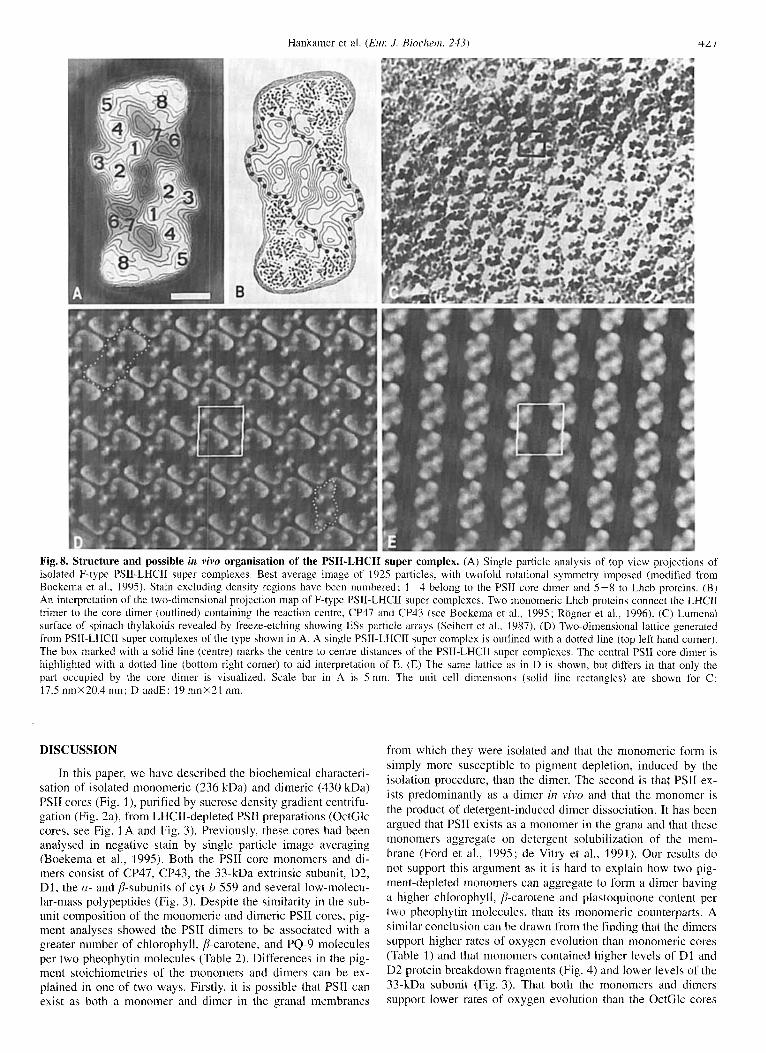
Fig. 8. Structure and possible in vivn organisation of the PSII-LHCII super complex. (A) Single particle analysis of top view projections of isolated F-type PSII-LHCII super complexes. Best average image of 1925 particles, with twofold rotational symmetry imposed (modified from Boekema et al., 1995). Stain excluding density regions have been numbered; 1-4 belong to the PSII core dimer and 5-8 to Lhcb proteins. (B) An interpretation of the two-dimensional projection map of F-type PSII-LHCII super complexes. Two monomeric Lhch proteins connect the LHCII trimer to the core dimer (outlined) containing the reaction centre, CP47 and CP43 (see Boekema et al., 1995; Rogner et al., 1996). (C) Lumenal surface of spinach thylakoids revealed by freeze-etching showing ESs particle arrays (Seibert et al., 1987). (D) Two-dimensional lattice generated from PSII-LHCIl super complexes of the type shown in A. A single PSII-LHCII super complex is outlined with a dotted line (top left hand corner). The box marked with a solid line (centre) marks the centre to centre distances of the PSII-LHCII super complexes. The central PSI1 core dimer is highlighted with a dotted line (bottom right corner) to aid interpretation of E. (E) The same lattice as in D is shown, hut differs in that only the part occupied by the core dimer is visualized. Scale bar in A is 5 nm. The unit cell dimensions (solid line rectangles) are shown for C: 17.5 nmX20.4 nm; D andE: 19 nmX21 nm.
DISCUSSION
In this paper, we have described the biochemical characteri- sation of isolated monomeric (236 kDa) and dimeric (430 kDa) PSII cores (Fig. I ) , purified by sucrose density gradient centrifu- gation (Fig. 2a), from LHCII-depleted PSII preparations (OctGlc cores, see Fig. 1 A and Fig. 3). Previously, these cores had been analysed in negative stain by single particle image averaging (Boekema et al., 1995). Both the PSII core monomers and di- mers consist of CP47, CP43, the 33-kDa extrinsic subunit, D2, D1, the a- and P-subunits of cyt b 559 and several low-molecu- lar-mass polypeptides (Fig. 3). Despite the similarity in the sub- unit composition of the monomeric and dimeric PSII cores, pig- ment analyses showed the PSII dimers to be associated with a greater number of chlorophyll, /?-carotene, and PQ-9 molecules per two pheophytin molecules (Table 2). Differences in the pig- ment stoichiometries of the monomers and dimers can be ex- plained in one of two ways. Firstly, it is possible that PSII can exist as both a monomer and dimer in the granal membranes
from which they were isolated and that the monomeric form is simply more susceptible to pigment depletion, induced by the isolation procedure, than the dimer. The second is that PSII ex- ists predominantly as a dimer in v i ~ o and that the monomer is the product of detergent-induced dimer dissociation. It has been argued that PSII exists as a monomer in the grana and that these monomers aggregate on detergent solubilization of the mem- brane (Ford et al., 1995; de Vitry et al., 1991). Our results do not support this argument as it is hard to explain how two pig- ment-depleted monomers can aggregate to form a dimer having a higher chlorophyll, p-carotene and plastoquinone content per two pheophytin molecules, than its monomeric counterparts. A similar conclusion can be drawn from the finding that the dimers support higher rates of oxygen evolution than monomeric cores (Table 1) and that monomers contained higher levels of D1 and D2 protein breakdown fragments (Fig. 4) and lower levels of the 33-kDa subunit (Fig. 3). That both the monomers and dimers support lower rates of oxygen evolution than the OctGlc cores
428 Hankamer et al. (ELK J. Biochem. 243)
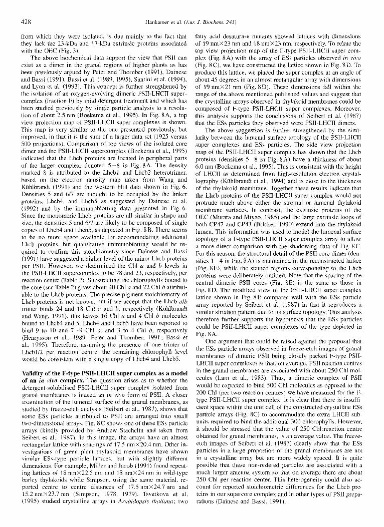
from which they were isolated, is due mainly to the fact that they lack the 23-kDa and 17-kDa extrinsic proteins associated with the OEC (Fig. 3).
The above biochemical data support the view that PSII can exist as a dimer in the granal regions of higher plants as has been previously argued by Peter and Thornber (1991), Dainese and Bassi (1991), Bassi et al. (1989, 1995), Santini et al. (1994), and Lyon et al. (1993). This concept is further strengthened by the isolation of an oxygen-evolving dimeric PSII-LHCII super- complex (fraction F) by mild detergent treatment and which has been studied previously by single particle analysis to a resolu- tion of about 2.5 nm (Boekema et al., 1995). In Fig. 8A, a top view projection map of PSII-LHCII super complexes is shown. This map is very similar to the one presented previously, but improved, in that it is the sum of a larger data set (1925 versus 500 projections). Comparison of top views of the isolated core dimer and the PSII-LHCII supercomplex (Boekema et al., 1995) indicated that the Lhcb proteins are located in peripheral parts of the larger complex, denoted 5-8 in Fig. SA. The density marked 8 is attributed to the Lhcbl and Lhcb2 heterotrimer, based on the electron density map taken from Wang and Kuhlbrandt (1991) and the western blot data shown in Fig. 6. Densities 5 and 6/7 are thought to be occupied by the linker proteins, Lhcb4, and Lhcb5 as suggested by Dainese et al. (1992) and by the immunoblotting data presented in Fig. 6. Since the monomeric Lhcb proteins are all similar in shape and size, the densities 5 and 6/7 are likely to be composed of single copies of Lhcb4 and LhcbS, as depicted in Fig. 8B. There seems to be no more space available for accommodating additional Lhcb proteins, but quantitative immunoblotting would be re- quired to confirm this stoichiometry since Dainese and Bassi (1991) have suggested a higher level of the minor Lhcb proteins per PSII. However, we determined the Chl a and b levels in the PSII-LHCII supercomplex to be 78 and 23, respectively, per reaction centre (Table 2). Substracting the chlorophylls bound to the core (see Table 2) gives about 40 Chl a and 22 Chl b attribut- able to the Lhcb proteins. The precise pigment stoichiometry of Lhcb proteins is not known, but if we accept that the Lhcb alb trimer binds 24 and 18 Chl a and b, respectively (Kuhlbrandt and Wang, 1991), this leaves 16 Chl u and 4 Chl b molecules bound to Lhcb4 and 5. Lhcb4 and LhcbS have been reported to bind 9 to 10 and 7-9 Chl a, and 3 to 4 Chl b, respectively (Henrysson et al., 2989; Peter and Thornber, 1991; Bassi et al., 1995). Therefore. assuming the presence of one trimer of Lhcbl/2 per reaction centre, the remaining chlorophyll level would be consistent with a single copy of Lhcb4 and Lhcb5.
Validity of the F-type PSII-LHCII super complex as a model of an in vivo complex. The question arises as to whether the detergent-solubilised PSII-LHCII super complex isolated from granal membranes is indeed an in vivo form of PSII. A closer examination of the lumenal surface of the granal membranes, as studied by freeze-etch analysis (Seibert et a]., 1987), shows that some ESs particles attributed to PSII are arranged into small two-dimensional arrays. Fig. 8 C shows one of these ESs particle arrays (kindly provided by Andrew Staehelin and taken from Seibert et al., 2987). In this image, the arrays have an almost rectangular lattice with spacings of 17.5 nmX20.4 nm. Other in- vestigations of green plant thylakoid membranes have shown similar ESs-type particle lattices, but with slightly different dimensions. For example, Miller and Jacob (1991) found repeat- ing lattices of 18 nmX22.5 nm and 18 nmX24 nm in wild-type barley thylakoids while Simpson, using the same material, re- ported centre to centre distances of 17.5 nmX24.7 nm and 15.2 nmX23.7 nm (Simpson, 1978, 1979). Tsvetkova et al. (1 995) studied crystalline arrays in Ambidopsis thaliarza ; two
fatty acid desaturase mutants showed lattices with dimensions of 19 nmX23 nm and 18 nmX23 nm, respectively. To relate the top view projection map of the F-type PSII-LHCII super com- plex (Fig. 8A) with the array of ESs particles observed in vivo (Fig. 8 C), we have constructed the lattice shown in Fig. 8 D. To produce this lattice, we placed the super complex at an angle of about 45 degrees in an almost rectangular array with dimensions of 19 nmX21 nm (Fig. SD). These dimensions fall within the range of the above mentioned published values and suggest that the crystalline arrays observed in thylakoid membranes could be composed of F-type PSII-LHCII super complexes. Moreover, this analysis supports the conclusions of Seibert et al. (1987) that the ESs particles they observed were PSII-LHCII dimers.
The above suggestion is further strengthened by the simi- larity between the lumenal surface topology of the PSII-LHCII super complexes and ESs particles. The side view projection map of the PSII-LHCII super complex has shown that the Lhcb proteins (densities 5-8 in Fig. 8A) have a thickness of about 6.0 nm (Boekema et al., 1995). This is consistent with the height of LHCII as determined from high-resolution electron crystal- lography (Kuhlbrandt et al., 1994) and is close to the thickness of the thylakoid membrane. Together these results indicate that the Lhcb proteins of the PSII-LHCII super complex would not protrude much above either the stromal or lumenal thylakoid membrane surfaces. In contrast, the extrinsic proteins of the OEC (Murata and Miyao, 1985) and the large extrinsic loops of both CP47 and CP43 (Bricker, 1990) extend into the thylakoid lumen. This information was used to model the lumenal surface topology of a F-type PSII-LHCII super complex array to allow a more direct comparison with the shadowing data of Fig. 8C. For this reason, the structural detail of the PSII core dimer (den- sities 1-4 in Fig. 8A) is maintained in the reconstructed lattice (Fig. SE), while the stained regions corresponding to the Lhcb proteins were deliberately omitted. Note that the spacing of the central dimeric PSII cores (Fig. 8E) is the same as those in Fig. 8D. The modified view of the PSII-LHCII super complex lattice shown in Fig. 8E compares well with the ESs particle array reported by Seibert et al. (2987) in that it reproduces a similar striation pattern due to its surface topology. This analysis therefore further supports the hypothesis that the ESs particles could be PSII-LHCII super complexes of the type depicted in Fig. 8A.
One argument that could be raised against the proposal that the ESs particle arrays observed in freeze-etch images of granal membranes of dimeric PSII being closely packed F-type PSII- LHCII super complexes is that, on average, PSII reaction centres in the granal membranes are associated with about 250 Chl mol- ecules (Lam et al., 1983). Thus, a dimeric complex of PSII would be expected to bind 500 Chl molecules as opposed to the 200 Chl (per two reaction centres) we have measured for the F- type PSII-LHCII super complex. I t is clear that there is insuffi- cient space within the unit cell of the constructed crystalline ESs particle arrays (Fig. 8C) to accommodate the extra LHCII sub- units required to bind the additional 300 chlorophylls. However, it should be stressed that the value of 250 Ch1:reaction centre obtained for granal membranes, is an average value. The freeze- etch images of Seibert et al. (1987) clearly show that the ESs particles in a large proportion of the granal membranes are not in a crystalline array but are more widely spaced. It is quite possible that these non-ordered particles are associated with a much larger antenna system so that on average there are about 250 Chl per reaction center. This heterogeneity could also ac- count for reported stoichiometric differences for the Lhcb pro- teins in our supercore complex and in other types of PSII prepa- rations (Dainese and Bassi. 1991).
Hankamer et al. (Eur J . Biochem. 243) 429
Studies with PSII-enriched membranes delipidated with Tri- ton X-l 00 often form two-dimensional arrays that have been analysed by electron crystallography to be PSII core dimers (Bassi et al., 1989; Santini et al., 1994; Lyon et al., 1993; Miller and Jacob, 1991), with a single exception, where a monomeric model was argued (Holzenberg et al., 1993). In the latter case, the unit cell was assumed to contain a full complement of Lhcb proteins. However, Lyon and colleagues (Lyon et al., 1993) showed that similar crystals contained little or no LHCII pro- teins (low Chl h level) but did have Lhcb4, 5 , and 6 (Marr et al., 1996). The detection of these linker-type proteins suggests their close association with the core in line with their position in Fig. 8 A and B. We did not, however, detect Lhcb6 in the F-type PSII-LHCII.
Regulatory implications of arrayed and non-arrayed PSII complexes. A more detailed analysis of the reconstructed crystal consisting of the F-type PSII-LHCII super complex (Fig. 8D) shows that each LHCII protein antenna set (densities 5-8 in Fig. 8A). is in close contact with the PSII core of a neighbouring PSII-LHCII super complex. I t is therefore reasonable to expect that excitation energy transfer can occur not only within the PSII super core, but between PSII-LHCII super complexes within such a crystalline array. The non-arrayed ESs particles are more widely spaced and presumably associated with larger antenna systems, more suitable for exciton trapping under low light conditions. These results are also in agreement with those of Spangfort and Anderson (1989) who suggested that Lhcbl and 2 form part of a mobile LHCII pool. The apparent heterogeneity of PSIULHCII organisation in the granal membranes may thus reflect a regulatory mechanism used by plants to adapt to changing levels of illumination. Biochemical fractionation studies could help to address how this highly dynamic system adapts to changing environmental conditions and in this context the work of Albertsson (1995) is relevant in that it emphasises the heterogeneity of PSII within the grana, particularly in terms of differing antenna sizes.
We wish to acknowledge the Biotechnology and Biological Sciences Research Council (BBSRC), The Research Institute of Innovative Tech- nology for the Earth (RITE), The Royal Society (DZ) for financial sup- port and a Biotechnology and Biological Sciences Research Council stu- dentship (JN). We are grateful to Dr Andrew Staehelin for supplying electron microscopy data to assist in the construction of Fig. 8. J. B. and B. H. also wish to thank the British Oxygen Company for their support. We would also like to extend our thanks to Dr Matthias Rogner, Dr Dirk Bald and Dr Jochen Kruip for many useful discussions and for initial size-exclusion HPLC measurements of our samples.
REFERENCES Albertsson, P.-A. (1995) Photosynth. Res. 46, 141 -149. Arnon, D. I. (1949) Plant Physiol. (Bethesdu) 24, 1-13. Barbato, R., Friso, G., Polverino De Laureto, P., Frizzo, A,, Rigoni, F. &
Giacometti, G. M. (1992) FEBS Lett. 311, 33-36. Barber, J., Chapman, D. J. & Telfer, A. (1987) FEBS Lett. 220, 67-73. Bassi, R., Ghiretti-Magaldhi, A., Tognon, G., Giacometti, G. M. &
Bassi, R., Marquardt, J. & Lavergne, J. (1995) Eur: J. Biochern. 233, Miller, K. R. (1989) Eur: J. Cell B i d . 50, 84-93.
709 -719.
Berthold, D. A., Babcock, G. T. & Yocutn, C. F. (1981) FEBS Lett. 134,
Boekema, E. J., Hankamer, B., Bald, D., Kruip, J., Nield, J.. Boonstra, A. F., Barber, J . & Rogner, M. (1995) Proc. Nut1 Acud. Sci. USA 92,
231 -234.
175- 179. Bricker, T. M. (1990) Photosynth. Res. 24, 1-13. Dainese, P. & Bassi, R. (1991) J . B id . Chenz. 266, 8136-8142. Dainese, P., Santini, C., Ghiretti-Magaldi, A,, Marquardt, J., Tidu, V.,
Mauro, S., Bergantino, E. & Bassi, R. (1992) in Research in photo- synthesis (Murata, N., ed.) vol. 2, pp. 13-20, Kluwer Academic Publishers, Dordrecht, The Netherlands.
Dekker, J. P., Boekema, E. J., Witt, H. T. & Rogner, M. (1988) Biochim. Biophys Actu 936, 307-318.
De Vitry, C., Diner, B. A. & Popot, J.-L. (1991) ,I. B i d . Chem. 266,
Falhel, T. G. & Staehelin, L. A. (1992) Pl~oto.~ynth. Res. 34, 249-262. Ford, R. C., Rosenberg, M. F., Shepherd, F. H., McPhie, P. & Holzen-
Henrysson, T., Schroder, W. P., Spangfort, M. & Akerlund, H.-E. (1989)
Holzenburg, A.. Bewley, M. C., Wilson, F. H., Nicholson, W. V. & Ford,
H@yer-Hansen, G., Bassi, R., H@nberg, L. S. & Simpson, D. J. (1988)
Ikeuchi, M., Yuasa, M. & Inoue, Y. (1985) FEBS Lett. 185, 316-322. Jansson, S. (1994) Biochim. Biophys. Actu 1/84, 1-19. Krol, M., Spangfort, M. D., Huner, N. P. A., Oquist, G., Gustafsson,
P. & Jansson, S. (1995) Plunt Physiol. (Bethesda) 107, 873-883. Kuhlbrandt, W. & Wang, D. N. (1991) Nature 350, 130-134. Kuhlbrandt, W., Wang, D. N. & Fujiyoshi, Y. (1994) Nuture 367, 614-
Laemmli, U. K. (1970) Nuture 277, 680-685. Lam, E., Baltimore, B., Ortiz, W., Chollar, S., Melis, A. & Malkin, R.
Lyon, M. K., Marr, K. M. & Furcinitti, P. S. (1993) J . Struct. B i d . 110,
Marder, J. B., Chapman, D. J., Telfer, A.. Nixon, P. J. & Barber, J. (1987)
Man, K. M., Mastronarde, D. N. & Lyon, M. K. (1996) J . Cell B i d .
Miller, K. R. & Cushman, R. A. (1978) Biochim. Biriphys. Acta 546,
Miller, K. R. & Jacob, J. S. (1991) in Proceedings 49th Annu. Meet. Electron Microscopy Soc. Am. (Bailey, G. W., ed.) p. 197, San Fran- cisco Press, Box 6800. San Francisco, CA 94101-6800, USA.
Murata, N. & Miyao, M. (1985) Trends Biochern. Sci. 10, 122-124. Nanba, 0. & Satoh, K. (1987) Proc. Nut1 Acad. Sci. USA 84, 109-112. Peter, G. F. & Thornber, J. P. (1991) Plant Cell Physiol. 32, 1237-1250. Porra, R. J. , Thompson, W. A. & Kriedemann, P. E. (1989) Biochim.
Redfeam, E. R. & Friend, J . (1962) Phytochemisrry (Ox$) 1, 147-151. RBgner, M., Boekema, E. J. & Barber, J. (1996) Trends Biochem. Sci.
Santini. C., Tidu, V., Tognon, G., Ghiretti Magaldi, A. & Bassi, R. (1994)
Seibert, M., DeWit, M. & Staehelin, L. A. (1987) J. Cell B i d . 105,
Simpson, D. J. (1978) Carlsherg Rex Cornmiin. 43, 365-389. Simpson, D. J. (1979) Carlsberg Rex Cornmun. 44, 305-336. Spangfort, M. & Anderson, B. (2989) Biochim. Biophys. Actu 977,
163- 170. Towbin, H., Staehelin, T. & Gordon, J . (1979) Proc. Nut1 Accirt. Sci. USA
76, 4350-4354. Tsvetkova, N. M., Apostolova, E. L., Brain, A. P. R., Willians, W. P. &
Quinn, P. J. (1995) Diochim. Biophys. Acta 1228, 201-210. Wang, D. N. & Kuhlbrandt, W. (1991) J. Mid. Bid . 217, 692-699. Zechmeister, L. & Polgir, A. (1943) J . Am. Chem. Soc. 65, 1522-1528.
16 614- 16 621.
burg, A. (1995) Micron 26, 133-140.
Biochim, Biophys. Actu 977, 301 -308.
R. C. (1993) Nuture 363, 470-472.
Planta (Heidelh.) 173, 13-21.
621.
(1983) Biochiin. Biophys. Artu 724, 201-211.
133 - 140.
Plant Mol. B i d . 9, 325-333.
132, 823-833.
481 -499.
Biophys. Acta 975, 384- 394.
21,44-49.
ELIK J. Biochenz. 221, 307-315.
2257-2265.