Embed Size (px)
Citation preview
Vol. 57, No. 11
Isolation, Purification, and Characterization of Fragment B, theNH2-Terminal Half of the Heavy Chain of Tetanus ToxinMORIHIRO MATSUDA,* DIAN-LIANG LEI,t NAKABA SUGIMOTO, KUNIHIRO OZUTSUMI,
AND TOSHIO OKABEDepartment of Tuberculosis Research I, Research Institute for Microbial Diseases, Osaka University,
3-1 Yamadaoka, Suita, Osaka 565, JapanReceived 22 May 1989/Accepted 3 August 1989
Fragment B, the N-terminal half of the heavy chain, an important domain of the tetanus neurotoxinmolecule, was isolated for the first time. Tetanus toxin (composed of three domains, A, B, and C) was preparedfrom culture filtrates. Fragment A-B, derived from the toxin treated mildly with papain, was used for theisolation of fragment B. Fragment A-B obtained was dissociated into fragments A and B by reduction with 100mM dithiothreitol and treatment with 2 M urea. Fragment B was separated from fragment A by ion-exchangecolumn chromatography on a Mono Q column equilibrated with 20 mM Tris hydrochloride buffer (pH 7.6),containing 1 mM dithiothreitol and 2 M urea, in a fast-protein liquid chromatography system by elution witha linear gradient of 0 to 0.5M NaCl. Fragment B was obtained in two forms having molecular weights of 48,000+ 2,000, which were indistinguishable by sodium dodecyl sulfate-gel electrophoresis or antigenic specfficity, butdistinguishable on polyacrylamide gel electrophoresis without sodium dodecyl sulfate and on isoelectric focusing(pl 6.7 and 7.3). The recovery of fragment B was 50 to 72% of that of fragment A-B on a molar basis. Purifiedfragment B was not toxic to mice on intravenous or intramuscular injection at doses of up to 100 ,ug, but wasfound to form channels (ca. 2.3 pS) in a lipid bilayer membrane by a patch clamp technique. The role of domainB of the tetanus toxin molecule in the mechanism of action of the toxin is discussed.
Tetanus toxin is one of the most poisonous substancesknown (39). The toxin, when injected intramuscularly at lowdoses, is taken up at the motor endplates and carried byretrograde axonal transport to the central nervous system,where it first blocks inhibitory synapses and then excitatorysynapses (36, 37), producing delayed spastic paralysis typi-cal of tetanus, and finally contracture. At enormous doses,the toxin blocks peripheral neuromuscular transmission,producing the acute botulinumlike flaccid paralysis (22, 23,34) observed in particular experimental conditions and insome cases of severe clinical tetanus. Recent findings haveindicated that the common underlying mechanism of thesesymptoms is inhibition of release of inhibitory and excitatoryneurotransmitters by the toxin at presynaptic sites of thenerve terminals (12). However, the exact structure-functionrelationship in the tetanus toxin molecule is unknown.The toxin (molecular weight [mol wt], ca. 150,000) is
composed of three domains (20, 21, 26), A, B, and C,according to a new nomenclature (8th International Confer-ence on Tetanus, Leningrad, 1987), each with a mol wt of ca.50,000 (Fig. 1). Two complementary nontoxic fragments, thelight chain, fragment A (mol wt, 52,000), and the heavychain, fragment B. C (mol wt, 98,000) (Fig. 1), were isolatedfrom extracellular, nicked toxin in sufficiently native formsto be reconstituted into a whole toxin (24, 25). Another set oftwo fragments, fragment A-B and fragment C (mol wt,52,000) (Fig. 1), were obtained by dissociating the toxin bymild treatment with papain (2, 14, 26). Fragment C isnontoxic and is known to bind gangliosides, putative tetanustoxin binding substances at motor endplates of animals, andsynaptic membranes and also to function in carrying thetoxin from the peripheral sites by retrograde axonal trans-
* Corresponding author.t Present address: National Institute for the Control of Pharma-
ceutical and Biological Products, Temple of Heaven, Beijing, China.
port to the central nervous system (2-4, 15). In contrast tothese isolated, nontoxic fragments (fragments A, B * C, andC), fragment A-B, which constitutes the NH2-terminal two-thirds of the toxin molecule, showed nonspastic toxicity (6,11, 13; K. Ozutsumi, D.-L. Lei, N. Sugimoto, and M.Matsuda, Toxicon, in press), inducing "weakness" (Ozut-sumi et al., in press). Furthermore, fragment A-B has beenreported to inhibit secretion or release of catecholaminewhen it was introduced into chromaffin cells (1, 30) and toblock synaptic transmissions when it was injected intraspi-nally into cats (38). Therefore, fragment A-B appears to playan important role for the toxin in eliciting toxicity at directtarget sites of sensitive cells.As for the roles of the domains of fragment A-B in the
action of tetanus toxin, domain B of the toxin molecules wasrecently concluded, from indirect studies on the neutralizingeffect of Fab fragments of polyclonal or monoclonal antibod-ies directed to the region of domain B in the tetanus toxinmolecule, to be critical for expression of the toxin action (5).For direct studies on the function of fragment B, its isolationis essential, but it has not previously been isolated (5). Thispaper reports the isolation of fragment B of the tetanus toxinmolecule from fragment A-B and its purification and char-acterization. The role of domain B of the toxin in themechanism of action of tetanus toxin is also discussed.
MATERIALS AND METHODS
Bacterial strain and preparation of tetanus toxin. A Bikensubstrain of the Harvard A47 strain of Clostridium tetaniwas used for toxin production. Extracellular toxin wasprepared from culture filtrates after incubation of the organ-isms in modified Latham medium at 35°C for 5 days asdescribed previously (24). The toxin was isolated and puri-fied by gel permeation chromatography as described previ-ously (24, 28).Fragment A-B. The purified tetanus toxin was subjected to
3588
INFECTION AND IMMUNITY, Nov. 1989, p. 3588-35930019-9567/89/113588-06$02.00/0Copyright C 1989, American Society for Microbiology
PURIFICATION OF FRAGMENT B OF TETANUS TOXIN
PA
I[lBI C COOHH2N-
Light chain
[A)IA-BJ
a [A)-- i
IBI--
5'i
Heavy chain
1BClIC)
lee
FIG. 1. Two-chain model of tetanus toxin composed of threedomains, A, B, and C (tripartite model), according to a new
nomenclature, and the fragments of the toxin. Tetanus toxin pre-
pared from bacterial cells is a single polypeptide chain (mol wt, ca.150,000). The toxin prepared from the culture filtrate (extracellulartoxin) is a nicked form, two-chain molecule consisting of a lightchain (fragment A; mol wt, ca. 52,000) and a heavy chain (fragmentB C; mol wt, ca. 98,000) linked by a disulfide bridge and nonco-
valent bonds. Mild treatment of the toxin with papain cleaved thetoxin at the site indicated by an arrow (PA) into two components,fragment A-B (mol wt, ca. 99,000) and fragment C (mol wt, ca.
52,000).
mild treatment with papain to dissociate it into fragmentsA-B and C as described previously (26, 29; Ozutsumi et al.,in press). Fragment A-B was then separated and purifiedfrom the digest by gel permeation chromatography on a TSKG3000SW column (Toyo Soda Co., Tokyo, Japan) equili-brated with 0.1 M NaK phosphate buffer (pH 6.8) in ahigh-performance liquid chromatography system (ToyoSoda Co.) as described previously (29; Ozutsumi et al., inpress).
Thiol reduction and urea treatment of fragment A-B. Frag-ment A-B (0.5 to 1.2 mg of protein per ml in 0.1 M NaKphosphate buffer [pH 6.8]) was reduced with dithiothreitol(DTT; final concentration, 100 mM) at 25°C for 60 min. Thereduced fragment A-B was treated with solid urea at variousconcentrations (1 to 8 M) for urea-polyacrylamide gel elec-trophoresis or at a final concentration of 2 M for ion-exchange chromatography.
Polyacrylamide gel electrophoresis. Conventional poly-acrylamide gel electrophoresis in 5% gel (column) or in 7%gel (slab) and urea-polyacrylamide gel electrophoresis in 5%gel (column) were carried out in 10 mM Tris-77 mM glycinebuffer (pH 8.6) as the electrode buffer by the methodsoriginally described by Davis (9) and Jovin et al. (18),respectively. Sodium dodecyl sulfate (SDS)-polyacrylamidegel electrophoresis in 8 to 25% gradient gel and isoelectricfocusing were carried out in a Phast System (PharmaciaLKB, Uppsala, Sweden).
Separation of fragment B from reduced, urea-treated frag-ment A-B. The reduced, urea-treated fragment A-B (0.5 to1.2 mg of protein per ml; 2.2 ml) was applied to a prepackedSephadex G-25 column (PD-10; Pharmacia LKB) equili-brated with 20 mM Tris hydrochloride buffer (pH 7.6)containing 1 mM DTT and 2 M urea (buffer A), eluted with3.5 ml of buffer A, and filtered through a membrane filter(Acrodisc; Gelman Sciences, Inc., Ann Arbor, Mich.) with apore size of 0.2 ,um (final volume, ca. 4 ml). Ion-exchangechromatography was carried out in a fast-protein liquidchromatography system (Pharmacia LKB) composed of two
model P-500 high-precision pumps, a gradient programmer(model GP-250), and a single-path UV monitor, model UV-1(Pharmacia LKB). The filtered sample (ca. 4 ml, 1.1 to 4 mgof protein) was applied on a prepacked column of Mono QHR5/5 (Pharmacia LKB) equilibrated with buffer A. Materialwas eluted with a linear gradient of NaCl (0.025 M NaClincrease per min) formed using buffers A and B (buffer B isbuffer A supplemented with 0.5 M NaCl) at a flow rate of 1ml/min. The protein content of the eluate was monitored at280 nm, and each peak was collected.
Immunodiffusion test. The double-diffusion precipitationmethod originally described by Ouchterlony (27) was em-ployed using 1% agarose containing 50 mM Tris-0.6 Mglycine buffer (pH 8.5) and 1 mM EDTA.
Toxicity test. Toxicity was examined in OF1 mice of bothsexes, weighing 20 to 25 g, by intravenous injection (0.1 ml)or intramuscular injection (0.1 to 0.5 ml) of samples.
Protein determination. Protein was measured by themethod of Lowry et al. (19).Fragment A and fragment B - C. Complementary frag-
ments of tetanus toxin, fragment A and fragment B - C, wereisolated and purified from the reduced, urea-treated extra-cellular toxin by gel permeation chromatography on anUltrogel AcA (Pharmacia LKB) column as described previ-ously (24).
Tetanus antitoxin serum. Horse antitoxin serum (lot no.0044) was a gift from the National Institute of Health,Tokyo, Japan.
Chemicals. DTT was obtained from Nakarai Tesque Co.,Kyoto, Japan; urea (for biochemistry) was obtained from E.Merck, Darmstadt, Federal Republic of Germany; andasolectin (soybean lecithin) was obtained from Daigo EiyoKagaku Co., Osaka.
Detection of channel formation in lipid biolayers by a patchclamp technique. Asolectin was suspended at a concentra-tion of 9 mg/ml in 10 mM potassium acetate buffer (pH 4.0)containing 150 mM KCl by sonication under continuousbubbling with N2 gas. Two hundred microliters of thesuspension was mixed with an equal volume of solution oftetanus toxin (100 ,ug/ml), fragment A-B (100 p.g/ml) orfragment B (50 ,g/ml) in the same buffer and sonicated at37°C for 2 min. Test mixtures were prepared by adding 1001Al of the resulting sonically treated material extract to 0.4 mlof the same buffer. The formation of lipid monolayers on theaqueous surface of the test mixture was confirmed byobserving drops of water running over the surface. Patchcapillary pipettes were filled with 10 mM HEPES (N-2-hy-droxyethylpiperazine-N'-2-ethanesulfonic acid)-KOH (pH7.0) containing 150 mM KCl. Patch clamping was performedas described previously (35) on a lipid bilayer formed at thetip of the patch pipettes (electrical resistance, 7 to 30 MfQ) bythe method of Suarez-Isla et al. (33), namely, dipping the tipinto the test mixture twice and then clamping the voltage ofthe inside of the patch pipette against the bath electrode andamplifying the current passed through the lipid bilayer at thetip of the patch pipette.
Determination of NH2-terminal sequence. The NH2-ter-minal sequence of the fragments of the toxin was analyzedon electroblotted proteins by the microsequence method ofHirano (16).
RESULTS
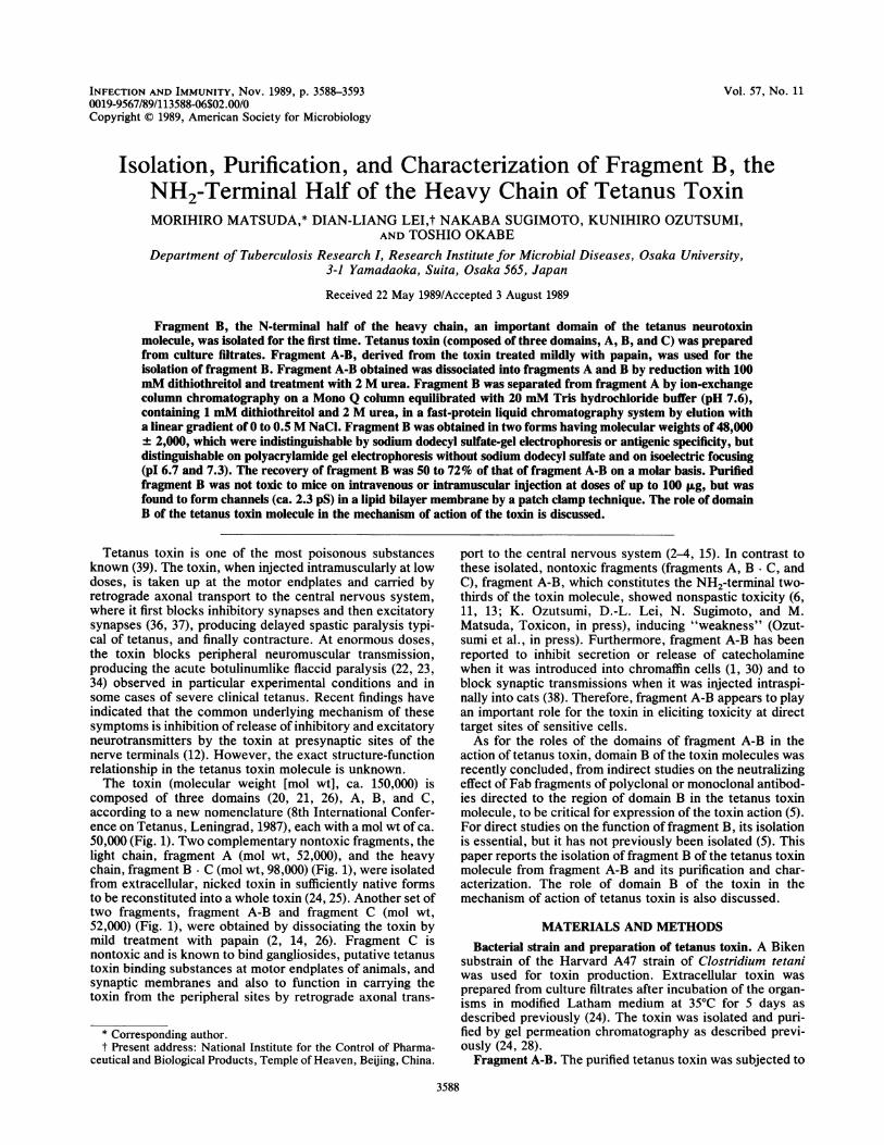
Dissociation of fragment A-B by reduction with DTT andtreatment with urea. To determine the optimal conditions fordissociation of fragment A-B, we first treated fragment A-B
r . F -.~~-1i
VOL. 57, 1989 3589
3590 MATSUDA ET AL.
<<..
...
-'S
a b c dFIG. 2. Conventional (lane a) and urea-polyacrylamide gel (lanes
b through d) electrophoresis. Lane a, Untreated fragment A-B; laneb, 2 M urea-treated fragment A-B in 2 M urea gel; lane c, reducedand 4 M urea-treated fragment A-B in 2 M urea gel; lane d, reducedand 2 M urea-treated fragment A-B in 2 M urea gel. Approximately15 ,ug of protein was applied to each gel (5% gel). Electrophoresiswas carried out at a constant current of 2 mA per gel at 4°C for 120min. Migration was from top to bottom.
(0.5 mg/ml in NaK phosphate buffer [pH 6.8]) with 100 mMDTT at 25°C for 60 min and then treated it with variousconcentrations of urea (0 to 8 M) and subjected the prepa-ration to electrophoresis in the presence of various concen-trations (0 to 8 M) of urea. Treatment of the reducede..... A 1D 1.L.o -A..J...iragmetin thecompoare linlcan be
Sepaurea-trraphy i
into itsciationtreatmchrom.mMDthe redent offractioiat 0.10peaks 1
Ec
00
c,J
C
0
4
FIG.
DTT-reBuffers
indicateand B.
protein
FIG. 4. Immunodiffusion patterns of fragment A-B, fractions I,II, and III, fragments A, B C, and C, and toxin against horseantitoxin. Panel A: [A], fragment A (0.5 mg/ml); [A-B], fragmentA-B (0.5 mg/ml); I, fraction 1(0.5 mg/ml); II, fraction 11 (0.5 mg/ml);III, fraction III (0.5 mg/ml); and AT, antitoxin (1,000 U/ml). Panel B:[A], fragment A (0.5 mg/ml); [A-B], fragment A-B (0.5 mg/ml);[B C], fragment B C (0.5 mg/ml); [C], fragment C (0.5 mg/ml); andTOX, toxin (1 mg/ml).
,nt A-Di witn L mV ure;a ana sutseq4uent eitectropnore;osis* *Antigenic specificities of the purified components derivedpresence of 2 M urea gave the best separation of from reduced, urea-treated fragment A-B. Figure 4 shows the~nents (Fig. 2). Thus the components in fragment A-B antigenic relationship of fragment A-B and the componentsked by a disulfide bridge and noncovalent bonds that (fractions I, II, and III) derived from the dissociated frag-dissociated with 2 M urea. ment A-B. The antigenic specificities of fractions I and IIuration of the components derived from the reduced and were identical, while that of fraction III was different from.eated fragment A-B by fast-protein liquid chromatog- these two fractions but identical to that of fragment A (Fig.on a Mono Q column. Fragment A-B was dissociated 4A). These components all showed partial antigenic identitycomponents under the optimal conditions for disso- with fragment A-B (Fig. 4A). Figure 4B shows the results ofdescribed above (reduction with 100 mM DTT and antigenic analyses of fraction I, using purified fragments A,
ent with 2 M urea) and then subjected to ion-exchange B C, and C: fraction I showed partial antigenic identitiesatography on a Mono Q column in the presence of 1 with fragment A-B and fragment B
- C, but differed inTT and 2 M urea. Figure 3 shows the elution profile of * * * fLuce,uea-reaedragent -B itha lnea grdi- antigenic specificity from fragment A and fragment C. Theluced_urea-treatedfrgment A-B withlineargradi same pattern of antigenic specificity as that of fraction I (Fig.0 to 0.5 M NaCl. Three sharp peaks (designated as 4B) was observed with fraction II. Therefore we concluded
ns I, II, and III in order of their elution) were obtained that fractions I and II had the same antigenic specificity and), 0.13, and 0.19 M NaCl (Fig. 3). The ratio of these that they were antigenically identical to domain B of tetanuswas approximately 1:1:2 on a protein basis. toxin, while fraction III had the same antigenicity as domain
A of the toxin.Properties of the purified components. Figure 5 shows the
0.5 electrophoretic patterns of the toxin, fragment A-B, andfractions I, II, and III in polyacrylamide gel in the presence(Fig. 5A) and absence (Fig. SB) of SDS. Fractions I and IIgave single protein bands with the same mol wt of 48,000 ±
X 2,000 on SDS-gel electrophoresis (Fig. 5A), but single bandswith different electrophoretic mobilities in gel without SDS
l (Fig. 5B). On isoelectric focusing, fractions I and II showedpI values of 7.3 and 6.7, respectively. The mol wt of fractionIII isolated from fragment A-B was 54,000 + 2,000 onSDS-gel electrophoresis. From their antigenic specificities
0o and mol wts and the difference in their electrophoreticmobilities, we concluded that fractions I and II corre-
1020 30 4sponded to domain B of fragment A-B with microheteroge-
Elution time (min) neity in molecular structure. Therefore, we named fractions
3. Elution profile (fast-protein liquid chromatography) of I and II fragments B1 and B2, respectively. The recovery ofduced and urea-treated fragment A-B on a Mono Q column. fragment B (fragments B1 plus B2) was 49.5 to 72% of that of
as described in Materials and Methods. The broken line fragment A-B on a protein basis. Preliminary analysis of thees the gradient from 0 to 0.5 M NaCl, obtained with buffers A NH2-terminal sequence of fragment B2 gave a single aminoFlow rate, 1 ml/min. Sample: Volume, 4 ml; amount, 4 mg of terminus, aspartic acid, confirming that the preparation was
highly purified. The NH2-terminal sequence of fragment B2
INFECT. IMMUN.
PURIFICATION OF FRAGMENT B OF TETANUS TOXIN
AkDa
94 _67 -_ -43 --30 -
20.1--;1 4.4
-9.4
t4
Ur T' A B, B2AB'AB C TAB A B, B2FIG. 5. Polyacrylamide gel electrophoresis of toxin, fraction I,
fraction II, and fraction III in the presence (A) and, absence (B) ofSDS. Panel A, SDS-polyacrylamide 8 to 25% gradient gel: Mr,protein markers for mol wt; T', reduced toxin; A, fraction III; B1,fraction I; B2, fraction II; AB', reduced fragment A-B; AB, un-treated fragment A-B; C, fragment C. Samples of approximately 10,ug of protein for lane Mr, 2 ,ug of protein each for lanes T', AB', andAB, and 1 ,ug of protein each for lanes A, B1, B2, and C wereapplied. Electrophoresis was carried out in a Phast System at aconstant current of 10 mA at room temperature for 1 h. Panel B, 7%polyacrylamide gel without SDS: T, untreated toxin; AB, untreatedfragment A-B; A, fraction III; B1, fraction I; B2, fraction II. Samplesof approximately 5 F±g of protein for lane T, 2 ,ug of protein for laneAB, and 1 ,g of protein each for lanes A, B1, and B2 were applied.Electrophoresis was carried out at a constant current of 10 mA for3 h. Migration was from top to bottom. Gels were stained withCoomassie brilliant blue R.
was D-L-G-G-E-L-X(C)-I-K-I-K-N-E-D-L, indicating thefragment begins with Asp-461 in the tetanus toxin molecule.
Toxicity of the purified preparations of fragment B. Wheninjected into mice intravenously or intramuscularly at dosesof up to 100 ,ug of protein, fragments B1 and B2 did notproduce symptoms of spastic paralysis typical of tetanus,botulinumlike flaccid paralysis (23, 34), which is observableon injection of an enormous dose of tetanus toxin, orweakness (13, 29; Ozutsumi et al., in press), as observed oninjection of fragment A-B.Fragment B forms channel in a lipid bilayer. Using the
patch clamp technique, we examined the channel activitiesof asolectin bilayer membranes with incorporated fragmentB by recording their membrane currents. Definite channelactivities were observed in 19 patches out of 146 trials in thelipid membranes with incorporated fragment B in the pres-ence of a pH gradient (pH 7.0 inside the patch capillarypipette and pH 4.0 outside), as in the case of lipid bilayerswith incorporated toxin or fragment A-B.
Figure 6 shows an example of the current in a membranewith incorporated fragment B1. The level of the membranecurrent shifted abruptly, indicating gating of the ion-perme-able channels in the lipid bilayer patch. The channel activi-ties in the membranes with incorporated fragment B (frag-ment B1 or B2) showed single conductance levels of 2.3 + 0.2PS.
DISCUSSION
In the present study we isolated fragment B for the firsttime. Dissociation of whole toxin into light chain (fragmentA) and heavy chain (fragment B * C) requires treatment with4 M urea and reduction with 100 mM DTT (24). In contrast,we found that a lower concentration of urea (2 M) gave thebest separation of fragment B from fragment A (Fig. 2).Fragments B and A could then be separated from each otheron an ion-exchange column because of their significantdifference in pls. Recently it was found that fragment A-B,
FIG. 6. Record of the membrane current obtained from anasolectin bilayer patch with incorporated fragment B preparation(fragment B1) of tetanus toxin. A patch pipette with an electricalresistance of 15 MQi was used. The asolectin bilayer patch formed atthe tip of the pipette showed a resistance of 12.5 GQi. The recordingwas made at a holding potential of 100 mV. The upper trace was arecord with a high-cut filter of 1 kHz on paper. The lower trace is aphotograph taken directly from the cathode ray oscilloscope. Cali-bration bars indicate 1.5 pA (ordinate) and 3 s (abscissa) in the uppertrace and 0.2 pA (ordinate) and 150 ms (abscissa) in the lower trace.
from which fragment B was isolated, is toxic (11, 13, 29;Ozutsumi et al., in press) and blocks synaptic transmissions(38). We confirmed a peculiar, delayed nonspastic toxicity offragment A-B in mice, which we termed weakness (Ozut-sumi et al., in press), and found further that fragment A-Bblocked both inhibitory and excitatory synapses simulta-neously in the central nervous system when injected directlyintraspinally into cats (38). Furthermore, fragment A-B wasreported to inhibit exocytosis in adrenal chromaffin cellsunder particular conditions (1, 30). Therefore, fragment A-Bappears to play an important role in the action of tetanustoxin.To define in more detail the region of the toxin molecular
responsible for its toxic effects, recently Bizzini et al. (5)examined the effects of masking various domains of the toxinwith Fab fragments of antibodies and concluded that the areacorresponding to fragment B is critical for expression of thetoxicity of tetanus toxin. However, they could not examinethe toxic effect of isolated fragment B as they were unable toisolate it (5). In this study we showed that the isolatedfragment B did not show any toxicity in mice even atenormous doses of up to 100 pLg. This is contrary to theconclusion of Bizzini et al. (5) that the toxic effect isassociated only with fragment B. Cooperation of fragment Bwith fragment A is probably required to elicit toxic effects invivo.Tetanus toxin forms transmembrane ion channels in lipid
bilayers (7, 8, 10, 17). Fragment B - C (17) and fragment A-B(10, 17, 32) of the toxin form ion channels with similarconductances. Since neither fragment A (17) nor fragment C(10) increases the ionic conductance of planar bilayers, it hasbeen suggested that the channel-forming domain of thetetanus toxin molecule must be located in the domaincorresponding to fragment B (10, 17). The present studygave, for the first time, direct evidence that fragment Bforms ion channels in a lipid bilayer. Gamble and Montal (10)attempted to observe the channel-forming activity of domainB by adding reduced fragment A-B to lipid bilayer. How-ever, under their conditions, fragment A-B had been reoxi-dized, or, even though it was reduced, fragments A and Bwere probably still united by noncovalent bonds as shown
VOL. 57, 1989 3591
i
3592 MATSUDA ET AL.
above (Fig. 2). Therefore it is possible that they did notobserve the activity of domain B. In the present study, theconcentration of fragment B tested for channel activityappeared high. This may be partly due to the fact that in our
assay system using patch pipettes, the effective areas are
much smaller than those in black membranes. The charac-teristics, including dose dependency and influence of lipidcomposition (10), of the channel formed by fragment B are
now being studied in our laboratory in comparison withthose of fragment A-B and tetanus toxin in this assay
system. The detailed results of the comparative study will bepublished elsewhere.The toxic action of tetanus toxin is thought to involve at
least the following steps: initial binding, uptake and retro-grade axonal transport to the target cell membranes, bindingto target cells, internalization into target cells, and finallyinteraction with target molecules resulting in inhibition of theprocess(es) of neurotransmitter release (20, 31). The chan-nel-forming property of fragment B demonstrated in thisstudy may be relevant to the mechanism by which an activetoxin fragment, probably the fragment A portion of thetoxin, is internalized into the cytoplasm of target cells. Themolecular (or enzymatic) mechanism by which the activefragment inhibits transmitter release remains to be investi-gated.
In the present study, fragment B was isolated in two forms(B1 and B2) which were not distinguishable by mol wt on
SDS-gel electrophoresis but which were distinguishable bytheir electric charges. This may explain the microheteroge-neity in structure of tetanus toxin molecules. To obtainfurther information on this difference in structure, we are
now determining the NH2- and COOH-terminal sequences offragments B1 and B2.With the isolation of fragment B in this study and of other
fragments in previously studies (2-4, 14, 20, 24, 26), it is nowpossible to investigate complete sets of delineated fragmentsof ca. 50 and ca. 100 kilodaltons of any combinations of thethree domains of the tetanus toxin molecule for investigatingits structure-function relationship.
ACKNOWLEDGMENT
We thank Hisashi Hirano, National Institute of AgrobiologicalResources, Tsukuba, Japan, for help in determining the NH2-terminal sequence of the fragment.
LITERATURE CITED
1. Bittner, M. A., and R. W. Holz. 1988. Effects of tetanus toxin oncatecholamine release from intact and digitonin-permeabilizedchromaffin cells. J. Neurochem. 51:451-456.
2. Bizzini, B., P. Grob, and K. Akert. 1981. Papain-derived frag-ment Ilc of tetanus toxin: its binding to isolated synapticmembranes and retrograde axonal transport. Brain Res. 210:291-299.
3. Bizzini, B., P. Grob, M. A. Glicksman, and K. Akert. 1980. Useof the B-IIb tetanus toxin-derived fragment as a specific neu-
ropharmacological transport agent. Brain Res. 193:221-227.4. Bizzini, B., K. Stoeckel, and M. Schwab. 1977. An antigenic
polypeptide fragment isolated from tetanus toxin: chemicalcharacterization, binding to gangliosides and retrograde axonaltransport in various neuron systems. J. Neurchem. 28:529-542.
5. Bizzini, B., P. Toth, and A. A. Fedinec. 1988. Defining a regionon tetanus toxin responsible for neuromuscular blockade. Tox-icon 26:309-318.
6. Bizzini, B., A. Turpin, and M. Raynaud. 1973. Immunochemis-try of tetanus toxin: the nitration of tyrosyl residues in tetanustoxin. Eur. J. Biochem. 39:171-181.
7. Boquet, P., and E. Duflot. 1982. Tetanus toxin fragment formschannels in lipid vesicles at low pH. Proc. Natl. Acad. Sci. USA79:7614-7618.
8. Borochov-Neori, H., E. Yavin, and M. Montal. 1984. Tetanustoxin forms channels in planar lipid bilayers containing gangli-osides. Biophys. J. 45:83-85.
9. Davis, B. J. 1964. Disc electrophoresis. II. Method and applica-tion to human serum proteins. Ann. N.Y. Acad. Sci. 121:404 427.
10. Gamble, F., and M. Montal. 1988. Characterization of thechannel properties of tetanus toxin in planar lipid bilayers.Biophys. J. 53:771-783.
11. Gawade, S., C. Bon, and B. Bizzini. 1985. The use of antibodyFab fragments specifically directed to two different complemen-tary parts of the tetanus toxin molecule for studying the mode ofaction of the toxin. Brain Res. 334:139-146.
12. Habermann, E., and F. Dreyer. 1986. Clostridial neurotoxins:handling and action at the cellular and molecular level. Curr.Top. Microbiol. Immunol. 129:93-179.
13. Helting, T. B., H. J. Ronneberger, R. Vollerthun, and V.Neubauer. 1978. Toxicity of papain-digested tetanus toxin.Pathological effect of Fragment B in absence of spastic paraly-sis. J. Biol. Chem. 253:125-129.
14. Helting, T. B., and 0. Zwisler. 1977. Structure of tetanus toxin.I. Breakdown of the toxin molecule and discrimination betweenpolypeptide fragments. J. Biol. Chem. 252:187-193.
15. Helting, T. B., 0. Zwisler, and H. Wiegandt. 1977. Structure oftetanus toxin. II. Toxin binding to ganglioside. J. Biol. Chem.252:194-198.
16. Hirano, H. 1989. Microsequence analysis of winged bean seedproteins electroblotted from two-dimensional gel. J. Prot.Chem. 8:115-130.
17. Hoch, D. H., M. Romero-Mira, B. E. Ehrlich, A. Finkelstein,B. R. DasGupta, and L. L. Simpson. 1985. Channels formed bybotulinum, tetanus and diphtheria toxin in planar lipid bilayers:relevance to translocation of proteins across membranes. Proc.Natl. Acad. Sci. USA 82:1692-1696.
18. Jovin, T., A. Chrambach, and M. A. Naughton. 1964. Anapparatus for preparative temperature-regulated polyacryl-amide gel electrophoresis. Anal. Biochem. 9:351-369.
19. Lowry, 0. H., N. J. Rosebrough, A. L. Farr, and R. J. Randall.1951. Protein measurement with the Folin phenol reagent. J.Biol. Chem. 193:265-275.
20. Matsuda, M. 1989. The structure of tetanus toxin, p. 69-92. InL. L. Simpson (ed.), Botulinum neurotoxin and tetanus toxin.Academic Press, Inc., New York.
21. Matsuda, M., G. Makinaga, and T. Hirai. 1983. Studies on theantibody composition and neutralizing activity of tetanus anti-toxin sera from various species of animals in relation to theantigenic substructure of the tetanus toxin molecule. Biken J.26:133-143.
22. Matsuda, M., N. Sugimoto, and K. Ozutsumi. 1982. Acutebotulinum-like intoxication by tetanus toxin in mice and thelocalization of the acute toxicity in the N-terminal papain-fragment of the toxin, p. 21-32. In Proceedings of the 6thInternational Conference on Tetanus, Lyon, France, 1981.Fondation Mdrieux, Lyon.
23. Matsuda, M., N. Sugimoto, K. Ozutsumi, and T. Hirai. 1982.Acute botulinum-like intoxication by tetanus neurotoxin inmice. Biochem. Biophys. Res. Commun. 104:799-805.
24. Matsuda, M., and M. Yoneda. 1975. Isolation and purification oftwo antigenically active, "complementary" polypeptide frag-ments of tetanus neurotoxin. Infect. Immun. 12:1147-1153.
25. Matsuda, M., and M. Yoneda. 1976. Reconstitution of tetanusneurotoxin from two antigenically active polypeptide fragments.Biochem. Biophys. Res. Commun. 68:668-674.
26. Matsuda, M., and M. Yoneda. 1977. Antigenic substructure oftetanus neurotoxin. Biochem. Biophys. Res. Commun. 77:268-274.
27. Ouchterlony, 0. 1953. Antigen-antibody reactions in gels. IV.Types of reactions in coordinated systems of diffusion. ActaPathol. Microbiol. Scand. 32:231-240.
28. Ozutsumi, K., N. Sugimoto, and M. Matsuda. 1985. Rapid,
INFECT. IMMUN.
PURIFICATION OF FRAGMENT B OF TETANUS TOXIN
simplified method for production and purification of tetanustoxin. Appl. Environ. Microbiol. 49:939-943.
29. Ozutsumi, K., N. Sugimoto, and M. Matsuda. 1985. Rapid andsimplified method of purification of tetanus toxin and toxinfragments by HPLC, p. 49-56. In G. Nistic6, P. Mastroeni, andM. Pitzurra (ed.), Proceedings of the 7th International Confer-ence on Tetanus, Copanello, Italy, 1984. Gangemi PublishingCo., Rome.
30. Penner, R., E. Neher, and F. Dreyer. 1986. Intracellularlyinjected tetanus toxin inhibits exocytosis in bovine adrenalchromaffin cells. Nature (London) 374:76-78.
31. Schmitt, A., F. Dreyer, and C. John. 1981. At least threesequential steps are involved in the tetanus-induced block ofneuromuscular transmission. Naunyn-Schmiedeberg's Arch.Pharmacol. 37:326-330.
32. Simpson, L. L., and D. H. Hoch. 1985. Neuropharmacologicalcharacterization of Fragment B from tetanus toxin. J. Pharma-col. Exp. Ther. 232:223-227.
33. Suarez-Isla, B. A., K. Wan, J. Lindstrom, and M. Montal. 1983.Single-channel recordings from purified acetylcholine receptorsreconstituted in bilayers formed at the tip of patch pipets.
Biochemistry 22:2319-2323.34. Sugimoto, N., M. Matusda, Y. Ohnuki, T. Nakayama, and K.
Imai. 1982. Neuromuscular blocking in acutely tetanus intoxi-cated mice. Biken J. 25:21-28.
35. Sugimoto, N., M. Takagi, K. Ozutsumi, S. Harada, and M.Matsuda. 1988. Enterotoxin of Clostridium perfringens type Aforms ion-permeable channels in a lipid bilayer membrane.Biochem. Biophys. Res. Commun. 156:551-556.
36. Takano, K. 1985. Neurophysiological aspects of tetanus toxineffects on the motor system. Eur. J. Epidemiol. 1:193-201.
37. Takano, K., and K. Kanda. 1981. Effect of tetanus toxin onexcitatory and inhibitory synapses of the motoneurones in thespinal cord. Adv. Physiol. Sci. 1:105-108.
38. Takano, K., F. Kirchner, A. Gremmelt, M. Matsuda, K. Ozut-sumi, and N. Sugimoto. 1989. Blocking effects of tetanus toxinand its fragment [A-B] on the excitatory and inhibitory synapsesof the spinal motoneurone of the cat. Toxicon 27:385-392.
39. van Heyningen, W. H., and J. H. Melianby. 1971. Tetanus toxin,p. 69-108. In S. Kadis, T. C. Montie, and S. J. Ajl (ed.),Microbial toxins, vol. II. Academic Press, Inc., New York.
VOL. 57, 1989 3593