Embed Size (px)
Citation preview
*Author for Correspondence. E-mail: [email protected]
www. ijamb2002.com
Molecular Characterization of Melanin Pigment Producing Actinomycetes
Radhakrishnan Srinivasan1, Varadharajan Mohan*1, Kannan Amaravathy2, Kalaivanan Saranya Devi1 and Chandrasekaran Ramprasath3
1Forest Pathology Lab, Forest Protection Division, Institute of Forest Genetics and Tree Breeding, Forest Campus, Coimbatore - 641002, India. 2Department of Biochemistry, VPMM Arts and Science College for Women, Krishnankovil - 626 190, India. 3Biocontrol and Microbial Metabolites Lab, Centre for Advanced Studies in Botany, University of Madras, Guindy Campus, Chennai - 600025, India. Abstract: The soil samples were collected from Melia dubia plantations in two different locations in Tamil Nadu. A total of twenty five actinomycetes isolates were isolated and they were screened for melanin pigment production using different culture media. Only two isolates were able to produce melanin pigment under in vitro study and one isolate showed positive reaction to L-tyrosine substrate. Based on morphological and bio-chemical characters the isolate was identified as Streptomyces sp. The DNA of the isolate was isolated and its 16S rDNA gene was amplified and sequenced. The phylogenetic analysis of Streptomyces puniciscabiei was carried out.
Key words: Actinomycetes, Melanin, 16S rDNA, Streptomyces puniciscabiei
Introduction Actinomycetes are the most widely distributed group of microorganisms in nature and are also well known as saprophytic soil inhabitants. They are aerobic, spore forming gram-positive bacteria, belonging to the order Actinomycetales characterized with substrate and aerial mycelium growth (1). It has a high G+C (>55%) content in their DNA. It has proven to be a rich source of
Indian Journal of Applied Microbiology ISSN (Online): 2454-289X, ISSN (Print): 2249-8400 Copyright © 2016 IJAM, Chennai, India Volume 19 Number 1
January - June 2016, pp. 9-20
10 RADHAKRISHNAN SRINIVASAN e t al
INDIAN JOURNAL OF APPLIED MICROBIOLOGY Vol. 19 No. 1 Jan.- Jun. 2016
important natural products. Approximately 7,000 metabolites derived from actinomycetes were reported in the Dictionary of Natural Products (2). Actinomycetes hold a prominent position in the production of novel antibiotics and of other therapeutically important compounds. Notably, they are responsible to produce about half of the discovered secondary metabolites (3-4), especially antibiotics (5), pigments (6-7), antitumor agents, immunosuppressive agents and enzymes (2).
Actinomycetes have also capable of producing dark-brown coloured substances called melanin or melanoid pigments. Melanin compounds are irregular, dark brown polymers. In biological life, they have broad spectrum properties including antioxidant (8), antimicrobial activity (9), antitumor activity (10), antivenin activity (11), anti-virus (12), hepatoprotective activity (13) and radio protective (14), etc. Effectively, it protects the living organisms from extreme temperature and ultraviolet radiation (15). Melanins are widely used in medicine, pharmacology and cosmetics preparations (2). These melanins are not essential for the growth and development of the organisms but play an important role in improving their survival and competitiveness. The synthetic pigments were harmful to human body so now the alternative things were using natural pigment in cosmetic and food industries. Searching of potential actinomycetes contributes an essential component in natural product. Hence, in the present study, an attempt was made for isolation, screening and molecular characterization of melanin producing actinomycetes from agro-forestry field soil samples.
Materials and methods
Sample collection Soil samples were collected from Melia dubia (Malabar Neem wood in English, Malai vembu in Tamil) fields at Chennimalai and Gopichettipalayam, Erode district, Tamil Nadu. The soil samples were collected from the depth of 15 cm by random method, brought to the laboratory in sterile polythene bags and used for further analysis.
Isolation of actinomycetes Soil suspension method described by Oskay et al. (16) was used, where 1g of pre-treated soil sample was taken and mixed with 100 ml of sterile distilled water (sdH2O). The soil suspension was shaken vigorously under room temperature (25±2°C) on an orbital shaker at 200 rpm for 30 min. 200 µl of the serially diluted soil suspension were pipette out from 10-2 to 10-5 and lawn culture onto Starch Casein Agar (SCA) plates and duplicates were done for each dilution. All the plates were incubated at 30°C for 1 – 2 weeks. Emerging actinomycetes were picked and streaked onto fresh SCA plates and incubated at 30°C for 1 week. Pure cultures were maintained at SCA slants.
Identification of isolated cultures Pure culture of actinomycetes isolates were identified using morphological and cultural characteristics by the methods as described in the International Streptomyces Project (ISP) (17).
Molecular Characterization of Melanin Pigment …….. 11
INDIAN JOURNAL OF APPLIED MICROBIOLOGY Vol. 19 No. 1 Jan.- Jun. 2016
Screening of melanin pigment producing isolates Melanin pigment producing actinomycetes isolates were screened in Synthetic Tyrosine Agar, Peptone – Iron Agar, ISP – 1, ISP – 2, Malt Yeast Extract Agar and Starch Casein Agar media. All six sterile different agar plates were prepared and actinomycetes isolates were streaked and incubated at 270 C for one week. After incubation, the plates were observed for the production of dark-brown to black colour pigment.
Detection of melanin pigment producing isolates 10 ml of suitable sterile broth medium was prepared in test tubes and inoculated with one loopful of the actinomycetes spores. Then, the inoculated tubes were subjected to stationary stage at 270 C for 7 days. Melanin pigment was estimated by adding 2 ml of the culture to 1 ml of 0.4% of substrate solution (L-tyrosine). The reaction mixture was incubated at for 30 minutes and red coloration was observed (18). If no coloration appeared within this period, the reaction mixture was further incubated for 2 hours (19).
Biochemical characterization of melanin producing actinomycetes isolates Culture characterization of melanin producing actinomycetes isolates was determined according to the International Streptomyces project (ISP) (17). The biochemical tests such as nitrate reduction test, utilization of various carbon and nitrogen sources, catalase production test, urease test, citrate utilization, oxidase test, starch hydrolysis, gelatin hydrolysis, casein hydrolysis and IMViC tests were performed.
Molecular characterization of actinomycetes isolates The melanin producing actinomycetes genomic DNA was isolated using the Insta Gene TM Matrix Genomic DNA isolation Kit. The 16S rDNA fragment was amplified by using 16S rRNA universal primers 27F-AGAGTTTGATCMTGGCTCAG and 1492R-TACGGYTACCTTGTTACGACTT in MJ Research Peltier Thermal Cycler (PCR). The amplified DNA fragments were purified by using Montage PCR Clean up kit (Millipore). Forward and reverse DNA sequencing reactions were performed using an ABI PRISM® BigDyeTM Terminator Cycle Sequencing Kits with AmpliTaq® DNA polymerase (FS enzyme) on ABI 3730xl genetic sequencer. Consensus sequence of 1446bp of 16S rDNA gene was generated from forward and reverse sequence data using aligner software. The 16S rDNA sequence was used to carry out BLAST with the NR data base of NCBI gene bank data base.
Nucleotide sequence accession The 16S rDNA sequences for the melanin producing actinomycetes isolate have been deposited in Gene Bank http://www.ncbi.nlm.nih.gov/genbank.
Phylogenetic analysis The 16S rDNA sequence of the melanin producing actinomycetes isolate was aligned manually with available actinomycetes nucleotide sequence retrieved from GENE/EMBL database by
12 RADHAKRISHNAN SRINIVASAN e t al
INDIAN JOURNAL OF APPLIED MICROBIOLOGY Vol. 19 No. 1 Jan.- Jun. 2016
MUSCLE 3.7 version program and PhyML 3.0 aLRT was used for phylogeny analysis. The program Tree Dyn 198.3 was used for tree rendering.
Secondary structure prediction of 16SrDNA The secondary structure of the 16S rDNA sequence of the melanin producing actinomycetes isolate was predicted using the bioinformatics tools available online http://www.genebee.msu.sulservices/rna2 reduced-html.
Restriction site analysis in 16SrDNA The restriction sites in 16S rDNA sequence of the melanin producing actinomycetes were analysed using NEB culture program version 2.0 tools in online www.neb.com.nebcutter2/index.php.
Results
Isolation of actinomycetes Two soil samples collected from Melia dubia fields at Chennimalai and Gopichettipalayam, Erode district, Tamil Nadu and used for the isolation of actinomycetes. A total of 25 actinomycetes isolates (Ac 1 – Ac 25) were isolated using Starch Casein Agar medium and they were identified as actinomycetes based on morphological, biochemical and cultural characters.
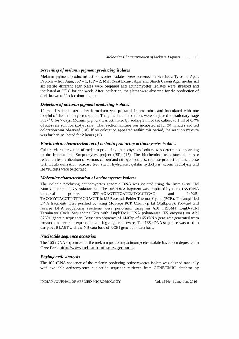
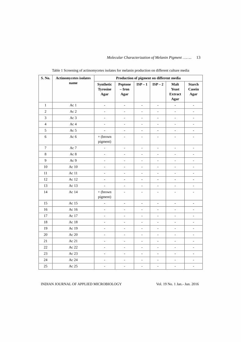
Screening and detection of melanin producing actinomycete isolates All the twenty five isolates of actinomycetes were screened for melanin production using different culture media such as Synthetic Tyrosine Agar, Peptone – Iron Agar, ISP – 1, ISP – 2, Malt Yeast extract Agar and Starch Casein Agar. Among these 25 isolates, only two actinomycetes (Ac 6 and Ac 14) isolates were able to produce brown colour pigment on Synthetic Tyrosine Agar and Peptone – Iron Agar media (Table 1; Fig. 1).
Fig. 1 Production of melanin pigment by isolates Ac 14
Molecular Characterization of Melanin Pigment …….. 13
INDIAN JOURNAL OF APPLIED MICROBIOLOGY Vol. 19 No. 1 Jan.- Jun. 2016
Table 1 Screening of actinomycetes isolates for melanin production on different culture media
S. No. Actinomycetes isolates name
Production of pigment on different media
Synthetic Tyrosine
Agar
Peptone – Iron Agar
ISP – 1 ISP – 2 Malt Yeast
Extract Agar
Starch Casein Agar
1 Ac 1 - - - - - -
2 Ac 2 - - - - - -
3 Ac 3 - - - - - -
4 Ac 4 - - - - - -
5 Ac 5 - - - - - -
6 Ac 6 + (brown pigment)
- - - - -
7 Ac 7 - - - - - -
8 Ac 8 - - - - - -
9 Ac 9 - - - - - -
10 Ac 10 - - - - - -
11 Ac 11 - - - - - -
12 Ac 12 - - - - - -
13 Ac 13 - - - - - -
14 Ac 14 + (brown pigment)
- - - - -
15 Ac 15 - - - - - -
16 Ac 16 - - - - - -
17 Ac 17 - - - - - -
18 Ac 18 - - - - - -
19 Ac 19 - - - - - -
20 Ac 20 - - - - - -
21 Ac 21 - - - - - -
22 Ac 22 - - - - - -
23 Ac 23 - - - - - -
24 Ac 24 - - - - - -
25 Ac 25 - - - - - -
14 RADHAKRISHNAN SRINIVASAN e t al
INDIAN JOURNAL OF APPLIED MICROBIOLOGY Vol. 19 No. 1 Jan.- Jun. 2016
The production of melanin was confirmed by using tyrosine as substrate this was achieved by adding 2ml of culture grown in Synthetic Tyrosine and Peptone – Iron broth with tyrosine solution. The formation of red coloration indicates melanin production. Formation of melanin was observed in AC 14 culture grown in Synthetic Tyrosine medium by red colour formation.
Characterization of the selected isolates Morphological and biochemical characterization of Ac 14 were recorded (Tables 2 and 3). Based on the observations, the Ac 14 was found to be Streptomyces sp. (MDCE2).
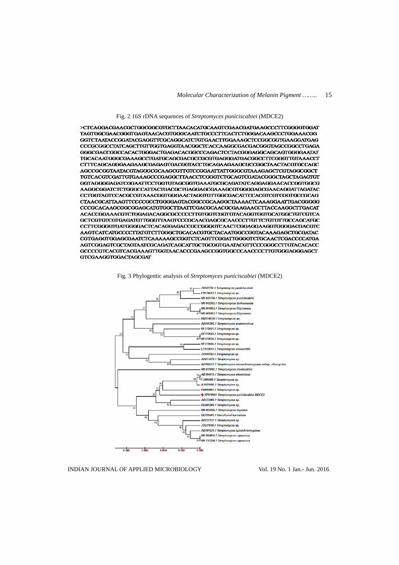
Molecular characterization of the selected Streptomyces sp. isolate Molecular characterization of Streptomyces sp. (MDCE2) was done by PCR amplification of 16S rDNA gene. The genomic DNA and amplified products were separated in agarose gel. The 16S rDNA genes of Streptomyces sp (MDCE2) isolated from collected soils was partially sequenced by using universal 16S rRNA sequence primers 518F-CCAGCAGCCGCGGTAATACG and 800R-TACCAGGGTATCTAATCC. The 16S rDNA sequences (Fig. 2) of Streptomyces sp. (MDCE2) was deposited in NCBI to get the accession number. The accession number of Streptomyces sp. (MDCE2) is KP970681.
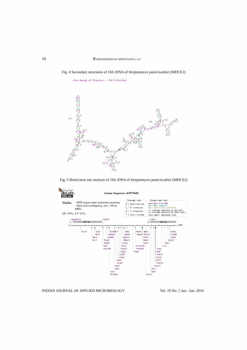
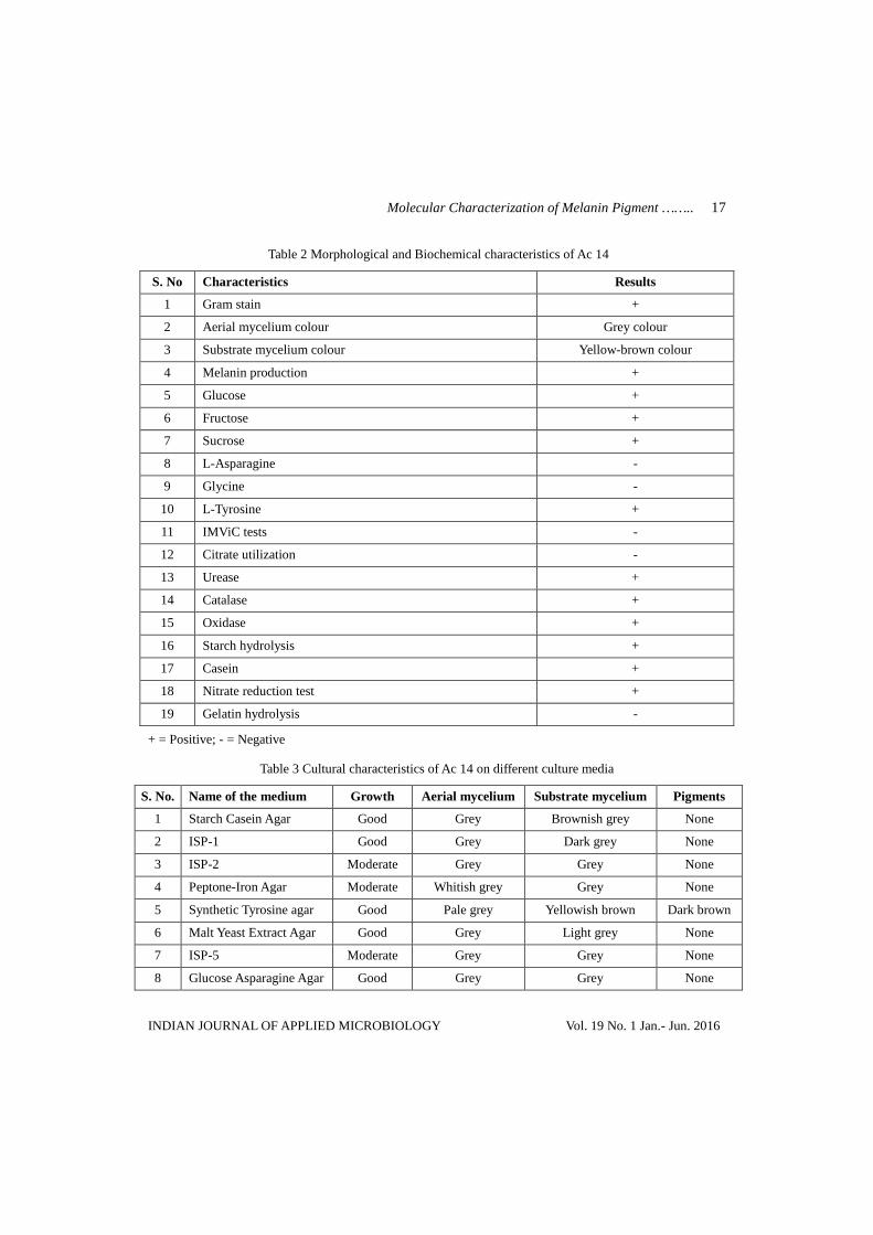
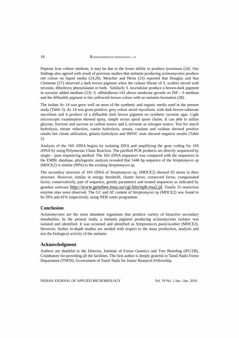
The construction of phylogentic tree was done by neighbour joining tree method by comparing the sequence with EMBL database (Fig. 3). The secondary structure of 16S rDNA of Streptomyces sp. (MDCE2) was predicted by using the bioinformatics tools available in http://www.genebee.msu.su/cgi-bin/nph-rna2.pl as showed in Figure 4. The restriction sites in 16S rDNA sequence of Streptomyces sp. (MDCE2) were analysed using NEB culture program (Fig. 5).
Discussion Microorganisms produce a diffusible, dark-brown pigment, melanin that can effectively protect the living organisms from ultraviolet radiation. Actinomycetes also produce melanin in different culture media and these melanin pigments were useful for taxonomical studies. In the present study, 25 actinomycetes isolates (Ac 1 – Ac 25) were isolated and screened for melanin pigment production. Among 24 isolate, only one (Ac 14) isolate was able to produce dark-brown colour pigment and it was identified as melanin. Similar screening was also obtained by several researchers (20-21, 6, 22). Mencher and Heim (23) confirmed that the dark brown soluble pigment was referred as melanoid or melanin in Streptomyces lavendulae. The method of testing for melanin production by L-tyrosine as a substrate may be good criterion for the identification and classification of Streptomyces (6). Many Streptomyces species produce a diffusible dark brown pigment on complex media, are considered to be a useful criterion for taxonomical studies (24-25, 6, 2). Streptomyces griseus ISP 5236 and S. ornatus ISP 5307, produce melanin pigment only on Synthetic Tyrosine Agar (25).
In the present study, formation of melanin was observed in actinomycetes 2:2 culture grown in Synthetic Tyrosine medium by red colour formation. It is difficult to determine red colouration in
Molecular Characterization of Melanin Pigment …….. 15
INDIAN JOURNAL OF APPLIED MICROBIOLOGY Vol. 19 No. 1 Jan.- Jun. 2016
Fig. 2 16S rDNA sequences of Streptomyces puniciscabiei (MDCE2)
Fig. 3 Phylogentic analysis of Streptomyces puniciscabiei (MDCE2)
16 RADHAKRISHNAN SRINIVASAN e t al
INDIAN JOURNAL OF APPLIED MICROBIOLOGY Vol. 19 No. 1 Jan.- Jun. 2016
Fig. 4 Secondary structures of 16S rDNA of Streptomyces puniciscabiei (MDCE2)
Fig. 5 Restriction site analysis of 16S rDNA of Streptomyces puniciscabiei (MDCE2)
Molecular Characterization of Melanin Pigment …….. 17
INDIAN JOURNAL OF APPLIED MICROBIOLOGY Vol. 19 No. 1 Jan.- Jun. 2016
Table 2 Morphological and Biochemical characteristics of Ac 14
S. No Characteristics Results
1 Gram stain +
2 Aerial mycelium colour Grey colour
3 Substrate mycelium colour Yellow-brown colour
4 Melanin production +
5 Glucose +
6 Fructose +
7 Sucrose +
8 L-Asparagine -
9 Glycine -
10 L-Tyrosine +
11 IMViC tests -
12 Citrate utilization -
13 Urease +
14 Catalase +
15 Oxidase +
16 Starch hydrolysis +
17 Casein +
18 Nitrate reduction test +
19 Gelatin hydrolysis -
+ = Positive; - = Negative
Table 3 Cultural characteristics of Ac 14 on different culture media
S. No. Name of the medium Growth Aerial mycelium Substrate mycelium Pigments
1 Starch Casein Agar Good Grey Brownish grey None
2 ISP-1 Good Grey Dark grey None
3 ISP-2 Moderate Grey Grey None
4 Peptone-Iron Agar Moderate Whitish grey Grey None
5 Synthetic Tyrosine agar Good Pale grey Yellowish brown Dark brown
6 Malt Yeast Extract Agar Good Grey Light grey None
7 ISP-5 Moderate Grey Grey None
8 Glucose Asparagine Agar Good Grey Grey None
18 RADHAKRISHNAN SRINIVASAN e t al
INDIAN JOURNAL OF APPLIED MICROBIOLOGY Vol. 19 No. 1 Jan.- Jun. 2016
Peptone Iron culture medium, it may be due to the lower ability to produce tyrosinase (24). Our findings also agreed with result of previous studies that melanin producing actinomycetes produce red colour on liquid media (24,26). Mencher and Heim (23) reported that Douglas and San Clemente (27) observed a dark brown pigment when the culture filtrate of S. scabies mixed with tyrosine, dihydroxy phenylalanin or both. Similarly S. lavendulae produce a brown-dark pigment in tyrosine added medium (23). S. albidoflavus-143 shows moderate growth on ISP – 6 medium and the diffusible pigment is fair yellowish brown colour with no melanin formation (28).
The isolate Ac 14 was grew well on most of the synthetic and organic media used in the present study (Table 3). Ac 14 was gram positive, grey colour aerial mycelium, with dark brown substrate mycelium and it produce of a diffusible dark brown pigment on synthetic tyrosine agar. Light microscopic examination showed spiny, simple rectus spiral spore chains. It can able to utilize glucose, fructose and sucrose as carbon source and L-tyrosine as nitrogen source. Test for starch hydrolysis, nitrate reduction, casein hydrolysis, urease, catalase and oxidase showed positive results but citrate utilization, gelatin hydrolysis and IMViC tests showed negative results (Table 2).
Analysis of the 16S rDNA begins by isolating DNA and amplifying the gene coding for 16S rDNA by using Polymerase Chain Reaction. The purified PCR products are directly sequenced by single – pass sequencing method. The 16S rDNA sequences was compared with the sequences in the EMBL database, phylogentic analysis revealed that 1446 bp sequence of the Streptomyces sp (MDCE2) is similar (99%) to the existing Streptomyces sp.
The secondary structure of 16S rDNA of Streptomyces sp. (MDCE2) showed 65 stems in their structure. However, similar in energy threshold, cluster factor, conserved factor, compensated factor, conservatively, part of sequence, greedy parameters and treated sequences as indicated by genebee software http://www.genebee.msu.su/cgi-bin/nph-rna2.pl. Totally 55 restriction enzyme sites were observed. The GC and AT content of Streptomyces sp (MDCE2) was found to be 59% and 41% respectively, using NEB cutter programme.
Conclusion Actinomycetes are the most abundant organisms that produce variety of bioactive secondary metabolites. In the present study, a melanin pigment producing actinomycetes isolates was isolated and identified. It was screened and identified as Streptomyces puniciscabiei (MDCE2). However, further in-depth studies are needed with respect to the mass production, analysis and test the biological activity of the melanin.
Acknowledgment Authors are thankful to the Director, Institute of Forest Genetics and Tree Breeding (IFGTB), Coimbatore for providing all the facilities. The first author is deeply grateful to Tamil Nadu Forest Department (TNFD), Government of Tamil Nadu for Junior Research Fellowship.
Molecular Characterization of Melanin Pigment …….. 19
INDIAN JOURNAL OF APPLIED MICROBIOLOGY Vol. 19 No. 1 Jan.- Jun. 2016
References 1. Lechevalier, H., Lechevalier, M.P., 1981, Introduction to the order Actinomycetales, In
The Prokaryotes, M.P. Starr, H. Stolp, H.G. Trüper, A. Balows, H.G. Schlegel, (Eds.), Springer-Verlag Berlin, Germany, pp, 1915–1922.
2. Manivasagan, P., Venkatesan, J., Sivakumar, K. and Se-Kwon Kim, 2013, Marine actinobacterial metabolites: Current status and future perspectives, World. J. Microb. Biot., 29, pp. 1737 – 1750.
3. Goncalves, R.C.R. and Pombeiro-Sponchiado, S.R., 2005, Antioxidant activity of the melanin pigment extracted from Aspergillus nidulans, Biol. Pharm. Bull., 28, pp. 1129–1131.
4. Casadevall, A., Rosas, A.L. and Nosanchuk, J.D., 2000, Melanin and virulence in Cryptococcus neoformans. Curr. Opin. Microbiol., 3, pp. 354–358.
5. El-Obeid, A., Al-Harbi, S., Al-Jomah, N. and Hassib, A., 2006, Herbal melanin modulates tumor necrosis factor alpha (TNF- a), interleukin 6 (IL-6) and vascular endothelial growth factor (VEGF) production, Phytomed., 13, pp. 324–333.
6. Bull, A.T., 2004, Microbial diversity and bioprospecting, ASM press, Washington, DC.
7. Berdy, J., 2005, Bioactive microbial metabolites, J. Antibiot., 58, pp. 1–26.
8. Strohl, W.R., 2004, Antimicrobials, In Microbial diversity and bioprospecting, A. T. Bull (Ed.), ASM Press, Washington, DC, pp. 336–355.
9. Dastager, S., Li, W.J., Dayanand, A., Tang, S.K., Tian, X.P., Zhi, X.Y., Xu, L.H. and Jiang, C.L., 2006, Separation, identification and analysis of pigment (melanin) production in Streptomyces, Afr. J. Biotech., 5, pp. 1131–1134.
10. Chaudhary, H.S., Soni, B., Shrivastava, A.R. and Shrivastava, S., 2013, Diversity and versatility of actinomycetes and its role in antibiotic production, J. Appl. Pharm. Sci., 3, pp. 83–94.
11. Hung, Y.C., Sava, V., Hong, M.Y. and Huang, G.S., 2004, Inhibitory effects on phospholipase A2 and antivenin activity of melanin extracted from Thea sinensis Linn, Life Sci., 74, pp. 2037–2047.
12. Montefiori, D.C. and Zhou, J., 1991, Selective antiviral activity of synthetic soluble L-Tyrosine and L-DOPA melanins against human immunodeficiency virus in vitro, Antiviral Res., 15, pp. 11–25.
13. Sava, V., Hung, Y., Blagodarsky, V., Hong, M.Y. and Huang, G., 2003. The liver-protecting activity of melanin-like pigment derived from black tea, Food. Res. Int., 36, pp. 505–511.
20 RADHAKRISHNAN SRINIVASAN e t al
INDIAN JOURNAL OF APPLIED MICROBIOLOGY Vol. 19 No. 1 Jan.- Jun. 2016
14. Dadachova, E., Bryan, R.A., Huang, X., Moadel, T., Schweitzer, A.D., Aisen, P., Nosanchuk, J.D. and Casadevall, A., 2007, Ionizing radiation changes the electronic properties of melanin and enhances the growth of melanized fungi. Plos One., 2, pp. 457.
15. Vinarov, A.U., Robysheva, Z.N., Sidorenko, T.E. and Dirina, E.N., 2002, Biotechnology of pigment melanin, In Proceedings of the 1st international congress biotechnology-state of the art and prospects of development, p 96.
16. Oskay, M., Same, A. and Azeri, C., 2004, Antibacterial activity of some actinomycetes isolated from farming soils of Turkey, Afr. J. Biotechol., 3, pp. 441-446.
17. Shirling, E.B. and Gottlieb, D., 1966, Methods for characterization of Streptomyces species, Int. J. Syst. Baceriol., 16, pp. 313-340.
18. Shirling, E.B. and Gottlieb, D., 1970, Report of the International Streptomyces Project-five years of collaborative research, In The Actinomycetales, Prauser, H. (Ed.), Gustav, Fischer Verlag, Jena, pp. 79-89.
19. Scribner, E., Tang, T. and Bradley, S.G., 1973, Production of a sporulation pigment by Streptomyces venezuelae, Appl. Microbiol., 25, pp. 873-879.
20. Zenova, G., 1965, Melanoid pigments of actinomycetes, Mikrobiologiia., 34, pp. 278–283.
21. Coelho, R.R.R. and Linhares, L.F., 1993, Melanogenic actinomycetes (Streptomyces sp.) from Brazilian soils, Biol. Fert. Soils., 15, pp. 220-224.
22. Kshitija, R.D., 2012, Isolation, characterization of melanin producing organism and extraction of melanin, Int. J.Sci. Eng. Res., 3, pp. 1-4.
23. Mencher, J.R. and Heim, A.H., 1962, Melanin biosynthesis by Streptomyces lavendulae, J. Gen. Microbiol., 28, pp. 665-670.
24. Arai, T. and Mikami, Y., 1972a, Chromogenicity of Streptomyces, Appl. Microbiol., pp. 402-406.
25. Arai, T. and Mikami, Y., 1972b, Chromogenesis mirabilis in Streptomyces griseus, Appl. Microbiol., 768-771.
26. Vasanthabharathi, V., Lakshminarayanan, R. and Jayalakshmi, S., 2011, Melanin production from marine Streptomyces, Afr. J. Biotech., 10, pp. 11224-11234.
27. Douglas, R.J. and San Clemente, C.L., 1956, Respiration of scab-producing strains of actinomycetes, Canad. J. Microbiol., 2, p. 407.
28. Atta, H.M., Bahobail, A.S. and El-Sehrawi, M.H., 2011, Studies on isolation, classification and phylogentic characterization of antifungal substance produced by Streptomyces albidoflavus-143, J. Science, 44, pp. 40-53.


![[EXOTIC] Blaptica Dubia Roaches Info_care Sheet](https://img.pdfslide.net/doc/110x75/55cf8fc8550346703b9fc8a7/exotic-blaptica-dubia-roaches-infocare-sheet.jpg)