Embed Size (px)
Citation preview
lable at ScienceDirect
Journal of Human Evolution 86 (2015) 32e42
Contents lists avai
Journal of Human Evolution
journal homepage: www.elsevier .com/locate/ jhevol
Three-dimensional kinematics of the pelvis and hind limbs inchimpanzee (Pan troglodytes) and human bipedal walking
Matthew C. O'Neill a, b, Leng-Feng Lee c, Brigitte Demes b, Nathan E. Thompson b,Susan G. Larson b, Jack T. Stern Jr b, Brian R. Umberger c, *
a Department of Basic Medical Sciences, University of Arizona College of Medicine-Phoenix, Phoenix, AZ 85004, USAb Department of Anatomical Sciences, Stony Brook University School of Medicine, Stony Brook, NY 11794, USAc Department of Kinesiology, University of Massachusetts, Amherst, MA 01003, USA
a r t i c l e i n f o
Article history:Received 6 September 2014Accepted 20 May 2015Available online 17 July 2015
Keywords:ChimpanzeeHumanKinematicsPelvisHind limbBipedalism
* Corresponding author.E-mail address: [email protected] (B.R. Um
1 While “lower limb” is typically preferred in hum“hind limb” to describe the thigh, shank and foot in bo
http://dx.doi.org/10.1016/j.jhevol.2015.05.0120047-2484/© 2015 Elsevier Ltd. All rights reserved.
a b s t r a c t
The common chimpanzee (Pan troglodytes) is a facultative biped and our closest living relative. As such,the musculoskeletal anatomies of their pelvis and hind limbs have long provided a comparative contextfor studies of human and fossil hominin locomotion. Yet, how the chimpanzee pelvis and hind limbactually move during bipedal walking is still not well defined. Here, we describe the three-dimensional(3-D) kinematics of the pelvis, hip, knee and ankle during bipedal walking and compare those values tohumans walking at the same dimensionless and dimensional velocities. The stride-to-stride and intra-specific variations in 3-D kinematics were calculated using the adjusted coefficient of multiple corre-lation. Our results indicate that humans walk with a more stable pelvis than chimpanzees, especially intilt and rotation. Both species exhibit similar magnitudes of pelvis list, but with segment motion that isopposite in phasing. In the hind limb, chimpanzees walk with a more flexed and abducted limb posture,and substantially exceed humans in the magnitude of hip rotation during a stride. The average stride-to-stride variation in joint and segment motion was greater in chimpanzees than humans, while theintraspecific variation was similar on average. These results demonstrate substantial differences betweenhuman and chimpanzee bipedal walking, in both the sagittal and non-sagittal planes. These new 3-Dkinematic data are fundamental to a comprehensive understanding of the mechanics, energetics andcontrol of chimpanzee bipedalism.
© 2015 Elsevier Ltd. All rights reserved.
1. Introduction
Humans are unique among apes and other primates in themusculoskeletal design of the pelvis and hind limbs.1 Our short,wide pelvis and long, heavy hind limbs reflect both our evolutionfrom an arboreal ape as well as selection pressures for aneconomical, two-legged walking stride (Rodman and McHenry,1980; Sockol et al., 2007). The common chimpanzee (Pan troglo-dytes) e a facultative biped and our closest living relative e uses amore expensive, flexed-limb gait when moving on two legs. Whilequalitative differences between human and chimpanzee bipedalwalking kinematics have been noted at least since the pioneering
berger).an-specific studies, we use
th chimpanzees and humans.
work of Elftman (1944), direct quantitative comparisons of theirpelvis and hind limb motions are quite limited. Yet, such data areessential for understanding how variation in musculoskeletalstructure affects locomotor performance.
The three-dimensional (3-D) kinematics of humanwalking havebeen examined and described in considerable detail (e.g. Apkarianet al., 1989; Kadaba et al., 1990; Rose and Gamble, 2006). Thesestudies have revealed important non-sagittal plane motions withdirect relevance for understanding joint and muscle-tendon me-chanics. For example, measurements of the 3-D motion of thepelvis and thigh are needed for the accurate determination of hipjoint kinetics (e.g. Eng and Winter, 1995) and associated skeletalloading (e.g. Stansfield et al., 2003a), as well as calculations ofmuscle-tendon force and fascicle length change during a stride (e.g.Arnold and Delp, 2011). Given this, accurate 3-D quantification ofsegment and joint motion has become fundamental to determiningthe mechanics, energetics and control of locomotor tasks. Inchimpanzee bipedal walking, qualitative observation indicates that
M.C. O'Neill et al. / Journal of Human Evolution 86 (2015) 32e42 33
e in addition to their well-known flexed-limb posturee substantial3-D motions occur about the pelvis and hips (Elftman, 1944;Jenkins, 1972; Stern and Susman, 1981; Stern and Larson, 1993).Yet, no comprehensive joint motion analysis has been undertaken.
Most previous studies of chimpanzee kinematics have beenlimited to spatio-temporal analyses that focus on a few quantitativemetrics, such as stride lengths and durations (Alexander andMaloiy, 1984; Kimura, 1987, 1990; Reynolds, 1987; Aerts et al.,2000; Kimura and Yaguramaki, 2009). Sagittal plane hip, kneeand ankle angles have been published for bonobos (D'Août et al.,2002) and, more recently, for common chimpanzees (Pontzeret al., 2014). However, to date, the only multi-plane investigationof chimpanzee pelvis and hind limb motion during bipedal walkingis that of Jenkins (1972). Therein, two-dimensional cineradiographytaken asynchronously in both sagittal and frontal planes was usedto reconstruct the motion of the pelvis, femur, tibia-fibula and footelements. This approach has the advantage of permitting the directtracking of skeletal motion, but the published report itself lacksmuch quantitative detail regarding the timing or duration of theobserved kinematics. Further, in this and other studies, no com-parable walking data were collected from humans.
Equivalent lab-based measurements of chimpanzees andhumans have the potential to improve our understanding of themechanics, energetics and control of facultative and habitualbipedalism. The aim of this study is to present the 3-D kinematics ofthe pelvis and hind limb of bipedal walking in both species, as wellas compare stride-to-stride, intraspecific and interspecific varia-tion. For completeness, our chimpanzee data are compared to thekinematics of humans walking at similar dimensionless (i.e.relative-speed match) and dimensional (i.e. absolute-speed match)speeds. The dimensionless comparisonminimizes the effects due todifferences in body size or speed, while emphasizing those arisingspecifically from differences in musculoskeletal design betweenchimpanzees and humans. The dimensional comparison, incontrast, permits an assessment of how sensitive the interspecificdifferences in 3-D kinematics are to walking speed.
2. Materials and methods
2.1. Chimpanzee and human subjects
Three-dimensional kinematic data were collected from thepelvis and hind limbs of three male common chimpanzeesP. troglodytes (age: 5.5 ± 0.2 yrs; Mb: 26.5 ± 6.7 kg) and three malehumans Homo sapiens (age: 24.3 ± 2.3 yrs; Mb: 79.2 ± 6.2 kg). Thenumber of human subjects was matched to the chimpanzee datasetto facilitate a comparison of interspecific movement variability.Each bipedal chimpanzeewalked across an 11m rigid, level runwayat self-selected speeds, following an animal trainer offering a food
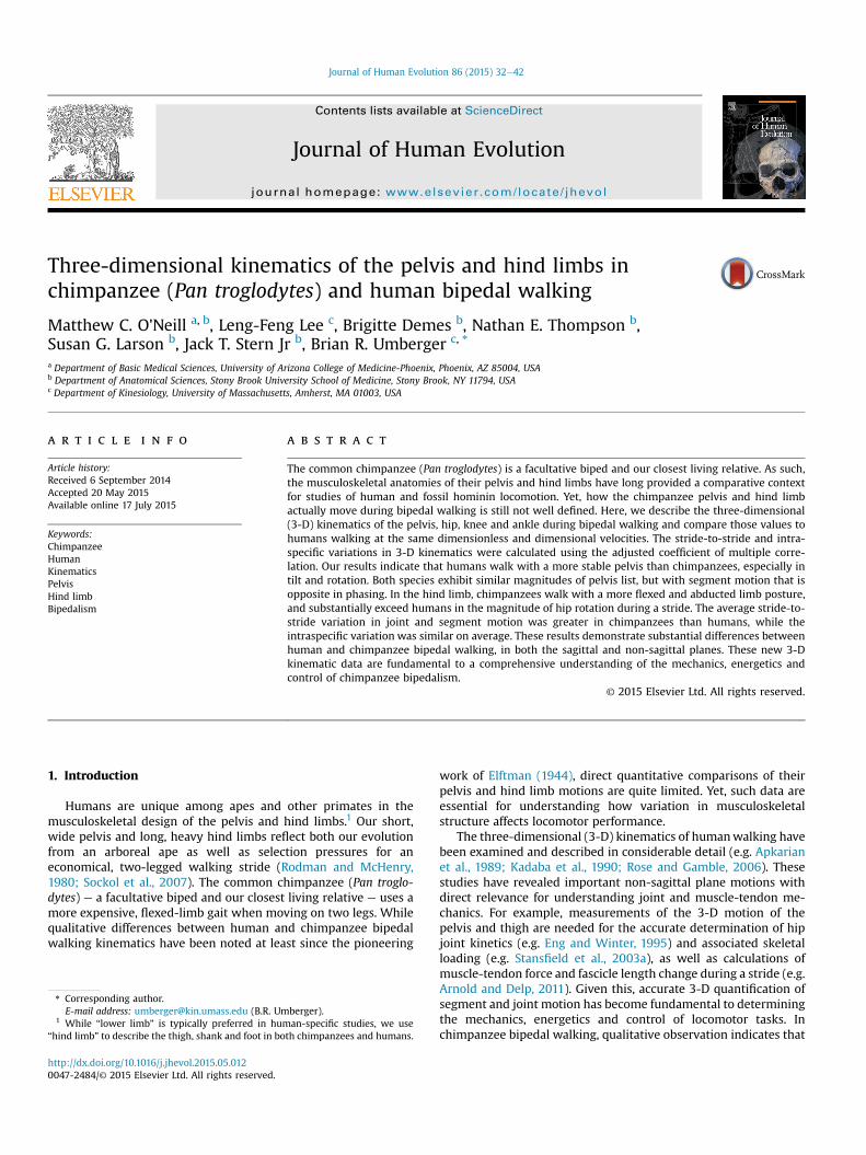
Figure 1. A full bipedal walking stride. A full stride includes both stance and swing phases.support, and the second double-support (double support 2) periods. In the first double-suppperiod the right hind limb is the trailing limb.
reward (Fig. 1). Human data were then collected during walkingalong a 20 m rigid, level runway at speeds matching the chim-panzee dataset in dimensionless (i.e. relative-speed match) anddimensional (i.e. absolute-speed match) forms. The Stony BrookUniversity Institutional Animal Care and Use Committee and theUniversity of Massachusetts Amherst Institutional Research Boardapproved all chimpanzee and human experiments, respectively.The human subjects each provided written informed consentbefore participating in the study.
2.2. Chimpanzee training
Each chimpanzee was trained to walk on its hind limbs acrossthe 11 m rigid, level runway at self-selected speeds using food re-wards and positive reinforcement. The training regime consisted ofmixed periods of walking and resting over approximately 1 h perday, 3e5 days per week for at least 6 months prior to the start ofdata collection. The aims of the training regime were to teach eachchimpanzee towalk bipedally for multiple strides on command andfollow a straight path along the runway through the calibratedrecording volume. Training familiarized the animals with theexperimental protocol, thereby reducing random kinematic vari-ance unrelated to musculoskeletal design and/or speed effects. Inour view, training was essential to maximizing the comparability ofour chimpanzee and human data sets.
2.3. Musculoskeletal modeling
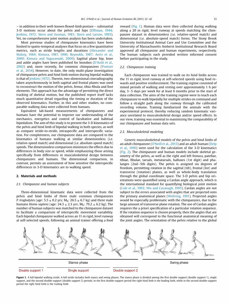
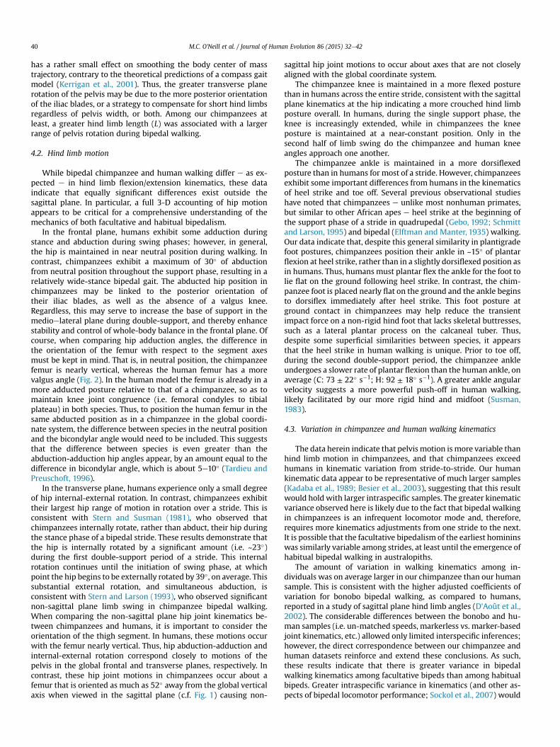
Generic musculoskeletal models of the pelvis and hind limbs ofan adult chimpanzee (O'Neill et al., 2013) and an adult human (Delpet al., 1990) were used for the calculation of the 3-D kinematics(Fig. 2). The chimpanzee and human models include skeletal ge-ometry of the pelvis, as well as the right and left femora, patellae,tibiae, fibulae, tarsals, metatarsals, halluxes (1st digit) and pha-langes (2nde5th digits). The pelvis is assigned six degrees offreedom, permitting rotation in the sagittal (tilt), frontal (list) andtransverse (rotation) planes, as well as whole-body translationthrough the global coordinate space. The 3-D pelvis and hip ori-entations were quantified using a Cardan angle approach, which isthe international standard for quantifying biological joint motion(Cole et al., 1993; Wu and Cavanagh, 1995). Cardan angles are notsubject to the errors associated with angles that are projected ontothe primary anatomical planes (Woltring, 1991). Projected angleswould be especially problematic with the chimpanzees, due to thelarge amount of transverse plane rotation. The use of Cardan anglesrequires the a priori specification of a particular rotation sequence.If the rotation sequence is chosen properly, then the angles that areobtained will correspond to the functional anatomical meaning ofthe joint angles. The orientation of the pelvis relative to the global
The stance phase is divided among the first double support (double support 1), singleort period the right hind limb is the leading limb, while in the second double-support
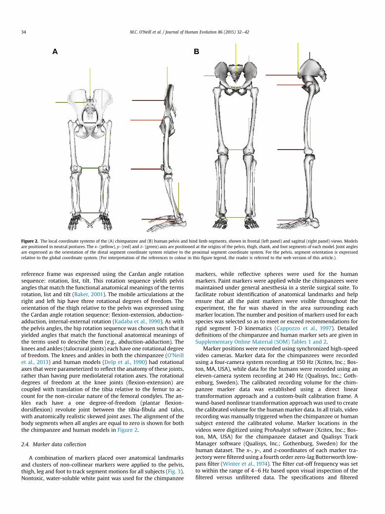
Figure 2. The local coordinate systems of the (A) chimpanzee and (B) human pelvis and hind limb segments, shown in frontal (left panel) and sagittal (right panel) views. Modelsare positioned in neutral postures. The x- (yellow), y- (red) and z- (green) axis are positioned at the origins of the pelvis, thigh, shank, and foot segments of each model. Joint anglesare expressed as the orientation of the distal segment coordinate system relative to the proximal segment coordinate system. For the pelvis, segment orientation is expressedrelative to the global coordinate system. (For interpretation of the references to colour in this figure legend, the reader is referred to the web version of this article.).
M.C. O'Neill et al. / Journal of Human Evolution 86 (2015) 32e4234
reference frame was expressed using the Cardan angle rotationsequence: rotation, list, tilt. This rotation sequence yields pelvisangles that match the functional anatomical meanings of the termsrotation, list and tilt (Baker, 2001). The mobile articulations at theright and left hip have three rotational degrees of freedom. Theorientation of the thigh relative to the pelvis was expressed usingthe Cardan angle rotation sequence: flexion-extension, abduction-adduction, internal-external rotation (Kadaba et al., 1990). As withthe pelvis angles, the hip rotation sequence was chosen such that ityielded angles that match the functional anatomical meanings ofthe terms used to describe them (e.g., abduction-adduction). Theknees and ankles (talocrural joints) each have one rotational degreeof freedom. The knees and ankles in both the chimpanzee (O'Neillet al., 2013) and human models (Delp et al., 1990) had rotationalaxes that were parameterized to reflect the anatomy of these joints,rather than having pure mediolateral rotation axes. The rotationaldegrees of freedom at the knee joints (flexion-extension) arecoupled with translation of the tibia relative to the femur to ac-count for the non-circular nature of the femoral condyles. The an-kles each have a one degree-of-freedom (plantar flexion-dorsiflexion) revolute joint between the tibia-fibula and talus,with anatomically realistic skewed joint axes. The alignment of thebody segments when all angles are equal to zero is shown for boththe chimpanzee and human models in Figure 2.
2.4. Marker data collection
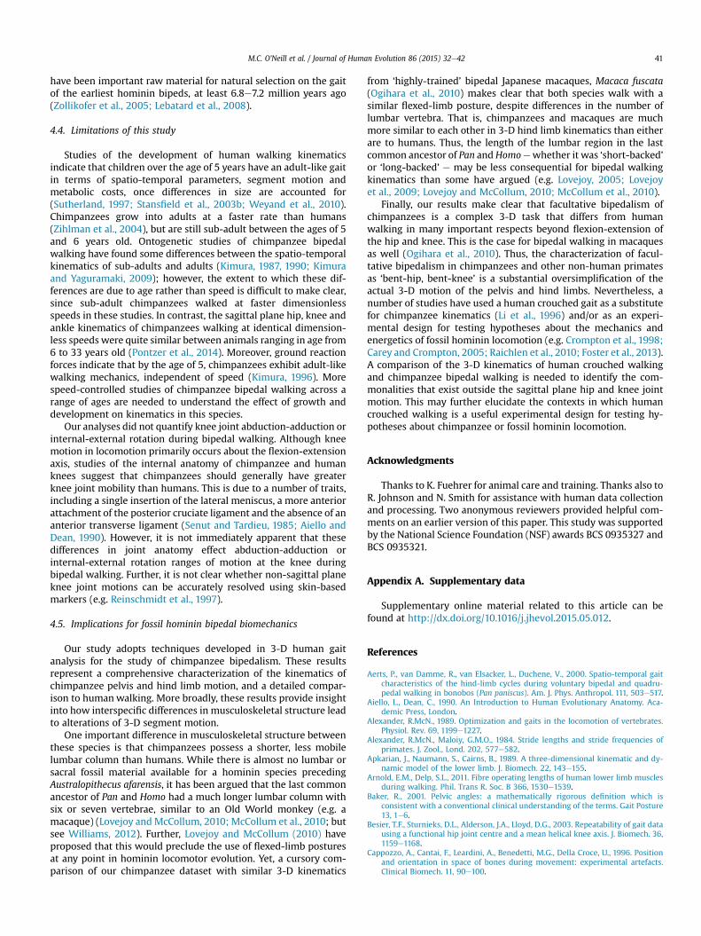
A combination of markers placed over anatomical landmarksand clusters of non-collinear markers were applied to the pelvis,thigh, leg and foot to track segment motions for all subjects (Fig. 3).Nontoxic, water-soluble white paint was used for the chimpanzee
markers, while reflective spheres were used for the humanmarkers. Paint markers were applied while the chimpanzees weremaintained under general anesthesia in a sterile surgical suite. Tofacilitate robust identification of anatomical landmarks and helpensure that all the paint markers were visible throughout theexperiment, the fur was shaved in the area surrounding eachmarker location. The number and position of markers used for eachspecies was selected so as to meet or exceed recommendations forrigid segment 3-D kinematics (Cappozzo et al., 1997). Detaileddefinitions of the chimpanzee and human marker sets are given inSupplementary Online Material (SOM) Tables 1 and 2.
Marker positions were recorded using synchronized high-speedvideo cameras. Marker data for the chimpanzees were recordedusing a four-camera system recording at 150 Hz (Xcitex, Inc.; Bos-ton, MA, USA), while data for the humans were recorded using aneleven-camera system recording at 240 Hz (Qualisys, Inc.; Goth-enburg, Sweden). The calibrated recording volume for the chim-panzee marker data was established using a direct lineartransformation approach and a custom-built calibration frame. Awand-based nonlinear transformation approach was used to createthe calibrated volume for the humanmarker data. In all trials, videorecording was manually triggered when the chimpanzee or humansubject entered the calibrated volume. Marker locations in thevideos were digitized using ProAnalyst software (Xcitex, Inc.; Bos-ton, MA, USA) for the chimpanzee dataset and Qualisys TrackManager software (Qualisys, Inc.; Gothenburg, Sweden) for thehuman dataset. The x-, y-, and z-coordinates of each marker tra-jectory were filtered using a fourth order zero-lag Butterworth low-pass filter (Winter et al., 1974). The filter cut-off frequency was setto within the range of 4e6 Hz based upon visual inspection of thefiltered versus unfiltered data. The specifications and filtered
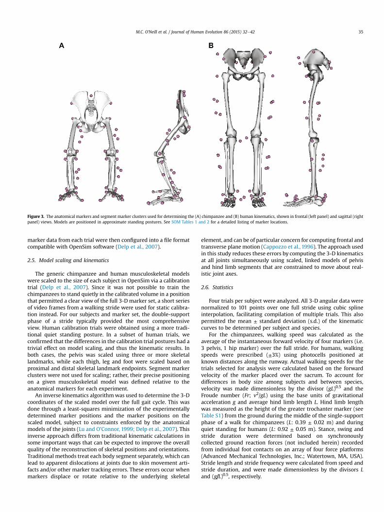
Figure 3. The anatomical markers and segment marker clusters used for determining the (A) chimpanzee and (B) human kinematics, shown in frontal (left panel) and sagittal (rightpanel) views. Models are positioned in approximate standing postures. See SOM Tables 1 and 2 for a detailed listing of marker locations.
M.C. O'Neill et al. / Journal of Human Evolution 86 (2015) 32e42 35
marker data from each trial were then configured into a file formatcompatible with OpenSim software (Delp et al., 2007).
2.5. Model scaling and kinematics
The generic chimpanzee and human musculoskeletal modelswere scaled to the size of each subject in OpenSim via a calibrationtrial (Delp et al., 2007). Since it was not possible to train thechimpanzees to stand quietly in the calibrated volume in a positionthat permitted a clear view of the full 3-D marker set, a short seriesof video frames from a walking stride were used for static calibra-tion instead. For our subjects and marker set, the double-supportphase of a stride typically provided the most comprehensiveview. Human calibration trials were obtained using a more tradi-tional quiet standing posture. In a subset of human trials, weconfirmed that the differences in the calibration trial postures had atrivial effect on model scaling, and thus the kinematic results. Inboth cases, the pelvis was scaled using three or more skeletallandmarks, while each thigh, leg and foot were scaled based onproximal and distal skeletal landmark endpoints. Segment markerclusters were not used for scaling; rather, their precise positioningon a given musculoskeletal model was defined relative to theanatomical markers for each experiment.
An inverse kinematics algorithmwas used to determine the 3-Dcoordinates of the scaled model over the full gait cycle. This wasdone through a least-squares minimization of the experimentallydetermined marker positions and the marker positions on thescaled model, subject to constraints enforced by the anatomicalmodels of the joints (Lu and O'Connor, 1999; Delp et al., 2007). Thisinverse approach differs from traditional kinematic calculations insome important ways that can be expected to improve the overallquality of the reconstruction of skeletal positions and orientations.Traditional methods treat each body segment separately, which canlead to apparent dislocations at joints due to skin movement arti-facts and/or other marker tracking errors. These errors occur whenmarkers displace or rotate relative to the underlying skeletal
element, and can be of particular concern for computing frontal andtransverse plane motion (Cappozzo et al., 1996). The approach usedin this study reduces these errors by computing the 3-D kinematicsat all joints simultaneously using scaled, linked models of pelvisand hind limb segments that are constrained to move about real-istic joint axes.
2.6. Statistics
Four trials per subject were analyzed. All 3-D angular data werenormalized to 101 points over one full stride using cubic splineinterpolation, facilitating compilation of multiple trials. This alsopermitted the mean ± standard deviation (s.d.) of the kinematiccurves to be determined per subject and species.
For the chimpanzees, walking speed was calculated as theaverage of the instantaneous forward velocity of four markers (i.e.3 pelvis, 1 hip marker) over the full stride. For humans, walkingspeeds were prescribed (±3%) using photocells positioned atknown distances along the runway. Actual walking speeds for thetrials selected for analysis were calculated based on the forwardvelocity of the marker placed over the sacrum. To account fordifferences in body size among subjects and between species,velocity was made dimensionless by the divisor (gL)0.5 and theFroude number (Fr; v2/gL) using the base units of gravitationalacceleration g and average hind limb length L. Hind limb lengthwas measured as the height of the greater trochanter marker (seeTable S1) from the ground during the middle of the single-supportphase of a walk for chimpanzees (L: 0.39 ± 0.02 m) and duringquiet standing for humans (L: 0.92 ± 0.05 m). Stance, swing andstride duration were determined based on synchronouslycollected ground reaction forces (not included herein) recordedfrom individual foot contacts on an array of four force platforms(Advanced Mechanical Technologies, Inc.; Watertown, MA, USA).Stride length and stride frequency were calculated from speed andstride duration, and were made dimensionless by the divisors Land (g/L)0.5, respectively.
M.C. O'Neill et al. / Journal of Human Evolution 86 (2015) 32e4236
To compare the stride-to-stride, intraspecific and interspecificvariation of the pelvis and hind limb angles of our chimpanzee andhuman samples, the adjusted coefficient of multiple correlation(CMC; Kadaba et al., 1989) was calculated. The CMC represents thecorrelation of the segment or joint motion among strides for eachindividual (i.e. stride-to-stride variation) or among individuals (i.e.intraspecific variation). Finally, the balanced chimpanzee and hu-man datasets permits a direct, interspecific comparison of CMCs.
3. Results
The self-selected, average walking speed was 1.09 ± 0.10 m s�1
for the chimpanzees, and the matched relative walking speed forthe human subjects was 1.66 ± 0.06 m s�1. These values correspondto identical dimensionless velocities (v) of 0.56 ± 0.06 and0.56 ± 0.01, and Froude numbers (Fr) of 0.31 ± 0.06 and 0.31 ± 0.02for each species, respectively (Table 1). These speeds are close to e
but slightly faster than e the preferred overground speeds for hu-man walking, but well below the expected walkerun transitionspeed (i.e. v ¼ 0.7; Fr ¼ 0.5; Alexander, 1989; Kram et al., 1997). Thechimpanzees and humans walked with stride lengths of0.78 ± 0.07 m and 1.69 ± 0.15 m and stride frequencies of1.43 ± 0.23 Hz and 1.00 ± 0.05 Hz, respectively. The stance andlimb-swing durations (of individual limbs) were 0.45 ± 0.09 s and0.27 ± 0.03 s for chimpanzees, and 0.64 ± 0.02 s and 0.36 ± 0.04 sfor humans. As such, the duty factors were nearly equivalent.
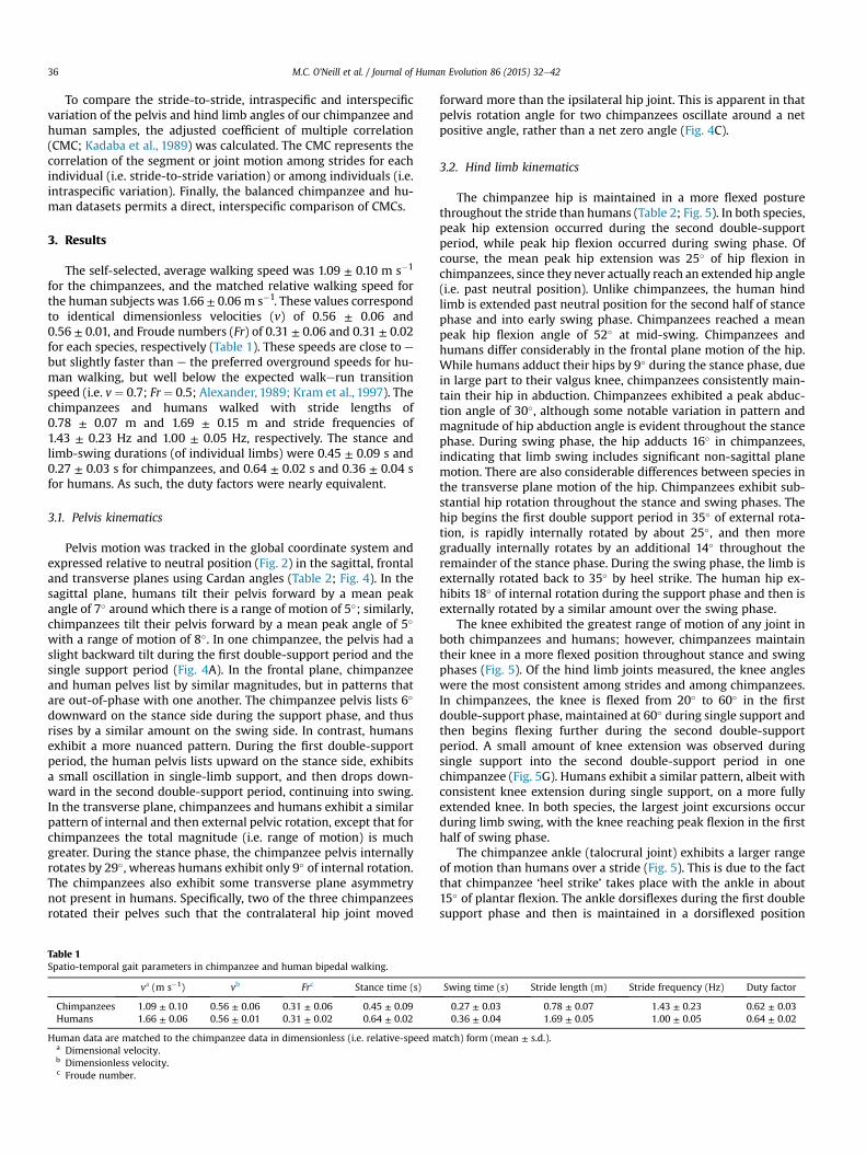
3.1. Pelvis kinematics
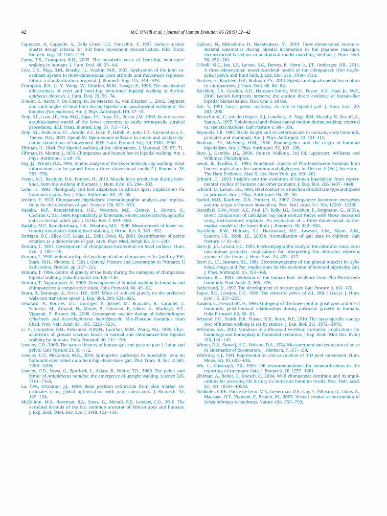
Pelvis motion was tracked in the global coordinate system andexpressed relative to neutral position (Fig. 2) in the sagittal, frontaland transverse planes using Cardan angles (Table 2; Fig. 4). In thesagittal plane, humans tilt their pelvis forward by a mean peakangle of 7� around which there is a range of motion of 5�; similarly,chimpanzees tilt their pelvis forward by a mean peak angle of 5�
with a range of motion of 8�. In one chimpanzee, the pelvis had aslight backward tilt during the first double-support period and thesingle support period (Fig. 4A). In the frontal plane, chimpanzeeand human pelves list by similar magnitudes, but in patterns thatare out-of-phase with one another. The chimpanzee pelvis lists 6�
downward on the stance side during the support phase, and thusrises by a similar amount on the swing side. In contrast, humansexhibit a more nuanced pattern. During the first double-supportperiod, the human pelvis lists upward on the stance side, exhibitsa small oscillation in single-limb support, and then drops down-ward in the second double-support period, continuing into swing.In the transverse plane, chimpanzees and humans exhibit a similarpattern of internal and then external pelvic rotation, except that forchimpanzees the total magnitude (i.e. range of motion) is muchgreater. During the stance phase, the chimpanzee pelvis internallyrotates by 29�, whereas humans exhibit only 9� of internal rotation.The chimpanzees also exhibit some transverse plane asymmetrynot present in humans. Specifically, two of the three chimpanzeesrotated their pelves such that the contralateral hip joint moved
Table 1Spatio-temporal gait parameters in chimpanzee and human bipedal walking.
va (m s�1) vb Frc Stance time (s)
Chimpanzees 1.09 ± 0.10 0.56 ± 0.06 0.31 ± 0.06 0.45 ± 0.09Humans 1.66 ± 0.06 0.56 ± 0.01 0.31 ± 0.02 0.64 ± 0.02
Human data are matched to the chimpanzee data in dimensionless (i.e. relative-speed ma Dimensional velocity.b Dimensionless velocity.c Froude number.
forward more than the ipsilateral hip joint. This is apparent in thatpelvis rotation angle for two chimpanzees oscillate around a netpositive angle, rather than a net zero angle (Fig. 4C).
3.2. Hind limb kinematics
The chimpanzee hip is maintained in a more flexed posturethroughout the stride than humans (Table 2; Fig. 5). In both species,peak hip extension occurred during the second double-supportperiod, while peak hip flexion occurred during swing phase. Ofcourse, the mean peak hip extension was 25� of hip flexion inchimpanzees, since they never actually reach an extended hip angle(i.e. past neutral position). Unlike chimpanzees, the human hindlimb is extended past neutral position for the second half of stancephase and into early swing phase. Chimpanzees reached a meanpeak hip flexion angle of 52� at mid-swing. Chimpanzees andhumans differ considerably in the frontal plane motion of the hip.While humans adduct their hips by 9� during the stance phase, duein large part to their valgus knee, chimpanzees consistently main-tain their hip in abduction. Chimpanzees exhibited a peak abduc-tion angle of 30�, although some notable variation in pattern andmagnitude of hip abduction angle is evident throughout the stancephase. During swing phase, the hip adducts 16� in chimpanzees,indicating that limb swing includes significant non-sagittal planemotion. There are also considerable differences between species inthe transverse plane motion of the hip. Chimpanzees exhibit sub-stantial hip rotation throughout the stance and swing phases. Thehip begins the first double support period in 35� of external rota-tion, is rapidly internally rotated by about 25�, and then moregradually internally rotates by an additional 14� throughout theremainder of the stance phase. During the swing phase, the limb isexternally rotated back to 35� by heel strike. The human hip ex-hibits 18� of internal rotation during the support phase and then isexternally rotated by a similar amount over the swing phase.
The knee exhibited the greatest range of motion of any joint inboth chimpanzees and humans; however, chimpanzees maintaintheir knee in a more flexed position throughout stance and swingphases (Fig. 5). Of the hind limb joints measured, the knee angleswere the most consistent among strides and among chimpanzees.In chimpanzees, the knee is flexed from 20� to 60� in the firstdouble-support phase, maintained at 60� during single support andthen begins flexing further during the second double-supportperiod. A small amount of knee extension was observed duringsingle support into the second double-support period in onechimpanzee (Fig. 5G). Humans exhibit a similar pattern, albeit withconsistent knee extension during single support, on a more fullyextended knee. In both species, the largest joint excursions occurduring limb swing, with the knee reaching peak flexion in the firsthalf of swing phase.
The chimpanzee ankle (talocrural joint) exhibits a larger rangeof motion than humans over a stride (Fig. 5). This is due to the factthat chimpanzee ‘heel strike’ takes place with the ankle in about15� of plantar flexion. The ankle dorsiflexes during the first doublesupport phase and then is maintained in a dorsiflexed position
Swing time (s) Stride length (m) Stride frequency (Hz) Duty factor
0.27 ± 0.03 0.78 ± 0.07 1.43 ± 0.23 0.62 ± 0.030.36 ± 0.04 1.69 ± 0.05 1.00 ± 0.05 0.64 ± 0.02
atch) form (mean ± s.d.).
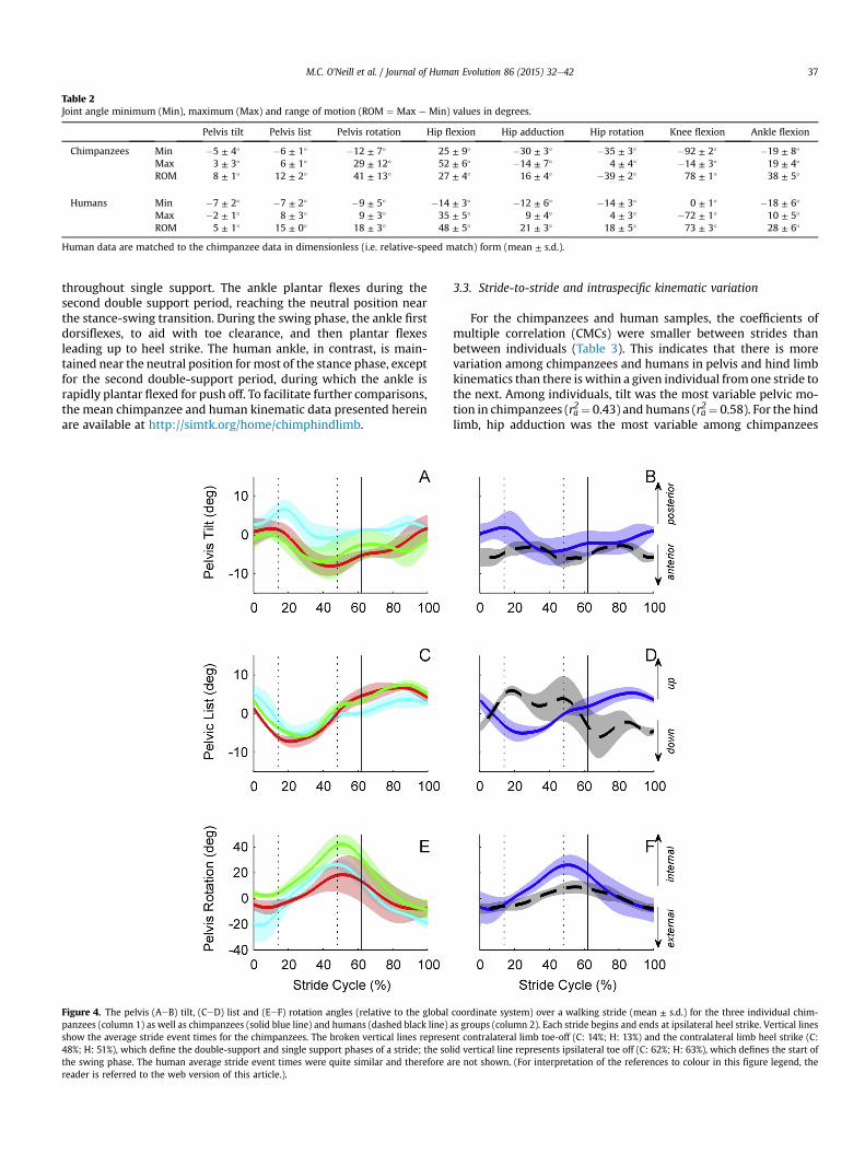
Table 2Joint angle minimum (Min), maximum (Max) and range of motion (ROM ¼ Max e Min) values in degrees.
Pelvis tilt Pelvis list Pelvis rotation Hip flexion Hip adduction Hip rotation Knee flexion Ankle flexion
Chimpanzees Min �5 ± 4� �6 ± 1� �12 ± 7� 25 ± 9� �30 ± 3� �35 ± 3� �92 ± 2� �19 ± 8�
Max 3 ± 3� 6 ± 1� 29 ± 12� 52 ± 6� �14 ± 7� 4 ± 4� �14 ± 3� 19 ± 4�
ROM 8 ± 1� 12 ± 2� 41 ± 13� 27 ± 4� 16 ± 4� �39 ± 2� 78 ± 1� 38 ± 5�
Humans Min �7 ± 2� �7 ± 2� �9 ± 5� �14 ± 3� �12 ± 6� �14 ± 3� 0 ± 1� �18 ± 6�
Max �2 ± 1� 8 ± 3� 9 ± 3� 35 ± 5� 9 ± 4� 4 ± 3� �72 ± 1� 10 ± 5�
ROM 5 ± 1� 15 ± 0� 18 ± 3� 48 ± 5� 21 ± 3� 18 ± 5� 73 ± 3� 28 ± 6�
Human data are matched to the chimpanzee data in dimensionless (i.e. relative-speed match) form (mean ± s.d.).
M.C. O'Neill et al. / Journal of Human Evolution 86 (2015) 32e42 37
throughout single support. The ankle plantar flexes during thesecond double support period, reaching the neutral position nearthe stance-swing transition. During the swing phase, the ankle firstdorsiflexes, to aid with toe clearance, and then plantar flexesleading up to heel strike. The human ankle, in contrast, is main-tained near the neutral position formost of the stance phase, exceptfor the second double-support period, during which the ankle israpidly plantar flexed for push off. To facilitate further comparisons,the mean chimpanzee and human kinematic data presented hereinare available at http://simtk.org/home/chimphindlimb.
Figure 4. The pelvis (AeB) tilt, (CeD) list and (EeF) rotation angles (relative to the globalpanzees (column 1) as well as chimpanzees (solid blue line) and humans (dashed black line)show the average stride event times for the chimpanzees. The broken vertical lines represe48%; H: 51%), which define the double-support and single support phases of a stride; the sothe swing phase. The human average stride event times were quite similar and therefore areader is referred to the web version of this article.).
3.3. Stride-to-stride and intraspecific kinematic variation
For the chimpanzees and human samples, the coefficients ofmultiple correlation (CMCs) were smaller between strides thanbetween individuals (Table 3). This indicates that there is morevariation among chimpanzees and humans in pelvis and hind limbkinematics than there is within a given individual fromone stride tothe next. Among individuals, tilt was the most variable pelvic mo-tion in chimpanzees (ra2¼ 0.43) and humans (ra2¼ 0.58). For the hindlimb, hip adduction was the most variable among chimpanzees
coordinate system) over a walking stride (mean ± s.d.) for the three individual chim-as groups (column 2). Each stride begins and ends at ipsilateral heel strike. Vertical linesnt contralateral limb toe-off (C: 14%; H: 13%) and the contralateral limb heel strike (C:lid vertical line represents ipsilateral toe off (C: 62%; H: 63%), which defines the start ofre not shown. (For interpretation of the references to colour in this figure legend, the
Figure 5. The hip (AeB) flexion, (CeD) adduction, (EeF) rotation, (GeH) knee flexion and (IeJ) ankle flexion angles over a walking stride (mean ± s.d.) for the three individualchimpanzees (column 1) as well as chimpanzees (solid blue line) and humans (dashed black line) as groups (column 2). Each stride begins and ends at ipsilateral heel strike. Verticallines show the average stride event times for the chimpanzees. The broken vertical lines represent contralateral limb toe-off (C: 14%; H: 13%) and the contralateral limb heel strike(C: 48%; H: 51%), which define the double-support and single support phases of a stride; the solid vertical line represents ipsilateral toe off (C: 62%; H: 63%), which defines the startof the swing phase. The human average stride event times were quite similar and therefore are not shown. (For interpretation of the references to colour in this figure legend, thereader is referred to the web version of this article.).
M.C. O'Neill et al. / Journal of Human Evolution 86 (2015) 32e4238
(ra2 ¼ 0.31), but in humans hip adduction, rotation and ankle flexionhad similar CMC values. Our human results appear to be represen-tative of larger human samples, as they are generally consistentwiththe CMC results of both Kadaba et al. (1989) and Besier et al. (2003).
Directly comparing the CMC values of our chimpanzee andhuman samples indicates that, on average, chimpanzees are more
variable in their kinematics stride-to-stride (i.e. chimp mean:ra2 ¼ 0.82, human mean: ra2 ¼ 0.97). That is, a human walking strideis more stereotyped than a bipedal stride of chimpanzees, in bothpelvis and hind limb motion. Among subjects, chimpanzees exhibitgreater variability than humans in pelvis and hind limb motion aswell (i.e. chimp mean: ra2 ¼ 0.71, human mean: ra2 ¼ 0.81), but not
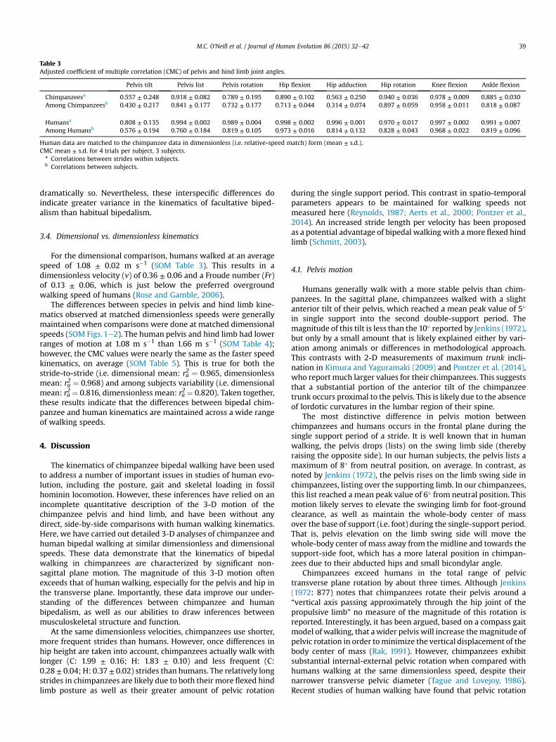
Table 3Adjusted coefficient of multiple correlation (CMC) of pelvis and hind limb joint angles.
Pelvis tilt Pelvis list Pelvis rotation Hip flexion Hip adduction Hip rotation Knee flexion Ankle flexion
Chimpanzeesa 0.557 ± 0.248 0.918 ± 0.082 0.789 ± 0.195 0.890 ± 0.102 0.563 ± 0.250 0.940 ± 0.036 0.978 ± 0.009 0.885 ± 0.030Among Chimpanzeesb 0.430 ± 0.217 0.841 ± 0.177 0.732 ± 0.177 0.713 ± 0.044 0.314 ± 0.074 0.897 ± 0.059 0.958 ± 0.011 0.818 ± 0.087
Humansa 0.808 ± 0.135 0.994 ± 0.002 0.989 ± 0.004 0.998 ± 0.002 0.996 ± 0.001 0.970 ± 0.017 0.997 ± 0.002 0.991 ± 0.007Among Humansb 0.576 ± 0.194 0.760 ± 0.184 0.819 ± 0.105 0.973 ± 0.016 0.814 ± 0.132 0.828 ± 0.043 0.968 ± 0.022 0.819 ± 0.096
Human data are matched to the chimpanzee data in dimensionless (i.e. relative-speed match) form (mean ± s.d.).CMC mean ± s.d. for 4 trials per subject, 3 subjects.
a Correlations between strides within subjects.b Correlations between subjects.
M.C. O'Neill et al. / Journal of Human Evolution 86 (2015) 32e42 39
dramatically so. Nevertheless, these interspecific differences doindicate greater variance in the kinematics of facultative biped-alism than habitual bipedalism.
3.4. Dimensional vs. dimensionless kinematics
For the dimensional comparison, humans walked at an averagespeed of 1.08 ± 0.02 m s�1 (SOM Table 3). This results in adimensionless velocity (v) of 0.36 ± 0.06 and a Froude number (Fr)of 0.13 ± 0.06, which is just below the preferred overgroundwalking speed of humans (Rose and Gamble, 2006).
The differences between species in pelvis and hind limb kine-matics observed at matched dimensionless speeds were generallymaintained when comparisons were done at matched dimensionalspeeds (SOM Figs. 1e2). The human pelvis and hind limb had lowerranges of motion at 1.08 m s�1 than 1.66 m s�1 (SOM Table 4);however, the CMC values were nearly the same as the faster speedkinematics, on average (SOM Table 5). This is true for both thestride-to-stride (i.e. dimensional mean: ra2 ¼ 0.965, dimensionlessmean: ra2 ¼ 0.968) and among subjects variability (i.e. dimensionalmean: ra2 ¼ 0.816, dimensionless mean: ra2 ¼ 0.820). Taken together,these results indicate that the differences between bipedal chim-panzee and human kinematics are maintained across a wide rangeof walking speeds.
4. Discussion
The kinematics of chimpanzee bipedal walking have been usedto address a number of important issues in studies of human evo-lution, including the posture, gait and skeletal loading in fossilhominin locomotion. However, these inferences have relied on anincomplete quantitative description of the 3-D motion of thechimpanzee pelvis and hind limb, and have been without anydirect, side-by-side comparisons with human walking kinematics.Here, we have carried out detailed 3-D analyses of chimpanzee andhuman bipedal walking at similar dimensionless and dimensionalspeeds. These data demonstrate that the kinematics of bipedalwalking in chimpanzees are characterized by significant non-sagittal plane motion. The magnitude of this 3-D motion oftenexceeds that of human walking, especially for the pelvis and hip inthe transverse plane. Importantly, these data improve our under-standing of the differences between chimpanzee and humanbipedalism, as well as our abilities to draw inferences betweenmusculoskeletal structure and function.
At the same dimensionless velocities, chimpanzees use shorter,more frequent strides than humans. However, once differences inhip height are taken into account, chimpanzees actually walk withlonger (C: 1.99 ± 0.16; H: 1.83 ± 0.10) and less frequent (C:0.28 ± 0.04; H: 0.37 ± 0.02) strides than humans. The relatively longstrides in chimpanzees are likely due to both their more flexed hindlimb posture as well as their greater amount of pelvic rotation
during the single support period. This contrast in spatio-temporalparameters appears to be maintained for walking speeds notmeasured here (Reynolds, 1987; Aerts et al., 2000; Pontzer et al.,2014). An increased stride length per velocity has been proposedas a potential advantage of bipedal walking with a more flexed hindlimb (Schmitt, 2003).
4.1. Pelvis motion
Humans generally walk with a more stable pelvis than chim-panzees. In the sagittal plane, chimpanzees walked with a slightanterior tilt of their pelvis, which reached a mean peak value of 5�
in single support into the second double-support period. Themagnitude of this tilt is less than the 10� reported by Jenkins (1972),but only by a small amount that is likely explained either by vari-ation among animals or differences in methodological approach.This contrasts with 2-D measurements of maximum trunk incli-nation in Kimura and Yaguramaki (2009) and Pontzer et al. (2014),who report much larger values for their chimpanzees. This suggeststhat a substantial portion of the anterior tilt of the chimpanzeetrunk occurs proximal to the pelvis. This is likely due to the absenceof lordotic curvatures in the lumbar region of their spine.
The most distinctive difference in pelvis motion betweenchimpanzees and humans occurs in the frontal plane during thesingle support period of a stride. It is well known that in humanwalking, the pelvis drops (lists) on the swing limb side (therebyraising the opposite side). In our human subjects, the pelvis lists amaximum of 8� from neutral position, on average. In contrast, asnoted by Jenkins (1972), the pelvis rises on the limb swing side inchimpanzees, listing over the supporting limb. In our chimpanzees,this list reached a mean peak value of 6� from neutral position. Thismotion likely serves to elevate the swinging limb for foot-groundclearance, as well as maintain the whole-body center of massover the base of support (i.e. foot) during the single-support period.That is, pelvis elevation on the limb swing side will move thewhole-body center of mass away from the midline and towards thesupport-side foot, which has a more lateral position in chimpan-zees due to their abducted hips and small bicondylar angle.
Chimpanzees exceed humans in the total range of pelvictransverse plane rotation by about three times. Although Jenkins(1972: 877) notes that chimpanzees rotate their pelvis around a“vertical axis passing approximately through the hip joint of thepropulsive limb” no measure of the magnitude of this rotation isreported. Interestingly, it has been argued, based on a compass gaitmodel of walking, that awider pelvis will increase themagnitude ofpelvic rotation in order tominimize the vertical displacement of thebody center of mass (Rak, 1991). However, chimpanzees exhibitsubstantial internal-external pelvic rotation when compared withhumans walking at the same dimensionless speed, despite theirnarrower transverse pelvic diameter (Tague and Lovejoy, 1986).Recent studies of human walking have found that pelvic rotation
M.C. O'Neill et al. / Journal of Human Evolution 86 (2015) 32e4240
has a rather small effect on smoothing the body center of masstrajectory, contrary to the theoretical predictions of a compass gaitmodel (Kerrigan et al., 2001). Thus, the greater transverse planerotation of the pelvis may be due to the more posterior orientationof the iliac blades, or a strategy to compensate for short hind limbsregardless of pelvis width, or both. Among our chimpanzees atleast, a greater hind limb length (L) was associated with a largerrange of pelvis rotation during bipedal walking.
4.2. Hind limb motion
While bipedal chimpanzee and human walking differ e as ex-pected e in hind limb flexion/extension kinematics, these dataindicate that equally significant differences exist outside thesagittal plane. In particular, a full 3-D accounting of hip motionappears to be critical for a comprehensive understanding of themechanics of both facultative and habitual bipedalism.
In the frontal plane, humans exhibit some adduction duringstance and abduction during swing phases; however, in general,the hip is maintained in near neutral position during walking. Incontrast, chimpanzees exhibit a maximum of 30� of abductionfrom neutral position throughout the support phase, resulting in arelatively wide-stance bipedal gait. The abducted hip position inchimpanzees may be linked to the posterior orientation oftheir iliac blades, as well as the absence of a valgus knee.Regardless, this may serve to increase the base of support in themedioelateral plane during double-support, and thereby enhancestability and control of whole-body balance in the frontal plane. Ofcourse, when comparing hip adduction angles, the difference inthe orientation of the femur with respect to the segment axesmust be kept in mind. That is, in neutral position, the chimpanzeefemur is nearly vertical, whereas the human femur has a morevalgus angle (Fig. 2). In the human model the femur is already in amore adducted posture relative to that of a chimpanzee, so as tomaintain knee joint congruence (i.e. femoral condyles to tibialplateau) in both species. Thus, to position the human femur in thesame abducted position as in a chimpanzee in the global coordi-nate system, the difference between species in the neutral positionand the bicondylar angle would need to be included. This suggeststhat the difference between species is even greater than theabduction-adduction hip angles appear, by an amount equal to thedifference in bicondylar angle, which is about 5e10� (Tardieu andPreuschoft, 1996).
In the transverse plane, humans experience only a small degreeof hip internal-external rotation. In contrast, chimpanzees exhibittheir largest hip range of motion in rotation over a stride. This isconsistent with Stern and Susman (1981), who observed thatchimpanzees internally rotate, rather than abduct, their hip duringthe stance phase of a bipedal stride. These results demonstrate thatthe hip is internally rotated by a significant amount (i.e. ~23�)during the first double-support period of a stride. This internalrotation continues until the initiation of swing phase, at whichpoint the hip begins to be externally rotated by 39�, on average. Thissubstantial external rotation, and simultaneous abduction, isconsistent with Stern and Larson (1993), who observed significantnon-sagittal plane limb swing in chimpanzee bipedal walking.When comparing the non-sagittal plane hip joint kinematics be-tween chimpanzees and humans, it is important to consider theorientation of the thigh segment. In humans, these motions occurwith the femur nearly vertical. Thus, hip abduction-adduction andinternal-external rotation correspond closely to motions of thepelvis in the global frontal and transverse planes, respectively. Incontrast, these hip joint motions in chimpanzees occur about afemur that is oriented as much as 52� away from the global verticalaxis when viewed in the sagittal plane (c.f. Fig. 1) causing non-
sagittal hip joint motions to occur about axes that are not closelyaligned with the global coordinate system.
The chimpanzee knee is maintained in a more flexed posturethan in humans across the entire stride, consistent with the sagittalplane kinematics at the hip indicating a more crouched hind limbposture overall. In humans, during the single support phase, theknee is increasingly extended, while in chimpanzees the kneeposture is maintained at a near-constant position. Only in thesecond half of limb swing do the chimpanzee and human kneeangles approach one another.
The chimpanzee ankle is maintained in a more dorsiflexedposture than in humans for most of a stride. However, chimpanzeesexhibit some important differences from humans in the kinematicsof heel strike and toe off. Several previous observational studieshave noted that chimpanzees e unlike most nonhuman primates,but similar to other African apes e heel strike at the beginning ofthe support phase of a stride in quadrupedal (Gebo, 1992; Schmittand Larson, 1995) and bipedal (Elftman and Manter, 1935) walking.Our data indicate that, despite this general similarity in plantigradefoot postures, chimpanzees position their ankle in ~15� of plantarflexion at heel strike, rather than in a slightly dorsiflexed position asin humans. Thus, humans must plantar flex the ankle for the foot tolie flat on the ground following heel strike. In contrast, the chim-panzee foot is placed nearly flat on the ground and the ankle beginsto dorsiflex immediately after heel strike. This foot posture atground contact in chimpanzees may help reduce the transientimpact force on a non-rigid hind foot that lacks skeletal buttresses,such as a lateral plantar process on the calcaneal tuber. Thus,despite some superficial similarities between species, it appearsthat the heel strike in human walking is unique. Prior to toe off,during the second double-support period, the chimpanzee ankleundergoes a slower rate of plantar flexion than the human ankle, onaverage (C: 73 ± 22� s�1; H: 92 ± 18� s�1). A greater ankle angularvelocity suggests a more powerful push-off in human walking,likely facilitated by our more rigid hind and midfoot (Susman,1983).
4.3. Variation in chimpanzee and human walking kinematics
The data herein indicate that pelvis motion is more variable thanhind limb motion in chimpanzees, and that chimpanzees exceedhumans in kinematic variation from stride-to-stride. Our humankinematic data appear to be representative of much larger samples(Kadaba et al., 1989; Besier et al., 2003), suggesting that this resultwould hold with larger intraspecific samples. The greater kinematicvariance observed here is likely due to the fact that bipedal walkingin chimpanzees is an infrequent locomotor mode and, therefore,requires more kinematics adjustments from one stride to the next.It is possible that the facultative bipedalism of the earliest homininswas similarly variable among strides, at least until the emergence ofhabitual bipedal walking in australopiths.
The amount of variation in walking kinematics among in-dividuals was on average larger in our chimpanzee than our humansample. This is consistent with the higher adjusted coefficients ofvariation for bonobo bipedal walking, as compared to humans,reported in a study of sagittal plane hind limb angles (D'Août et al.,2002). The considerable differences between the bonobo and hu-man samples (i.e. un-matched speeds, markerless vs. marker-basedjoint kinematics, etc.) allowed only limited interspecific inferences;however, the direct correspondence between our chimpanzee andhuman datasets reinforce and extend these conclusions. As such,these results indicate that there is greater variance in bipedalwalking kinematics among facultative bipeds than among habitualbipeds. Greater intraspecific variance in kinematics (and other as-pects of bipedal locomotor performance; Sockol et al., 2007) would
M.C. O'Neill et al. / Journal of Human Evolution 86 (2015) 32e42 41
have been important raw material for natural selection on the gaitof the earliest hominin bipeds, at least 6.8e7.2 million years ago(Zollikofer et al., 2005; Lebatard et al., 2008).
4.4. Limitations of this study
Studies of the development of human walking kinematicsindicate that children over the age of 5 years have an adult-like gaitin terms of spatio-temporal parameters, segment motion andmetabolic costs, once differences in size are accounted for(Sutherland, 1997; Stansfield et al., 2003b; Weyand et al., 2010).Chimpanzees grow into adults at a faster rate than humans(Zihlman et al., 2004), but are still sub-adult between the ages of 5and 6 years old. Ontogenetic studies of chimpanzee bipedalwalking have found some differences between the spatio-temporalkinematics of sub-adults and adults (Kimura, 1987, 1990; Kimuraand Yaguramaki, 2009); however, the extent to which these dif-ferences are due to age rather than speed is difficult to make clear,since sub-adult chimpanzees walked at faster dimensionlessspeeds in these studies. In contrast, the sagittal plane hip, knee andankle kinematics of chimpanzees walking at identical dimension-less speeds were quite similar between animals ranging in age from6 to 33 years old (Pontzer et al., 2014). Moreover, ground reactionforces indicate that by the age of 5, chimpanzees exhibit adult-likewalking mechanics, independent of speed (Kimura, 1996). Morespeed-controlled studies of chimpanzee bipedal walking across arange of ages are needed to understand the effect of growth anddevelopment on kinematics in this species.
Our analyses did not quantify knee joint abduction-adduction orinternal-external rotation during bipedal walking. Although kneemotion in locomotion primarily occurs about the flexion-extensionaxis, studies of the internal anatomy of chimpanzee and humanknees suggest that chimpanzees should generally have greaterknee joint mobility than humans. This is due to a number of traits,including a single insertion of the lateral meniscus, a more anteriorattachment of the posterior cruciate ligament and the absence of ananterior transverse ligament (Senut and Tardieu, 1985; Aiello andDean, 1990). However, it is not immediately apparent that thesedifferences in joint anatomy effect abduction-adduction orinternal-external rotation ranges of motion at the knee duringbipedal walking. Further, it is not clear whether non-sagittal planeknee joint motions can be accurately resolved using skin-basedmarkers (e.g. Reinschmidt et al., 1997).
4.5. Implications for fossil hominin bipedal biomechanics
Our study adopts techniques developed in 3-D human gaitanalysis for the study of chimpanzee bipedalism. These resultsrepresent a comprehensive characterization of the kinematics ofchimpanzee pelvis and hind limb motion, and a detailed compar-ison to humanwalking. More broadly, these results provide insightinto how interspecific differences in musculoskeletal structure leadto alterations of 3-D segment motion.
One important difference in musculoskeletal structure betweenthese species is that chimpanzees possess a shorter, less mobilelumbar column than humans. While there is almost no lumbar orsacral fossil material available for a hominin species precedingAustralopithecus afarensis, it has been argued that the last commonancestor of Pan and Homo had a much longer lumbar column withsix or seven vertebrae, similar to an Old World monkey (e.g. amacaque) (Lovejoy and McCollum, 2010; McCollum et al., 2010; butsee Williams, 2012). Further, Lovejoy and McCollum (2010) haveproposed that this would preclude the use of flexed-limb posturesat any point in hominin locomotor evolution. Yet, a cursory com-parison of our chimpanzee dataset with similar 3-D kinematics
from ‘highly-trained’ bipedal Japanese macaques, Macaca fuscata(Ogihara et al., 2010) makes clear that both species walk with asimilar flexed-limb posture, despite differences in the number oflumbar vertebra. That is, chimpanzees and macaques are muchmore similar to each other in 3-D hind limb kinematics than eitherare to humans. Thus, the length of the lumbar region in the lastcommon ancestor of Pan andHomoewhether it was ‘short-backed’or ‘long-backed’ e may be less consequential for bipedal walkingkinematics than some have argued (e.g. Lovejoy, 2005; Lovejoyet al., 2009; Lovejoy and McCollum, 2010; McCollum et al., 2010).
Finally, our results make clear that facultative bipedalism ofchimpanzees is a complex 3-D task that differs from humanwalking in many important respects beyond flexion-extension ofthe hip and knee. This is the case for bipedal walking in macaquesas well (Ogihara et al., 2010). Thus, the characterization of facul-tative bipedalism in chimpanzees and other non-human primatesas ‘bent-hip, bent-knee’ is a substantial oversimplification of theactual 3-D motion of the pelvis and hind limbs. Nevertheless, anumber of studies have used a human crouched gait as a substitutefor chimpanzee kinematics (Li et al., 1996) and/or as an experi-mental design for testing hypotheses about the mechanics andenergetics of fossil hominin locomotion (e.g. Crompton et al., 1998;Carey and Crompton, 2005; Raichlen et al., 2010; Foster et al., 2013).A comparison of the 3-D kinematics of human crouched walkingand chimpanzee bipedal walking is needed to identify the com-monalities that exist outside the sagittal plane hip and knee jointmotion. This may further elucidate the contexts in which humancrouched walking is a useful experimental design for testing hy-potheses about chimpanzee or fossil hominin locomotion.
Acknowledgments
Thanks to K. Fuehrer for animal care and training. Thanks also toR. Johnson and N. Smith for assistance with human data collectionand processing. Two anonymous reviewers provided helpful com-ments on an earlier version of this paper. This study was supportedby the National Science Foundation (NSF) awards BCS 0935327 andBCS 0935321.
Appendix A. Supplementary data
Supplementary online material related to this article can befound at http://dx.doi.org/10.1016/j.jhevol.2015.05.012.
References
Aerts, P., van Damme, R., van Elsacker, L., Duchene, V., 2000. Spatio-temporal gaitcharacteristics of the hind-limb cycles during voluntary bipedal and quadru-pedal walking in bonobos (Pan paniscus). Am. J. Phys. Anthropol. 111, 503e517.
Aiello, L., Dean, C., 1990. An Introduction to Human Evolutionary Anatomy. Aca-demic Press, London.
Alexander, R.McN., 1989. Optimization and gaits in the locomotion of vertebrates.Physiol. Rev. 69, 1199e1227.
Alexander, R.McN., Maloiy, G.M.O., 1984. Stride lengths and stride frequencies ofprimates. J. Zool., Lond. 202, 577e582.
Apkarian, J., Naumann, S., Cairns, B., 1989. A three-dimensional kinematic and dy-namic model of the lower limb. J. Biomech. 22, 143e155.
Arnold, E.M., Delp, S.L., 2011. Fibre operating lengths of human lower limb musclesduring walking. Phil. Trans R. Soc. B 366, 1530e1539.
Baker, R., 2001. Pelvic angles: a mathematically rigorous definition which isconsistent with a conventional clinical understanding of the terms. Gait Posture13, 1e6.
Besier, T.F., Sturnieks, D.L., Alderson, J.A., Lloyd, D.G., 2003. Repeatability of gait datausing a functional hip joint centre and a mean helical knee axis. J. Biomech. 36,1159e1168.
Cappozzo, A., Cantai, F., Leardini, A., Benedetti, M.G., Della Croce, U., 1996. Positionand orientation in space of bones during movement: experimental artefacts.Clinical Biomech. 11, 90e100.
M.C. O'Neill et al. / Journal of Human Evolution 86 (2015) 32e4242
Cappozzo, A., Cappello, A., Della Croce, U.D., Pensalfini, F., 1997. Surface-markercluster design criteria for 3-D bone movement reconstruction. IEEE Trans.Biomed. Eng. 44, 1165e1174.
Carey, T.S., Crompton, R.H., 2005. The metabolic costs of ‘bent-hip, bent-knee’walking in humans. J. Hum. Evol. 48, 25e44.
Cole, G.K., Nigg, B.M., Ronsky, J.L., Yeadon, M.R., 1993. Application of the joint co-ordinate system to three-dimensional joint attitude and movement represen-tation: a standardization proposal. J. Biomech. Eng. 115, 344e349.
Crompton, R.H., Li, Y., Wang, W., Günther, M.M., Savage, R., 1998. The mechanicaleffectiveness of erect and ‘bent-hip, bent-knee’ bipedal walking in Austral-opithecus afarensis. J. Hum. Evol. 35, 55e74.
D'Août, K., Aerts, P., De Clercq, D., De Meester, K., Van Elsacker, L., 2002. Segmentand joint angles of hind limb during bipedal and quadrupedal walking of thebonobo (Pan paniscus). Am. J. Phys. Anthropol. 119, 37e51.
Delp, S.L., Loan, J.P., Hoy, M.G., Zajac, F.E., Topp, E.L., Rosen, J.M., 1990. An interactivegraphics-based model of the lower extremity to study orthopaedic surgicalprocedures. IEEE Trans. Biomed. Eng. 37, 757e767.
Delp, S.L., Anderson, F.C., Arnold, A.S., Loan, P., Habib, A., John, C.T., Guendelman, E.,Thelen, D.G., 2007. OpenSIM: Open-source software to create and analyze dy-namic simulations of movement. IEEE Trans. Biomed. Eng. 54, 1940e1950.
Elftman, H., 1944. The bipedal walking of the chimpanzee. J. Mammal. 25, 67e71.Elftman, H., Manter, J., 1935. Chimpanzee and human feet in bipedal walking. Am. J.
Phys. Anthropol. 1, 69e79.Eng, J.J., Winter, D.A., 1995. Kinetic analysis of the lower limbs during walking: what
information can be gained from a three-dimensional model? J. Biomech. 28,753e758.
Foster, A.D., Raichlen, D.A., Pontzer, H., 2013. Muscle force production during bent-knee, bent-hip walking in humans. J. Hum. Evol. 65, 294e302.
Gebo, D., 1992. Plantigrady and foot adaptation in African apes: Implications forhominid origins. Am. J. Phys. Anthropol. 89, 29e58.
Jenkins, F., 1972. Chimpanzee bipedalism: cineradiographic analysis and implica-tions for the evolution of gait. Science 178, 877e879.
Kadaba, M.P., Ramakrishnan, H.K., Wootten, M.E., Gainey, J., Gorton, G.,Cochran, G.V.B., 1989. Repeatability of kinematic, kinetic and electromyographicdata in normal adult gait. J. Ortho. Res. 7, 849e860.
Kadaba, M.P., Ramakrishnan, H.K., Wootten, M.E., 1990. Measurement of lower ex-tremity kinematics during level walking. J. Ortho. Res. 8, 383e392.
Kerrigan, D.C., Riley, O.P., Lelas, J.L., Della Croce, U., 2001. Quantification of pelvicrotation as a determinate of gait. Arch. Phys. Med. Rehab 82, 217e220.
Kimura, T., 1987. Development of chimpanzee locomotion on level surfaces. Hum.Evol. 2, 107e119.
Kimura, T., 1990. Voluntary bipedal walking of infant chimpanzees. In: Jouffroy, F.H.,Stack, M.H., Niemitz, C. (Eds.), Gravity, Posture and Locomotion in Primates. IlSedicesimo, Firenze, pp. 237e251.
Kimura, T., 1996. Centre of gravity of the body during the ontogeny of chimpanzeebipedal walking. Folia Primatol. 66, 126e136.
Kimura, T., Yaguramaki, N., 2009. Development of bipedal walking in humans andchimpanzees: a comparative study. Folia Primatol. 80, 45e62.
Kram, R., Domingo, A., Ferris, D.P., 1997. Effect of reduced gravity on the preferredwalk-run transition speed. J. Exp. Biol. 200, 821e826.
Lebatard, A., Bourl�es, D.L., Duringer, P., Jolivet, M., Braucher, R., Caraillet, J.,Schuster, M., Arnaud, N., Moni�e, P., Lihoreau, F., Likius, A., Mackaye, H.T.,Vignaud, P., Brunet, M., 2008. Cosmogenic nuclide dating of Sahelanthropustchadensis and Australopithecus bahrelghazali: Mio-Pliocene hominids fromChad. Proc. Natl. Acad. Sci. 105, 3226e3231.
Li, Y., Crompton, R.H., Alexander, R.McN., Gunther, M.M., Wang, W.J., 1996. Char-acteristics of ground reaction forces in normal and chimpanzee-like bipedalwalking by humans. Folia Primatol. 66, 137e159.
Lovejoy, C.O., 2005. The natural history of human gait and posture part 1. Spine andpelvis. Gait Posture 21, 95e112.
Lovejoy, C.O., McCollum, M.A., 2010. Spinopelvic pathways to bipedality: why nohominids ever relied on a bent-hipebent-knee gait. Phil. Trans. R. Soc. B 365,3289e3299.
Lovejoy, C.O., Suwa, G., Spurlock, L., Asfaw, B., White, T.D., 2009. The pelvis andfemur of Ardipithecus ramidus: the emergence of upright walking. Science 326,71e1e71e6.
Lu, T.W., O'Connor, J.J., 1999. Bone position estimation from skin marker co-ordinates using global optimization with joint constraints. J. Biomech. 32,129e134.
McCollum, M.A., Roseman, B.A., Suwa, G., Meindl, R.S., Lovejoy, C.O., 2010. Thevertebral formula of the last common ancestor of African apes and humans.J. Exp. Zool. (Mol. Dev. Evol.) 314B, 123e134.
Ogihara, N., Makishima, H., Nakatsukasa, M., 2010. Three-dimensional musculo-skeletal kinematics during bipedal locomotion in the Japanese macaque,reconstructed based on an anatomical model-matching method. J. Hum. Evol.58, 252e261.
O'Neill, M.C., Lee, L.F., Larson, S.G., Demes, B., Stern Jr., J.T., Umberger, B.R., 2013.A three-dimensional musculoskeletal model of the chimpanzee (Pan troglo-dytes) pelvis and hind limb. J. Exp. Biol. 216, 3709e3723.
Pontzer, H., Raichlen, D.A., Rodman, P.S., 2014. Bipedal and quadrupedal locomotionin chimpanzees. J. Hum. Evol. 66, 64e82.
Raichlen, D.A., Gordon, A.D., Harcourt-Smith, W.E.H., Foster, A.D., Haas Jr., W.R.,2010. Laetoli footprints preserve the earliest direct evidence of human-likebipedal biomechanics. PLoS One 5, e9769.
Rak, Y., 1991. Lucy's pelvic anatomy: its role in bipedal gait. J. Hum. Evol. 20,283e290.
Reinschmidt, C., van den Bogert, A.J., Lundberg, A., Nigg, B.M., Murphy, N., Stacoff, A.,Stano, A., 1997. Tibiofemoral and tibiocalcaneal motion during walking: externalvs. skeletal markers. Gait Posture 6, 98e109.
Reynolds, T.R., 1987. Stride length and its determinants in humans, early hominids,primates and mammals. Am. J. Phys. Anthropol. 72, 101e115.
Rodman, P.S., McHenry, H.M., 1980. Bioenergetics and the origin of hominidbipedalism. Am. J. Phys. Anthropol. 52, 103e106.
Rose, J., Gamble, J.G., 2006. Human Walking, 3rd Ed. Lippincott, Williams andWilkings, Philadelphia.
Senut, B., Tardieu, C., 1985. Functional aspects of Plio-Pleistocene hominid limbbones: implications for taxonomy and phylogeny. In: Delson, E. (Ed.), Ancestors:The Hard Evidence. Alan R. Liss, New York, pp. 193e201.
Schmitt, D., 2003. Insights into the evolution of human bipedalism from experi-mental studies of humans and other primates. J. Exp. Biol. 206, 1437e1448.
Schmitt, D., Larson, S.G., 1995. Heel contact as a function of substrate type and speedin primates. Am. J. Phys. Anthropol. 96, 39e50.
Sockol, M.D., Raichlen, D.A., Pontzer, H., 2007. Chimpanzee locomotor energeticsand the origin of human bipedalism. Proc. Natl. Acad. Sci. 104, 12265e12269.
Stansfield, B.W., Nicol, A.C., Paul, J.P., Kelly, I.G., Graichen, F., Bergmann, G., 2003a.Direct comparison of calculated hip joint contact forces with those measuredusing instrumented implants. An evaluation of a three-dimensional mathe-matical model of the lower limb. J. Biomech. 36, 929e936.
Stansfield, B.W., Hillman, S.J., Hazlewood, M.E., Lawson, A.M., Mann, A.M.,Loudon, I.R., Robb, J.E., 2003b. Normalisation of gait data in children. GaitPosture 17, 81e87.
Stern Jr., J.T., Larson, S.G., 1993. Electromyographic study of the obturator muscles innon-human primates: implications for interpreting the obturator externusgroove of the femur. J. Hum. Evol. 24, 403e427.
Stern Jr., J.T., Susman, R.L., 1981. Electromyography of the gluteal muscles in Hylo-bates, Pongo, and Pan: implications for the evolution of hominid bipedality. Am.J. Phys. Anthropol. 55, 153e166.
Susman, R.L., 1983. Evolution of the human foot: evidence from Plio-Pleistocenehominids. Foot Ankle 3, 365e376.
Sutherland, D., 1997. The development of mature gait. Gait Posture 6, 163e170.Tague, R.G., Lovejoy, C.O., 1986. The obstetric pelvis of A.L. 288-1 (Lucy). J. Hum.
Evol. 15, 237e255.Tardieu, C., Preuschoft, H., 1996. Ontogeny of the knee-joint in great apes and fossil
hominids: pelvi-femoral relationships during postnatal growth in humans.Folia Primatol. 66, 68e81.
Weyand, P.G., Smith, B.R., Puyau, M.R., Butte, N.F., 2010. The mass-specific energycost of human walking is set by stature. J. Exp. Biol. 213, 3972e3979.
Williams, S.A., 2012. Variation in anthropoid vertebral formulae: implications forhomology and homoplasy in hominoid evolution. J. Exp. Zool. (Mol. Dev. Evol.)318, 134e147.
Winter, D.A., Siswall, H.G., Hobson, D.A., 1974. Measurement and reduction of noisein kinematics of locomotion. J. Biomech. 7, 157e159.
Woltring, H.J., 1991. Representation and calculation of 3-D joint movement. Hum.Move. Sci. 10, 603e616.
Wu, G., Cavanagh, P.R., 1995. ISB recommendations for standardization in thereporting of kinematic data. J. Biomech. 28, 1257e1261.
Zihlman, A., Bolter, D., Boesch, C., 2004. Wild chimpanzee dentition and its impli-cations for assessing life history in immature hominin fossils. Proc. Natl. Acad.Sci. 101, 10541e10543.
Zollikofer, C.P.E., Ponce de Le�on, M.S., Lieberman, D.E., Guy, F., Pilbeam, D., Likius, A.,Mackaye, H.T., Vignaud, P., Brunet, M., 2005. Virtual cranial reconstruction ofSahelanthropus tchandensis. Nature 434, 755e759.