Embed Size (px)
Citation preview
RESEARCH ARTICLE
JSI-124 Suppresses Invasion andAngiogenesis of Glioblastoma Cells In VitroGuang Yuan1,2,4, Shaofeng Yan1, Hao Xue1, Ping Zhang1, Jintang Sun3, Gang Li1,2*
1 Department of Neurosurgery, Qilu Hospital of Shandong University, 107Wenhua Xi Road, Jinan, 250012,P.R. China, 2 Brain Science Research Institute, Shandong University, 44Wenhua Xi Road, Jinan, 250012,P.R. China, 3 Institute of Basic Medical Sciences and Key Laboratory of Cardiovascular Proteomics ofShandong Province, Qilu Hospital of Shandong University, 44Wenhua Xi Road, Jinan, 250012, P.R. China,4 Department of Neurosurgery, Central Hospital of Zibo City, 54 Gongqingtuan Xi Road, Zibo, 255036, P.R.China
AbstractGlioblastoma multiforme (GBM) is one of the utmost malignant tumors. Excessive angio-
genesis and invasiveness are the major reasons for their uncontrolled growth and resis-
tance toward conventional strategies resulting in poor prognosis. In this study, we found
that low-dose JSI-124 reduced invasiveness and tumorigenicity of GBM cells. JSI-124 ef-
fectively inhibited VEGF expression in GBM cells. In a coculture study, JSI-124 completely
prevented U87MG cell–mediated capillary formation of HUVECs and the migration of
HUVECs when cultured alone or cocultured with U87MG cells. Furthermore, JSI-124 inhib-
ited VEGF-induced cell proliferation, motility, invasion and the formation of capillary-like
structures in HUVECs in a dose-dependent manner. JSI-124 suppressed VEGF-induced p-
VEGFR2 activity through STAT3 signaling cascade in HUVECs. Immunohistochemistry
analysis showed that the expression of CD34, Ki67, p-STAT3 and p-VEGFR2 protein in xe-
nografts was remarkably decreased. Taken together, our findings provide the first evidence
that JSI-124 effectively inhibits tumor angiogenesis and invasion, which might be a viable
drug in anti-angiogenesis and anti-invasion therapies.
IntroductionGlioblastoma multiforme (GBM), the most aggressive and accounts for 54% of all gliomas [1],is considered incurable largely due to sustained and excessive angiogenesis and invasiveness,and approximately 77% of glioma patients die within the first year of their diagnosis [2–4]. An-giogenesis, considered crucial for the transition of tumors from a dormant to malignant state[5,6], is now established as one of the hallmarks of cancer and responsible for over 90% of allcancer deaths [7]. Angiogenesis is a rate-limiting process including the destabilization of inte-grated blood vessel, endothelial cell proliferation, migration, and tubulogenesis. Disruptingtumor angiogenesis has been shown effective tumor growth and metastasis inhibition [8].
Moreover, accumulating evidence shows that the STAT3 is highly expressed in manlignantgliomas and strongly linked to tumor angiogenesis and metastasis [9–12]. As a latent self-
PLOSONE | DOI:10.1371/journal.pone.0118894 March 19, 2015 1 / 18
OPEN ACCESS
Citation: Yuan G, Yan S, Xue H, Zhang P, Sun J, LiG (2015) JSI-124 Suppresses Invasion andAngiogenesis of Glioblastoma Cells In Vitro. PLoSONE 10(3): e0118894. doi:10.1371/journal.pone.0118894
Academic Editor: Ramani Ramchandran, MedicalCollege of Wisconsin, UNITED STATES
Received: January 28, 2014
Accepted: January 13, 2015
Published: March 19, 2015
Copyright: © 2015 Yuan et al. This is an openaccess article distributed under the terms of theCreative Commons Attribution License, which permitsunrestricted use, distribution, and reproduction in anymedium, provided the original author and source arecredited.
Funding: This work was supported by NaturalScience Foundation of China (81172403 and81372719) and Promotive Research Fund forExcellent Young and Middle-aged Scientists ofShandong Province (BS2010SW013). The fundershad no role in study design, data collection andanalysis, decision to publish, or preparation of themanuscript.
Competing Interests: The authors have declaredthat no competing interests exist.
signaling transcription factor, STAT3 is activated by certain interleukins and growth factors.Compelling evidence has established that constitutive and aberrant activation of STAT3 occurin malignant gliomas and play a pivotal role in malignant transformation, tumor cell survivaland angiogenesis [13]. Furthermore, recent studies have identified STAT3 as a direct transcrip-tional activator of VEGF and hypoxia- inducible factor 1α (HIF-1α) under hypoxia, which arekey stimuli known to initiate endothelial cell migration, invasion and differentiation [14].Activated STAT3 leads to transcription of various target genes, such as cyclin D1, Bcl-2, Bcl-xL, matrix metalloproteinase 2 (MMP2), and VEGF, to regulate cell survival, angiogenesis, im-mune evasion, and inflammation in tumor microenvironment [15,16]. Inhibiting activatedSTAT3 signaling contributes to angiogenesis inhibition, tumor growth arrest, and metastasissuppression [17–19]. Currently, several strategies have been already reported to block the ac-tion of STAT3 pathway, including natural compounds, peptidomimetic compounds, smallmolecules, and oligonucleotides which have been developed and are undergoing into clinicalstages [8,20]. Therefore, agents that interfere with activated STAT3 are promising for preven-tion and treatment of cancer.
JSI-124 (cucurbitacin I), a natural chemical compound belonging to the cucurbitacin family,was discovered as a potent STAT3 inhibitor and exhibited anticancer potential through theinduction of apoptosis in a wide variety of human tumor cell lines in multiple cancer cell lines,such as breast cancer, lung cancer, glioma, and melanoma [19,21,22]. However, the exactmechanism of JSI-124 is not fully elucidated.
In this study, we screened a number of natural compounds and found that JSI-124 exertedits invasion inhibition property at low dose and its anti-angiogenesis characteristic. We provideevidence that JSI-124 dose dependently suppresses the activation of STAT3 in human endothe-lial cells. Our results indicate that JSI-124 could potentially be beneficial as a promising thera-peutic agent for GBM.
Materials and Methods
Ethics StatementThe experiments conformed to the Animal Management Rule of the Chinese Ministry ofHealth (documentation 55, 2001), and the experimental protocol was approved by the AnimalCare and Use Committee of Shandong University.
ReagentsJSI-124 (Cucurbitacin I) was purchased from Sigma. A 1 mg/ml JSI-124 stock solution was pre-pared in dimethyl sulfoxide (DMSO; Sigma), stored at −20°C and then diluted as needed in cellculture medium. Recombinant human VEGF165 was purchased from R&D Systems. Matrigeland transwell chambers were obtained from BD Biosciences. Antibodies against JAK2, STAT3,phospho-STAT3 (Ser727),VEGFR2, phospho-VEGFR2 (Tyr1175), Bcl-2, Bcl-xL, Caspase-3,GAPDH and poly (ADP-ribose) polymerase (PARP) were obtained from Cell Signaling Tech-nology. Phospho-JAK2 (Y1007/Y1008) was purchased from Abcam.
Cell lines and cell cultureHuman umbilical vein endothelial cells (HUVECs) were obtained from the American TypeCulture Collection (ATCC). HUVECs were cultured in endothelial cell medium (ECM):M199medium (Life Technologies, Invitrogen) supplemented with 20% fetal bovine serum (Hyclone,USA), 20 μg/mL bovine endothelial cell growth factor (Roche), 0.1 mg/mL heparin (Sigma) at37°C with 5% CO2. All human glioblastoma cells were obtained from ATCC and incubated in
Invasion and Angiogenesis Inhibition by JSI-124 in GBM
PLOSONE | DOI:10.1371/journal.pone.0118894 March 19, 2015 2 / 18
DMEM (GIBCO, USA) supplemented with 10% fetal bovine serum (Hyclone, USA), 100 units/ml penicillin, and 100μg/ml streptomycin in a humidified air of 5% CO2 at 37°C.
Cell viability assayThe cytotoxic effect of JSI-124 on GBM cells and HUVECs were determined using CCK-8assay (Dojindo, Japan). Cells in medium containing 20% FBS or 10% FBS were seeded into 96-well flat-bottomed plates at 5×10³ cells/well and incubated at 37°C overnight. After the desiredtreatment, the cells were incubated for an additional 4 h with 100μl serum free DMEM with10μl CCK-8 at 37°C. The absorbance at 450 nm was measured using a microplate reader. Theabsorbance was measured at 450 nm wavelength.
Western blot analysisAfter the desired treatment, cells were washed twice with cold phosphate-buffered saline (PBS)and harvested with a rubber scraper. Cell pellets were lysed and kept on ice for at least 30 minin a buffer containing 50 mM TrisHCl (pH 7.4), 150 mM NaCl, 0.5% Nonidet P-40, 50 mMNaF, 1 mM Na3VO4, 1 mM phenylmethylsulfonyl fluoride and 1mM PMSF. The lysates werecleared by centrifugation and the supernatants were collected. Cell lysates were then separatedby sodium dodecyl sulfate-polyacrylamide gel electrophoresis (SDS-PAGE) and subjected towestern blot analysis with the primary antibodies and horseradish peroxidase-labeled second-ary antibodies.
VEGF Enzyme-Linked Immunosorbent AssayThe VEGF protein that released into the conditioned medium of U87MG cells was determinedusing an ELISA kit (R&D Systems, Minneapolis, MN, USA). U87MG cells (5×10⁵) were seededin six-well plates in 2ml of complete growth medium. Twenty-four hours later, cells wereserum-starved for 24 hours and then exposed to JSI-124 (100 nM) with 1 ml of DMEM con-taining 2% FBS. After 24 hours of incubation in 5% CO2 at 37°C and 95% humidified air toallow VEGF protein secretion, the conditioned medium was collected, and 1 mM phenyl meth-yl sulphonyl fluoride (PMSF) was added. The supernatant was clarified by centrifugation for5 minutes at 14,000 rpm, aliquoted, and stored at -80°C until analysis.
Clonogenicity assayU251 and U87MG cells were pretreated with DMSO (<0.1%) or JSI-124 (100 nM) for 2 h. Thepretreated cells were throughoutly washed with serum-free medium for three times to removeall drugs. U251 (1×10³) and U87MG (1×10³) cells then were plated onto a 6-well tissue cultureplate in complete medium and incubated at 37°C. Cells were allowed to grow in complete me-dium for 5 days. Then cells were fixed and stained with 1% Toluidine Blue in 1% borax andcounted under the microscope (×50 magnification). Five random fields were counted under alight microscope at ×50 magnification.
Endothelial cell capillary-like tube formation assayMatrigel was thawed at 4°C, and each well of prechilled 96-well plates was coated with 30 μlmatrigel and incubated at 37°C for 45 min. HUVECs (4×10⁴) were added in 200 μl ECM withvarious concentrations of JSI-124. After 4 h of incubation at 37°C, 5% CO2, tubular structureformation was captured under microscope and measured length of tube by using Image-ProPlus software.
Invasion and Angiogenesis Inhibition by JSI-124 in GBM
PLOSONE | DOI:10.1371/journal.pone.0118894 March 19, 2015 3 / 18
To examine the effect of JSI-124 on tumor cell–induced tube formation of HUVECs, a con-ditioned medium was collected from U87MG cells and used as the growth medium forHUVECs. Briefly, cells were seeded at 70% confluency; after overnight incubation, cells weretreated in the presence or in the absence of JSI-124, as indicated, for 8 hours. After 8 hours,cells were washed thoroughly with phosphate-buffered saline (PBS) and further incubated inreduced serum containing DMEM for another 24 hours and collected as a conditioned medi-um. The conditioned medium was then used to study the in vitro tube formation assay inHUVECs, as described above.
To examine the effect of JSI-124 on VEGF-induced tube formation, HUVECs suspended inendothelial cell basal medium containing 0.5% fetal bovine serum (FBS) were seeded on a cul-ture plate coated with growth factor–reduced Matrigel. JSI-124 was added to the cell suspen-sion 30 minutes before plating the cells, and recombinant human VEGF165 (20 ng/ml) wasadded at the time of seeding as indicated.
Transwell migration and invasion assayThe motility and invasion of HUVECs and GBM cells were determined using a transwell assay(Corning, Inc.) with 6.5-mm-diameter polycarbonate filters (8-μm pore size). The chambers oftranswell invasion assay were coated 50% matrigel, while transwell migration assay coated nomatrigel. The two chambers were coated with 0.1% gelatin for 30 min in cell incubator. Thebottom transwell chambers were filled with ECM or DMEM with 0.5% FBS supplemented withor without 20ng/mL VEGF, and the top chambers were seeded 4×10⁴ cells/per well HUVECsor GBM cells in 100 μL ECM or DMEM (0.5% FBS) plus different concentrations of JSI-124.Cells were allowed to migrate for 8 h. To assay for glioblastoma cell–induced migration of en-dothelial cells, we performed a coculture assay using migration chambers as described by Tsujiiet al [23]. Nonmigrated or noninvasive cells were removed with cotton swabs, and migrated orinvasive cells were fixed with cold 4% paraformaldehyde and stained with eosine or crystal vio-let. Images were taken using an inverted microscope.
Cell death detection ELISAPlus assayCell death detection ELISAPlus assay (Roche) was performed to determine apoptosis by quanti-fication of histone-complexed DNA fragments according to the manufacturer’s instructionand absorbance was determined at 405 nm wavelength.
Flow cytometry assayAfter treating cells with various treatments, we measured apoptosis using Alexa Fluor 488annexin V/Dead Cell Apoptosis Kit (Invitrogen, Ltd, USA) according to the manufacturer’s in-structions. Briefly, cells were washed twice with cold PBS and collected and then resuspendedin Annexin V binding buffer at a concentration of 5× 10⁶ cells/ml. Afterward, 1×10⁶ cells weretransferred to a tube and subsequently stained with 5μl Alexa Fluor 488 annexin V and 100μg/ml propidium iodide (PI) to each 100μl of cell suspension. After incubated in the dark for15 min at room temperature, stained cells were then analyzed by flow cytometry.
Tumor xenograft modelBalb/c nude (nu/nu) female mice were purchased from Vital River Laboratories. U87MG cells(5×10⁶ cells in 50μl of serum-free DMEM) were inoculated subcutaneously into the right flankof five-week-old female mice after acclimated for a week. Tumor growth was measured dailywith calipers. Tumor volume was calculated as (L×W2)/2, where L is the length in millimeters,
Invasion and Angiogenesis Inhibition by JSI-124 in GBM
PLOSONE | DOI:10.1371/journal.pone.0118894 March 19, 2015 4 / 18
andW is the width in millimeters. When the tumors reached a mean volume of 90 to 120mm3, 12 mice were randomly assigned to JSI-124 (1 mg/kg/day, in 20% DMSO in PBS) ordrug vehicle control (20% DMSO in PBS) and dosed i.p. with 100μl vehicle of drug once dailyfor 18 days. Tumors were dissected and frozen in liquid nitrogen or fixed in formalin.
Statistical analysisThe data were expressed as means ±S.D. Statistical analysis was performed with the two-tailedStudent’s test. The criterion for statistical significance was set at P<0.05.
Results
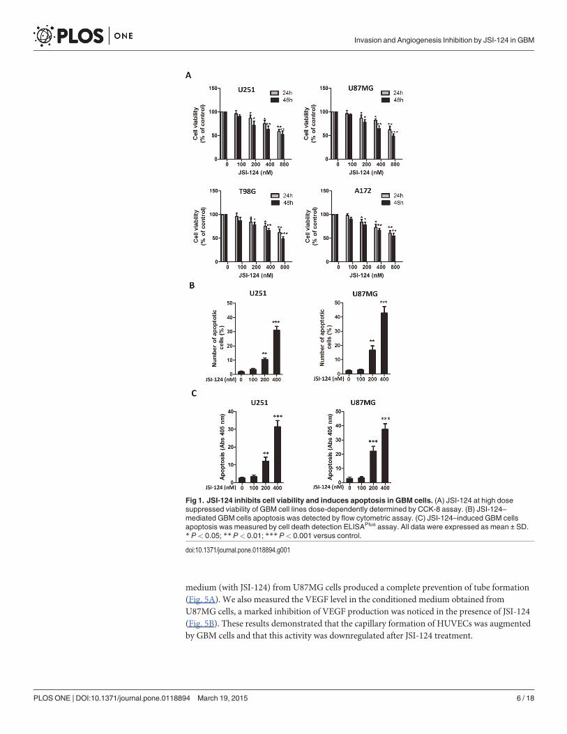
JSI-124 inhibits cell viability and induces apoptosis in GBM cellsTo assess the effect of JSI-124 on GBM cells, we first examined several GBM cell lines treatedwith various doses of JSI-124 by CCK-8 assay. JSI-124 inhibited cell viability in a dose- andtime-dependent manner, and significant cell viability inhibitory effect of JSI-124 was observedat concentrations more than 100nmol/L (Fig. 1A). JSI-124 exhibited anticancer potentialthrough the induction of apoptosis in a wide variety of human tumor cell lines [19,21,22]. Toverify apoptotic property on GBM cells, flow cytometry assay and cell death detection ELISA-Plus assay were performed. No change in apoptosis was observed at 100nM JSI-124 for 48 h, buthigher concentration JSI-124 significantly induced GBM cells apoptosis dose-dependently(Fig. 1B and S1 Fig.). The similar phenomenon was observed in GBM cells determined by celldeath detection ELISAPlus assay (Fig. 1C).
JSI-124 treatment at low dose inhibits invasiveness of GBM cellsWe next examined if the effect of JSI-124 at low dose on invasiveness of GBM cells. As show inFig. 2, transwell invasion assay revealed 100 nM JSI-124 treatment significantly inhibited GBMcells invasion ability at 8 h.
Exposure of GBM cells to JSI-124 at low dose reduces clonogenicityWe further examined if low-dose JSI-124 would be sufficient to prevent GBM cells tumorige-nicity. Using an in vitro clonogenic assay, we found that just brief exposure of GBM cells toJSI-124 (100 nM) for 2 h was sufficient to significantly reduce the number of tumor colonies ofU251 and U87MG cells (Fig. 3), suggesting that JSI-124 may inhibit GBM tumorigenicity.
JSI-124 inhibits VEGF expression in GBM cellsVEGF is a critical factor in new blood vessel formation [6,24]. In a tumor microenvironment,cancer cells secrete a high level of VEGF that binds to receptors on surrounding endothelialcells, promoting endothelial cell migration, proliferation, and differentiation, as well as tubeformation [25,26]. In this experiment, we measure the effect of JSI-124 on the VEGF level inGBM cells at different concentrations by ELISA. The treatment of JSI-124 at 100nM for 24hours markedly reduced the secretion of VEGF by GBM cells (Fig. 4).
JSI-124 inhibites the U87MG cells–induced tube formation of HUVECsIn this experiment, we used conditioned media from U87MG cells treated with or withoutJSI-124 to determine whether GBM cells could induce the capillary formation of HUVECs andexamine the effect of on this event. It is clearly seen that the conditioned medium (without JSI-124) from U87MG cells induced capillary formation of HUVECs. However, the conditioned
Invasion and Angiogenesis Inhibition by JSI-124 in GBM
PLOSONE | DOI:10.1371/journal.pone.0118894 March 19, 2015 5 / 18
medium (with JSI-124) from U87MG cells produced a complete prevention of tube formation(Fig. 5A). We also measured the VEGF level in the conditioned medium obtained fromU87MG cells, a marked inhibition of VEGF production was noticed in the presence of JSI-124(Fig. 5B). These results demonstrated that the capillary formation of HUVECs was augmentedby GBM cells and that this activity was downregulated after JSI-124 treatment.
Fig 1. JSI-124 inhibits cell viability and induces apoptosis in GBM cells. (A) JSI-124 at high dosesuppressed viability of GBM cell lines dose-dependently determined by CCK-8 assay. (B) JSI-124–mediated GBM cells apoptosis was detected by flow cytometric assay. (C) JSI-124–induced GBM cellsapoptosis was measured by cell death detection ELISAPlus assay. All data were expressed as mean ± SD.* P< 0.05; ** P< 0.01; *** P< 0.001 versus control.
doi:10.1371/journal.pone.0118894.g001
Invasion and Angiogenesis Inhibition by JSI-124 in GBM
PLOSONE | DOI:10.1371/journal.pone.0118894 March 19, 2015 6 / 18
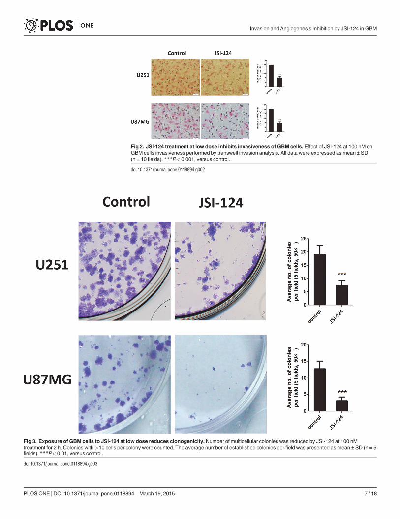
Fig 2. JSI-124 treatment at low dose inhibits invasiveness of GBM cells. Effect of JSI-124 at 100 nM onGBM cells invasiveness performed by transwell invasion analysis. All data were expressed as mean ± SD(n = 10 fields). ***P< 0.001, versus control.
doi:10.1371/journal.pone.0118894.g002
Fig 3. Exposure of GBM cells to JSI-124 at low dose reduces clonogenicity.Number of multicellular colonies was reduced by JSI-124 at 100 nMtreatment for 2 h. Colonies with>10 cells per colony were counted. The average number of established colonies per field was presented as mean ± SD (n = 5fields). ***P< 0.01, versus control.
doi:10.1371/journal.pone.0118894.g003
Invasion and Angiogenesis Inhibition by JSI-124 in GBM
PLOSONE | DOI:10.1371/journal.pone.0118894 March 19, 2015 7 / 18
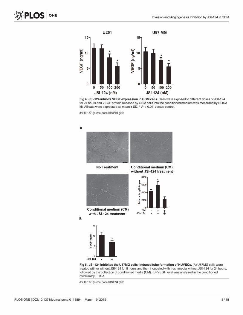
Fig 4. JSI-124 inhibits VEGF expression in GBM cells.Cells were exposed to different doses of JSI-124for 24 hours and VEGF protein released by GBM cells into the conditioned medium was measured by ELISAkit. All data were expressed as mean ± SD. * P< 0.05, versus control.
doi:10.1371/journal.pone.0118894.g004
Fig 5. JSI-124 inhibites the U87MG cells–induced tube formation of HUVECs. (A) U87MG cells weretreated with or without JSI-124 for 8 hours and then incubated with fresh media without JSI-124 for 24 hours,followed by the collection of conditioned media (CM). (B) VEGF level was analyzed in the conditionedmedium by ELISA.
doi:10.1371/journal.pone.0118894.g005
Invasion and Angiogenesis Inhibition by JSI-124 in GBM
PLOSONE | DOI:10.1371/journal.pone.0118894 March 19, 2015 8 / 18
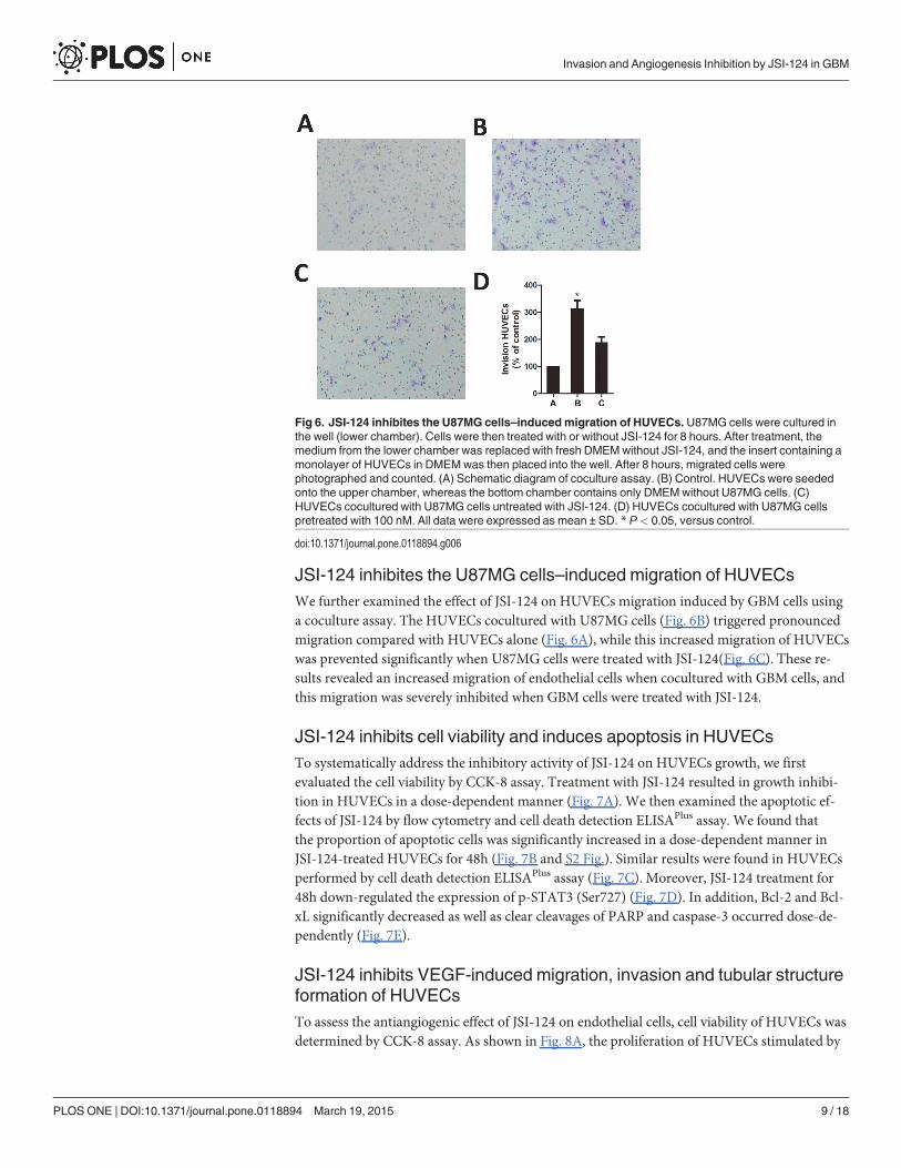
JSI-124 inhibites the U87MG cells–induced migration of HUVECsWe further examined the effect of JSI-124 on HUVECs migration induced by GBM cells usinga coculture assay. The HUVECs cocultured with U87MG cells (Fig. 6B) triggered pronouncedmigration compared with HUVECs alone (Fig. 6A), while this increased migration of HUVECswas prevented significantly when U87MG cells were treated with JSI-124(Fig. 6C). These re-sults revealed an increased migration of endothelial cells when cocultured with GBM cells, andthis migration was severely inhibited when GBM cells were treated with JSI-124.
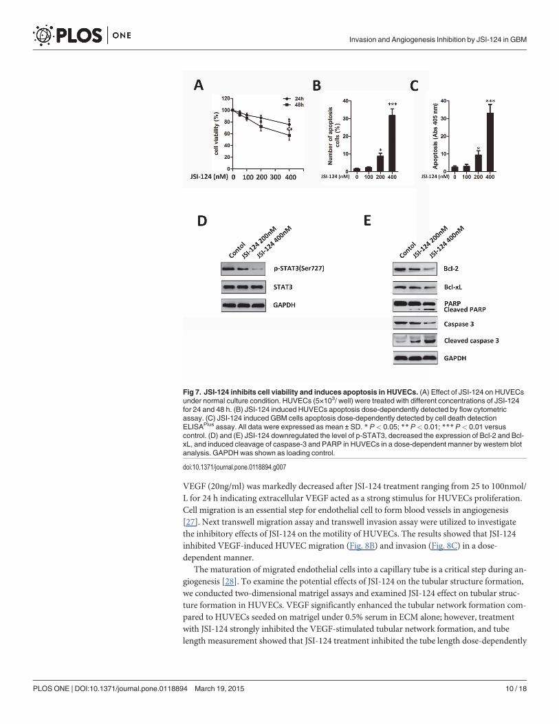
JSI-124 inhibits cell viability and induces apoptosis in HUVECsTo systematically address the inhibitory activity of JSI-124 on HUVECs growth, we firstevaluated the cell viability by CCK-8 assay. Treatment with JSI-124 resulted in growth inhibi-tion in HUVECs in a dose-dependent manner (Fig. 7A). We then examined the apoptotic ef-fects of JSI-124 by flow cytometry and cell death detection ELISAPlus assay. We found thatthe proportion of apoptotic cells was significantly increased in a dose-dependent manner inJSI-124-treated HUVECs for 48h (Fig. 7B and S2 Fig.). Similar results were found in HUVECsperformed by cell death detection ELISAPlus assay (Fig. 7C). Moreover, JSI-124 treatment for48h down-regulated the expression of p-STAT3 (Ser727) (Fig. 7D). In addition, Bcl-2 and Bcl-xL significantly decreased as well as clear cleavages of PARP and caspase-3 occurred dose-de-pendently (Fig. 7E).
JSI-124 inhibits VEGF-induced migration, invasion and tubular structureformation of HUVECsTo assess the antiangiogenic effect of JSI-124 on endothelial cells, cell viability of HUVECs wasdetermined by CCK-8 assay. As shown in Fig. 8A, the proliferation of HUVECs stimulated by
Fig 6. JSI-124 inhibites the U87MG cells–inducedmigration of HUVECs.U87MG cells were cultured inthe well (lower chamber). Cells were then treated with or without JSI-124 for 8 hours. After treatment, themedium from the lower chamber was replaced with fresh DMEMwithout JSI-124, and the insert containing amonolayer of HUVECs in DMEMwas then placed into the well. After 8 hours, migrated cells werephotographed and counted. (A) Schematic diagram of coculture assay. (B) Control. HUVECs were seededonto the upper chamber, whereas the bottom chamber contains only DMEMwithout U87MG cells. (C)HUVECs cocultured with U87MG cells untreated with JSI-124. (D) HUVECs cocultured with U87MG cellspretreated with 100 nM. All data were expressed as mean ± SD. * P< 0.05, versus control.
doi:10.1371/journal.pone.0118894.g006
Invasion and Angiogenesis Inhibition by JSI-124 in GBM
PLOSONE | DOI:10.1371/journal.pone.0118894 March 19, 2015 9 / 18
VEGF (20ng/ml) was markedly decreased after JSI-124 treatment ranging from 25 to 100nmol/L for 24 h indicating extracellular VEGF acted as a strong stimulus for HUVECs proliferation.Cell migration is an essential step for endothelial cell to form blood vessels in angiogenesis[27]. Next transwell migration assay and transwell invasion assay were utilized to investigatethe inhibitory effects of JSI-124 on the motility of HUVECs. The results showed that JSI-124inhibited VEGF-induced HUVEC migration (Fig. 8B) and invasion (Fig. 8C) in a dose-dependent manner.
The maturation of migrated endothelial cells into a capillary tube is a critical step during an-giogenesis [28]. To examine the potential effects of JSI-124 on the tubular structure formation,we conducted two-dimensional matrigel assays and examined JSI-124 effect on tubular struc-ture formation in HUVECs. VEGF significantly enhanced the tubular network formation com-pared to HUVECs seeded on matrigel under 0.5% serum in ECM alone; however, treatmentwith JSI-124 strongly inhibited the VEGF-stimulated tubular network formation, and tubelength measurement showed that JSI-124 treatment inhibited the tube length dose-dependently
Fig 7. JSI-124 inhibits cell viability and induces apoptosis in HUVECs. (A) Effect of JSI-124 on HUVECsunder normal culture condition. HUVECs (5×10³/ well) were treated with different concentrations of JSI-124for 24 and 48 h. (B) JSI-124 induced HUVECs apoptosis dose-dependently detected by flow cytometricassay. (C) JSI-124 induced GBM cells apoptosis dose-dependently detected by cell death detectionELISAPlus assay. All data were expressed as mean ± SD. * P< 0.05; ** P< 0.01; *** P< 0.01 versuscontrol. (D) and (E) JSI-124 downregulated the level of p-STAT3, decreased the expression of Bcl-2 and Bcl-xL, and induced cleavage of caspase-3 and PARP in HUVECs in a dose-dependent manner by western blotanalysis. GAPDH was shown as loading control.
doi:10.1371/journal.pone.0118894.g007
Invasion and Angiogenesis Inhibition by JSI-124 in GBM
PLOSONE | DOI:10.1371/journal.pone.0118894 March 19, 2015 10 / 18
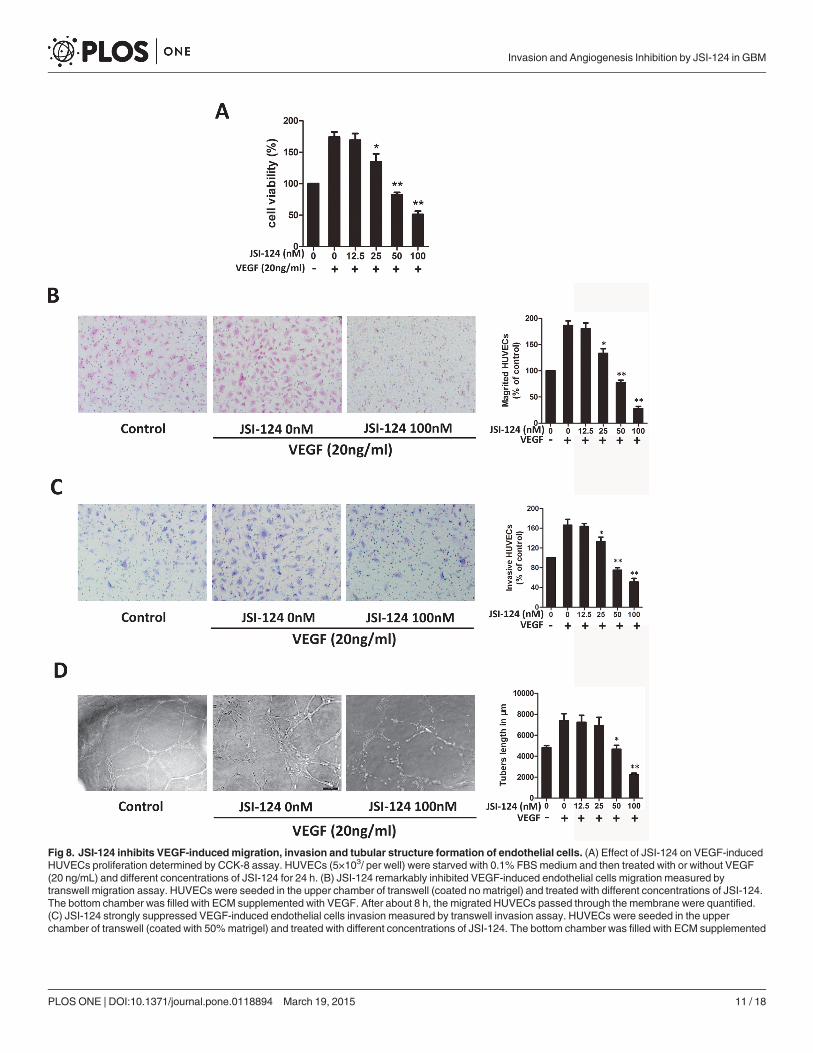
Fig 8. JSI-124 inhibits VEGF-inducedmigration, invasion and tubular structure formation of endothelial cells. (A) Effect of JSI-124 on VEGF-inducedHUVECs proliferation determined by CCK-8 assay. HUVECs (5×10³/ per well) were starved with 0.1% FBSmedium and then treated with or without VEGF(20 ng/mL) and different concentrations of JSI-124 for 24 h. (B) JSI-124 remarkably inhibited VEGF-induced endothelial cells migration measured bytranswell migration assay. HUVECs were seeded in the upper chamber of transwell (coated no matrigel) and treated with different concentrations of JSI-124.The bottom chamber was filled with ECM supplemented with VEGF. After about 8 h, the migrated HUVECs passed through the membrane were quantified.(C) JSI-124 strongly suppressed VEGF-induced endothelial cells invasion measured by transwell invasion assay. HUVECs were seeded in the upperchamber of transwell (coated with 50%matrigel) and treated with different concentrations of JSI-124. The bottom chamber was filled with ECM supplemented
Invasion and Angiogenesis Inhibition by JSI-124 in GBM
PLOSONE | DOI:10.1371/journal.pone.0118894 March 19, 2015 11 / 18
(Fig. 8D). Overall, these results indicated that JSI-124 could suppress VEGF-induced angiogen-esis by inhibiting migration, invasion, and tube formation of HUVECs.
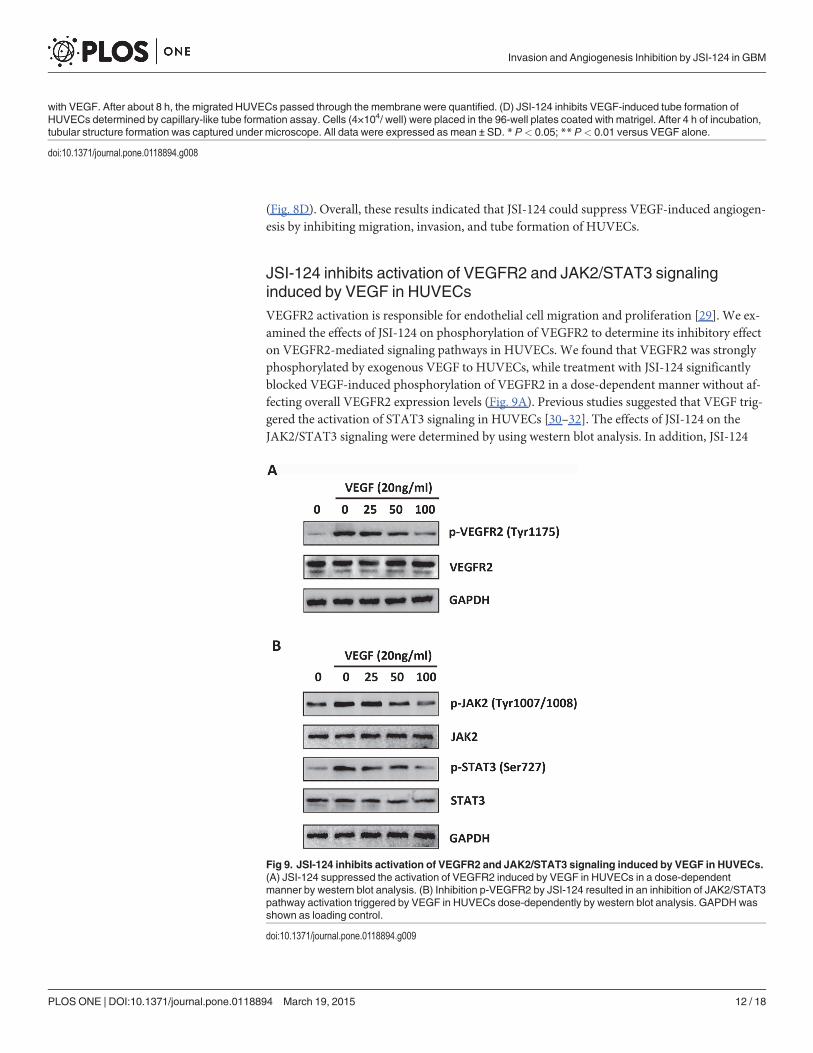
JSI-124 inhibits activation of VEGFR2 and JAK2/STAT3 signalinginduced by VEGF in HUVECsVEGFR2 activation is responsible for endothelial cell migration and proliferation [29]. We ex-amined the effects of JSI-124 on phosphorylation of VEGFR2 to determine its inhibitory effecton VEGFR2-mediated signaling pathways in HUVECs. We found that VEGFR2 was stronglyphosphorylated by exogenous VEGF to HUVECs, while treatment with JSI-124 significantlyblocked VEGF-induced phosphorylation of VEGFR2 in a dose-dependent manner without af-fecting overall VEGFR2 expression levels (Fig. 9A). Previous studies suggested that VEGF trig-gered the activation of STAT3 signaling in HUVECs [30–32]. The effects of JSI-124 on theJAK2/STAT3 signaling were determined by using western blot analysis. In addition, JSI-124
with VEGF. After about 8 h, the migrated HUVECs passed through the membrane were quantified. (D) JSI-124 inhibits VEGF-induced tube formation ofHUVECs determined by capillary-like tube formation assay. Cells (4×10⁴/ well) were placed in the 96-well plates coated with matrigel. After 4 h of incubation,tubular structure formation was captured under microscope. All data were expressed as mean ± SD. * P< 0.05; ** P< 0.01 versus VEGF alone.
doi:10.1371/journal.pone.0118894.g008
Fig 9. JSI-124 inhibits activation of VEGFR2 and JAK2/STAT3 signaling induced by VEGF in HUVECs.(A) JSI-124 suppressed the activation of VEGFR2 induced by VEGF in HUVECs in a dose-dependentmanner by western blot analysis. (B) Inhibition p-VEGFR2 by JSI-124 resulted in an inhibition of JAK2/STAT3pathway activation triggered by VEGF in HUVECs dose-dependently by western blot analysis. GAPDHwasshown as loading control.
doi:10.1371/journal.pone.0118894.g009
Invasion and Angiogenesis Inhibition by JSI-124 in GBM
PLOSONE | DOI:10.1371/journal.pone.0118894 March 19, 2015 12 / 18
significantly suppressed the phosphorylation of JAK2 (Tyr1007/1008) and STAT3 (Ser727)stimulated by VEGF in a dose-dependent manner (Fig. 9B). These results provide evidencethat JSI-124 blocked angiogenesis by targeting JAK2/STAT3 signaling pathway.
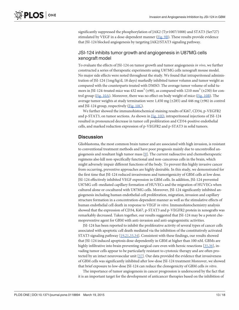
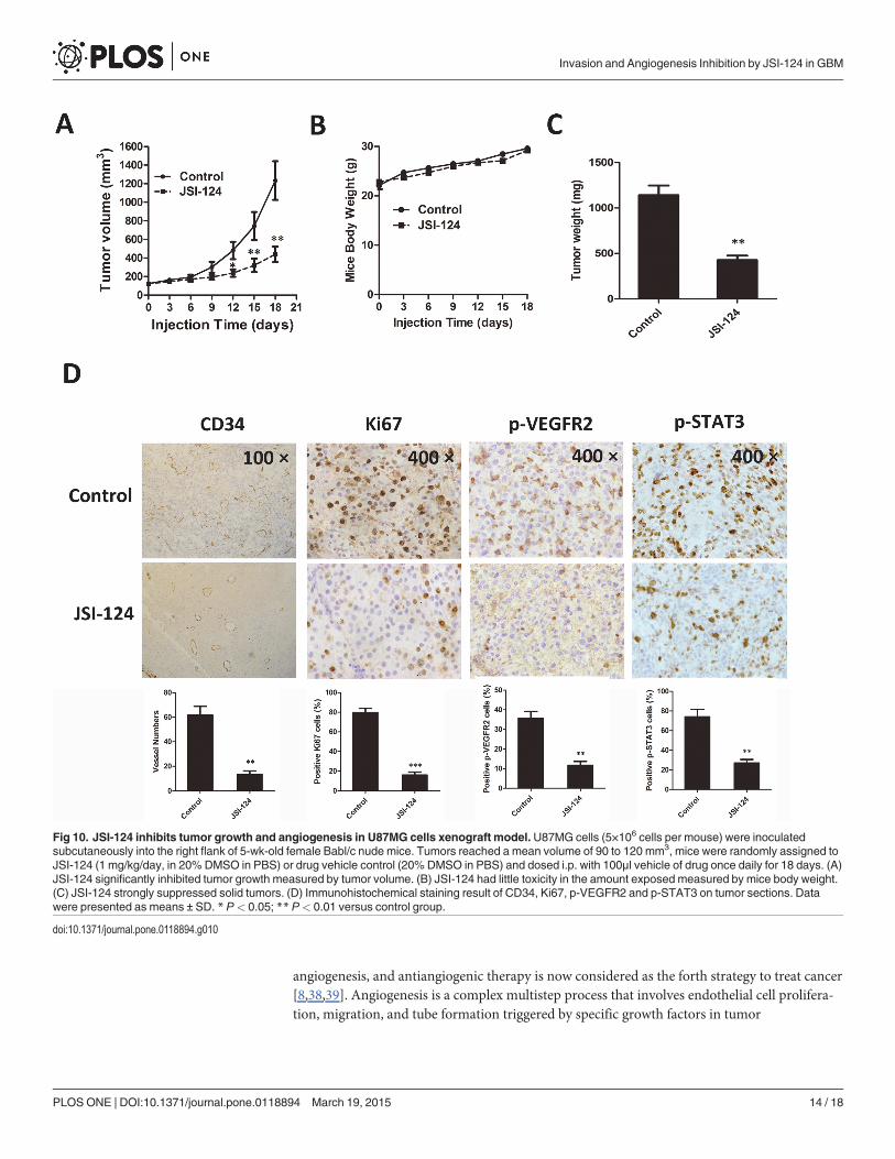
JSI-124 inhibits tumor growth and angiogenesis in U87MG cellsxenograft modelTo evaluate the effects of JSI-124 on tumor growth and tumor angiogenesis in vivo, we furtherconstructed a series of therapeutic experiments using U87MG cells xenograft mouse model.No major side effects were noted throughout the study. We found that intraperitoneal adminis-tration of JSI-124 (1mg/kg/d, 18 days) markedly inhibited tumor volumn and tumor weight ascompared with the counterparts treated with DMSO. The average tumor volume of solid tu-mors in JSI-124-treated mice was 432 mm3 (±90), as compared with 1210 mm3 (±230) for con-trol group (Fig. 10A). Moreover, there was no effect on body weight of mice (Fig. 10B). Theaverage tumor weights at study termination were 1,450 mg (±285) and 446 mg (±96) in controland JSI-124 group, respectively (Fig. 10C).
We further showed the immunohistochemical staining results of Ki67, CD34, p-VEGFR2and p-STAT3, on tumor sections. As shown in Fig. 10D, intraperitoneal injections of JSI-124resulted in pronounced decrease in tumor cell proliferation and CD34-positive endothelialcells, and marked reduction expression of p-VEGFR2 and p-STAT3 in solid tumors.
DiscussionGlioblastoma, the most common brain tumor and are associated with high invasion, is resistantto conventional treatment methods and have poor prognosis mainly due to uncontrolled an-giogenesis and resultant high tumor mass [9]. The current radioactive and chemotherapeuticregimens also kill non-specifically functional and non-cancerous cells in the brain, whichmight adversely impair different functions of the body. To prevent this highly invasive cancerfrom occurring, preventive approaches are highly desirable. In this study, we demonstrated forthe first time that JSI-124 reduced invasiveness and tumorigenicity of GBM cells at low dose.JSI-124 effectively inhibited VEGF expression in GBM cells. In addition, JSI-124 preventedU87MG cell–mediated capillary formation of HUVECs and the migration of HUVECs whencultured alone or cocultured with U87MG cells. Moreover, JSI-124 significantly inhibited an-giogenesis including human endothelial cell proliferation, migration, invasion and capillarystructure formation in a concentration-dependent manner as well as the stimulative effects ofhuman endothelial cell death in response to VEGF in vitro. Immunohistochemistry analysisshowed that the expression of CD34, Ki67, p-STAT3 and p-VEGFR2 protein in xenografts wasremarkably decreased. Taken together, our results suggested that JSI-124 may be a potent che-mopreventive agent for GBM with anti-invasion and anti-angiogenisitic activities.
JSI-124 has been reported to inhibit the proliferative activity of several types of cancer cellsassociated with apoptotic cell death mediated via the inhibition of the constitutively activatedSTAT3 signaling pathway [19,21,33,34]. Consistent with these findings, our results showedthat JSI-124 induced apoptosis dose-dependently in GBM at higher than 100 nM. GBMs arehighly infiltrative into brain preventing surgical cure even with heroic resections [35,36], in-vading tumor cells appear to be particularly resistant to cytotoxic therapy and are often pro-tected by an intact neurovascular unit [37]. Our data provided the evidence that invasivenessof GBM cells was significantly inhibited after low-dose JSI-124 treatment Moreover, we showedthat brief exposure to low-dose JSI-124 can reduce the clonogenicity of GBM cells in vitro.
The importance of tumor angiogenesis in cancer progression is underscored by the fact thatit is an important target for the development of anticancer therapies based on the inhibition of
Invasion and Angiogenesis Inhibition by JSI-124 in GBM
PLOSONE | DOI:10.1371/journal.pone.0118894 March 19, 2015 13 / 18
angiogenesis, and antiangiogenic therapy is now considered as the forth strategy to treat cancer[8,38,39]. Angiogenesis is a complex multistep process that involves endothelial cell prolifera-tion, migration, and tube formation triggered by specific growth factors in tumor
Fig 10. JSI-124 inhibits tumor growth and angiogenesis in U87MG cells xenograft model. U87MG cells (5×10⁶ cells per mouse) were inoculatedsubcutaneously into the right flank of 5-wk-old female Babl/c nude mice. Tumors reached a mean volume of 90 to 120 mm3, mice were randomly assigned toJSI-124 (1 mg/kg/day, in 20% DMSO in PBS) or drug vehicle control (20% DMSO in PBS) and dosed i.p. with 100μl vehicle of drug once daily for 18 days. (A)JSI-124 significantly inhibited tumor growth measured by tumor volume. (B) JSI-124 had little toxicity in the amount exposed measured by mice body weight.(C) JSI-124 strongly suppressed solid tumors. (D) Immunohistochemical staining result of CD34, Ki67, p-VEGFR2 and p-STAT3 on tumor sections. Datawere presented as means ± SD. * P< 0.05; ** P< 0.01 versus control group.
doi:10.1371/journal.pone.0118894.g010
Invasion and Angiogenesis Inhibition by JSI-124 in GBM
PLOSONE | DOI:10.1371/journal.pone.0118894 March 19, 2015 14 / 18
microenvironment [39]. Cancer cells produce numerous angiogenic factors, including VEGF,FGF, EGF, PDGF etc, which play a pivotal role in the development of tumor angiogenesis bystimulating endothelial cell proliferation, migration, and capillary tube formation [40–42].Among all angiogenic factors, VEGF is identified as a key mediator of angiogenesis [6]. Glio-blastomas secrete a very large quantity of VEGF protein into the surrounding microenviron-ment, thereby allowing endothelial cell proliferation, migration, and tube formation [43,44]. Inthis study, we found that the basal VEGF levels in U87MG and U251 cells were significantlyhigh and that JSI-124 dose-dependently downregulated protein levels of VEGF. Using a cocul-ture assay, we examined the effect of this inhibitor on glioblastoma cell–induced migration andon the tube formation of endothelial cells. Treatment with JSI-124 decreased VEGF secretionby U87MG cells and thereby reduced endothelial cell migration and tube formation.
Moreover, we showed that JSI-124 blocked the proliferation, capillary formation, and mi-gration of endothelial cells, all of which are critical steps for angiogenesis. We further showedthat induction of apoptosis in endothelial cells by JSI-124 was due to the downregulation ofBcl-2 and Bcl-xL, both of which are apoptotic proteins overexpressed in endothelial cells andassociated with resistance to apoptosis. Overexpression of VEGF and VEGF receptors corre-lates with increased microvessel density, proliferation, and tumor growth rate, which lead topoor patient prognosis in a variety of malignancies [45,46]. We provided first evidence thatJSI-124 effectively abrogated VEGF-induced HUVECs proliferation, invasion, migration, andcapillary-like structures formation in vitro. Phosphorylation of VEGFR2 is critical for VEGF-mediated microvascular permeability, endothelial cell proliferation, invasion and migration[47–49]. In the present study, we found that JSI-124 blocked the activity of VEGFR2 (Tyr1175)by down-regulation of VEGF-induced phosphorylation of VEGFR-2 expression. A close asso-ciation between STAT3 activation and glioma growth and vascularization has been reportedpreviously [11,12,50], and activation of STAT3 has been directly correlated with VEGF pro-duction [13]. Our present investigation showed that the expression of VEGF was dose-depen-dently suppressed by JSI-124 via STAT3 (Ser727) inhibition in HUVECs. STAT3 is principallyactivated by nonreceptor tyrosine kinase JAK2 [51]. Our results showed that phosphorylationof JAK2 (Tyr1007/1008) was dose-dependently blocked by JSI-124 in HUVECs, indicating thatthe direct effects of JSI-124 on angiogenesis might be through inhibiting the VEGFR2/STAT3signaling pathway.
To evaluate the antitumor activity of JSI-124 in vivo, Babl/c nude mice transplanted withU87MG cells were treated with JSI-124. We found that JSI-124 (1 mg/kg/day) significantly sup-pressed tumor volume and tumor weight without any side effects on mice, and remarkably re-duced neovascularization accompanied by down-regulation expression of p-STAT3 and p-VEGFR2. In addition to the effective antiangiogenesis of JSI-124 in vitro, we presume that JSI-124 suppresses GBM growth in vivo through not only directly tumor cell proliferation inhibi-tion but also tumor angiogenesis suppression.
Taken together, our results indicate that JSI-24 may be an effective preventive agent forGBM with anti-invasion and anti-angiogenesitic activity. It is hoped that in the future, patientsharboring GBM can be offered an affordable preventive agent to reduce the incidence of GBM.
Supporting InformationS1 Fig. JSI-124 induced GBM cells apoptosis dose-dependently detected by flow cytometricassay.(TIF)
Invasion and Angiogenesis Inhibition by JSI-124 in GBM
PLOSONE | DOI:10.1371/journal.pone.0118894 March 19, 2015 15 / 18
S2 Fig. JSI-124 induced HUVECs apoptosis dose-dependently detected by flow cytometricassay.(TIF)
Author ContributionsConceived and designed the experiments: GY. Performed the experiments: GY SY. Analyzedthe data: HX. Contributed reagents/materials/analysis tools: PZ JS. Wrote the paper: GY GL.
References1. Louis DN, Ohgaki H, Wiestler OD, CaveneeWK, Burger PC, et al. (2007) The 2007WHO classification
of tumours of the central nervous system. Acta Neuropathol 114: 97–109. PMID: 17618441
2. Fan S, Sun Z, Jiang D, Dai C, Ma Y, et al. (2010) BmKCT toxin inhibits glioma proliferation and tumormetastasis. Cancer Lett 291: 158–166. doi: 10.1016/j.canlet.2009.10.011 PMID: 19906483
3. Louis DN, Pomeroy SL, Cairncross JG (2002) Focus on central nervous system neoplasia. Cancer Cell1: 125–128. PMID: 12086870
4. Omuro AM, Faivre S, Raymond E (2007) Lessons learned in the development of targeted therapy formalignant gliomas. Mol Cancer Ther 6: 1909–1919. PMID: 17620423
5. Skobe M, Rockwell P, Goldstein N, Vosseler S, Fusenig NE (1997) Halting angiogenesis suppressescarcinoma cell invasion. Nat Med 3: 1222–1227. PMID: 9359696
6. Folkman J (1995) Angiogenesis in cancer, vascular, rheumatoid and other disease. Nat Med 1: 27–31.PMID: 7584949
7. Hanahan D, Weinberg RA (2000) The hallmarks of cancer. Cell 100: 57–70. PMID: 10647931
8. Cook KM, FiggWD (2010) Angiogenesis inhibitors: current strategies and future prospects. CA CancerJ Clin 60: 222–243. doi: 10.3322/caac.20075 PMID: 20554717
9. Onishi M, Ichikawa T, Kurozumi K, Date I (2011) Angiogenesis and invasion in glioma. Brain TumorPathol 28: 13–24. doi: 10.1007/s10014-010-0007-z PMID: 21221826
10. Jain RK, di Tomaso E, Duda DG, Loeffler JS, Sorensen AG, et al. (2007) Angiogenesis in brain tu-mours. Nat Rev Neurosci 8: 610–622. PMID: 17643088
11. Doucette TA, Kong LY, Yang Y, Ferguson SD, Yang J, et al. (2012) Signal transducer and activator oftranscription 3 promotes angiogenesis and drives malignant progression in glioma. Neuro Oncol 14:1136–1145. doi: 10.1093/neuonc/nos139 PMID: 22753228
12. de Groot J, Liang J, Kong LY, Wei J, Piao Y, et al. (2012) Modulating antiangiogenic resistance by inhib-iting the signal transducer and activator of transcription 3 pathway in glioblastoma. Oncotarget 3:1036–1048. PMID: 23013619
13. Chen Z, Han ZC (2008) STAT3: a critical transcription activator in angiogenesis. Med Res Rev 28:185–200. PMID: 17457812
14. Jung JE, Lee HG, Cho IH, Chung DH, Yoon SH, et al. (2005) STAT3 is a potential modulator of HIF-1-mediated VEGF expression in human renal carcinoma cells. FASEB J 19: 1296–1298. PMID:15919761
15. Buettner R, Mora LB, Jove R (2002) Activated STAT signaling in human tumors provides novel molecu-lar targets for therapeutic intervention. Clin Cancer Res 8: 945–954. PMID: 11948098
16. Gamero AM, Young HA, Wiltrout RH (2004) Inactivation of Stat3 in tumor cells: releasing a brake on im-mune responses against cancer? Cancer Cell 5: 111–112. PMID: 14998485
17. Kim MJ, NamHJ, Kim HP, Han SW, Im SA, et al. (2013) OPB-31121, a novel small molecular inhibitor,disrupts the JAK2/STAT3 pathway and exhibits an antitumor activity in gastric cancer cells. Cancer Lett335: 145–152. doi: 10.1016/j.canlet.2013.02.010 PMID: 23402820
18. Pathak AK, Bhutani M, Nair AS, Ahn KS, Chakraborty A, et al. (2007) Ursolic acid inhibits STAT3 acti-vation pathway leading to suppression of proliferation and chemosensitization of human multiple mye-loma cells. Mol Cancer Res 5: 943–955. PMID: 17855663
19. Su Y, Li G, Zhang X, Gu J, Zhang C, et al. (2008) JSI-124 inhibits glioblastoma multiforme cell prolifera-tion through G(2)/M cell cycle arrest and apoptosis augment. Cancer Biol Ther 7: 1243–1249. PMID:18487947
20. Heath VL, Bicknell R (2009) Anticancer strategies involving the vasculature. Nat Rev Clin Oncol 6:395–404. doi: 10.1038/nrclinonc.2009.52 PMID: 19424102
Invasion and Angiogenesis Inhibition by JSI-124 in GBM
PLOSONE | DOI:10.1371/journal.pone.0118894 March 19, 2015 16 / 18
21. Blaskovich MA, Sun J, Cantor A, Turkson J, Jove R, et al. (2003) Discovery of JSI-124 (cucurbitacin I),a selective Janus kinase/signal transducer and activator of transcription 3 signaling pathway inhibitorwith potent antitumor activity against human and murine cancer cells in mice. Cancer Res 63: 1270–1279. PMID: 12649187
22. Jing N, Tweardy DJ (2005) Targeting Stat3 in cancer therapy. Anticancer Drugs 16: 601–607. PMID:15930886
23. Tsujii M, Kawano S, Tsuji S, Sawaoka H, Hori M, et al. (1998) Cyclooxygenase regulates angiogenesisinduced by colon cancer cells. Cell 93: 705–716. PMID: 9630216
24. Ferrara N (1999) Molecular and biological properties of vascular endothelial growth factor. J Mol Med(Berl) 77: 527–543. PMID: 10494799
25. Grunstein J, Roberts WG, Mathieu-Costello O, Hanahan D, Johnson RS (1999) Tumor-derived expres-sion of vascular endothelial growth factor is a critical factor in tumor expansion and vascular function.Cancer Res 59: 1592–1598. PMID: 10197634
26. Holash J, Maisonpierre PC, Compton D, Boland P, Alexander CR, et al. (1999) Vessel cooption, regres-sion, and growth in tumors mediated by angiopoietins and VEGF. Science 284: 1994–1998. PMID:10373119
27. Shibuya M (2006) Vascular endothelial growth factor (VEGF)-Receptor2: its biological functions, majorsignaling pathway, and specific ligand VEGF-E. Endothelium 13: 63–69. PMID: 16728325
28. Patan S (2004) Vasculogenesis and angiogenesis. Cancer Treat Res 117: 3–32. PMID: 15015550
29. Ferrara N, Gerber HP, LeCouter J (2003) The biology of VEGF and its receptors. Nat Med 9: 669–676.PMID: 12778165
30. Chen J, Wang J, Lin L, He L, Wu Y, et al. (2012) Inhibition of STAT3 signaling pathway by nitidine chlo-ride suppressed the angiogenesis and growth of human gastric cancer. Mol Cancer Ther 11: 277–287.doi: 10.1158/1535-7163.MCT-11-0648 PMID: 22203730
31. Kandala PK, Srivastava SK (2012) Diindolylmethane suppresses ovarian cancer growth and potenti-ates the effect of cisplatin in tumor mouse model by targeting signal transducer and activator of tran-scription 3 (STAT3). BMCMed 10: 9. doi: 10.1186/1741-7015-10-9 PMID: 22280969
32. Lu J, Zhang K, Nam S, Anderson RA, Jove R, et al. (2010) Novel angiogenesis inhibitory activity in cin-namon extract blocks VEGFR2 kinase and downstream signaling. Carcinogenesis 31: 481–488. doi:10.1093/carcin/bgp292 PMID: 19969552
33. Ishdorj G, Johnston JB, Gibson SB (2010) Inhibition of constitutive activation of STAT3 by curcurbita-cin-I (JSI-124) sensitized human B-leukemia cells to apoptosis. Mol Cancer Ther 9: 3302–3314. doi:10.1158/1535-7163.MCT-10-0550 PMID: 21159613
34. Yuan G, Yan SF, Xue H, Zhang P, Sun JT, et al. (2014) Cucurbitacin I induces protective autophagy inglioblastoma in vitro and in vivo. J Biol Chem 289: 10607–10619. doi: 10.1074/jbc.M113.528760PMID: 24599950
35. Salhia B, Tran NL, SymonsM, Winkles JA, Rutka JT, et al. (2006) Molecular pathways triggering gliomacell invasion. Expert Rev Mol Diagn 6: 613–626. PMID: 16824034
36. Kwiatkowska A, Symons M (2013) Signaling determinants of glioma cell invasion. Adv Exp Med Biol986: 121–141. doi: 10.1007/978-94-007-4719-7_7 PMID: 22879067
37. Furnari FB, Fenton T, Bachoo RM, Mukasa A, Stommel JM, et al. (2007) Malignant astrocytic glioma:genetics, biology, and paths to treatment. Genes Dev 21: 2683–2710. PMID: 17974913
38. Kerbel RS (2008) Tumor angiogenesis. N Engl J Med 358: 2039–2049. doi: 10.1056/NEJMra0706596PMID: 18463380
39. Bellou S, Pentheroudakis G, Murphy C, Fotsis T (2013) Anti-angiogenesis in cancer therapy: Herculesand hydra. Cancer Lett 338: 219–228. doi: 10.1016/j.canlet.2013.05.015 PMID: 23707856
40. Carmeliet P (2005) VEGF as a key mediator of angiogenesis in cancer. Oncology 69 Suppl 3: 4–10.PMID: 16301830
41. Chaudhry IH, O'Donovan DG, Brenchley PE, Reid H, Roberts IS (2001) Vascular endothelial growthfactor expression correlates with tumour grade and vascularity in gliomas. Histopathology 39: 409–415. PMID: 11683943
42. Dunn IF, Heese O, Black PM (2000) Growth factors in glioma angiogenesis: FGFs, PDGF, EGF, andTGFs. J Neurooncol 50: 121–137. PMID: 11245272
43. Bao S, Wu Q, Sathornsumetee S, Hao Y, Li Z, et al. (2006) Stem cell-like glioma cells promote tumorangiogenesis through vascular endothelial growth factor. Cancer Res 66: 7843–7848. PMID:16912155
44. Machein MR, Plate KH (2000) VEGF in brain tumors. J Neurooncol 50: 109–120. PMID: 11245271
Invasion and Angiogenesis Inhibition by JSI-124 in GBM
PLOSONE | DOI:10.1371/journal.pone.0118894 March 19, 2015 17 / 18
45. Ferrara N, Gerber HP (2001) The role of vascular endothelial growth factor in angiogenesis. Acta Hae-matol 106: 148–156. PMID: 11815711
46. Dvorak HF, Detmar M, Claffey KP, Nagy JA, van deWater L, et al. (1995) Vascular permeability factor/vascular endothelial growth factor: an important mediator of angiogenesis in malignancy and inflamma-tion. Int Arch Allergy Immunol 107: 233–235. PMID: 7542074
47. Dvorak HF, Nagy JA, Feng D, Brown LF, Dvorak AM (1999) Vascular permeability factor/vascular en-dothelial growth factor and the significance of microvascular hyperpermeability in angiogenesis. CurrTop Microbiol Immunol 237: 97–132. PMID: 9893348
48. Zachary I, Gliki G (2001) Signaling transduction mechanismsmediating biological actions of the vascu-lar endothelial growth factor family. Cardiovasc Res 49: 568–581. PMID: 11166270
49. Saraswati S, Agrawal SS (2013) Brucine, an indole alkaloid from Strychnos nux-vomica attenuatesVEGF-induced angiogenesis via inhibiting VEGFR2 signaling pathway in vitro and in vivo. Cancer Lett332: 83–93. doi: 10.1016/j.canlet.2013.01.012 PMID: 23348691
50. Siegelin MD, Raskett CM, Gilbert CA, Ross AH, Altieri DC (2010) Sorafenib exerts anti-glioma activityin vitro and in vivo. Neurosci Lett 478: 165–170. doi: 10.1016/j.neulet.2010.05.009 PMID: 20470863
51. Ren Z, Schaefer TS (2002) ErbB-2 activates Stat3 alpha in a Src- and JAK2-dependent manner. J BiolChem 277: 38486–38493. PMID: 11940572
Invasion and Angiogenesis Inhibition by JSI-124 in GBM
PLOSONE | DOI:10.1371/journal.pone.0118894 March 19, 2015 18 / 18


![cells inhibits angiogenesis in glioblastoma · cells * glioma Downregulation INTRODUCTION Angiogenesis is a key event in the progression of malignant gliomas [1,2]. It is a highly](https://img.pdfslide.net/doc/110x75/5ecd7b084c46b638be2fbb49/cells-inhibits-angiogenesis-in-glioblastoma-cells-glioma-downregulation-introduction.jpg)