Embed Size (px)
Citation preview
1
UNIT-VI (Pheretima)Concentrate on these VSAQs ::1. Distinguish between perichaetine and octochaetine arrangement. (Mar - 2009)Ans. Perichaetine Arrangement: If setae are arranged across the midline of each segment, around the body wall
the arrangement is called perichaetine. Eg:- Pheretima posthuma.Octochaetine arrangement: In this type eight setae are closely or widely or more widely paired in four pairsor separate. Eg:- Lumbricus and Octochaetus
2. What is a clitellum? Write its significance in Pheretima (March - 2010)Ans. 1) In mature earthworm there is a prominent circular band around the segments 14 to 16, called clitellum or
cingulum.2) Clitellum helps in cocoon formation during breeding.
3. What are the nephridial forests? Where are they present ? (2010 June)Ans: Large number of integumentary nephridia in the clitellar region are called forests of nephridia.These nephridia
in large number are present in the segments 14, 15 and 16. These are exonephric and closed type.4. Why are the earthworms called friends of farmers?Ans. * Earthworms make burrows into the soil and keep the soil loose. Air enters into
the loose soil and increases the soil aeration.* They bring out the nutrient rich subsurface soil to the surface by their burrowing activity.* Worm castings of earthworms are used as natural manure as they contain urea.
5. Distinguish between vermicompost and vermiwashAns. Vermicompost: Vermi compost is an organic manure produced by growing earthworms on beds with
kitchen wastes, cattle dung, agriculture wastes etc. It contains 60% nitrogen, 5. 04% phosphorous and0.8% proteins and micronutrients.It is useful for the plant growth as a good natural manure.Vermiwash: It is a liquid fertilizer produced by passing water through columns of vermiculture beds. It isused as foliar spray.
6. Where are the lymph glands present in Pheretima? What is their function?Ans: In Pheretima, in each segment from 26th segment onwards, a pair of whitish lymph glands occur. Each
gland arises by the complex folding of the anterior face of the septum. These glands produce phagocytes.7. Name the part in Pheretima, which acts as a suction pump helping in nutrition. Where is it present
in the animal?Ans: Pharynx with the help of its radial dilator muscles works as a suction pump in feeding. It is present from the
middle of the 3rd segment to the end of the 4th segment.
JUNIOR INTER ZOOLOGY REFRESHER EXERCISE - 2
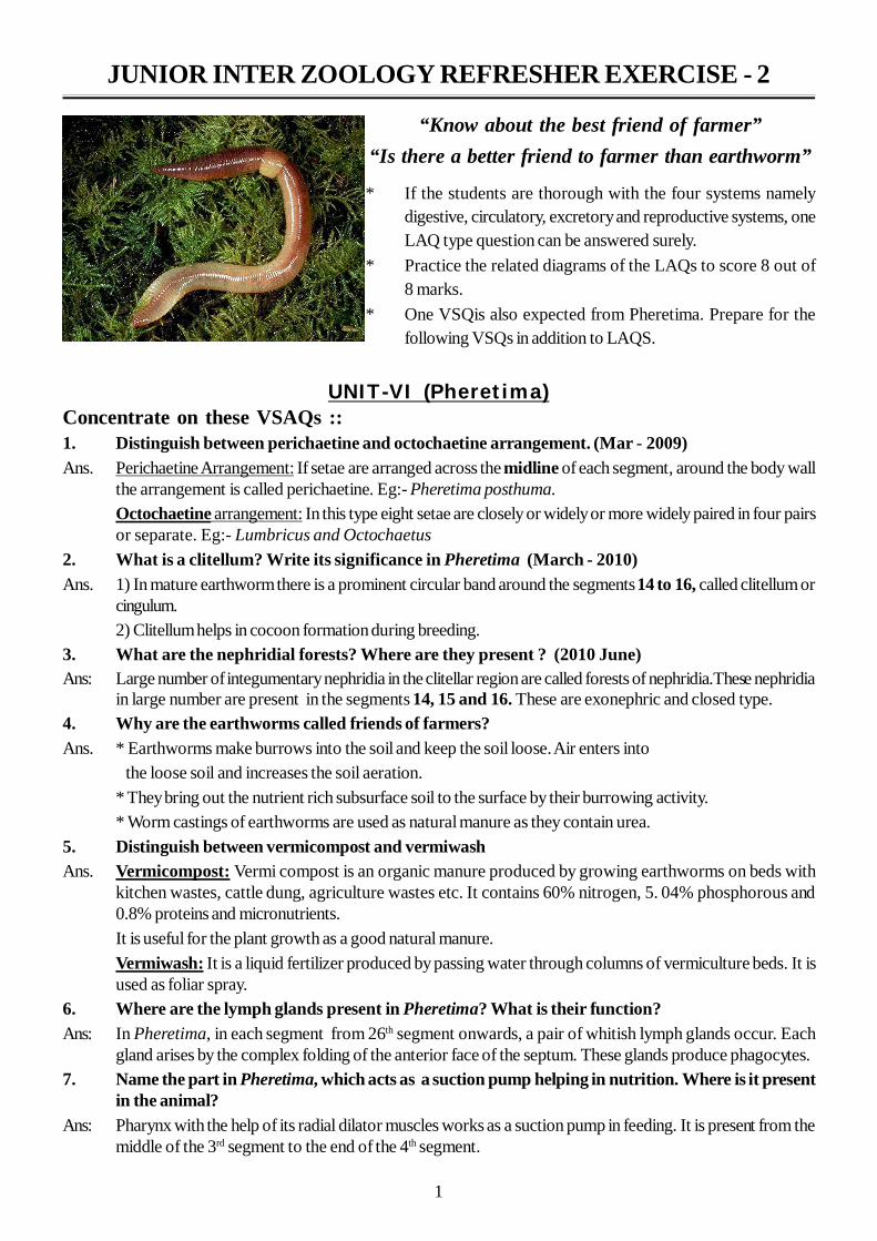
“Know about the best friend of farmer”“Is there a better friend to farmer than earthworm”
* If the students are thorough with the four systems namelydigestive, circulatory, excretory and reproductive systems, oneLAQ type question can be answered surely.
* Practice the related diagrams of the LAQs to score 8 out of8 marks.
* One VSQis also expected from Pheretima. Prepare for thefollowing VSQs in addition to LAQS.
2
8. Write any two structural differences between the lateral hearts and lateraloesophageal hearts.
Ans: Lateral heats Lateral oesophageal hearts1. Present in the segment 7 and 9 1. Present in the segments 12 and 132. They carry blood from dorsal vessel 2 2. They carry blood from the dorsal blood vessel and theto ventral blood vessel supra oesophageal vessel to the ventral blood vessel3. Each heart has four pairs of valves 3. Each heart has three pairs of valves
9. What is Phaeosome? Where is it present in Pheretima?Ans: * Phaeosome is ‘L’ shaped lens present in the photoreceptor cells of epidermis.It is also called optic
organelle.* These photoreceptors are abundant in the epidermis of the dorsal side of prostomium and peristomium10. In an experiment, male gential apertures of an earthworm A are sealed and it is allowed tocopulate with a normal earthworm B. What happen to ova of the earthworms A and B whetherthey are fertilized or not?
Ans. During copulation there is reciprocal transfer of sperms between the two copulating worms. In this casewhen the male genital apertures of A are closed and if it is allowed to copulate with normal earthworm B,sperms from B will be transferred into the spermethecal aperture of A but sperms of A have no chance to betransferred into spermethecal apertures of B.Eggs of A will get fertilized but eggs of B cannot be fertilized.
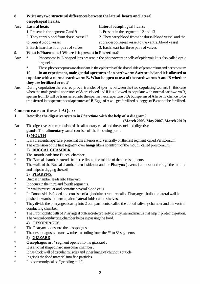
Concentrate on these LAQs ::1. Describe the digestive system in Pheretima with the help of a diagram?
(March 2005, May 2007, March 2010)Ans The digestive system consists of the alimentary canal and the associated digestive
glands. The alimentary canal consists of the following parts.1) MOUTH
* It is a crescentic aperture present at the anterior end, ventrally on the first segment called Peristomium* The extension of the first segment over hangs like a lip infront of the mouth, called prostomium.
2) BUCCAL CHAMBER* The mouth leads into Buccal chamber.* The Buccal chamber extends from the first to the middle of the third segments* The walls of the Buccal chamber turn inside out and the Pharynx ( everts ) comes out through the mouth
and helps in digging the soil.3) PHARYNX
* Buccal chamber leads into Pharynx.* It occurs in the third and fourth segments.* Its wall is muscular and contains several blood cells.* Its Dorsal side is folded and consists of a glandular structure called Pharyngeal bulb, thelateral wall is
pushed inwards to form a pair of lateral folds called shelves.* They divide the pharyngeal cavity into 2 compartments, called the dorsal salivary chamber and the ventral
conducting chamber.* The chromophilic cells of Pharyngeal bulb secrete proteolytic enzymes and mucus that help in protein digestion.* The ventral conducting chamber helps in passing the food.
4) OESOPHAGUS* The Pharynx opens into the oesophagus.* The oesophagus is a narrow tube extending from the 5th to 8th segments.
5) GIZZARD* Oesophagus in 8th segment opens into the gizzzard .* It is an oval shaped hard muscular chamber .* It has thick wall of circular muscles and inner lining of chitinous cuticle.* It grinds the food material into fine particles.* It is commonly called “ grinding mill “.
3
6) STOMACH* The gizzard leads into the stomach.* The stomach extends from the 9th to the 14th segment.* It is a highly vascular and glandular tube.* Its inner foldings contain gland cells, which secrete proteolytic enzymes, that help in protein digestion.
7) INTESTINE :* Stomach opens into the long and wide intestine which extends from the 15th to the last segment.* It’s mid dorsal fold is called typhlosole. This is poorly developed in Pheretima.* It increases the area of absorption of food.* On the basis of the presence of typhlosole the intestine is divisible into 3 regions.
1) Pre-typhlosolar region 2) Typhlosolar region 3) Post-typhlosolar region1) Pre-typhlosolar region :* This is the proximal part of the intestine located from 15th to 25th segments.* In the 26th segment, a pair of conical outgrowths, the intestinal caecae arise form this region laterally
and extend forward over three or four segments.* They secrete digestive juices, which contain amylase enzyme.* Typhlosole is absent in this part of intestine.2) Typhlosolar region :* It starts from 26th segment and terminates in 23 or 25 segments ahead of the hind end of the body.* It is distinguished by the presence of small mid dorsal projection, the typhlosole, hanging into the
lumen of the intestine. Typhlosole is poorly developed in Pheretima.* Digested food is absorbed in this region.3) Post-typhlosolar region :* It is the distal part of the intestine and it occupies the last 23 or 25 segments.* It is without a typhlosole.* It is also called rectum.* The undigested food is converted into small pellets.8) ANUS* It is present in the last segment. The undigested food is sent out through the anus in the form of
pellets. These pellets are called “worm castings”.
4
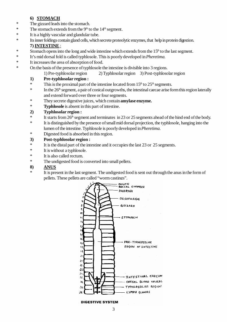
2. Explain the blood circulation in the first thirteen segments in Pheretima with a suitable diagram.(2006)
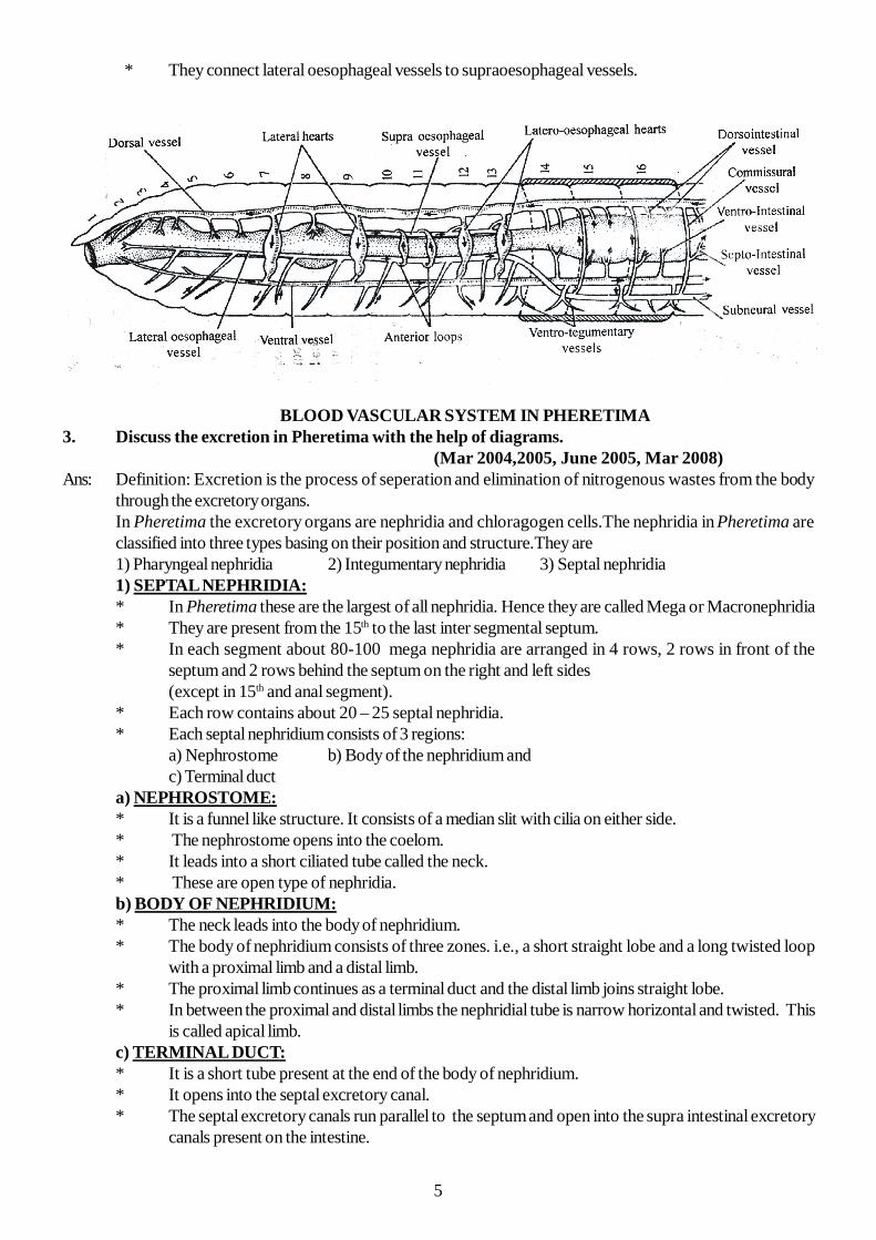
Ans: * In the first thirteen segments of Pheretima the longitudinal blood vessels are1) Dorsal blood vessel.2) Ventral blood vessel.3) Lateral oesophageal blood vessel.4) Supra oesophageal blood vessel.
1) Dorsal blood vessel: * It is present on the dorsal side of the alimentary canal. * In this vessel, blood flows from the posterior to the anterior side. * This is a pulsatile , muscular , valvular blood vessel. * It has paired valves in each segment which allows the blood to flow towards the anterior side only. * In the first thirteen segments this is a distributing blood vessel. * The dorsal blood vessel distributes blood to the buccal cavity, the pharynx, pharyngeal
nephridia, oesophagus, gizzard and stomach.2) Ventral blood vessel :
* It is present on the ventral side of the alimentary canal, and dorsal to the nerve cord. * It is present from second segment onwards. * It is non-muscular, non-valvular and non-pulsatile blood vessel. It is completely distributing blood
vessel. * In each segment one pair of ventro tegumentary blood vessels arise from the ventral blood vessel
and distribute blood to body wall, integumentary nephridia, reproductive organs and septa. * In the ventral vessel blood flows from the anterior side to the posterior side.3) Lateral oesophageal blood vessels:
* These are one pair of blood vessels present on either side of the alimentary canal in the first 13segments.
* These are completely collecting blood vessels, which collect blood from the body wall, septa,reproductive organs, pharyngeal nephridia and other visceral organs present in the first 13 segments.
4) The supra oesophageal vessel:* This is a single blood vessel present on the dorsal side of the stomach. extending in the segments
9 to 13. * It is a collecting blood vessel that collects blood from the gizzard and the stomach. * It receives blood through the anterior loops and ring vessels from the lateral oesophageal blood
vessels. Lateral hearts:
* These are four pairs. Present in the segments 7, 9, 12 and 13 * These are arranged on the right and left sides of the alimentary canal. * The lateral hearts present in 7th and 9th segements carry blood from the dorsal blood vessel to the
ventral blood vessel. * Each lateral heart has four pairs of valves. * The lateral hearts present in 12th and 13th segments carry blood from the dorsal blood vessel and
the supraoesophageal blood vessel to the ventral blood vessel. * These two pairs of hearts present in 12th and 13th segment are called lateral oesophageal hearts.
They possess three pairs of valves each.The anterior loops:
* These are two pairs of blood vessels. Present one pair in each of 10th and 11th segments. * They connect lateral oesophageals with supraoesophageal blood vessel . * These are non-pulsatile and non-valvular blood vessels.
Ring vessels:* These are twelve rings in each of the segments from 10 to 13,
5
* They connect lateral oesophageal vessels to supraoesophageal vessels.
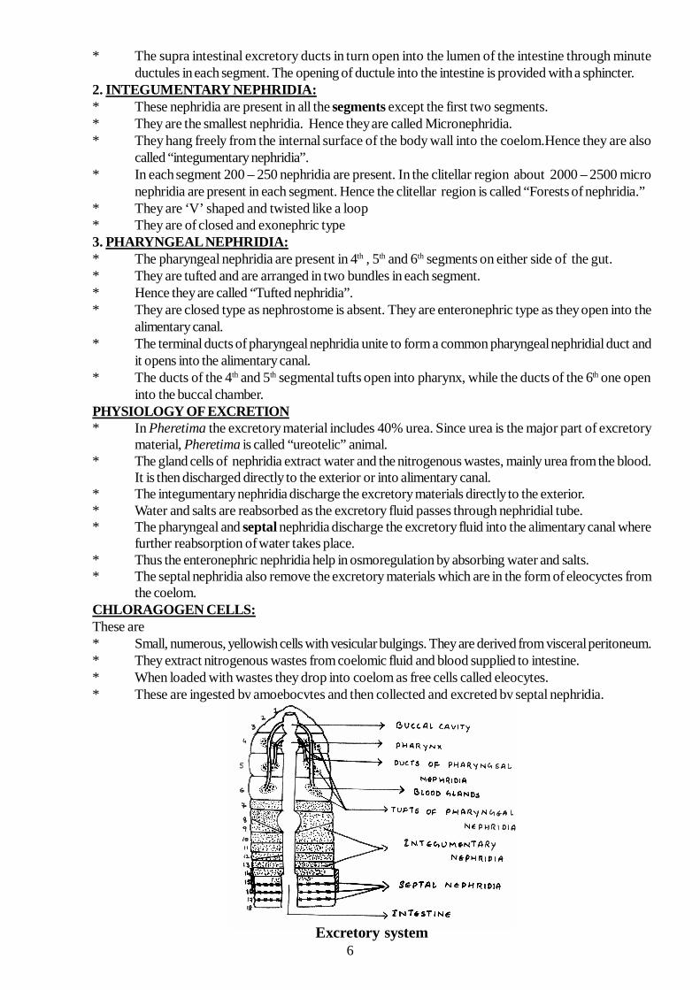
BLOOD VASCULAR SYSTEM IN PHERETIMA3. Discuss the excretion in Pheretima with the help of diagrams.
(Mar 2004,2005, June 2005, Mar 2008)Ans: Definition: Excretion is the process of seperation and elimination of nitrogenous wastes from the body
through the excretory organs.In Pheretima the excretory organs are nephridia and chloragogen cells.The nephridia in Pheretima areclassified into three types basing on their position and structure.They are1) Pharyngeal nephridia 2) Integumentary nephridia 3) Septal nephridia1) SEPTAL NEPHRIDIA:* In Pheretima these are the largest of all nephridia. Hence they are called Mega or Macronephridia* They are present from the 15th to the last inter segmental septum.* In each segment about 80-100 mega nephridia are arranged in 4 rows, 2 rows in front of the
septum and 2 rows behind the septum on the right and left sides(except in 15th and anal segment).
* Each row contains about 20 – 25 septal nephridia.* Each septal nephridium consists of 3 regions:
a) Nephrostome b) Body of the nephridium andc) Terminal duct
a) NEPHROSTOME:* It is a funnel like structure. It consists of a median slit with cilia on either side.* The nephrostome opens into the coelom.* It leads into a short ciliated tube called the neck.* These are open type of nephridia.b) BODY OF NEPHRIDIUM:* The neck leads into the body of nephridium.* The body of nephridium consists of three zones. i.e., a short straight lobe and a long twisted loop
with a proximal limb and a distal limb.* The proximal limb continues as a terminal duct and the distal limb joins straight lobe.* In between the proximal and distal limbs the nephridial tube is narrow horizontal and twisted. This
is called apical limb.c) TERMINAL DUCT:* It is a short tube present at the end of the body of nephridium.* It opens into the septal excretory canal.* The septal excretory canals run parallel to the septum and open into the supra intestinal excretory
canals present on the intestine.
6
* The supra intestinal excretory ducts in turn open into the lumen of the intestine through minuteductules in each segment. The opening of ductule into the intestine is provided with a sphincter.
2. INTEGUMENTARY NEPHRIDIA:* These nephridia are present in all the segments except the first two segments.* They are the smallest nephridia. Hence they are called Micronephridia.* They hang freely from the internal surface of the body wall into the coelom.Hence they are also
called “integumentary nephridia”.* In each segment 200 – 250 nephridia are present. In the clitellar region about 2000 – 2500 micro
nephridia are present in each segment. Hence the clitellar region is called “Forests of nephridia.”* They are ‘V’ shaped and twisted like a loop* They are of closed and exonephric type3. PHARYNGEAL NEPHRIDIA:* The pharyngeal nephridia are present in 4th , 5th and 6th segments on either side of the gut.* They are tufted and are arranged in two bundles in each segment.* Hence they are called “Tufted nephridia”.* They are closed type as nephrostome is absent. They are enteronephric type as they open into the
alimentary canal.* The terminal ducts of pharyngeal nephridia unite to form a common pharyngeal nephridial duct and
it opens into the alimentary canal.* The ducts of the 4th and 5th segmental tufts open into pharynx, while the ducts of the 6th one open
into the buccal chamber.PHYSIOLOGY OF EXCRETION* In Pheretima the excretory material includes 40% urea. Since urea is the major part of excretory
material, Pheretima is called “ureotelic” animal.* The gland cells of nephridia extract water and the nitrogenous wastes, mainly urea from the blood.
It is then discharged directly to the exterior or into alimentary canal.* The integumentary nephridia discharge the excretory materials directly to the exterior.* Water and salts are reabsorbed as the excretory fluid passes through nephridial tube.* The pharyngeal and septal nephridia discharge the excretory fluid into the alimentary canal where
further reabsorption of water takes place.* Thus the enteronephric nephridia help in osmoregulation by absorbing water and salts.* The septal nephridia also remove the excretory materials which are in the form of eleocyctes from
the coelom.CHLORAGOGEN CELLS:These are* Small, numerous, yellowish cells with vesicular bulgings. They are derived from visceral peritoneum.* They extract nitrogenous wastes from coelomic fluid and blood supplied to intestine.* When loaded with wastes they drop into coelom as free cells called eleocytes.* These are ingested by amoebocytes and then collected and excreted by septal nephridia.
Excretory system
7
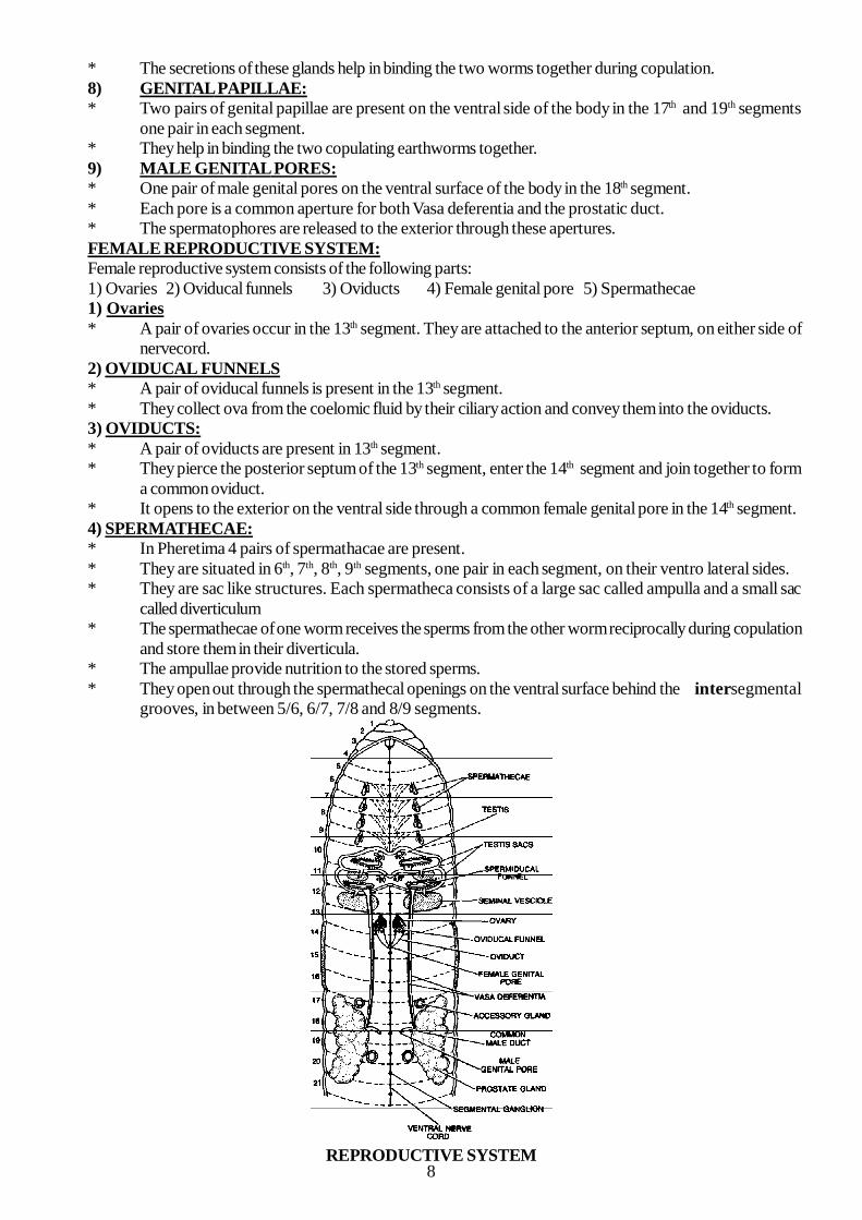
4. Give an account of reproductive organs in Pheretima. Draw a neat labelled diagram of thereproductive system? ( June 2004, Mar 2006 & Mar 2009, June 2010)
Ans. 1. Pheretima is a hermaphrodite. Both male and female reproductive organs are present in each individual.2. The gonads are formed from the coelomic epithelium (mesoderm)3. The male reproductive system matures first. Hence it is called “protandrous”.4. Self fertilization is prevented by the protandrous nature.Male reproductive system:1) Testes 2) Testis Sacs 3) Seminal Vesicles4) Spermiducal funnels 5) Vasa deferentia 6) Prostate glands7) Accessory glands 8) Genital papillae 9) Male genital aperture
1) TESTES:* Two pairs of testes are present in the 10th and 11th segmenst (one pair each) below the stomach.* Each testis consists of four to eight finger like processes.* They produce spermatogonia and release them into testis sacs.2) TESTIS SACS:* Testis sacs are derived from the coelomic epithelium of 10th and 11th segments.* The testis sac of 10th segment encloses a pair of spermiducal funnels beside the testes.* The testis sac of the 11th segment is large and it encloses a pair of seminal vesicles also.* The testis sacs protect the testis.3) SEMINAL VESICLES:* Two pairs of seminal vesicles are situated in the 11th and 12th segments (one pair in each segment)
attached to the anterior septa.* The testis sacs of the 10th segment open into the seminal vesicles of the 11th segment. While that of the
11th segment opens into the seminal vesicles of the 12th segment.* The spermatogonia released from the testis undergo reduction division in the seminal vesicles and form
the sperms.* The seminal vesicles of the 11th segment are situated in the testis sac, while those of the 12th segment are
free.* Since the seminal vesicles are formed from the septa these are called septal pouches.4) SPERMIDUCAL FUNNELS:* Two pairs of funnel like structures called spermiducal funnels are present in 10th and 11th segments, one
pair in each segment .* The margin of the funnel is ciliated.* They are located below the testes, enclosed in testis sacs.* They collect the sperms produced in seminal vesicles and transfer them to the vasa deferentia.5) VASADEFERENTIA:* The spermiducal funnels lead into ciliated ducts called vasa deferentia.* Two vasa deferentia of each side run closely together posteriorly on the ventral side, upto 18thsegment.* They join the prostate gland duct and enter a muscular sheath called common spermatic and prostatic
duct.* The ducts remain separated in this muscular sheath.6) PROSTATE GLANDS:* A pair of prostate glands are present on the ventral side of the body from the 16th or 17th to 20th or 21st
segment.* They are white and irregular in shape.* From each gland a small duct called prostatic duct arises and opens into a muscular sheath called
common spermatic and prostatic duct, on each side.* These two ducts bend towards mid ventral region and open to the exterior through the male genital
pore.* The secretions of these glands help in binding the sperms into bundles called “Spermatophores” in the
other earthworms.In Pheretima its function is not reported.
7) ACCESSORY GLANDS:* Two pairs of accessory glands, one pair each in 17the and 19th segments are present.* They open on the genital papillae through small ducts.
8
* The secretions of these glands help in binding the two worms together during copulation.8) GENITAL PAPILLAE:* Two pairs of genital papillae are present on the ventral side of the body in the 17th and 19th segments
one pair in each segment.* They help in binding the two copulating earthworms together.9) MALE GENITAL PORES:* One pair of male genital pores on the ventral surface of the body in the 18th segment.* Each pore is a common aperture for both Vasa deferentia and the prostatic duct.* The spermatophores are released to the exterior through these apertures.FEMALE REPRODUCTIVE SYSTEM:Female reproductive system consists of the following parts:1) Ovaries 2) Oviducal funnels 3) Oviducts 4) Female genital pore 5) Spermathecae1) Ovaries* A pair of ovaries occur in the 13th segment. They are attached to the anterior septum, on either side of
nervecord.2) OVIDUCAL FUNNELS* A pair of oviducal funnels is present in the 13th segment.* They collect ova from the coelomic fluid by their ciliary action and convey them into the oviducts.3) OVIDUCTS:* A pair of oviducts are present in 13th segment.* They pierce the posterior septum of the 13th segment, enter the 14th segment and join together to form
a common oviduct.* It opens to the exterior on the ventral side through a common female genital pore in the 14th segment.4) SPERMATHECAE:* In Pheretima 4 pairs of spermathacae are present.* They are situated in 6th, 7th, 8th, 9th segments, one pair in each segment, on their ventro lateral sides.* They are sac like structures. Each spermatheca consists of a large sac called ampulla and a small sac
called diverticulum* The spermathecae of one worm receives the sperms from the other worm reciprocally during copulation
and store them in their diverticula.* The ampullae provide nutrition to the stored sperms.* They open out through the spermathecal openings on the ventral surface behind the intersegmental
grooves, in between 5/6, 6/7, 7/8 and 8/9 segments.
REPRODUCTIVE SYSTEM