Embed Size (px)
DESCRIPTION
jurnal urologi
Citation preview
Hindawi Publishing CorporationCase Reports in UrologyVolume 2012, Article ID 430746, 4 pagesdoi:10.1155/2012/430746
Case Report
Delayed Presentation of Traumatic Intraperitoneal Rupture ofUrinary Bladder
Hazim H. Alhamzawi,1 Husham M. Abdelrahman,1 Khalid M. Abdelrahman,2
Ayman El-Menyar,3, 4 Hassan Al-Thani,1 and Rifat Latifi1, 4
1 Trauma Surgery Section, Department of Surgery, Hamad Medical Corporation, P.O. Box 3050, Doha, Qatar2 Urology Section, Surgery Department, Hamad Medical Corporation, P.O. Box 3050, Doha, Qatar3 Clinical Research, Trauma Surgery Section, Hamad Medical Corporation, Doha, Qatar4 Clinical Medicine, Weill Cornell Medical School, P.O. Box 24144, Doha, Qatar
Correspondence should be addressed to Hazim H. Alhamzawi, [email protected]
Received 2 November 2012; Accepted 21 November 2012
Academic Editors: P. H. Chiang, L. Henningsohn, and F. M. Solivetti
Copyright © 2012 Hazim H. Alhamzawi et al. This is an open access article distributed under the Creative Commons AttributionLicense, which permits unrestricted use, distribution, and reproduction in any medium, provided the original work is properlycited.
Blunt injury of the urinary bladder is well known and usually associates pelvic fractures. Isolated bladder injury is a rare conditionand on the other hand, delayed bladder perforation is an extremely rare entity. Herein, we described an unusual case of isolateddelayed intraperitoneal bladder rupture that occurred on the third post injury day in a young male in the absence of freeintraperitoneal fluid and pelvic fracture. The diagnostic workup, course and the need for surgical repair of the injury is presented.
1. Introduction
Around 60% to 85% of all bladder injuries result from bluntabdominal trauma (BAT) but the incidence of intraperi-toneal urinary bladder (UB) rupture is relatively uncommonfrom blunt injuries [1]. Isolated UB rupture following blunttrauma has an insidious presentation, and often results indelayed diagnosis and management [2–8]. The mechanism ofinjury include sudden compression of the full bladder, shearforces, or a pelvic fracture [2, 3, 9].
Rupture of bladder may be presented with lower abdom-inal pain, inability to void, and perineal ecchymoses [3].The cardinal sign of injury to the bladder is gross hematuria[6], which is present in more than 95% of cases, while onlyabout 5% of the patients have microscopic hematuria alone[6, 7]. Over 80% of the patients with UB rupture had anassociated pelvic fracture in centers with high percentage ofblunt trauma. On the other hand around 6% of patients withpelvic fracture sustain a bladder injury [3, 6].
Diagnosis of bladder injury, several days after admission,could be either a missed diagnosis or a truly delayed rupture.Delayed diagnosis of bladder rupture may be associated with
laboratory abnormalities such as metabolic derangements,and leukocytosis. Delay in the presentation and treatmentmay substantially increases mortality [7–10]. Therefore, earlyand accurate diagnosis with imaging techniques is imper-ative. Computed tomographic cystography (CTC) and/orretrograde cystography (RGC) are the standard imaging toolsfor the diagnosis of bladder injury [4–10]. We present acase of delayed rupture of UB due to blunt trauma withoutassociated injuries.
2. Case Report
A twenty three-year old male patient sustained BAT due tofall from a 3-meter height. Initial vital signs were: bloodpressure136/80 mmHg, heart rate 64 BPM, respiratory rate20 per minute, oxygen saturation of 100% on room air,and temperature of 36.9◦C. Patient was fully consciouswith neither external bleeding nor neurological deficits.Abdominal examination showed mild generalized tendernessand voluntary guarding at epigastric and suprapubic regions.Pelvic, genitourinary & rectal examinations revealed noabnormality.
2 Case Reports in Urology
Figure 1: Pelvic X-ray of the patient showing no fracture.
(a)
(b)
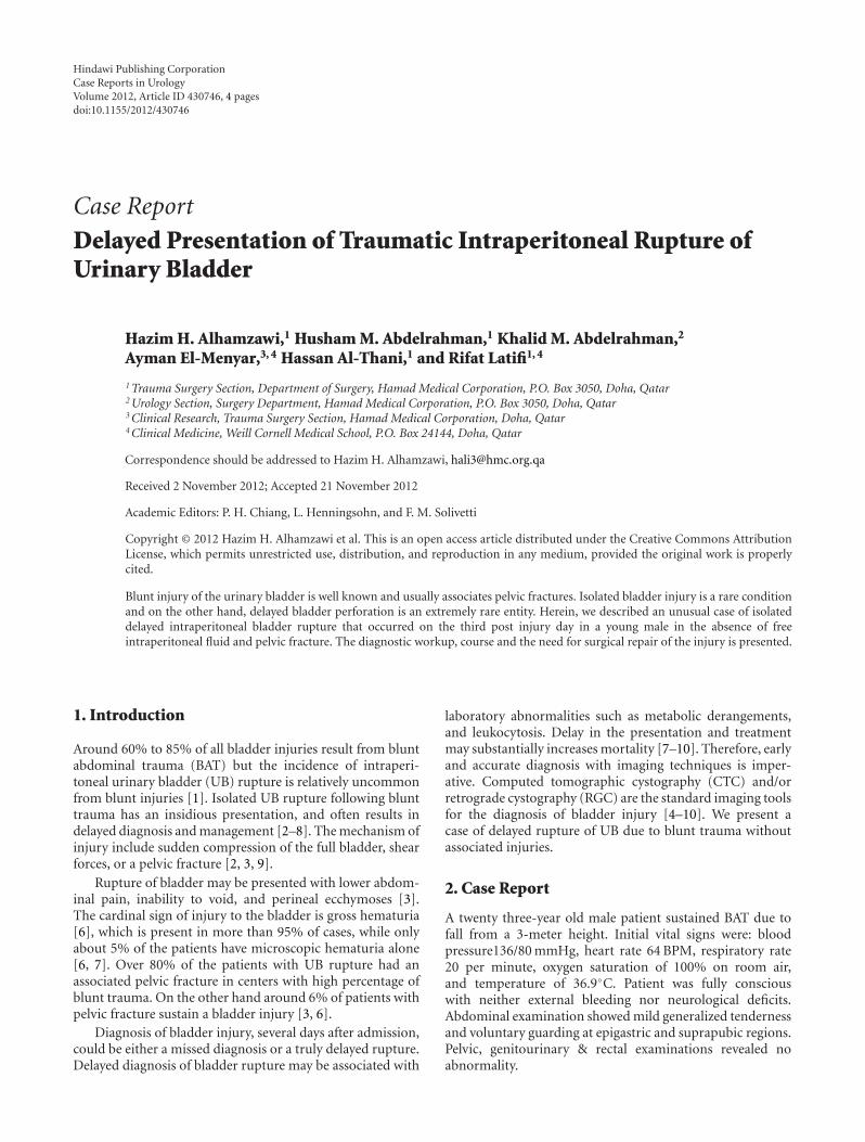
Figure 2: (a) CT image outlines the distended urinary bladder(UB). (b) CT image showing irregularity and thickening on theright side of UB wall.
(a)
(b)
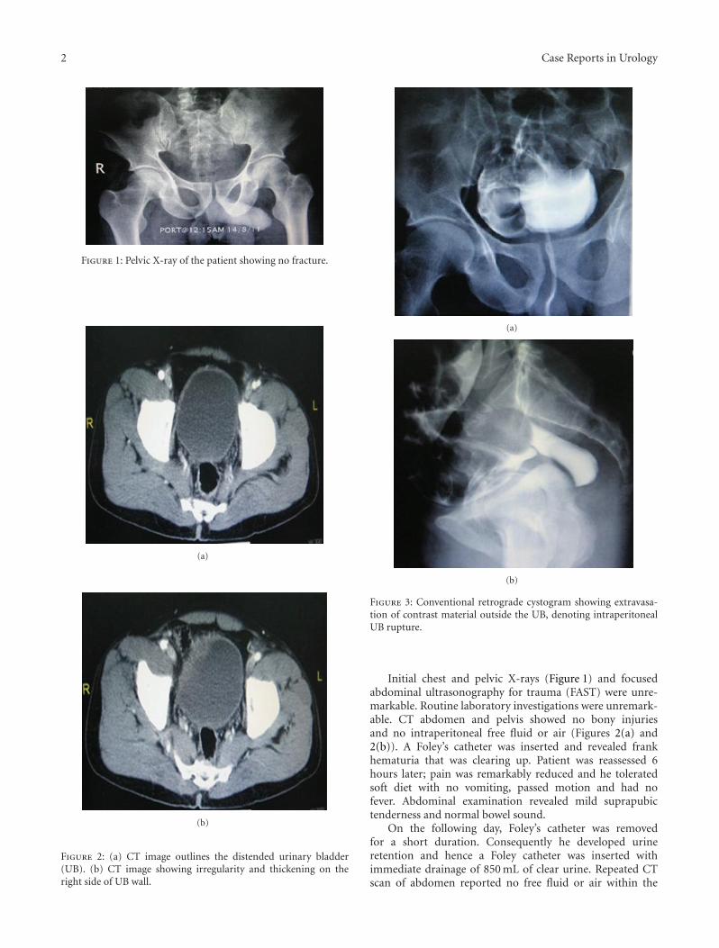
Figure 3: Conventional retrograde cystogram showing extravasa-tion of contrast material outside the UB, denoting intraperitonealUB rupture.
Initial chest and pelvic X-rays (Figure 1) and focusedabdominal ultrasonography for trauma (FAST) were unre-markable. Routine laboratory investigations were unremark-able. CT abdomen and pelvis showed no bony injuriesand no intraperitoneal free fluid or air (Figures 2(a) and2(b)). A Foley’s catheter was inserted and revealed frankhematuria that was clearing up. Patient was reassessed 6hours later; pain was remarkably reduced and he toleratedsoft diet with no vomiting, passed motion and had nofever. Abdominal examination revealed mild suprapubictenderness and normal bowel sound.
On the following day, Foley’s catheter was removedfor a short duration. Consequently he developed urineretention and hence a Foley catheter was inserted withimmediate drainage of 850 mL of clear urine. Repeated CTscan of abdomen reported no free fluid or air within the
Case Reports in Urology 3
(a)
(b)
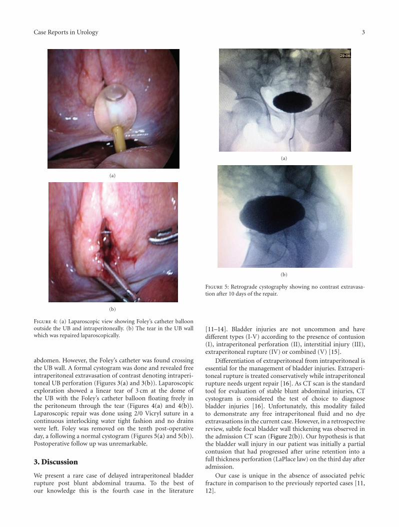
Figure 4: (a) Laparoscopic view showing Foley’s catheter balloonoutside the UB and intraperitoneally. (b) The tear in the UB wallwhich was repaired laparoscopically.
abdomen. However, the Foley’s catheter was found crossingthe UB wall. A formal cystogram was done and revealed freeintraperitoneal extravasation of contrast denoting intraperi-toneal UB perforation (Figures 3(a) and 3(b)). Laparoscopicexploration showed a linear tear of 3 cm at the dome ofthe UB with the Foley’s catheter balloon floating freely inthe peritoneum through the tear (Figures 4(a) and 4(b)).Laparoscopic repair was done using 2/0 Vicryl suture in acontinuous interlocking water tight fashion and no drainswere left. Foley was removed on the tenth post-operativeday, a following a normal cystogram (Figures 5(a) and 5(b)).Postoperative follow up was unremarkable.
3. Discussion
We present a rare case of delayed intraperitoneal bladderrupture post blunt abdominal trauma. To the best ofour knowledge this is the fourth case in the literature
(a)
(b)
Figure 5: Retrograde cystography showing no contrast extravasa-tion after 10 days of the repair.
[11–14]. Bladder injuries are not uncommon and havedifferent types (I-V) according to the presence of contusion(I), intraperitoneal perforation (II), interstitial injury (III),extraperitoneal rupture (IV) or combined (V) [15].
Differentiation of extraperitoneal from intraperitoneal isessential for the management of bladder injuries. Extraperi-toneal rupture is treated conservatively while intraperitonealrupture needs urgent repair [16]. As CT scan is the standardtool for evaluation of stable blunt abdominal injuries, CTcystogram is considered the test of choice to diagnosebladder injuries [16]. Unfortunately, this modality failedto demonstrate any free intraperitoneal fluid and no dyeextravasations in the current case. However, in a retrospectivereview, subtle focal bladder wall thickening was observed inthe admission CT scan (Figure 2(b)). Our hypothesis is thatthe bladder wall injury in our patient was initially a partialcontusion that had progressed after urine retention into afull thickness perforation (LaPlace law) on the third day afteradmission.
Our case is unique in the absence of associated pelvicfracture in comparison to the previously reported cases [11,12].
4 Case Reports in Urology
4. Conclusion
Although rare, delayed bladder injury presentation is possi-ble and one should have high index of suspicion in traumapatient with unexplained abdominal findings and or urinaryretention.
References
[1] A. S. Cass and M. Luxenberg, “Features of 164 bladderruptures,” Journal of Urology, vol. 138, no. 4, pp. 743–745,1987.
[2] T. Mokoena and A. G. Naidu, “Diagnostic difficulties inpatients with a ruptured bladder,” British Journal of Surgery,vol. 82, no. 1, pp. 69–70, 1995.
[3] R. G. Gomez, L. Ceballos, M. Coburn et al., “Consensusstatement on bladder injuries,” BJU International, vol. 94, no.1, pp. 27–32, 2004.
[4] P. V. Quagliano, S. M. Delair, and A. K. Malhotra, “Diagnosisof blunt bladder injury: a prospective comparative study ofcomputed tomography cystography and conventional retro-grade cystography,” Journal of Trauma, vol. 61, no. 2, pp. 410–421, 2006.
[5] A. S. Cass, “Diagnostic studies in bladder rupture. Indicationsand techniques,” Urologic Clinics of North America, vol. 16, no.2, pp. 267–273, 1989.
[6] J. H. Ahn, A. F. Morey, and J. W. McAninch, “Workup andmanagement of traumatic hematuria,” Emergency MedicineClinics of North America, vol. 16, no. 1, pp. 145–164, 1998.
[7] H. Tezval, M. Tezval, C. von Klot et al., “Urinary tract injuriesin patients with multiple trauma,” World Journal of Urology,vol. 25, no. 2, pp. 177–184, 2007.
[8] S. Deem, C. D. Lavender, and S. Agarwal, “Delayed presenta-tion of traumatic bladder injury: a case report and review ofcurrent treatment trends,” The Internet Journal of Urology, vol.5, no. 1, p. 8, 2007.
[9] J. P. Vaccaro and J. M. Brody, “CT cystography in theevaluation of major bladder trauma,” Radiographics, vol. 20,no. 5, pp. 1373–1381, 2000.
[10] C. H. Hsieh, R. J. Chen, J. F. Fang et al., “Diagnosis andmanagement of bladder injury by trauma surgeons,” AmericanJournal of Surgery, vol. 184, no. 2, pp. 143–147, 2002.
[11] D. Brown, H. L. Magill, and T. L. Black, “Delayed presentationof traumatic intraperitoneal bladder rupture,” Pediatric Radi-ology, vol. 16, no. 3, pp. 252–253, 1986.
[12] M. Laufik, D. Buono, G. Casola, and C. Sirlin, “Delayed trau-matic bladder rupture,” American Journal of Roentgenology,vol. 184, no. 3, pp. S99–S101, 2005.
[13] A. R. Turnbull, C. J. Smart, and J. D. Jenkins, “Delayed ruptureof the bladder,” British Journal of Urology, vol. 50, no. 3, pp.162–163, 1978.
[14] J. N. Corriere Jr. and C. M. Sandler, “Delayed post-traumaticrupture of the bladder,” Journal of the Royal College of Surgeonsof Edinburgh, vol. 19, no. 4, pp. 247–248, 1974.
[15] C. M. Sandler, J. T. Hall, M. B. Rodriguez, and J. N. Corriere Jr.,“Bladder injury in blunt pelvic trauma,” Radiology, vol. 158,no. 3, pp. 633–638, 1986.
[16] P. V. Quagliano, S. M. Delair, and A. K. Malhotra, “Diagnosisof blunt bladder injury: a prospective comparative study ofcomputed tomography cystography and conventional retro-grade cystography,” Journal of Trauma, vol. 61, no. 2, pp. 410–421, 2006.

Submit your manuscripts athttp://www.hindawi.com
Stem CellsInternational
Hindawi Publishing Corporationhttp://www.hindawi.com Volume 2014
Hindawi Publishing Corporationhttp://www.hindawi.com Volume 2014
MEDIATORSINFLAMMATION
of
Hindawi Publishing Corporationhttp://www.hindawi.com Volume 2014
Behavioural Neurology
EndocrinologyInternational Journal of
Hindawi Publishing Corporationhttp://www.hindawi.com Volume 2014
Hindawi Publishing Corporationhttp://www.hindawi.com Volume 2014
Disease Markers
Hindawi Publishing Corporationhttp://www.hindawi.com Volume 2014
BioMed Research International
OncologyJournal of
Hindawi Publishing Corporationhttp://www.hindawi.com Volume 2014
Hindawi Publishing Corporationhttp://www.hindawi.com Volume 2014
Oxidative Medicine and Cellular Longevity
Hindawi Publishing Corporationhttp://www.hindawi.com Volume 2014
PPAR Research
The Scientific World JournalHindawi Publishing Corporation http://www.hindawi.com Volume 2014
Immunology ResearchHindawi Publishing Corporationhttp://www.hindawi.com Volume 2014
Journal of
ObesityJournal of
Hindawi Publishing Corporationhttp://www.hindawi.com Volume 2014
Hindawi Publishing Corporationhttp://www.hindawi.com Volume 2014
Computational and Mathematical Methods in Medicine
OphthalmologyJournal of
Hindawi Publishing Corporationhttp://www.hindawi.com Volume 2014
Diabetes ResearchJournal of
Hindawi Publishing Corporationhttp://www.hindawi.com Volume 2014
Hindawi Publishing Corporationhttp://www.hindawi.com Volume 2014
Research and TreatmentAIDS
Hindawi Publishing Corporationhttp://www.hindawi.com Volume 2014
Gastroenterology Research and Practice
Hindawi Publishing Corporationhttp://www.hindawi.com Volume 2014
Parkinson’s Disease
Evidence-Based Complementary and Alternative Medicine
Volume 2014Hindawi Publishing Corporationhttp://www.hindawi.com