Embed Size (px)
Citation preview
Title A novel transmission pathway : first report of a larvaltrematode in a tanaidacean crustacean
Author(s) Kakui, Keiichi
Citation Fauna Ryukyuana, 17: 13-22
Issue Date 2014-12-04
URL http://hdl.handle.net/20.500.12000/38637
Rights
Fauna Ryukyuana ISSN 2187-6657 http://w3.u-ryukyu.ac.jp/naruse/lab/Fauna_Ryukyuana.html
13
A novel transmission pathway: first report of a larval trematode
in a tanaidacean crustacean
Keiichi Kakui Faculty of Science, Hokkaido University, Sapporo 060-0810, Japan
E-mail: [email protected]
Abstract. This study reports on the first trematode parasite observed in any species in the crustacean order Tanaidacea. Metacercariae occupying the body cavity of the host (Longiflagrum nasutus) were detected in 13 of 37 tanaidaceans collected from Manko Wetland, a Ramsar site on Okinawa Island, Japan. The morphology of the trematode and analyses of its 18S rRNA, 28S rRNA, and ITS1 nucleotide sequences suggest that it belongs in the family Microphallidae. Definitive hosts are not highly specific in microphallids, and are chiefly birds. Tanaidaceans have been reported in stomach contents from wading birds; L. nasutus was abundant at Manko Wetland, and is a likely prey for shorebirds there. This circumstantial evidence suggests an avian definitive host for the trematode in L. nasutus.
Introduction
The life cycles of most digenean trematodes are unknown, as they involves multiple hosts. In the typical life cycle, digeneans utilise two intermediate hosts and a definitive host. The first intermediate host is usually a gastropod, although a few species’ life cycles involve a bivalve, scaphopod, or polychaete worm (Cribb et al. 2001). Second intermediate hosts are more diverse, including crustaceans, annelids, molluscs, fishes, amphibians, and other groups (cf., Lefebvre & Poulin 2005). The discovery of representatives of additional higher taxa acting as second intermediate hosts will facilitate solving the life cycles of digeneans that remained intractable because the second intermediate host has been unknown.
Most species in the crustacean order Tanaidacea are marine, although a few occur in brackish habitats or fresh water. Tanaidaceans are typically a few millimetres long, and primarily inhabit bottom sediments. They can occur at high densities (e.g., 146,000 individuals/m2; Delille et al. 1985) and appear to be important prey items in marine food webs, where they are consumed by other crustaceans, fishes, and migratory birds (Larsen 2005). Curiously, though tanaidaceans are abundant, there are few records of their endoparasites: only
nematodes and acanthocephalan larvae have been reported (Larsen 2005).
During qualitative sampling for benthic animals at Manko Wetland, a Ramsar wetland site on Okinawa Island in subtropical southern Japan, I collected many specimens of the tanaidacean Longiflagrum nasutus (Nunomura, 2005), some of which contained trematode metacercariae. In this study, I examined the morphology of these metacercariae and attempted to identify them through molecular phylogenetic analyses.
Materials and methods
Host sampling. Tanaidaceans were collected from muddy bottom sediment in shallow water on Manko Wetland, Okinawa Island, Japan (26°11'41.50"N 127°40'56"E) on 20 November 2013, by washing sediment through a sieve (0.5 mm mesh). Thirty-seven individuals were fixed in 4% paraformaldehyde in phosphate-buffered saline (PBS, pH 7.4) and preserved in PBS. Two infected animals found among captive tanaidaceans in the laboratory on 13 January 2014 were fixed in absolute ethanol and preserved at –20°C.
Morphological observation. Tanaidaceans were examined for parasite infection by ventral observation under a Nikon SMZ-10 stereoscopic microscope. Infected tanaidaceans stored in PBS were dehydrated in an ethanol series. Two specimens were transferred into dimethyl sulfoxide, frozen at 4°C, cut transversely with a hand-held razor, mounted in glycerine, and observed using autofluorescence with a Zeiss LSM 510 confocal laser scanning microscope (CLSM) under the default setting, the DPSS 561 nm laser. Another six specimens were dissected with chemically sharpened tungsten needles to extract parasites. The parasites recovered were mounted in glycerine and observed with an Olympus BX51 light microscope (LM) equipped with Nomarski interference optics. The eight dissected and five intact tanaidacean specimens harbouring the parasite were deposited in the Hokkaido University Museum, Sapporo, Japan (catalogue number ZIHU 4929) and in the University Museum Fujukan, University of the
14 [報告] 角井: タナイス寄生性吸虫類の初報告
Ryukyus (catalogue number RUMF-ZC-3678). Molecular phylogenetic analyses. One of the
two infected tanaidaceans stored in absolute ethanol was dissected to extract the parasite. The parasite was cleaned by brief sonication and pierced with a needle before DNA extraction with a DNeasy Blood & Tissue Kit (Qiagen GmbH). Table 1 lists the primers used for PCR and cycle sequencing. DNA amplification conditions were 95°C for 1 min; 30 cycles of 95°C for 30 sec, 50°C for 30 sec, and 72°C for 3 min (18S) or 1 min (28S and ITS1); and 72°C for 7 min. All nucleotide sequences were determined by direct sequencing with a BigDye Terminator Kit ver. 3.1 and a 3730 DNA analyzer (Life Technologies).
Three phylogenetic analyses were conducted to identify the parasite. In the first, a combined 18S+28S dataset was analysed by maximum-likelihood (ML) to place the parasite in a superfamily. This analysis utilised the aligned dataset of Olson et al. (2003), which included 170 trematode terminal taxa (available at http://ftp.ebi.ac.uk in directory/pub/databases/embl/align: ALIGN_00525 and _00526; see also table 1 in Olson et al. 2003), sequence data for Collyriclum faba (Heneberg & Literák 2013), and data for unidentified species. Alignments were initially carried out independently for the 18S and 28S datasets, as follows. First, a pre-alignment was performed with MAFFT version 7 (Katoh & Standley 2013) with the “Auto”
strategy (FFT-NS-i method; Katoh et al. 2002). Ambiguous sites were then removed by using trimAl (Capella-Gutiérrez et al. 2009) with the option “automated1.” Finally, the aligned sequences were trimmed in MEGA 5.2 (Tamura et al. 2011) to match the shortest length. Concatenation of the aligned datasets and selection of the optimal substitution model were performed with Kakusan4 version 4.0.2012.12.14 (Tanabe 2011). The ML analysis was conducted in RAxML version 8.0.0 (Stamatakis 2014), assisted by phylogears2 version 2.0.2013.10.22 (Tanabe 2008), and nodal support values were obtained through ML analyses of 1000 bootstrap pseudoreplicates (Felsenstein 1985).
A second ML analysis was conducted with a 28S dataset that included representatives of the superfamily identified in the first analysis and appropriate outgroup taxa (50 terminal taxa; listed in Appendix 1), to determine the family identity of the trematode. All procedures were as detailed for the first analysis above.
A third analysis utilized an ITS1 dataset (17 terminal taxa; listed in Appendix 1) to identify the trematode’s phylogenetic position within the family indicated by the second analysis. Sequence alignment was performed as above. Model selection, the ML analysis, and bootstrap analysis of 1000 bootstrap pseudoreplicates were carried out with TREEFINDER, March 2011 version (Jobb 2011).
The optimal substitution models for the three
Table 1. List of PCR and cycle sequencing (CS) primers used in this study. 表 1. PCR反応とサイクルシーケンシング (CS) で用いたプライマー一覧.
Marker Primer Sequence (5´ to 3´) Reaction Source 18S SR1 TACCTGGTTGATCCTGCCAG PCR & CS Nakayama et al. (1996) SR6 GTCAGAGGTGAAATTCTTGG CS Nakayama et al. (1996) SR8 GGATTGACAGATTGAGAGCT CS Nakayama et al. (1996) SR9 AACTAAGAACGGCCATGCAC CS Nakayama et al. (1996) SR10 AGGTCTGTGATGCCCTTAGA CS Nakayama et al. (1996) SR11 CGCTTACTAGGAATTCCTCG CS Nakayama et al. (1996) SR12 CCTTCCGCAGGTTCACCTAC PCR & CS Nakayama et al. (1996) EU60F GAAACTGCGAATGGCTCATT CS Puitika et al. (2007) EU929R TTGGCAAATGCTTTCGC CS Puitika et al. (2007) 18S554f AAGTCTGGTGCCAGCAGCGCG CS Maraun et al. (2009) 18S614r TCCAACTACGAGCTTTTTAACC CS Maraun et al. (2009) 28S U178 GCACCCGCTGAAYTTAAG PCR & CS Lockyer et al. (2003) 300F CAAGTACCGTGAGGGAAAGTTG CS Lockyer et al. (2003) 900F CCGTCTTGAAACACGGACCAAG CS Lockyer et al. (2003) U1148 GACCCGAAAGATGGTGAA CS Lockyer et al. (2003) L1642 CCAGCGCCATCCATTTTCA PCR & CS Lockyer et al. (2003) ITS1 1780F ACACCGCCCGTCGCTACTA PCR & CS Galaktionov et al. (2012) M5-8 GGCTGCGCTCTTCATCGACA PCR & CS Galaktionov et al. (2012)
Fauna Ryukyuana, 17: 13–22.
15 [Report] Kakui: Trematode metacercaria found in a tanaidacean
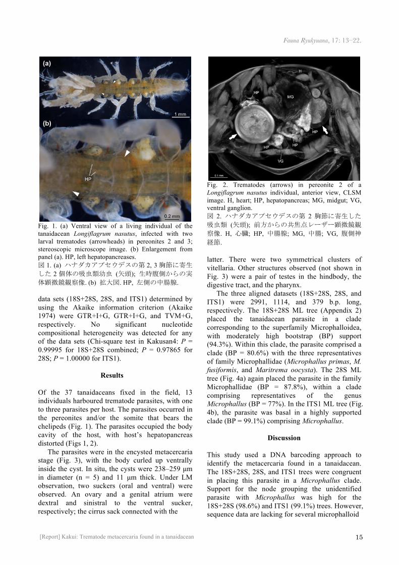
Fig. 1. (a) Ventral view of a living individual of the tanaidacean Longiflagrum nasutus, infected with two larval trematodes (arrowheads) in pereonites 2 and 3; stereoscopic microscope image. (b) Enlargement from panel (a). HP, left hepatopancreases. 図 1. (a) ハナダカアプセウデスの第 2, 3胸節に寄生した 2 個体の吸虫類幼虫 (矢頭); 生時腹側からの実体顕微鏡観察像. (b) 拡大図. HP, 左側の中腸腺. data sets (18S+28S, 28S, and ITS1) determined by using the Akaike information criterion (Akaike 1974) were GTR+I+G, GTR+I+G, and TVM+G, respectively. No significant nucleotide compositional heterogeneity was detected for any of the data sets (Chi-square test in Kakusan4: P = 0.99995 for 18S+28S combined; P = 0.97865 for 28S; P = 1.00000 for ITS1).
Results
Of the 37 tanaidaceans fixed in the field, 13 individuals harboured trematode parasites, with one to three parasites per host. The parasites occurred in the pereonites and/or the somite that bears the chelipeds (Fig. 1). The parasites occupied the body cavity of the host, with host’s hepatopancreas distorted (Figs 1, 2).
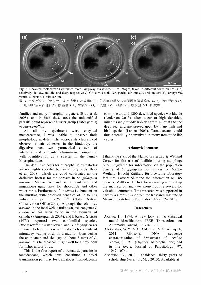
The parasites were in the encysted metacercaria stage (Fig. 3), with the body curled up ventrally inside the cyst. In situ, the cysts were 238–259 µm in diameter (n = 5) and 11 µm thick. Under LM observation, two suckers (oral and ventral) were observed. An ovary and a genital atrium were dextral and sinistral to the ventral sucker, respectively; the cirrus sack connected with the
Fig. 2. Trematodes (arrows) in pereonite 2 of a Longiflagrum nasutus individual, anterior view, CLSM image. H, heart; HP, hepatopancreas; MG, midgut; VG, ventral ganglion. 図 2. ハナダカアプセウデスの第 2 胸節に寄生した吸虫類 (矢頭); 前方からの共焦点レーザー顕微鏡観察像. H, 心臓; HP, 中腸腺; MG, 中腸; VG, 腹側神経節. latter. There were two symmetrical clusters of vitellaria. Other structures observed (not shown in Fig. 3) were a pair of testes in the hindbody, the digestive tract, and the pharynx.
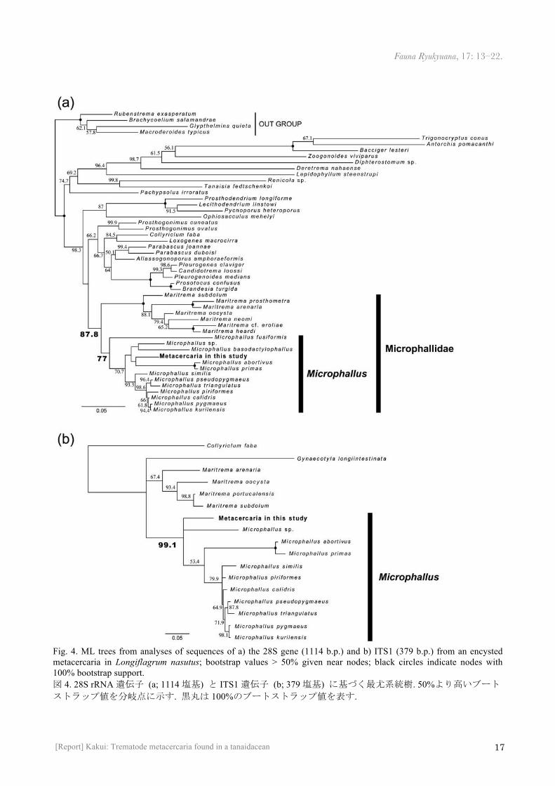
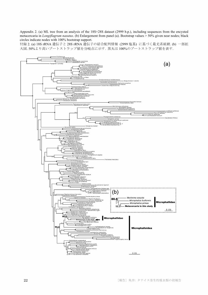
The three aligned datasets (18S+28S, 28S, and ITS1) were 2991, 1114, and 379 b.p. long, respectively. The 18S+28S ML tree (Appendix 2) placed the tanaidacean parasite in a clade corresponding to the superfamily Microphalloidea, with moderately high bootstrap (BP) support (94.3%). Within this clade, the parasite comprised a clade (BP = 80.6%) with the three representatives of family Microphallidae (Microphallus primas, M. fusiformis, and Maritrema oocysta). The 28S ML tree (Fig. 4a) again placed the parasite in the family Microphallidae (BP = 87.8%), within a clade comprising representatives of the genus Microphallus (BP = 77%). In the ITS1 ML tree (Fig. 4b), the parasite was basal in a highly supported clade (BP = 99.1%) comprising Microphallus.
Discussion
This study used a DNA barcoding approach to identify the metacercaria found in a tanaidacean. The 18S+28S, 28S, and ITS1 trees were congruent in placing this parasite in a Microphallus clade. Support for the node grouping the unidentified parasite with Microphallus was high for the 18S+28S (98.6%) and ITS1 (99.1%) trees. However, sequence data are lacking for several microphalloid
16 [報告] 角井: タナイス寄生性吸虫類の初報告
Fig. 3. Encysted metacercaria extracted from Longiflagrum nasutus, LM images, taken in different focus planes (a–c, relatively shallow, middle, and deep, respectively). CS, cirrus sack; GA, genital atrium; OS, oral sucker; OV, ovary; VS, ventral sucker; VT, vitellarium. 図 3. ハナダカアプセウデスより摘出した被嚢幼虫; 焦点面の異なる光学顕微鏡観察像 (a–c, それぞれ浅い, 中間, 深い焦点面像). CS, 陰茎嚢; GA, 生殖腔; OS, 口吸盤; OV, 卵巣; VS, 腹吸盤; VT, 卵黄腺.
families and many microphallid genera (Bray et al. 2008), and in both these trees the unidentified parasite could represent a sister group (sister genus) to Microphallus.
As all my specimens were encysted metacercariae, I was unable to observe their morphology in detail. The various structures I did observe—a pair of testes in the hindbody, the digestive tract, two symmetrical clusters of vitellaria, and a genital atrium—are compatible with identification as a species in the family Microphallidae.
The definitive hosts for microphallid trematodes are not highly specific, but are chiefly birds (Bray et al. 2008), which are good candidates as the definitive host(s) for the parasite in Longiflagrum nasutus. Manko Wetland is a wintering and migration-staging area for shorebirds and other water birds. Furthermore, L. nasutus is abundant on the mudflat, with observed densities of up to 523 individuals per 0.0625 m2 (Naha Nature Conservation Office 2009). Although the role of L. nasutus in the food web is unknown, the congener L. koyonense has been found in the stomach of catfishes (Angsupanich 2004), and Băcescu & Guţu (1975) reported two confamilial species, Discapseudes surinamensis and Halmyrapseudes spaansi, to be common in the stomach contents of migratory wading birds on a mudflat. Considering the abundance and size (up to about 8 mm) of L. nasutus, this tanaidacean might well be a prey item for fishes and/or birds.
This is the first report of a trematode parasite in tanaidaceans, which thus constitute a novel transmission pathway for trematodes. Tanaidaceans
comprise around 1200 described species worldwide (Anderson 2013), often occur at high densities, inhabit sandy/muddy habitats from mudflats to the deep sea, and are preyed upon by many fish and bird species (Larsen 2005). Tanaidaceans could thus potentially be involved in many trematode life cycles.
Acknowledgements
I thank the staff of the Manko Waterbird & Wetland Center for the use of facilities during sampling; Shoji Sugiyama for information on the population density of Longiflagrum nasutus on the Manko Wetland; Hiroshi Kajihara for providing laboratory facilities; Satoshi Shimano for information on 18S primers; Matthew H. Dick for reviewing and editing the manuscript; and two anonymous reviewers for valuable comments. This research was supported in part by a Grant-in-Aid from the Research Institute of Marine Invertebrates Foundation (FY2012–2013).
References
Akaike, H., 1974. A new look at the statistical model identification. IEEE Transactions on Automatic Control, 19: 716–723.
Al-Kandari, W.Y., S.A. Al-Bustan & M. Alnaqeeb, 2011. Ribosomal DNA sequence characterization of Maritrema cf. eroliae Yamaguti, 1939 (Digenea: Microphallidae) and its life cycle. Journal of Parasitology, 97: 1067–1074.
Anderson, G., 2013. Tanaidacea- thirty years of scholarship (vers. 1.1, May 2013). Available at
Fauna Ryukyuana, 17: 13–22.
17 [Report] Kakui: Trematode metacercaria found in a tanaidacean
Fig. 4. ML trees from analyses of sequences of a) the 28S gene (1114 b.p.) and b) ITS1 (379 b.p.) from an encysted metacercaria in Longiflagrum nasutus; bootstrap values > 50% given near nodes; black circles indicate nodes with 100% bootstrap support. 図 4. 28S rRNA遺伝子 (a; 1114塩基) と ITS1遺伝子 (b; 379塩基) に基づく最尤系統樹. 50%より高いブートストラップ値を分岐点に示す. 黒丸は 100%のブートストラップ値を表す.
18 [報告] 角井: タナイス寄生性吸虫類の初報告
http://peracarida.usm.edu/TanaidaceaText.pdf (accessed 11 April 2014).
Angsupanich, S., 2004. A new species of Longiflagrum (Tanaidacea, Parapseudidae), from Songkhla Lagoon, Thailand. Crustaceana, 77: 849–860.
Băcescu, M. & M. Guţu, 1975. A new genus (Discapseudes n.g.) and three new species of Apseudidae (Crustacea, Tanaidacea) from the northeastern coast of South America. Zoologische Mededelingen, 49: 95–113.
Bray, R.A., D.I. Gibson & A. Jones, 2008. Keys to the Trematoda volume 3. CABI Publishing and the Natural History Museum, London.
Capella-Gutiérrez, S., J.M. Silla-Martínez & T. Gabaldón, 2009. trimAl: a tool for automated alignment trimming in large-scale phylogenetic analyses. Bioinformatics, 25: 1972–1973.
Cribb, T.H., R.A. Bray & D.T.J. Littlewood, 2001. The nature and evolution of the association among digeneans, molluscs and fishes. International Journal for Parasitology, 31: 997–1011.
Delille, D., L.D. Guidi & J. Soyer, 1985. Nutrition of Allotanais hirsutus (Crustacea: Tanaidacea) at Kerguelen Island. In: W.R. Siegfried, R.R. Condy & R.M. Laws (eds.), Antarctic Nutrient Cycles and Food Webs. Pp. 378–380, Springer-Verlag, Berlin.
Felsenstein, J., 1985. Phylogenies and the comparative method. American Naturalist, 125: 1–15.
Galaktionov, K.V., I. Blasco-Costa & P.D. Olson, 2012. Life cycles, molecular phylogeny and historical biogeography of the ‘pygmaeus’ microphallids (Digenea: Microphallidae): widespread parasites of marine and coastal birds in the Holarctic. Parasitology, 139: 1346–1360.
Heneberg, P. & I. Literák, 2013. Molecular phylogenetic characterization of Collyriclum faba with reference to its three host-specific ecotypes. Parasitology International, 62: 262–267.
Jobb, G., 2011. TREEFINDER version of March 2011. Distributed by the author at http://www.treefinder.de (accessed 11 April 2014).
Katoh, K., K. Misawa, K. Kuma & T. Miyata, 2002. MAFFT: a novel method for rapid multiple sequence alignment based on fast Fourier transform. Nucleic Acids Research, 30: 3059–3066.
Katoh, K. & D.M. Standley, 2013. MAFFT
multiple sequence alignment software version 7: improvements in performance and usability. Molecular Biology and Evolution, 30: 772–780.
Larsen, K., 2005. Deep-sea Tanaidacea (Peracarida) from the Gulf of Mexico. Brill, Leiden.
Lefebvre, F. & R. Poulin, 2005. Progenesis in digenean trematodes: a taxonomic and synthetic overview of species reproducing in their second intermediate hosts. Parasitology, 130: 587–605.
Lockyer, A.E., P.D. Olson & D.T.J. Littlewood, 2003. Utility of complete large and small subunit rRNA genes in resolving the phylogeny of the Neodermata (Platyhelminthes): implications and a review of the cercomer theory. Biological Journal of the Linnean Society, 78: 155–171.
Maraun, M., G. Erdmann, G. Schulz, R.A. Norton, S. Scheu & K. Domes, 2009. Multiple convergent evolution of arboreal life in oribatid mites indicates the primacy of ecology. Proceedings of the Royal Society B, 276: 3219–3227.
Naha Nature Conservation Office, 2009. Heisei 20 nendo kunishitei Mankochojuhogoku niokeru hozenjigyokentochosagyomu (Surveys in 2008 for conservation projects in Manko Wetland, a government-designated wildlife preserve). Naha Nature Conservation Office, Ministry of the Environment, Okinawa (in Japanese).
Nakayama, T., S. Watanabe, K. Mitsui, H. Uchida & I. Inoue, 1996. The phylogenetic relationship between the Chlamydomonadales and Chlorococcales inferred from 18SrDNA sequence data. Phycological Research, 44: 47–55.
Olson, P.D., T.H. Cribb, V.V. Tkach, R.A. Bray & D.T.J. Littlewood, 2003. Phylogeny and classification of the Digenea (Platyhelminthes: Trematoda). International Journal for Parasitology, 33: 733–755.
Pina, S., F. Russell-Pinto & P. Rodrigues, 2007. Clarification of Cercaria sevillana (Digenea: Microphallidae) life cycle using morphological and molecular data. Journal of Parasitology, 93: 318–322.
Pina, S., F. Russell-Pinto & P. Rodrigues, 2011a. Description of Maritrema portucalensis sp. nov. (Digenea, Microphallidae) parasite of Carcinus maenas (Crustacea, Decapoda) from Aveiro estuary, northern Portugal. Acta Parasitologica, 56: 377–384.
Pina, S., F. Russell-Pinto & P. Rodrigues, 2011b. Morphological and molecular study of
Fauna Ryukyuana, 17: 13–22.
19 [Report] Kakui: Trematode metacercaria found in a tanaidacean
Microphallus primas (Digenea: Microphallidae) metacercaria, infecting the shore crab Carcinus maenas from northern Portugal. Folia Parasitologica, 58: 48–54.
Puitika, T., Y. Kasahara, N. Miyoshi, Y. Sato & S. Shimano, 2007. A taxon-specific oligonucleotide primer set for PCR-based detection of soil ciliate. Microbes and Environments, 22: 78–81.
Stamatakis, A., 2014. RAxML version 8: a tool for phylogenetic analysis and post-analysis of large phylogenies. Bioinformatics, 30: 1312–1313.
Tamura, K., D. Peterson, N. Peterson, G. Stecher, M. Nei & S. Kumar, 2011. MEGA5: Molecular evolutionary genetics analysis using maximum likelihood, evolutionary distance, and maximum parsimony methods. Molecular Biology and Evolution, 28: 2731–2739.
Tanabe, A.S., 2008. Phylogears2 version 2.0.2013.10.22. Distributed by the author at http://www.fifthdimension.jp (accessed 11 April 2014).
Tanabe, A.S., 2011. Kakusan4 and Aminosan: two programs for comparing nonpartitioned, proportional and separate models for combined molecular phylogenetic analyses of multilocus sequence data. Molecular Ecology Resources, 11: 914–921.
Tkach, V.V., D.T.J. Littlewood, P.D. Olson, J.M. Kinsella & Z. Swiderski, 2003. Molecular phylogenetic analysis of the Microphalloidea Ward, 1901 (Trematoda: Digenea). Systematic Parasitology, 56: 1–15.
新規の感染経路: タナイス目甲殻類を利用する吸虫類幼虫の初報告 角井敬知 北海道大学大学院理学研究院. 〒060-0810 札幌市北区北10条西8丁目. E-mail: [email protected]
要旨 . 吸虫の被嚢幼虫をタナイス目甲殻類 (ハナダカアプセウデス Longiflagrum nasutus) から初めて発見した. 寄生部位は胸節体腔内であり, 宿主 37個体中 13個体に感染が認められた. 形態的特徴および 3 遺伝子 (18S rRNA, 28S rRNA, ITS1) の配列情報から , 本吸虫はMicrophallidae 科の 1 種だと判断された . Microphallidae 科吸虫の終宿主特異性は高くないが, 主として鳥類だと考えられている. タナイス類は鳥類の胃内容物から見出されること
があり, ハナダカアプセウデスは採集地, ラムサール条約登録湿地である漫湖干潟に多産す
る種類である. このことから, 今回見つかった吸虫類幼虫の終宿主は鳥類であることが考え
られる.
投稿日: 2014年 8月 14日 受理日: 2014年 11月 13日 発行日: 2014年 12月 4日
20 [報告] 角井: タナイス寄生性吸虫類の初報告
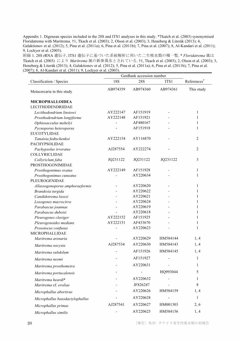
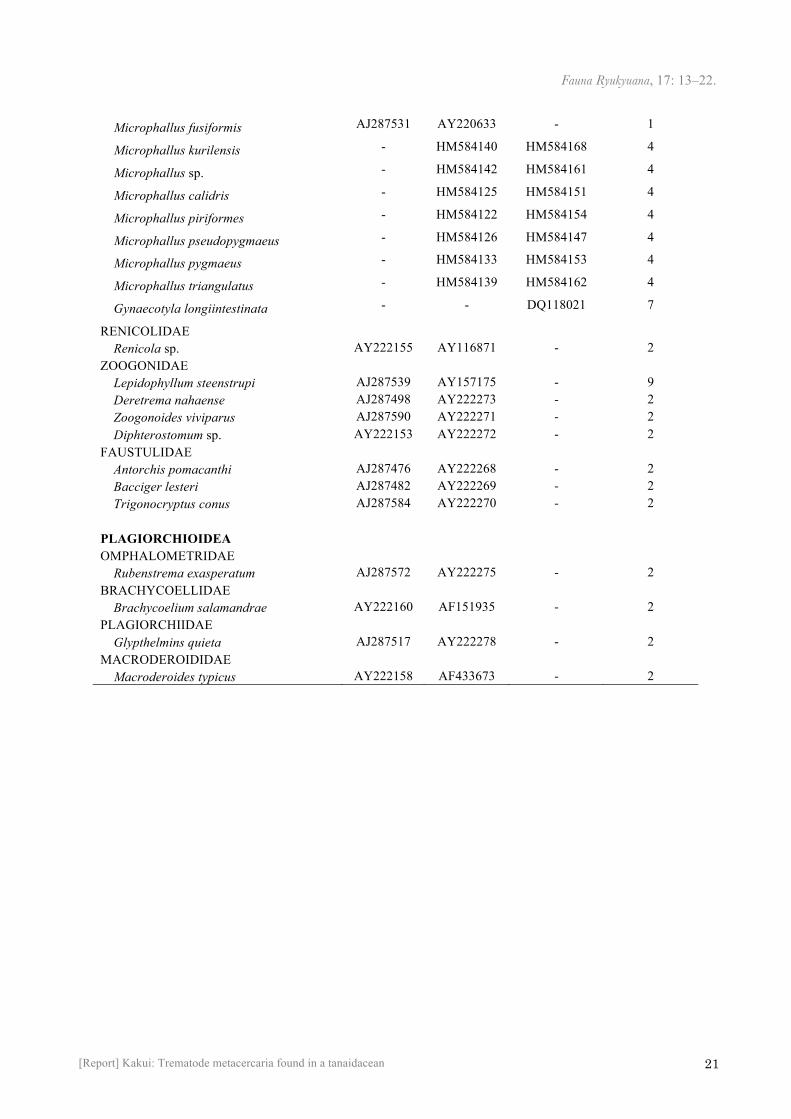
Appendix 1. Digenean species included in the 28S and ITS1 analyses in this study. *Tkatch et al. (2003) synonymised Floridatrema with Maritrema. †1, Tkach et al. (2003); 2, Olson et al. (2003); 3, Heneberg & Literák (2013); 4, Galaktionov et al. (2012); 5, Pina et al. (2011a); 6, Pina et al. (2011b); 7, Pina et al. (2007); 8, Al-Kandari et al. (2011); 9, Lockyer et al. (2003). 附録 1. 28S rRNA遺伝子, ITS1遺伝子に基づいた系統解析に用いた二生吸虫類の種一覧. * Floridatrema属はTkatch et al. (2003) により Maritrema属の新参異名とされている. †1, Tkach et al. (2003); 2, Olson et al. (2003); 3, Heneberg & Literák (2013); 4, Galaktionov et al. (2012); 5, Pina et al. (2011a); 6, Pina et al. (2011b); 7, Pina et al. (2007); 8, Al-Kandari et al. (2011); 9, Lockyer et al. (2003).
GenBank accession number Classification / Species 18S 28S ITS1 References† Metacercaria in this study AB974359 AB974360 AB974361 This study
MICROPHALLOIDEA LECITHODENDRIIDAE Lecithodendrium linstowi AY222147 AF151919 - 1 Prosthodendrium longiforme AY222148 AF151921 - 1 Ophiosacculus mehelyi - AF480167 - 1 Pycnoporus heteroporus - AF151918 - 1 EUCOTYLIDAE Tanaisia fedtschenkoi AY222154 AY116870 - 2 PACHYPSOLIDAE Pachypsolus irroratus AJ287554 AY222274 - 2 COLLYRICLIDAE Collyriclum faba JQ231122 JQ231122 JQ231122 3 PROSTHOGONIMIDAE Prosthogonimus ovatus AY222149 AF151928 - 1 Prosthogonimus cuneatus - AY220634 - 1 PLEUROGENIDAE Allassogonoporus amphoraeformis - AY220620 - 1 Brandesia turgida - AY220622 - 1 Candidotrema loossi - AY220621 - 1 Loxogenes macrocirra - AY220624 - 1 Parabascus joannae - AY220619 - 1 Parabascus duboisi - AY220618 - 1 Pleurogenes claviger AY222152 AF151925 - 1 Pleurogenoides medians AY222151 AF433670 - 1 Prosotocus confusus - AY220623 - 1 MICROPHALLIDAE Maritrema arenaria - AY220629 HM584144 1, 4
Maritrema oocysta AJ287534 AY220630 HM584143 1, 4
Maritrema subdolum - AF151926 HM584145 1, 4
Maritrema neomi - AF151927 - 1
Maritrema prosthometra - AY220631 - 1
Maritrema portucalensis - - HQ993044 5
Maritrema heardi* - AY220632 - 1 Maritrema cf. eroliae - JF826247 - 8
Microphallus abortivus - AY220626 HM584159 1, 4
Microphallus basodactylophallus - AY220628 - 1
Microphallus primas AJ287541 AY220627 HM001303 2, 6
Microphallus similis - AY220625 HM584156 1, 4
Fauna Ryukyuana, 17: 13–22.
21 [Report] Kakui: Trematode metacercaria found in a tanaidacean
Microphallus fusiformis AJ287531 AY220633 - 1
Microphallus kurilensis - HM584140 HM584168 4
Microphallus sp. - HM584142 HM584161 4
Microphallus calidris - HM584125 HM584151 4
Microphallus piriformes - HM584122 HM584154 4
Microphallus pseudopygmaeus - HM584126 HM584147 4
Microphallus pygmaeus - HM584133 HM584153 4
Microphallus triangulatus - HM584139 HM584162 4
Gynaecotyla longiintestinata - - DQ118021 7
RENICOLIDAE Renicola sp. AY222155 AY116871 - 2 ZOOGONIDAE Lepidophyllum steenstrupi AJ287539 AY157175 - 9 Deretrema nahaense AJ287498 AY222273 - 2 Zoogonoides viviparus AJ287590 AY222271 - 2 Diphterostomum sp. AY222153 AY222272 - 2 FAUSTULIDAE Antorchis pomacanthi AJ287476 AY222268 - 2 Bacciger lesteri AJ287482 AY222269 - 2 Trigonocryptus conus AJ287584 AY222270 - 2 PLAGIORCHIOIDEA OMPHALOMETRIDAE Rubenstrema exasperatum AJ287572 AY222275 - 2 BRACHYCOELLIDAE Brachycoelium salamandrae AY222160 AF151935 - 2 PLAGIORCHIIDAE Glypthelmins quieta AJ287517 AY222278 - 2 MACRODEROIDIDAE Macroderoides typicus AY222158 AF433673 - 2
22 [報告] 角井: タナイス寄生性吸虫類の初報告
Appendix 2. (a) ML tree from an analysis of the 18S+28S dataset (2999 b.p.), including sequences from the encysted metacercaria in Longiflagrum nasutus. (b) Enlargement from panel (a). Bootstrap values > 50% given near nodes; black circles indicate nodes with 100% bootstrap support. 付録 2. (a) 18S rRNA遺伝子と 28S rRNA遺伝子の結合配列情報 (2999塩基) に基づく最尤系統樹. (b) 一部拡大図. 50%より高いブートストラップ値を分岐点に示す. 黒丸は 100%のブートストラップ値を表す.
![Keiichi Nagao and Holger Bech Nielsen arXiv:1105.1294v5 ...arXiv:1105.1294v5 [quant-ph] 1 Feb 2018 IU-TH-9 Momentum and Hamiltonian in Complex Action Theory Keiichi Nagaoa,b1 and Holger](https://img.pdfslide.net/doc/110x75/5e3d492124c7c75fca66135b/keiichi-nagao-and-holger-bech-nielsen-arxiv11051294v5-arxiv11051294v5-quant-ph.jpg)


![Fauna Ryukyuana, 36: 13-17 Issue Dateir.lib.u-ryukyu.ac.jp/bitstream/20.500.12000/38801/1/FR...Fauna Ryukyuana, 36: 13–17. [報告] 嶋津•河内:多良間島におけるヤエヤマイシガメの分布状況](https://img.pdfslide.net/doc/110x75/5e2c41d904e7d7043a0f247f/fauna-ryukyuana-36-13-17-issue-fauna-ryukyuana-36-13a17-aeefffffc.jpg)


![Fauna Ryukyuananh.kanagawa-museum.jp/files/data/pdf/whatsnew/FR32-1...Fauna Ryukyuana, 32: 1–4. [記録] 渡辺•瑤寺裕:ハナダカツチスガリの再発見 3 図7. ハナダカツチスガリCerceris](https://img.pdfslide.net/doc/110x75/5e2b5bfaabfcbf638139b6f5/fauna-fauna-ryukyuana-32-1a4-eeoe-eaceffffffce.jpg)


![Keiichi Wada, arXiv:1909.06748v3 [astro-ph.GA] 10 Oct 2019](https://img.pdfslide.net/doc/110x75/61803771bd41536d4950c2f4/keiichi-wada-arxiv190906748v3-astro-phga-10-oct-2019.jpg)