Embed Size (px)
Citation preview
Keratins as the main component for the mechanicalintegrity of keratinocytesLena Rammsa,1, Gloria Fabrisa,1, Reinhard Windofferb, Nicole Schwarzb, Ronald Springera, Chen Zhouc, Jaroslav Lazarc,Simone Stiefela, Nils Herscha, Uwe Schnakenbergc, Thomas M. Magind, Rudolf E. Leubeb, Rudolf Merkela,and Bernd Hoffmanna,2
aInstitute of Complex Systems, ICS-7: Biomechanics, Forschungszentrum Jülich, 52425 Jülich, Germany; bInstitute of Molecular and Cellular Anatomy,Rheinisch-Westfaelische Technische Hochschule, Aachen University, 52057 Aachen, Germany; cInstitute of Materials in Electrical Engineering 1, RWTH AachenUniversity, 52074 Aachen, Germany; and dTranslational Centre for Regenerative Medicine and Institute of Biology, University of Leipzig, 04103 Leipzig,Germany
Edited by David A. Weitz, Harvard University, Cambridge, MA, and approved October 8, 2013 (received for review July 17, 2013)
Keratins are major components of the epithelial cytoskeleton andare believed to play a vital role for mechanical integrity at thecellular and tissue level. Keratinocytes as the main cell type of theepidermis express a differentiation-specific set of type I and type IIkeratins forming a stable network and are major contributors ofkeratinocyte mechanical properties. However, owing to compen-satory keratin expression, the overall contribution of keratins tocell mechanics was difficult to examine in vivo on deletion of singlekeratin genes. To overcome this problem, we used keratinocyteslacking all keratins. The mechanical properties of these cells wereanalyzed by atomic forcemicroscopy (AFM) andmagnetic tweezersexperiments. We found a strong and highly significant softening ofkeratin-deficient keratinocytes when analyzed by AFM on the cellbody and above the nucleus. Magnetic tweezers experiments fullyconfirmed these results showing, in addition, high viscous contri-butions to magnetic bead displacement in keratin-lacking cells.Keratin loss neither affected actin or microtubule networks northeir overall protein concentration. Furthermore, depolymerizationof actin preserves cell softening in the absence of keratin. Onreexpression of the sole basal epidermal keratin pair K5/14, thekeratin filament network was reestablished, and mechanicalproperties were restored almost to WT levels in both experimentalsetups. The data presented here demonstrate the importance ofkeratin filaments for mechanical resilience of keratinocytes andindicate that expression of a single keratin pair is sufficient foralmost complete reconstitution of their mechanical properties.
Formation of a barrier capable of protecting tissue from ex-ternal damage, chemical factors, and pathogens while resist-
ing mechanical stress, external pressure, or shear force is one ofthe main functions of epithelial tissues. Keratinocytes representthe major cell type of mammalian epidermis and are mainlyresponsible for barrier functionality (1, 2). On the molecularlevel, mechanical cell properties mainly depend on cytoskeletalfibrous structures (3), namely actin filaments, microtubules, andintermediate filaments (IFs). Although the contribution of actinfilaments and microtubules to the resilience of many cell types iswidely accepted (4), it has been hypothesized for many years thatthe resilience of epithelia against various types of deformationdepends largely on keratins (5–7). These form a stable networkspanning from the cell periphery to the nucleus. Peripheral fila-ments dynamically enlarge into thicker filaments and progressivelyintermingle with the preexisting network by continuously movingcentripetally until, in the center of the cell, a dense network ofkeratin filaments encircles the nucleus (8, 9).Keratins are encoded by a large multigene family of more than
50 genes that are specifically expressed depending on distinctdevelopmental pathways and physiological requirements (10, 11).Based on their amino acid sequence, type I keratins display anoverall acidic character and differ considerably from the morebasic type II keratins (12). Filament assembly requires both ker-atin types because of the obligatory heterodimer composition ofkeratin IFs (13). Given that most epithelia express 4–10 different
keratin subunits (14), the total and isotype-specific contributionof the overall keratin network to mechanical properties of epi-thelia remains highly challenging to analyze in vivo. An additionaldifficulty in experimentation is the absence of drugs to specificallydisrupt the keratin IF system.Most results on the mechanical properties and functions of IFs
are based on biomimetic systems and disease models. One of thehallmarks of IFs is their low bending stiffness. In conjunctionwith nonlinear strain stiffening observed in networks at largedeformations, these features supposedly enable IFs to serve asa mechanical buffer system protecting cells from environmentalstress (15–17). High tensional loads have already been observedfor keratins in vivo (18). Strain stiffening goes along with a pre-dominantly elastic behavior in biomimetic systems (19). A similarelastic response was also found in particle-tracking microrheologyexperiments on epithelial cells (20). Furthermore, on the level oftransgenic mice and patients with heritable fragility of the epi-dermis, a clear correlation between molecular integrity of keratinfilaments (K5 and K14) and mechanical toughness of epithelia wasshown (21, 22).Generation of keratin KO mouse strains by deleting either the
type I or type II keratin gene cluster (23) results in complete lossof keratin filaments in epidermal cells and provides the uniqueopportunity to accomplish accurate and exact biomechanicalanalyses of keratin-free keratinocytes for the very first time.Until now, these cell systems have been characterized for various
Significance
For decades, researchers have been trying to unravel one of thekey questions in cell biology regarding keratin intermediate fil-ament function in protecting epithelial cells against mechanicalstress. For many different reasons, however, this fundamentalhypothesis was still unproven. Here we answer this pivotalquestion by the use of keratin KO cells lacking complete keratingene clusters to result in total loss of keratin filaments. This lacksignificantly softens cells, reduces cell viscosity, and elevatesplastic cell deformation on force application. Reexpression ofsingle keratin genes facilitates biomechanical complementationof complete cluster loss. Our manuscript therefore makes a verystrong case for the crucial contribution of keratins to cell me-chanics, with far-reaching implications for epithelial patho-physiology.
Author contributions: R.W., U.S., T.M.M., R.E.L., R.M., and B.H. designed research; L.R., G.F.,R.W., N.S., C.Z., J.L., S.S., and N.H. performed research; C.Z., J.L., U.S., and T.M.M. contrib-uted new reagents/analytic tools; L.R., G.F., R.W., R.S., and R.M. analyzed data; and R.E.L.,R.M., and B.H. wrote the paper.
The authors declare no conflict of interest.
This article is a PNAS Direct Submission.1L.R. and G.F. contributed equally to this work.2To whom correspondence should be addressed. E-mail: [email protected].
This article contains supporting information online at www.pnas.org/lookup/suppl/doi:10.1073/pnas.1313491110/-/DCSupplemental.
www.pnas.org/cgi/doi/10.1073/pnas.1313491110 PNAS | November 12, 2013 | vol. 110 | no. 46 | 18513–18518
BIOPH
YSICSAND
COMPU
TATIONALBIOLO
GY
PHYS
ICS

cell biological and biochemical functions (24, 25). Of note, in-creased cell motility and altered desmosomal and hemidesmosomaladhesion were identified in these studies.In the current work, we analyze the keratin type I and type II
mutant keratinocyte cell lines and compare them to WT controlsestablished in parallel and rescue cell lines reexpressing keratins 5and 14. Mechanical properties are examined in living cells fromoutside by atomic force microscopy (AFM) (26, 27) and frominside by magnetic tweezers (28) (Fig. S1). The data providecompelling evidence for the hypothesis that keratins determinethe mechanical stability and integrity of epithelial cells by directeffects on various mechanical parameters such as elasticity, vis-cosity, spring constant, and relaxation time.
ResultsLoss of Keratin Filaments Leads to Significant Cell Softening. Keratinfilaments are believed to strengthen epithelial cells against varioustypes of mechanical stress, mainly indentation, local deformation,and strain. To investigate this hypothesis, we characterized themechanical properties of WT and keratin-lacking cell lines bycomplementary approaches, i.e., by AFM and magnetic tweezersexperiments. To consolidate our data beyond doubt, both keratintype I−/− mutant cells (KtyI−/−) and keratin type II mutant cells(KtyII−/−), as well as corresponding rescue cell lines, were exam-ined in parallel.In a first series of experiments, WT and KtyI−/− cell lines were
characterized for their resilience against external force by AFMindentation. Immunocytochemistry stainings revealed well-developed keratin networks in WT cells with thin keratin fila-ments in the cell periphery and more prominent keratin bundleswithin the cell body surrounding the nucleus as a stable cage(Fig. 1). In contrast, keratin filaments were completely absent inKtyI−/− cells. AFM indentation experiments were performedabove the nucleus and the cell body (Fig. 1 D–F and Fig. S1A).Subsequently, cell elasticities E were calculated for increasingindentation depths (Fig. S2 and SI Text, AFM Apparent ModulusPlots). Plateau regions indicating ideal indentation depths for cell
elasticity analysis were identified in an indentation range of 300–600 nm above the nucleus and estimated around 200 nm for thecell body. These indentation depths were always less than 10% ofthe respective overall cell thickness [7.0 ± 1.4 (SD) μm for WT(n = 6) and 5.0 ± 0.8 μm for KtyI−/− cells (n = 4) on the nucleus and∼4 μm for both cell types on the cell body]. In all AFM experi-ments, at least three force-distance curves were recorded for eachcell position. The total number of analyzed cells was >100 forWTand KtyI−/−. Superposition of the resulting raw data curves thatare exemplarily given for one position in Fig. 2A indicated reliablereproducibility. The data showed that cells responded elasticallywith no permanent plastic deformation or active response toprobing. Calculating the applied forces necessary to indent 100–600 nm directly from the raw data revealed significant differencesbetweenWT and KtyI−/− already for indentation depths of 200 nmfor nuclear and cell body regions (Fig. 2 B and C). The differencesbecame even more pronounced at higher indentation depths.To accurately calculate cellular elasticities, force-distance
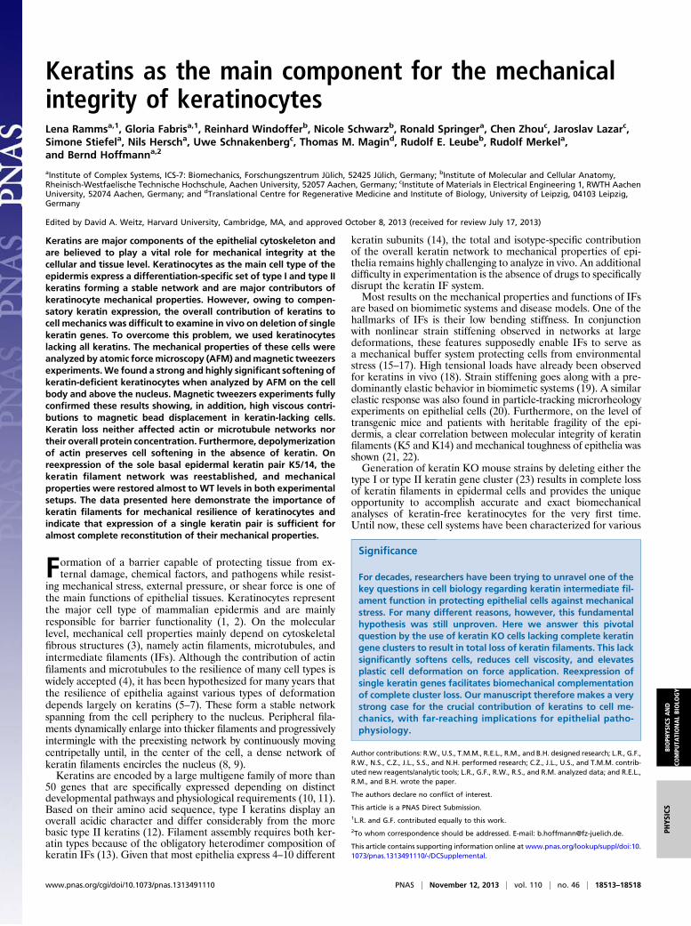
curves were analyzed by the classical Hertzian spherical indentermodel (Eq. 1) as implemented by the AFM manufacturer (Fig.3A). For all areas analyzed, the resulting Young’s moduli in-dicated a significantly higher (Fig. 3 B and C) stiffness of WTcells compared with KtyI−/− cells [nucleus: EWT = 459 ± 31 Pa(SEM) (n = 125) and EKtyI
−/− = 343 ± 18 Pa (n = 109); signif-icance level, 0.0030; cell body: EWT = 752 ± 100 Pa (n = 117) andEKtyI
−/− = 412 ± 75 Pa (n = 113); significance level, 0.0002].Force maps from whole WT and KtyI−/− cells also demonstratedan overall softened cell phenotype on keratin loss, most promi-nently above the nucleus (Fig. 4 A–D).Using an alternative approach for force-distance curve analysis
by accurately determining the indenter contact points and bydescribing curves via a standard power law function, the pre-sented results were fully confirmed with an even better fit ac-curacy (SI Text, Alternative Force-Distance Curve Analysis andFigs. S3–S6).Identical AFM experiments on keratin KtyII−/− mutant cells
lacking the complete set of type II keratins resulted in very
Fig. 1. Keratin network characterization and AFM analysis regions. (A–C)Immunfluorescence micrographs of the keratin network in WT, KtyI−/− KO,and K14 rescue (RES) keratinocytes. The KtyI−/− cell outline is given bya dashed line. (Scale bars, 10 μm.) (D) Top view phase contrast image of anAFM tip-less cantilever aligned with the cell body of a living adherent ker-atinocyte. (Scale bar, 10 μm.) (E) Side view diagram of a cantilever positionedabove the nucleus and (F) the cell body.
Fig. 2. AFM force-distance curves and force-indentation plots. (A) Plotshowing the superposition of three AFM force-distance curves (green, red,blue) recorded successively at 5-s intervals on the same position above thenucleus of a WT keratinocyte. (B) Plot showing the average forces needed toreach a certain indentation depth above the nucleus (n = 109–125,depending on indentation depth) and (C) cell body (n = 54–117). Significantdifferences between WT and KtyI−/− cells (KO) are indicated (*P = 0.05, **P =0.01, ***P = 0.001); error bars are SEM.
18514 | www.pnas.org/cgi/doi/10.1073/pnas.1313491110 Ramms et al.
similar data. Mutant cells were significantly softer (∼40%) thanthe corresponding WT cells (n ≥ 10 cells each).
Reexpression of Keratin 14 Rescues Keratin Network and CellElasticity. To prove that the absence of keratins was responsiblefor altered cell stiffness, rescue cells stably expressing keratin 14-YFP (yellow fluorescent protein) were generated (K14), leadingto IF formation by pairing with the endogenously produced K5.Cells were analyzed by the above-described AFM protocol.Proper keratin network formation was assessed by fluorescencemicroscopy (Fig. 1C) before indentation in each of the analyzedcells (n = 46). Elastic moduli were retrieved by fitting theresulting force distance curves with the Hertz model. To mini-mize errors, WT and KtyI−/− cells (n = 57 and n = 51, re-spectively) were measured in parallel using the same cantilever(Fig. 4 E and F). We found that the average cell elasticity was
significantly increased on reexpression of K14. Interestingly,Young’s moduli of rescue cells showed a biphasic distribution,with ∼50% of cells presenting reduced elasticity as in KtyI−/−
cells and the other ∼50% presenting WT or even more elevatedstiffness.
Keratin Stiffens Cells Against Intracellular Deformation and ElevatesCell Elasticity. Although AFM analyses are based on the appli-cation of extracellular indentation forces, we sought for an al-ternative method to probe for intracellular force measurementsof adherent cells. To this end, we adapted magnetic tweezers (fora scheme, see Fig. S1B) to examine intracellular deformation bylocal straining in keratin-free cells. In this set of experiments,KtyII−/− cells, corresponding WT cells and K5-YFP expressingrescue cells, were used. The KtyII−/− cells much like the KtyI−/−
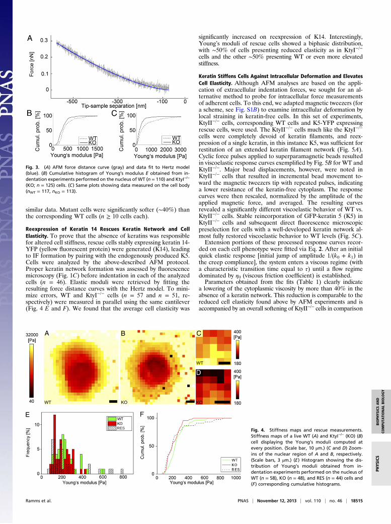
cells were completely devoid of keratin filaments, and reex-pression of a single keratin, in this instance K5, was sufficient forrestitution of an extended keratin filament network (Fig. 5A).Cyclic force pulses applied to superparamagnetic beads resultedin viscoelastic response curves exemplified by Fig. 5B for WT andKtyII−/−. Major bead displacements, however, were noted inKtyII−/− cells that resulted in incremental bead movement to-ward the magnetic tweezers tip with repeated pulses, indicatinga lower resistance of the keratin-free cytoplasm. The responsecurves were then rescaled, normalized by the amplitude of theapplied magnetic force, and averaged. The resulting curvesrevealed a significantly different viscoelastic behavior of WT vs.KtyII−/− cells. Stable reincorporation of GFP-keratin 5 (K5) inKtyII−/− cells and subsequent direct fluorescence microscopicpreselection for cells with a well-developed keratin network al-most fully restored viscoelastic behavior to WT levels (Fig. 5C).Extension portions of these processed response curves recor-
ded on each cell phenotype were fitted via Eq. 2. After an initialquick elastic response [initial jump of amplitude 1/(k0 + k1) inthe creep compliance], the system enters a viscous regime (witha characteristic transition time equal to τ) until a flow regimedominated by η0 (viscous friction coefficient) is established.Parameters obtained from the fits (Table 1) clearly indicate
a lowering of the cytoplasmic viscosity by more than 40% in theabsence of a keratin network. This reduction is comparable to thereduced cell elasticity found above by AFM experiments and isaccompanied by an overall softening of KtyII−/− cells in comparison
Fig. 3. (A) AFM force distance curve (gray) and data fit to Hertz model(blue). (B) Cumulative histogram of Young’s modulus E obtained from in-dentation experiments performed on the nucleus of WT (n = 110) and KtyI−/−
(KO; n = 125) cells. (C) Same plots showing data measured on the cell body(nWT = 117, nKO = 113).
Fig. 4. Stiffness maps and rescue measurements.Stiffness maps of a live WT (A) and KtyI−/− (KO) (B)cell displaying the Young’s moduli computed atevery position. (Scale bar, 10 μm.) (C and D) Zoom-ins of the nuclear region of A and B, respectively.(Scale bars, 3 μm.) (E) Histogram showing the dis-tribution of Young’s moduli obtained from in-dentation experiments performed on the nucleus ofWT (n = 58), KO (n = 48), and RES (n = 44) cells and(F) corresponding cumulative histograms.
Ramms et al. PNAS | November 12, 2013 | vol. 110 | no. 46 | 18515
BIOPH
YSICSAND
COMPU
TATIONALBIOLO
GY
PHYS
ICS
with WT and rescue cells. The relaxation times remaincomparable.
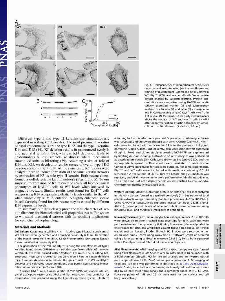
Keratin Loss Does Not Affect Actin and Microtubule Networks. Toverify that reduced cell resilience and viscosity in the absence ofkeratins were not significantly influenced by altered expression ofactin and tubulin, Western blot analysis was performed (24).Overall protein levels for all cell lines analyzed here indicated nomajor changes in protein concentration of actin and tubulin (Fig.6B). In agreement with previous findings, staining for actin andtubulin demonstrated that the overall actin and microtubule or-ganization was unaffected by the absence of keratins (Fig. 6A). Tofurther exclude a putative influence of actin on altered mechan-ical properties of KO cells, actin filaments were disassembled bylatrunculin A in WT and KtyI−/− cells. Subsequent AFM mea-surements on those cells identified a continuous, basically un-changed difference in elasticity and therefore significantly(significance level, 0.003) stiffer cell properties for WT cells com-pared with KtyI−/− cells, strongly supporting the dominant role ofthe keratin network in determining cell stiffness (Fig. 6C).
DiscussionThe elasticity of cells and their resistance to external mechanicalstress have been intensively analyzed for decades because oftheir crucial contribution to many cell functions such as differ-entiation (29) or cancer progression (30). Mechanical stability isespecially relevant for epidermal keratinocytes, which are sub-jected to intense deformation in all directions. Although one ofthe main characteristics of epithelial cytoskeleton is the pre-dominance of keratin filaments spanning the entire cytoplasmand surrounding the nucleus as a dense network (8), mechanicalanalyses have largely focused on actin filaments. On the otherhand, it has been proposed that IFs serve as a major mechanicalbuffer system protecting cells from environmental stress (5).Seminal publications have tried to shed light on this hypothesisby use of biomimetic model systems on artificial IF networks(15), extracted keratin networks (20, 31), and analyses on wholecells or mouse models (32, 33). In essence, many of these anal-yses identified a very low bending stiffness for keratin filamentsand a minor effect of keratin on cell elasticity. At the same time,however, rearrangements of the keratin network in various
cancer cell lines were hypothesized to induce significant changesin cell elasticity (5).Using two independent keratinocyte cell lines, both lacking
the entire keratin cytoskeleton, and matched rescue cell linesreexpressing the basal cell–specific keratin pair K5/14, we con-clusively demonstrate that elasticity, effective spring constant,and cell viscosity are significantly affected by keratins, leading tosoftened, less viscous cells in their absence. The use of two in-dependent keratin-free cell models, the previously establishedkeratin type II−/− mutant cells and the newly isolated keratintype I−/− keratinocyte mutant cells, in combination with matchedWT controls and rescue cell lines largely excludes cell line–specific artifacts and considerably strengthens the tenacity of ourfindings. Both mutant cell lines lacked keratin filaments com-pletely, showed very similar cell morphologies, and presentedvery similar biomechanical deficiencies as reflected by almostidentical reductions in Young’s moduli that were identified inour AFM analyses. Furthermore, all alterations in mechanicalcell properties were shown to be fully dependent on keratin loss.Here, neither actin nor MT protein levels nor correspondingcytoskeletal structures were affected. These results are well inline with results on protein levels found before for KtyII−/− cells(23, 24). Additionally, pharmacological actin network disruptionreduced the overall cell elasticity as one would expect on re-moval of cortical actin but did not influence the clear bio-mechanical differences between WT and KtyI−/− cells. Thesedata therefore clearly show that our AFM measurements weresufficiently deep to deform not only cortical actin but also thekeratin network beneath.Keratin material properties additionally explain the even more
drastic keratin mutant phenotypes observed in magnetic tweezersstraining experiments. Although AFM-induced indentationswork primarily against the comparatively low IF bending stiffnessof only 4–12 × 10−27 Nm2 (15), microrheological magnetictweezers experiments locally stretch the keratin network to suchan extent that they work against increasing stiffness at high strain.Keratin filament extensions of more than 2.5-fold have been ob-served in vitro (34), and stretched keratins have been imaged incultured keratinocytes that were elongated by 133% (18) withoutfilament rupture. The absence of plastic deformations on appli-cation of successive magnetic tweezers pulses in WT cells in thecurrent study provides further compelling evidence for the ef-fectiveness of keratin networks as mechanical buffer system.Consequently, the absence of this buffer system in keratin-deficient cells resulted in reduced viscosity, lowered effectivespring constants, and plastic cell deformation. These observationsare in line with reports on other IF systems. For example,vimentin-deficient fibroblasts presented a reduced stiffness onmechanical stress application by extracellular magnetic twistingcytometry (35). In another study, Brown et al. (36) noted that therigidity of T lymphocytes depends primarily on intact vimentinfilaments as determined by flow cytometry. On the other hand,AFM measurements on rat fibroblasts with a disrupted vimentincytoskeleton identified localized stiffening (37).
Fig. 5. Keratin network localization and magnetic tweezers analyses. (A)Immunfluorescence micrographs of the keratin network of WT, KO (KtyII−/−),and K5 rescue (Res) cells. (Scale bars, 10 μm.) (B) Example of raw magnetictweezers data: displacement of the superparamagnetic bead incorporated in thecytoplasm of a WT and a KtyII−/− cell (KO) following the application of 5-s forcepulses exerted at 10-s intervals. (C) Average overall rescaled peaks recorded onthe three phenotypes as a function of time (nWT = 40 for 9 cells; nKO = 39 for 10cells; nRES = 22 for 6 cells). In black, fits of Eq. 3. Error bars are SEM.
Table 1. Viscoelastic cell parameters
Parameter KO RES WT
keff = k0 + k1 (pN/μm) 8 11.8 12.3SD 1.1 3.2 1.8η (pN s/μm) 10.8 18.5 18.3SD 1.3 4.2 3.0τ (s) 0.4 0.43 0.48SD 0.11 0.19 0.17
Viscoelastic parameters obtained from fitting Eq. 2 to the averaged nor-malized magnetic tweezers data of WT, K5 rescue (RES), and KtyII−/− mutantcells (KO). keff, effective spring constant; η, viscous friction coefficient; τ,characteristic relaxation time.
18516 | www.pnas.org/cgi/doi/10.1073/pnas.1313491110 Ramms et al.
Different type I and type II keratins are simultaneouslyexpressed in resting keratinocytes. The most prominent keratinsof basal epidermal cells are the type II K5 and the type I keratinsK14 and K15 (14). K5 deletion results in pronounced cytolysisand neonatal lethality (38), whereas K14 depletion leads toepidermolysis bullosa simplex-like disease where mechanicaltrauma exacerbates blistering (39). Assuming a similar role ofK14 and K15, we decided to test for rescue of overall type I KOby reexpression of K14 only. At the same time, K5 rescues wereanalyzed here to induce formation of the same keratin networkby expression of K5 as sole type II keratin. Both rescue clonesformed a well-detectable keratin network (Figs. 1 and 5). To oursurprise, reexpression of K5 rescued basically all biomechanicalphenotypes of KtyII−/− cells to WT levels when analyzed bymagnetic tweezers. Similar results were found for KtyI−/− cellsreexpressing K14 recuperating elasticity levels similar to the WTwhen analyzed by AFM indentation. A slightly enhanced spreadin cell elasticity found for this rescue may be caused by differentK14 expression levels.In summary, our data clearly prove the vital character of ker-
atin filaments for biomechanical cell properties as a buffer systemto withstand mechanical stresses with far-reaching implicationsfor epithelial pathophysiology.
Materials and MethodsCell Culture. Keratinocyte cell line KtyII−/− lacking type II keratins and controlWT cell lines were generated and described previously (23, 24). Generationof the type II rescue cell line KII KO K5-GFP reexpressing GFP-tagged keratin5 was described in previously (25).
For generation of the cell line KtyI−/− lacking the complete set of type Ikeratins, homozygous C57B16 mice harboring two floxed alleles of the type Ikeratin cluster were crossed with 129S1hprt Cre mice. The resulting het-erozygous mice were crossed to get 25% type I keratin cluster-deficientmice. Keratinocytes were isolated from the epidermis of E18.5 WT and KtyI−/−
embryos and cultivated under conditions that permit spontaneous immor-talization as described in Turksen (40).
To rescue KtyI−/− cells, human keratin 14-YFP cDNA was cloned into len-tiviral pLVX-puro vector using XhoI and NotI restriction sites. Lentivirus fortransduction was produced using the Lenti-X expression system (Clontech)
according to the manufacturers’ protocol. Supernatant containing lentiviruswas harvested, and titers were checked with Lenti-X GoStix (Clontech). KtyI−/−
cells were incubated with lentivirus for 24 h in the presence of 8 μg/mLpolybrene (Sigma-Aldrich). Subsequently, cells were selected with puromycin(8 μg/mL; PAA), and clones stably expressing hK14-YFP were generatedby limiting dilution cloning. Cultivation of keratinocytes was performedas described previously (25). Cells were grown at 5% (vol/vol) CO2 and theappropriate temperature. Rescue cells were incubated in medium con-taining 8 μg/mL puromycin for selection purposes. For some experiments,KtyI−/− and WT cells were incubated with medium containing 0.5 μMlatrunculin A for 60 min at 37 °C. Directly before analysis, medium wasreplaced, and AFM measurements were performed within the next 60 min.The effectiveness of actin depolymerization was verified by immunocyto-chemistry on identically incubated cells.
Western Blotting. SDS/PAGE on crude protein extracts of all cell lines analyzedin this work was performed as described previously (41). Separation of totalprotein extracts was performed by standard procedures (4–20% SDS-PAGE).Using GAPDH as constitutively expressed marker (antibody G8795; Sigma-Aldrich), overall protein levels of actin and tubulin were determined usingmAB8457 (CST) and MAB1864 (Millipore) as antibodies.
Immunocytochemistry. For immunocytochemical experiments, 2.5 × 10⁴ cellswere grown on collagen I–coated glass coverslips for 48 h. Labelings wereperformed as described previously (25) using fluorescently labeled phalloidin(Invitrogen) for actin and antibodies against tubulin (see above) or keratin(rabbit anti-pan keratin; ProGen Biotechnik). Images were recorded eitheron an ApoTome.2 (Zeiss) using AxioVision LE software (Zeiss) or acquiredusing a laser scanning confocal microscope (LSM 710; Zeiss), both equippedwith a Plan-Apochromat 63×/1.4 oil immersion objective.
AFM Measurements. AFM imaging and force spectroscopy were performedusing a JPK Nanowizard Life Science version instrument (JPK) equipped witha fluid chamber (Biocell; JPK) for live cell analysis and an inverted opticalmicroscope (Axiovert 200; Zeiss) for sample observation. AFM imaging offixed and live cells was performed as described in SI Text, AFM Measure-ments. During indentation experiments, each analyzed position was recor-ded by at least three force curves and a cantilever speed of v = 1.5 μm/s.Force set points of 1.48 and 0.5 nN were used for the nucleus and cellbody, respectively.
Fig. 6. Independency of biomechanical deficiencieson actin and microtubules. (A) Immunofluorescentstaining of microtubules (Upper) and actin (Lower) inWT, KtyI−/− (KO), and rescue cells. (B) Crude proteinextract analysis by Western blotting. Protein con-centrations were equalized using GAPDH as consti-tutively expressed marker (1) and subsequentlyanalyzed for tubulin (2) and actin (3) expression. (aand b) Corresponding WTs. (c) KtyI−/−. (d) KtyII−/−. (e)K14 rescue. (f) K5 rescue. (C) Elasticity measurementsabove the nucleus of WT and KtyI−/− cells by AFMafter depolymerization of actin filaments by latrun-culin A. n = 30 cells each. (Scale bars, 20 μm.)
Ramms et al. PNAS | November 12, 2013 | vol. 110 | no. 46 | 18517
BIOPH
YSICSAND
COMPU
TATIONALBIOLO
GY
PHYS
ICS
AFM Data Analysis. The approaching parts of the recorded force-distancecurves were analyzed using the JPK DP software (Hertz fit, sphere model) toobtain values of Young’s modulus E (Fig. 2A). With this option, the softwareuses the fit function described by Sneddon (42)
FðaÞ= E1− ν2
�a2 +R2
2ln
R+aR−a
− aR�, [1]
δ=a2ln
R+ aR− a
Here, δ is the indentation depth, a is the contact radius of the indenter, R isthe measured silica bead radius, ν is the sample’s Poisson ratio (set to 0.5assuming cell’s incompressibility), and E is the cell’s Young’s modulus. In thefits, Young’s modulus E and the contact point were used as fit parameters.For estimating the optimal indentation depth, so-called “apparent modu-lus” plots were used based on ref. 43 (Fig. S2 and SI Text, AFM ApparentModulus Plots).
Because infinite sample thickness is assumed in the Hertz model, welimited the indentation to at most 5–10% of the sample thickness. Accord-ingly, curves recorded when indenting on the nuclei were fit for a range of0 to 500 nm after the contact point. For the cell body, comparably thinner,this distance was taken to be 200 nm.
Distributions of resulting elasticities E were analyzed for differences withthe parameter-free two-sided Mann-Whitney-Wilcoxon test.
Magnetic Tweezers Setup and Measurements. Construction and calibration ofthe self-made magnetic tweezers and measurement procedures are de-scribed in detail in Fig. S7 and in SI Text, Magnetic Tweezers Measurements.
In short, the instrument is based on a self-etched, water-cooled magneticcore of an electromagnet attached to a micromanipulator. M-270 Dyna-beads (Invitrogen) were incorporated as superparamagnetic beads usinga PDS-1000/He particle delivery system (BioRad).
Magnetic Tweezers Data Analysis. Single peaks were normalized by themagnetic force applied at every time frame [calculated from the calibrationcurve F = F(d), d being the bead-tip distance; Fig. S8 and SI Text, MagneticTweezers Measurements] and averaged for every cell type. Following Bauschet al. (28), the extensional parts of these averaged creep-response curves(i.e., distance divided by force) were fitted by a four-element mechanicalequivalent circuit composed of a dashpot in series with a Zener body
JðtÞ= 1k0
�1−
k1k0 + k1
exp�−tτ
��+
tη0, [2]
where k0 + k1 describes the effective spring constant of the system, η0 indi-cates the viscous friction coefficient of the bead, and τ is the relaxation timecharacteristic of the transition from an elastic to a viscous regime. Uncer-tainties of fit results were determined as SDs from 500 times fitting syntheticdata created by adding to the fit result normally distributed random num-bers with scatter taken from the experiment.
ACKNOWLEDGMENTS. We thank Florian Kumpfe (JPK) for technical adviceand constructive discussions. This work was supported by the START Programof the Faculty of Medicine, Rheinisch-Westfaelische Technische HochschuleAachen, and by the German Research Council (LE566/18-1, WI1731/6-1,WI1731-8-1, MA1316-9/3, MA1316-15, INST 268/230-1, Translational Centrefor Regenerative Medicine, and Leipzig No. 0315883).
1. Fuchs E (1995) Keratins and the skin. Annu Rev Cell Dev Biol 11:123–153.2. Sun TT, Shih C, Green H (1979) Keratin cytoskeletons in epithelial cells of internal
organs. Proc Natl Acad Sci USA 76(6):2813–2817.3. Kasza KE, et al. (2007) The cell as a material. Curr Opin Cell Biol 19(1):101–107.4. Stricker J, Falzone T, Gardel ML (2010) Mechanics of the F-actin cytoskeleton.
J Biomech 43(1):9–14.5. Beil M, et al. (2003) Sphingosylphosphorylcholine regulates keratin network archi-
tecture and visco-elastic properties of human cancer cells. Nat Cell Biol 5(9):803–811.6. Kreplak L, Fudge D (2007) Biomechanical properties of intermediate filaments: From
tissues to single filaments and back. BioEssays 29(1):26–35.7. Lulevich V, Yang HY, Isseroff RR, Liu GY (2010) Single cell mechanics of keratinocyte
cells. Ultramicroscopy 110(12):1435–1442.8. Windoffer R, Beil M, Magin TM, Leube RE (2011) Cytoskeleton in motion: The dy-
namics of keratin intermediate filaments in epithelia. J Cell Biol 194(5):669–678.9. Windoffer R, Wöll S, Strnad P, Leube RE (2004) Identification of novel principles of
keratin filament network turnover in living cells. Mol Biol Cell 15(5):2436–2448.10. Bragulla HH, Homberger DG (2009) Structure and functions of keratin proteins in
simple, stratified, keratinized and cornified epithelia. J Anat 214(4):516–559.11. Hesse M, Zimek A, Weber K, Magin TM (2004) Comprehensive analysis of keratin gene
clusters in humans and rodents. Eur J Cell Biol 83(1):19–26.12. Steinert PM, Jones JC, Goldman RD (1984) Intermediate filaments. J Cell Biol 99(1 Pt 2):
22s–27s.13. Eichner R, Sun TT, Aebi U (1986) The role of keratin subfamilies and keratin pairs in
the formation of human epidermal intermediate filaments. J Cell Biol 102(5):1767–1777.
14. Magin TM, Vijayaraj P, Leube RE (2007) Structural and regulatory functions of kera-tins. Exp Cell Res 313(10):2021–2032.
15. Janmey PA, Euteneuer U, Traub P, Schliwa M (1991) Viscoelastic properties of vi-mentin compared with other filamentous biopolymer networks. J Cell Biol 113(1):155–160.
16. Lin YC, et al. (2010) Origins of elasticity in intermediate filament networks. Phys RevLett 104(5):058101.
17. Storm C, Pastore JJ, MacKintosh FC, Lubensky TC, Janmey PA (2005) Nonlinear elas-ticity in biological gels. Nature 435(7039):191–194.
18. Fudge D, et al. (2008) The intermediate filament network in cultured human kera-tinocytes is remarkably extensible and resilient. PLoS ONE 3(6):e2327.
19. Yamada S, Wirtz D, Coulombe PA (2003) The mechanical properties of simple epi-thelial keratins 8 and 18: Discriminating between interfacial and bulk elasticities.J Struct Biol 143(1):45–55.
20. Sivaramakrishnan S, DeGiulio JV, Lorand L, Goldman RD, Ridge KM (2008) Micro-mechanical properties of keratin intermediate filament networks. Proc Natl Acad SciUSA 105(3):889–894.
21. Fuchs E, Cleveland DW (1998) A structural scaffolding of intermediate filaments inhealth and disease. Science 279(5350):514–519.
22. Simpson CL, Patel DM, Green KJ (2011) Deconstructing the skin: Cytoarchitecturaldeterminants of epidermal morphogenesis. Nat Rev Mol Cell Biol 12(9):565–580.
23. Vijayaraj P, et al. (2009) Keratins regulate protein biosynthesis through localization ofGLUT1 and -3 upstream of AMP kinase and Raptor. J Cell Biol 187(2):175–184.
24. Kröger C, et al. (2013) Keratins control intercellular adhesion involving PKC-α-medi-ated desmoplakin phosphorylation. J Cell Biol 201(5):681–692.
25. Seltmann K, et al. (2013) Keratins mediate localization of hemidesmosomes and re-press cell motility. J Invest Dermatol 133(1):181–190.
26. Azeloglu EU, Costa KD (2011) Atomic force microscopy in mechanobiology: Measur-ing microelastic heterogeneity of living cells. Methods Mol Biol 736:303–329.
27. Radmacher M, Fritz M, Kacher CM, Cleveland JP, Hansma PK (1996) Measuring theviscoelastic properties of human platelets with the atomic force microscope. Biophys J70(1):556–567.
28. Bausch AR, Ziemann F, Boulbitch AA, Jacobson K, Sackmann E (1998) Local mea-surements of viscoelastic parameters of adherent cell surfaces by magnetic beadmicrorheometry. Biophys J 75(4):2038–2049.
29. Engler AJ, Sen S, Sweeney HL, Discher DE (2006) Matrix elasticity directs stem celllineage specification. Cell 126(4):677–689.
30. Suresh S (2007) Biomechanics and biophysics of cancer cells. Acta Biomater 3(4):413–438.
31. Walter N, Busch T, Seufferlein T, Spatz JP (2011) Elastic moduli of living epithelialpancreatic cancer cells and their skeletonized keratin intermediate filament network.Biointerphases 6(2):79–85.
32. Cao T, Longley MA, Wang XJ, Roop DR (2001) An inducible mouse model for epi-dermolysis bullosa simplex: Implications for gene therapy. J Cell Biol 152(3):651–656.
33. Hesse M, Franz T, Tamai Y, Taketo MM, Magin TM (2000) Targeted deletion of ker-atins 18 and 19 leads to trophoblast fragility and early embryonic lethality. EMBO J19(19):5060–5070.
34. Kreplak L, Bär H, Leterrier JF, Herrmann H, Aebi U (2005) Exploring the mechanicalbehavior of single intermediate filaments. J Mol Biol 354(3):569–577.
35. Wang N, Stamenovi�c D (2000) Contribution of intermediate filaments to cell stiffness,stiffening, and growth. Am J Physiol Cell Physiol 279(1):C188–C194.
36. Brown MJ, Hallam JA, Colucci-Guyon E, Shaw S (2001) Rigidity of circulating lym-phocytes is primarily conferred by vimentin intermediate filaments. J Immunol166(11):6640–6646.
37. Plodinec M, et al. (2011) The nanomechanical properties of rat fibroblasts are mod-ulated by interfering with the vimentin intermediate filament system. J Struct Biol174(3):476–484.
38. Peters B, Kirfel J, Büssow H, Vidal M, Magin TM (2001) Complete cytolysis and neo-natal lethality in keratin 5 knockout mice reveal its fundamental role in skin integrityand in epidermolysis bullosa simplex. Mol Biol Cell 12(6):1775–1789.
39. Chan Y, et al. (1994) A human keratin 14 “knockout”: The absence of K14 leads tosevere epidermolysis bullosa simplex and a function for an intermediate filamentprotein. Genes Dev 8(21):2574–2587.
40. Turksen K (2005) Epidermal Cells: Methods and Protocols (Springer, New York), p 480.41. Waschbüsch D, et al. (2009) Presenilin 1 affects focal adhesion site formation and cell
force generation via c-Src transcriptional and posttranslational regulation. J BiolChem 284(15):10138–10149.
42. Sneddon IN (1965) The relation between load and penetration in the axisymmetricBoussinesq problem for a punch of arbitrary profile. Int J Eng Sci 3(1):47–57.
43. Rosenbluth MJ, Lam WA, Fletcher DA (2006) Force microscopy of nonadherent cells:a comparison of leukemia cell deformability. Biophys J 90(8):2994–3003.
18518 | www.pnas.org/cgi/doi/10.1073/pnas.1313491110 Ramms et al.


![Differentiation-Dependent Expression of Keratins in Human Oral … · 2017. 2. 1. · [ 16, 17]. The expression of specific keratins appears to depend on the type of tissue, as well](https://img.pdfslide.net/doc/110x75/5ff979cead588c6cd35f8d9b/differentiation-dependent-expression-of-keratins-in-human-oral-2017-2-1-16.jpg)