Embed Size (px)
Citation preview
Jour
nal o
f Cel
l Sci
ence
• A
dvan
ce a
rtic
le
© 2016. Published by The Company of Biologists Ltd.
KIF17 regulates RhoA-dependent actin remodeling at epithelial cell-cell
adhesions
Bipul R. Acharya1∞¥, Cedric Espenel1≡¥, Fotine Libanje2¥, Joel Raingeaud2, Jessica Morgan1§, Fanny Jaulin2* and Geri Kreitzer1*
1Department of Cell and Developmental Biology, Weill Cornell Medical College of Cornell
University, New York, NY
2Gustave Roussy Institute, UMR-8126, 114 rue Edouard Vaillant, 94805 Villejuif, France
*Address correspondence to:
Geri Kreitzer: [email protected] Weill Cornell Medical College, 1300 York Ave., New York, NY, 10065 Fanny Jaulin: [email protected] Gustave Roussy Institute, 114 rue Edouard Vaillant, 94805 Villejuif, France ¥ These authors made equally essential contributions ∞ Current address: University of Queensland, Building 80, Brisbane, Australia ≡ Current address: Stanford University, 443 Via Ortega, Stanford, CA. § Current address: University of California, Santa Cruz, Graduate program in Chemistry and
Biochemistry, Santa Cruz, CA.
JCS Advance Online Article. Posted on 12 January 2016
Jour
nal o
f Cel
l Sci
ence
• A
dvan
ce a
rtic
le
ABSTRACT
The kinesin KIF17 localizes at MT plus-ends and contributes to regulation of MT
stabilization, and epithelial polarization. We now show that KIF17 localizes at cell-cell
adhesions and that KIF17 depletion inhibits accumulation of actin at the apical pole of cells
grown in 3D organotypic cultures and alters the distribution of actin and E-cadherin in cells
cultured in 2D on solid supports. Overexpression of full-length KIF17 constructs or truncation
mutants containing the N-terminal motor domain resulted in accumulation of newly
incorporated GFP-actin into junctional actin foci, cleared E-cadherin from cytoplasmic vesicles
and stabilized cell-cell adhesions to challenge with calcium depletion. Expression of these
KIF17 constructs also increased cellular levels of active RhoA, while active RhoA was
diminished in KIF17-depleted cells. Inhibition of Rho or its effector ROCK, or expression of
LIMK1 kinase-dead or activated cofilinS3A inhibited KIF17-induced junctional actin
accumulation. Interestingly, KIF17 activity toward actin depends on the motor domain but is
independent of MT binding. Together, these data show that KIF17 can modify Rho-GTPase
signaling to influence junctional actin and the stability of the apical junctional complex of
epithelial cells.
Jour
nal o
f Cel
l Sci
ence
• A
dvan
ce a
rtic
le
INTRODUCTION
Epithelia play key roles in tissue homeostasis by establishing transport systems for
vectorial secretion and absorption and by forming a physical barrier between the internal milieu
and the outside environment. Adherens junctions (AJs) and tight junctions (TJs), formed by
trans-cellular interactions of transmembrane adhesion proteins linked to the cytoskeleton, are
essential for epithelial morphogenesis and function. Known collectively as the apical junctional
complex (AJC), AJs couple adjacent cells physically while TJs set boundaries between apical
and basolateral membranes and control paracellular permeability (Guillot and Lecuit, 2013).
Components of the AJC are delivered to the membrane by transport along microtubules (MTs)
(Chen et al., 2003; Ivanov et al., 2006; Mary et al., 2002; Nekrasova et al., 2011; Portereiko et
al., 2004; Yanagisawa et al., 2004) and are anchored at adhesive sites by their association
with actin and MT adaptors. As cell-cell adhesions mature, signaling molecules that also
associate with the AJC induce changes in actin and MT arrays by modifying polymer dynamics
and stability (Brieher and Yap, 2013; Chausovsky et al., 2000; Mege et al., 2006; Ratheesh et
al., 2012). Thus the cytoskeleton affects AJC formation and maturation, and signaling at the
AJC reciprocally affects actin and MTs; together, these processes direct morphogenetic
responses to numerous cues (Brieher and Yap, 2013; Mack and Georgiou, 2014). Although
many of the components involved in remodeling of AJCs and the cytoskeleton are known, the
mechanisms employed to coordinate these events are still incompletely defined.
Rho family GTPases and their effectors comprise a major class of signaling molecules at
the AJC (Citi et al., 2014; Fukata and Kaibuchi, 2001); many are regulated by cell-cell
adhesion. Signaling by Cdc42, Rac1 and RhoA regulates AJC formation, maturation and
remodeling. They also regulate actin and MT arrays (Samarin and Nusrat, 2009; Wojnacki et
al., 2014). Rac1 and Cdc42 regulate Arp2/3 to effect branched actin filament formation
(Kraemer et al., 2007; Otani et al., 2006) and RhoA regulates formins in the generation of actin
cables (Carramusa et al., 2007; Kher et al., 2014; Kobielak et al., 2004). RhoA signaling,
through its effector Rho kinase (ROCK), also exerts indirect effects on branched actin
formation by inactivating the actin severing protein cofilin. In addition, RhoA activation of formin
leads to MT capture and stabilization in migrating fibroblasts (Bartolini et al., 2008; Cook et al.,
1998; Palazzo et al., 2001) and plays a role in regulating MT stability in epithelial cells (Nakaya
et al., 2008). Combined, these functions allow Rho-GTPases to orchestrate the remodeling of
cytoskeletal arrays and cell-cell junctions that accompanies epithelial polarization.
Rho GTPases are regulated upstream and downstream of the AJC by guanine-nucleotide
exchange factors (GEFs) and GTPase activating proteins (GAPs) that control spatiotemporal
activation of Rho effectors (Quiros and Nusrat, 2014). How Rho-GTPase effectors and
regulators are targeted to discrete sites for selective activation is still a topic of intense study.
Jour
nal o
f Cel
l Sci
ence
• A
dvan
ce a
rtic
le
Dynamic MT plus-ends can interact with proteins at the cortex and can deliver MT plus-end
associated proteins that regulate cytoskeletal and junctional organization, leading to the
concentration of E-cadherin at cell-cell contacts (Ligon and Holzbaur, 2007; Ligon et al., 2001;
Stehbens et al., 2006). MT capture and stabilization at the cortex may also provide specialized
tracks for targeted delivery of cytoplasmic and membrane proteins important for junction
maturation and remodeling (Waterman-Storer et al., 2000). As such, delivery of Rho-GTPase
effectors and regulators by MT motors can be envisioned as playing a role in regulation of
localized signaling cascades at the AJC.
KIF17 is a multifunctional, homodimeric MT motor with roles in vesicular transport (Chu et
al., 2006; Jenkins et al., 2006; Setou et al., 2000), transport of RNA granules (Chennathukuzhi
et al., 2003; Kotaja et al., 2006; Saade et al., 2007; Takano et al., 2007), regulation of
transcriptional activators (Kotaja et al., 2005; Macho et al., 2002), and in building sensory cilia
(Dishinger et al., 2010; Fan et al., 2011; Insinna et al., 2008; Jenkins et al., 2006; Ou et al.,
2005; Pan et al., 2006; Snow et al., 2004). In epithelial cells, KIF17 colocalizes and interacts
with components of the MT plus-end cortical capture machinery, promoting MT stabilization
and cell polarization (Acharya et al., 2013; Espenel et al., 2013; Jaulin and Kreitzer, 2010).
This can influence cell architecture, but it is not yet known if MT modification is the only means
by which KIF17 contributes to epithelial polarization. In this study, we provide evidence that
KIF17 activates a RhoA signaling pathway at cell-cell contacts that influences both cortical
actin and cell-cell junctions. This, in concert with its effects on MT stabilization, may serve to
integrate cytoskeletal remodeling with maturation and stabilization of the AJC. A role for KIF17
in local RhoA activation also provides an additional potential mechanism by which this kinesin-
2 motor exerts effects on epithelial polarization.
Jour
nal o
f Cel
l Sci
ence
• A
dvan
ce a
rtic
le
RESULTS
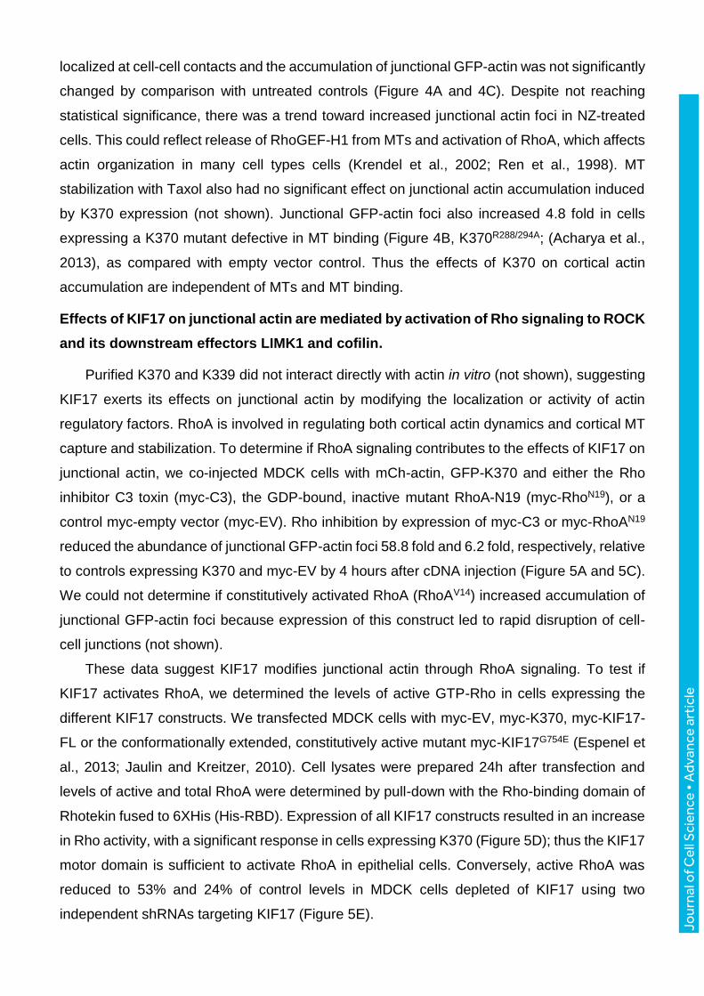
KIF17 contributes to actin organization in epithelial cells
We showed previously that KIF17 localizes to MT plus-ends with EB1 and contributes to
MT stabilization and polarization of epithelial cells (Jaulin and Kreitzer, 2010). In our analysis
of KIF17 distribution in MDCK and Caco-2 epithelial cells, we also identified a pool of KIF17
localized at sites of cell-cell contact that is lost after KIF17 depletion by shRNA (Figure 1A,
Supplemental Figure 1A and (Jaulin and Kreitzer, 2010)). KIF17 co-localized with E-cadherin
and actin at these cell-cell junctions (Figure 1A), and with -actinin, a junctional actin-binding
protein (Figure 1B).
In 3D organotypic MDCK cultures, 85% (+/-2.6%; n=322) of cysts that form have a single
layer of cells surrounding a central lumen, and actin is enriched at the apical pole of individual
cells (Figure 1C,D; shNC, short hairpin negative control). KIF17 depletion increased the
percentage of cysts with either no lumens or multiple lumens as described previously (Jaulin
and Kreitzer, 2010), and reduced the percentage of cysts with one lumen to 38% (+/-5.4%;
n=332). We used line-scan analysis of individual cells in cysts with a single lumen to measure
enrichment of actin at the apical pole (dotted line) relative to the basal pole (dashed line) in
control (shNC) and KIF17-depleted cells (shKIF17, Figure 1D). In shNC, the apical/basal actin
ratio is 6.74, but only 1.93 in shKIF17, demonstrating that apical actin enrichment is
compromised by KIF17 depletion. Thus, KIF17 colocalizes with and contributes to organizing
the distribution of actin in epithelial cells.
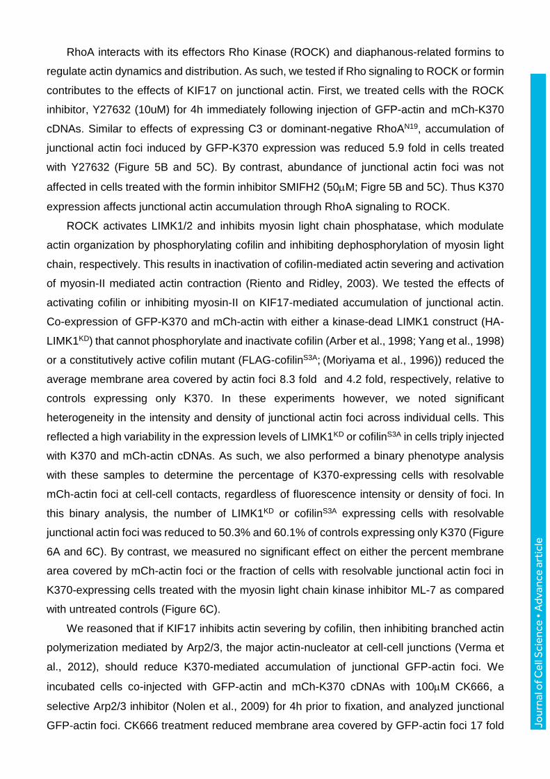
KIF17 localization at cell-cell contacts is mediated by its N-terminal motor domain.
To examine how KIF17 could affect actin organization, we prepared and analyzed the
localization of GFP-tagged KIF17 full-length and C-terminal truncation mutants in MDCK cells
(Figure 2A). Proteins were expressed acutely by intranuclear cDNA injection and their
localization was analyzed 3h after injection. Full-length KIF17 appeared primarily as diffuse
cytoplasmic fluorescence but a population of the protein localized as discrete puncta, a large
proportion of which accumulated at MT plus-ends in protruding regions of the cells (Figure 2A;
GFP-KIF17-FL) and as described previously (Jaulin and Kreitzer, 2010). The soluble pool of
KIF17-FL represents kinesin in a compact, auto-inhibited conformation (Espenel et al., 2013;
Hammond et al., 2010); this auto-inhibited conformation is disrupted by a single point mutation
in the hinge region between KIF17 coiled-coil domains (G754E), and when expressed in
epithelial cells GFP-KIF17-FLG754E localizes robustly at MT plus-ends and in cell protrusions
(Espenel et al., 2013; Jaulin and Kreitzer, 2010). Protein expressed from a construct in which
the tail and last coiled-coil are deleted, GFP-K490, localized at MT plus ends in cell protrusions
Jour
nal o
f Cel
l Sci
ence
• A
dvan
ce a
rtic
le
(Supplemental Figure 1B), but was also detected at cell-cell contacts in 24.3% of expressing
cells (Figure 2A and 2C). Proteins synthesized from two shorter constructs, GFP-K370
(encoding motor and neck) and GFP-K339 (encoding motor alone) can be detected along MTs
when cells are permeabilized briefly before fixation to release soluble protein (not shown;
(Jaulin and Kreitzer, 2010)) and when expressed at low levels (Supplemental Figure 1C). They
also localize at the centrosome with -tubulin (Supplemental Figure 1D). Moreover, K370 and
K339 localized prominently at cell-cell contacts in, respectively, 59.7% and 54.7% of the
injected cells (Figure 2A, 2B and 2C). The junctional localization of K339 and K370 is also
observed in other epithelial cell types such as MCF10A and Caco-2, is independent of the tag
identity (GFP, mCherry, myc, HA), and is seen with both N- and C-terminal fusion constructs
(Supplemental Figure 1E, (Jaulin and Kreitzer, 2010) and not shown). Together, this analysis
reveals that the motor domain is sufficient to target KIF17 to cell-cell contacts and that deletion
of C-terminus favors this subcellular localization. Like endogenous KIF17, K370 and K339
colocalized with actin, E-cadherin and -actinin at cell-cell contacts (Figure 2B and not shown).
We preferentially use K370 going forward since it behaves as a dimer in vitro. K339 behaves
as a monomer in vitro (Acharya et al., 2013), but had nearly identical impact in all experiments
where it was tested relative to K370.
Auto-inhibitory interactions of the KIF17 N-terminal motor and C-terminal tail domains
regulate KIF17 activity (Espenel et al., 2013; Hammond et al., 2010; Jaulin and Kreitzer, 2010).
In vitro, the KIF17 tail can bind directly to the KIF17 motor and reduces its MT-stimulated
ATPase activity (Acharya et al. 2013); as such, it may influence K370 and K339 localization at
the cortex. To test this directly, we co-expressed GFP-K339 or GFP-K370 with mCh-KIF17-
Tail (Figure 2A) and analyzed their localizations 3h after cDNA injection. Co-expression of
KIF17-Tail reduced the number of cells with junctional K339 and K370 to 45% and 33%,
respectively, of cells expressing these motor domains alone (Figure 2D). This effect of KIF17-
Tail on localization of K339 and K370 could result from either competition with proteins that
anchor KIF17 at cell-cell contacts, or by inhibition of the motor domain ATPase activity, which
would prevent movement along MTs (Acharya et al. 2013).
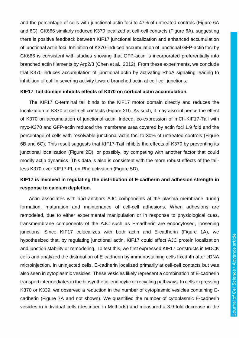
KIF17 motor domain enhances incorporation of actin at cell-cell contacts independently
of MT binding and ATPase activity.
The colocalization of KIF17 motor domains with junctional actin, and the effects of KIF17
depletion on actin distribution in cells cultured in 3D prompted us to examine if expression of
K370 affects actin organization. We expressed mCh-K370 or mCh-empty vector control (mCh-
EV) for 4hr after cDNA injection and analyzed the distribution of actin by immunofluorescence
microscopy. In K370 expressing cells, we observed a subtle but consistent enrichment of actin
Jour
nal o
f Cel
l Sci
ence
• A
dvan
ce a
rtic
le
in discrete foci at cell-cell contacts. This enrichment was best detected by applying a Sobel
edge detection filter to images (Supplemental Figure 2A). These actin foci were not detected
by phalloidin-labeling, probably because phalloidin strongly labels stress fibers and bundled
actin. This can mask signal from non-bundled and branched filaments, which are detected very
well with actin antibodies (Lessard, 1988; Nagasaki et al., 1994).
To further examine the change in junctional actin induced by K370, we monitored
incorporation of newly synthesized, fluorescently-tagged actin probes (GFP-actin or mCh-
LifeAct) into actin filaments by time-lapse fluorescence microscopy. We co-injected cells with
GFP-actin and mCh-K370 or mCh-EV cDNAs. One hour after injection, cells were transferred
to the microscope and images of GFP-actin were acquired at 10 min intervals for 4 hr at 37°C.
Newly synthesized GFP-actin (Figure 3A) and mCh-LifeAct (Supplemental Figure 2B)
accumulated in discrete foci along cell-cell contacts in control and K370 expressing cells.
However, co-expression of K370 accelerated the rate at which these new filaments became
apparent (Supplemental Video 1, and Supplemental Figure 3) and increased the number of
cells displaying these junctional actin filaments. Furthermore, K370 colocalized with GFP-actin
or mCh-LifeAct in these junctional foci (Figure 3A and Supplemental Figure 2B). In the time
course of these experiments, fluorescently tagged actin incorporated into more resolvable foci
at cell-cell contacts than mCh-LifeAct. For this reason, we used GFP-actin or mCh-actin in
experiments going forward to determine how KIF17 can impact junctional actin organization.
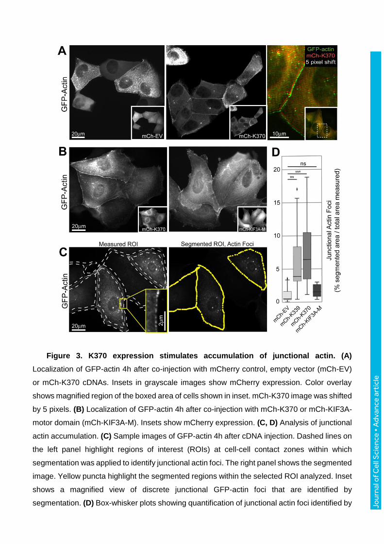
We quantified junctional accumulation of GFP-actin from images of cells by drawing
polylines along cell-cell contacts of microinjected cells. These regions of interest (ROIs) were
further processed and segmented to identify actin foci within these ROIs (Figure 3C). We then
measured the percentage of each ROI that was segmented as a readout of the junctional
region covered by GFP-actin foci (% segmented area / total area measured, Figure 3D) for
control and experimental data. This analysis revealed a 4.9 and 5.5 fold increase in junctional
GFP-actin foci in cells expressing mCh-K339 or mCh-K370 respectively, as compared with
controls expressing mCh-EV. By contrast, junctional GFP-actin foci were not increased in cells
expressing the heterodimeric kinesin-2 motor KIF3A (mCh-KIF3A-M, Figure 3B and 3D) as
compared with controls, demonstrating a selective effect of K370 or K339 on junctional actin
accumulation in MDCK cells. The effects of expressing KIF17 constructs on actin in 3D
cultured cells could not be determined as they induced substantial changes in cell shape over
extended times needed for cysts to develop.
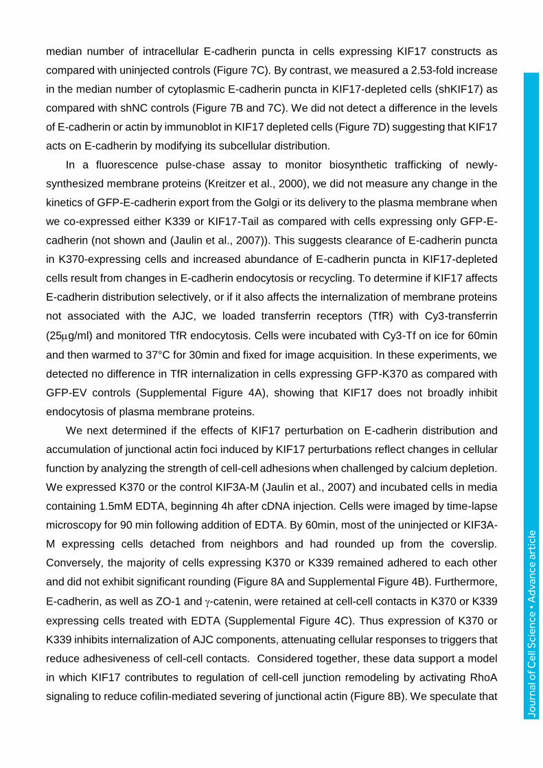
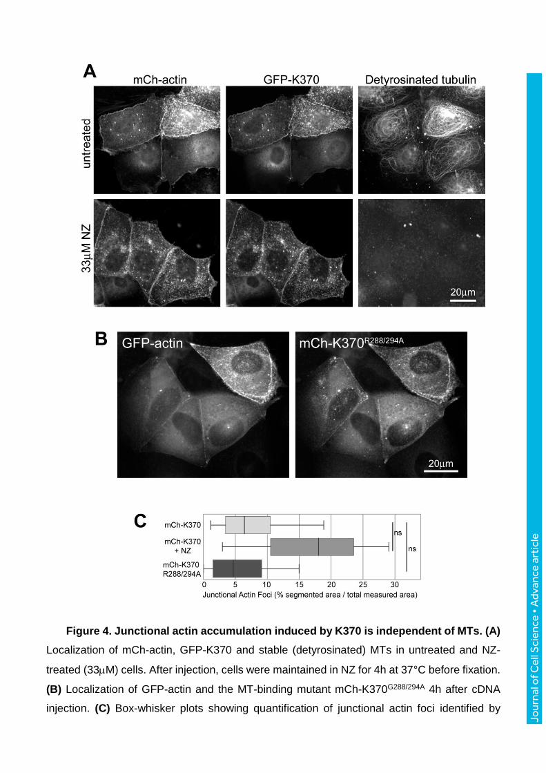
We next tested if the effects of KIF17 on cortical actin accumulation are MT dependent.
We pre-incubated cells with 33M nocodazole (NZ) to break down MTs prior to injecting GFP-
actin and K370 cDNAs. After an additional 4h with continuous NZ exposure, cells were fixed
and analyzed for accumulated GFP-actin at the cell periphery. In the absence of MTs, K370
Jour
nal o
f Cel
l Sci
ence
• A
dvan
ce a
rtic
le
localized at cell-cell contacts and the accumulation of junctional GFP-actin was not significantly
changed by comparison with untreated controls (Figure 4A and 4C). Despite not reaching
statistical significance, there was a trend toward increased junctional actin foci in NZ-treated
cells. This could reflect release of RhoGEF-H1 from MTs and activation of RhoA, which affects
actin organization in many cell types cells (Krendel et al., 2002; Ren et al., 1998). MT
stabilization with Taxol also had no significant effect on junctional actin accumulation induced
by K370 expression (not shown). Junctional GFP-actin foci also increased 4.8 fold in cells
expressing a K370 mutant defective in MT binding (Figure 4B, K370R288/294A; (Acharya et al.,
2013), as compared with empty vector control. Thus the effects of K370 on cortical actin
accumulation are independent of MTs and MT binding.
Effects of KIF17 on junctional actin are mediated by activation of Rho signaling to ROCK
and its downstream effectors LIMK1 and cofilin.
Purified K370 and K339 did not interact directly with actin in vitro (not shown), suggesting
KIF17 exerts its effects on junctional actin by modifying the localization or activity of actin
regulatory factors. RhoA is involved in regulating both cortical actin dynamics and cortical MT
capture and stabilization. To determine if RhoA signaling contributes to the effects of KIF17 on
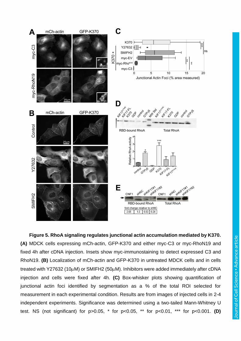
junctional actin, we co-injected MDCK cells with mCh-actin, GFP-K370 and either the Rho
inhibitor C3 toxin (myc-C3), the GDP-bound, inactive mutant RhoA-N19 (myc-RhoN19), or a
control myc-empty vector (myc-EV). Rho inhibition by expression of myc-C3 or myc-RhoAN19
reduced the abundance of junctional GFP-actin foci 58.8 fold and 6.2 fold, respectively, relative
to controls expressing K370 and myc-EV by 4 hours after cDNA injection (Figure 5A and 5C).
We could not determine if constitutively activated RhoA (RhoAV14) increased accumulation of
junctional GFP-actin foci because expression of this construct led to rapid disruption of cell-
cell junctions (not shown).
These data suggest KIF17 modifies junctional actin through RhoA signaling. To test if
KIF17 activates RhoA, we determined the levels of active GTP-Rho in cells expressing the
different KIF17 constructs. We transfected MDCK cells with myc-EV, myc-K370, myc-KIF17-
FL or the conformationally extended, constitutively active mutant myc-KIF17G754E (Espenel et
al., 2013; Jaulin and Kreitzer, 2010). Cell lysates were prepared 24h after transfection and
levels of active and total RhoA were determined by pull-down with the Rho-binding domain of
Rhotekin fused to 6XHis (His-RBD). Expression of all KIF17 constructs resulted in an increase
in Rho activity, with a significant response in cells expressing K370 (Figure 5D); thus the KIF17
motor domain is sufficient to activate RhoA in epithelial cells. Conversely, active RhoA was
reduced to 53% and 24% of control levels in MDCK cells depleted of KIF17 using two
independent shRNAs targeting KIF17 (Figure 5E).
Jour
nal o
f Cel
l Sci
ence
• A
dvan
ce a
rtic
le
RhoA interacts with its effectors Rho Kinase (ROCK) and diaphanous-related formins to
regulate actin dynamics and distribution. As such, we tested if Rho signaling to ROCK or formin
contributes to the effects of KIF17 on junctional actin. First, we treated cells with the ROCK
inhibitor, Y27632 (10uM) for 4h immediately following injection of GFP-actin and mCh-K370
cDNAs. Similar to effects of expressing C3 or dominant-negative RhoAN19, accumulation of
junctional actin foci induced by GFP-K370 expression was reduced 5.9 fold in cells treated
with Y27632 (Figure 5B and 5C). By contrast, abundance of junctional actin foci was not
affected in cells treated with the formin inhibitor SMIFH2 (50M; Figre 5B and 5C). Thus K370
expression affects junctional actin accumulation through RhoA signaling to ROCK.
ROCK activates LIMK1/2 and inhibits myosin light chain phosphatase, which modulate
actin organization by phosphorylating cofilin and inhibiting dephosphorylation of myosin light
chain, respectively. This results in inactivation of cofilin-mediated actin severing and activation
of myosin-II mediated actin contraction (Riento and Ridley, 2003). We tested the effects of
activating cofilin or inhibiting myosin-II on KIF17-mediated accumulation of junctional actin.
Co-expression of GFP-K370 and mCh-actin with either a kinase-dead LIMK1 construct (HA-
LIMK1KD) that cannot phosphorylate and inactivate cofilin (Arber et al., 1998; Yang et al., 1998)
or a constitutively active cofilin mutant (FLAG-cofilinS3A; (Moriyama et al., 1996)) reduced the
average membrane area covered by actin foci 8.3 fold and 4.2 fold, respectively, relative to
controls expressing only K370. In these experiments however, we noted significant
heterogeneity in the intensity and density of junctional actin foci across individual cells. This
reflected a high variability in the expression levels of LIMK1KD or cofilinS3A in cells triply injected
with K370 and mCh-actin cDNAs. As such, we also performed a binary phenotype analysis
with these samples to determine the percentage of K370-expressing cells with resolvable
mCh-actin foci at cell-cell contacts, regardless of fluorescence intensity or density of foci. In
this binary analysis, the number of LIMK1KD or cofilinS3A expressing cells with resolvable
junctional actin foci was reduced to 50.3% and 60.1% of controls expressing only K370 (Figure
6A and 6C). By contrast, we measured no significant effect on either the percent membrane
area covered by mCh-actin foci or the fraction of cells with resolvable junctional actin foci in
K370-expressing cells treated with the myosin light chain kinase inhibitor ML-7 as compared
with untreated controls (Figure 6C).
We reasoned that if KIF17 inhibits actin severing by cofilin, then inhibiting branched actin
polymerization mediated by Arp2/3, the major actin-nucleator at cell-cell junctions (Verma et
al., 2012), should reduce K370-mediated accumulation of junctional GFP-actin foci. We
incubated cells co-injected with GFP-actin and mCh-K370 cDNAs with 100M CK666, a
selective Arp2/3 inhibitor (Nolen et al., 2009) for 4h prior to fixation, and analyzed junctional
GFP-actin foci. CK666 treatment reduced membrane area covered by GFP-actin foci 17 fold
Jour
nal o
f Cel
l Sci
ence
• A
dvan
ce a
rtic
le
and the percentage of cells with junctional actin foci to 47% of untreated controls (Figure 6A
and 6C). CK666 similarly reduced K370 localized at cell-cell contacts (Figure 6A), suggesting
there is positive feedback between KIF17 junctional localization and enhanced accumulation
of junctional actin foci. Inhibition of K370-induced accumulation of junctional GFP-actin foci by
CK666 is consistent with studies showing that GFP-actin is incorporated preferentially into
branched actin filaments by Arp2/3 (Chen et al., 2012). From these experiments, we conclude
that K370 induces accumulation of junctional actin by activating RhoA signaling leading to
inhibition of cofilin severing activity toward branched actin at cell-cell junctions.
KIF17 Tail domain inhibits effects of K370 on cortical actin accumulation.
The KIF17 C-terminal tail binds to the KIF17 motor domain directly and reduces the
localization of K370 at cell-cell contacts (Figure 2D). As such, it may also influence the effect
of K370 on accumulation of junctional actin. Indeed, co-expression of mCh-KIF17-Tail with
myc-K370 and GFP-actin reduced the membrane area covered by actin foci 1.9 fold and the
percentage of cells with resolvable junctional actin foci to 30% of untreated controls (Figure
6B and 6C). This result suggests that KIF17-Tail inhibits the effects of K370 by preventing its
junctional localization (Figure 2D), or possibly, by competing with another factor that could
modify actin dynamics. This data is also is consistent with the more robust effects of the tail-
less K370 over KIF17-FL on Rho activation (Figure 5D).
KIF17 is involved in regulating the distribution of E-cadherin and adhesion strength in
response to calcium depletion.
Actin associates with and anchors AJC components at the plasma membrane during
formation, maturation and maintenance of cell-cell adhesions. When adhesions are
remodeled, due to either experimental manipulation or in response to physiological cues,
transmembrane components of the AJC such as E-cadherin are endocytosed, loosening
junctions. Since KIF17 colocalizes with both actin and E-cadherin (Figure 1A), we
hypothesized that, by regulating junctional actin, KIF17 could affect AJC protein localization
and junction stability or remodeling. To test this, we first expressed KIF17 constructs in MDCK
cells and analyzed the distribution of E-cadherin by immunostaining cells fixed 4h after cDNA
microinjection. In uninjected cells, E-cadherin localized primarily at cell-cell contacts but was
also seen in cytoplasmic vesicles. These vesicles likely represent a combination of E-cadherin
transport intermediates in the biosynthetic, endocytic or recycling pathways. In cells expressing
K370 or K339, we observed a reduction in the number of cytoplasmic vesicles containing E-
cadherin (Figure 7A and not shown). We quantified the number of cytoplasmic E-cadherin
vesicles in individual cells (described in Methods) and measured a 3.9 fold decrease in the
Jour
nal o
f Cel
l Sci
ence
• A
dvan
ce a
rtic
le
median number of intracellular E-cadherin puncta in cells expressing KIF17 constructs as
compared with uninjected controls (Figure 7C). By contrast, we measured a 2.53-fold increase
in the median number of cytoplasmic E-cadherin puncta in KIF17-depleted cells (shKIF17) as
compared with shNC controls (Figure 7B and 7C). We did not detect a difference in the levels
of E-cadherin or actin by immunoblot in KIF17 depleted cells (Figure 7D) suggesting that KIF17
acts on E-cadherin by modifying its subcellular distribution.
In a fluorescence pulse-chase assay to monitor biosynthetic trafficking of newly-
synthesized membrane proteins (Kreitzer et al., 2000), we did not measure any change in the
kinetics of GFP-E-cadherin export from the Golgi or its delivery to the plasma membrane when
we co-expressed either K339 or KIF17-Tail as compared with cells expressing only GFP-E-
cadherin (not shown and (Jaulin et al., 2007)). This suggests clearance of E-cadherin puncta
in K370-expressing cells and increased abundance of E-cadherin puncta in KIF17-depleted
cells result from changes in E-cadherin endocytosis or recycling. To determine if KIF17 affects
E-cadherin distribution selectively, or if it also affects the internalization of membrane proteins
not associated with the AJC, we loaded transferrin receptors (TfR) with Cy3-transferrin
(25g/ml) and monitored TfR endocytosis. Cells were incubated with Cy3-Tf on ice for 60min
and then warmed to 37°C for 30min and fixed for image acquisition. In these experiments, we
detected no difference in TfR internalization in cells expressing GFP-K370 as compared with
GFP-EV controls (Supplemental Figure 4A), showing that KIF17 does not broadly inhibit
endocytosis of plasma membrane proteins.
We next determined if the effects of KIF17 perturbation on E-cadherin distribution and
accumulation of junctional actin foci induced by KIF17 perturbations reflect changes in cellular
function by analyzing the strength of cell-cell adhesions when challenged by calcium depletion.
We expressed K370 or the control KIF3A-M (Jaulin et al., 2007) and incubated cells in media
containing 1.5mM EDTA, beginning 4h after cDNA injection. Cells were imaged by time-lapse
microscopy for 90 min following addition of EDTA. By 60min, most of the uninjected or KIF3A-
M expressing cells detached from neighbors and had rounded up from the coverslip.
Conversely, the majority of cells expressing K370 or K339 remained adhered to each other
and did not exhibit significant rounding (Figure 8A and Supplemental Figure 4B). Furthermore,
E-cadherin, as well as ZO-1 and -catenin, were retained at cell-cell contacts in K370 or K339
expressing cells treated with EDTA (Supplemental Figure 4C). Thus expression of K370 or
K339 inhibits internalization of AJC components, attenuating cellular responses to triggers that
reduce adhesiveness of cell-cell contacts. Considered together, these data support a model
in which KIF17 contributes to regulation of cell-cell junction remodeling by activating RhoA
signaling to reduce cofilin-mediated severing of junctional actin (Figure 8B). We speculate that

Jour
nal o
f Cel
l Sci
ence
• A
dvan
ce a
rtic
le
by shifting the balance between actin polymerization and severing, KIF17 reduces the
internalization of AJC proteins and thereby increases the strength of cell-cell adhesions.
Jour
nal o
f Cel
l Sci
ence
• A
dvan
ce a
rtic
le
DISCUSSION
The data presented here provide evidence that KIF17 contributes to regulation of branched
actin stability at cell-cell contacts, stabilization of E-cadherin at the plasma membrane, and to
intercellular adhesion strength. Remarkably, these functions are dependent on the kinesin
motor domain but independent of MTs. In concert with EB1 and APC, components of the MT
plus-end capture machinery, KIF17 also promotes MT stabilization in epithelial cells and is
sufficient to stabilize MTs in vitro (Acharya et al., 2013; Espenel et al., 2013; Jaulin and
Kreitzer, 2010). Considering that KIF17 depletion also compromises apical actin recruitment
and lumen formation in 3D culture (Figure 1 and (Jaulin and Kreitzer, 2010)), our findings
suggest KIF17 plays a central role in coordinating actin and MT remodeling with formation and
remodeling of cell-cell junctions to promote morphogenesis and epithelial polarization.
During expansion of primordial cell-cell contacts, distinct arrays of branched and
unbranched actin associate with E-cadherin as spot junctions are remodeled into mature,
junctional complexes at the apico-lateral membrane domain of polarized cells. Experiments
monitoring actin incorporation by FRAP showed that 80-90% of filaments are very dynamic
(Yamada et al., 2005) (Kovacs et al., 2011) and are generated by Arp2/3 dependent branched
actin nucleation (Kovacs et al., 2002; Otani et al., 2006; Tang and Brieher, 2012). This is
consistent with our data in MDCK cells showing that the accumulation of GFP-actin at cell-cell
contacts is attenuated by inhibiting Arp2/3. Although circumferential, formin-dependent
unbranched actin arrays contribute to maturation of AJs, they do not appear to be regulated
by KIF17, and are likely utilized downstream of the initial establishment of cell-cell junctions.
Formation and organization of branched actin and actin cables are regulated by a combination
of actin nucleation, elongation and severing activities coordinated by junction-associated Rho-
GTPases, their regulators and effectors; these concentrate E-cadherin at AJCs during
polarization and modulate junction assembly and maintenance (Citi et al., 2014; Mack and
Georgiou, 2014). As such, the effects of KIF17 on cortical actin and intercellular junctions can
be attributed at least in part, to activation of RhoA signaling since levels of active RhoA are
increased by expression of KIF17 constructs and reduced by KIF17 knock-down. Although
Rho has no reported role in regulating nucleation of branched actin filaments, signaling to its
effector ROCK activates LIMK1, which then phosphorylates and inhibits cofilin-mediated
severing of branched-actin. This would be expected to shift the balance between actin
polymerization and depolymerization at cell-cell contacts. In support of a role for KIF17 in this
pathway, we found that pharmacological inhibitors of ROCK, or expression of either kinase-
dead LIMK1 or a cofilin phospho-mimic, inhibited the effect of KIF17 expression on
accumulation of junctional actin.
Jour
nal o
f Cel
l Sci
ence
• A
dvan
ce a
rtic
le
KIF17 may have additional functions in regulating establishment and remodeling of the
AJC. MT capture and cortical stabilization by KIF17 (Jaulin and Kreitzer, 2010) could generate
specialized tracks comprised of post-translationally modified, stable MTs, for targeted delivery
of cytoplasmic and membrane proteins important for junction formation and remodeling
(Waterman-Storer et al., 2000). However, neither MT depolymerization nor expression of a
K370 mutant that cannot bind MTs (K370R288/294A) impinges on the ability of K370 to induce
accumulation of junctional actin foci. Based on this, we believe that the effects of KIF17 on
junctional actin are independent of its effects on MTs.
In contacting, but not yet polarized cells, MTs are organized primarily in radial arrays
emanating from the MTOC, with a subset of plus-ends localizing in close proximity to the
developing AJC. Kinesin-mediated transport on MTs is used to both deliver and retrieve
cadherin and other adhesion components to and from the plasma membrane (Chen et al.,
2003; Ivanov et al., 2006; Krylyshkina et al., 2002; Mary et al., 2002; Nekrasova et al., 2011;
Portereiko et al., 2004; Yanagisawa et al., 2004). Dynamic MT plus-ends, where KIF17
localizes with EB1 and APC (Jaulin and Kreitzer, 2010), can interact with proteins at the cortex
and deposit MT plus-end associated proteins that regulate cytoskeletal and junctional
organization, such as APC, leading to the concentration of E-cadherin at cell-cell contacts
(Ligon and Holzbaur, 2007; Ligon et al., 2001; Stehbens et al., 2006). We show here that KIF17
overexpression clears E-cadherin-containing vesicles from the cytoplasm, and conversely, that
KIF17 depletion results in an increase in cytoplasmic E-cadherin. As there is no evidence
implicating KIF17 in biosynthetic trafficking of E-cadherin, we speculate that by modifying
cortical actin through RhoA signaling, KIF17 stabilizes E-cadherin in the plasma membrane
and affects junction stability by enhancing anchorage of AJC proteins to the underlying cortical
cytoskeleton. This would be expected to attenuate endocytosis of E-cadherin and AJC
components both at steady-state, and in response to signals that induce junction remodeling
and is consistent with our data showing that internalization of E-cadherin induced by calcium
depletion is blocked by expression of K370 and K339 motor domain constructs.
We do not yet fully understand how KIF17 and K370 activate RhoA signaling to promote
junctional actin accumulation and stabilization of cell-cell adhesions. KIF17 may activate Rho-
GEFs, or inhibit Rho-GAPs, to maintain high levels of active Rho, either on MT plus-ends or at
the cortex when it contacts the plasma membrane. In one possible scenario, the KIF17 motor
could interact with a Rho-GEF and deliver it to cortex, where it would be activated when off-
loaded from MTs (Enomoto, 1996). Candidates that activate Rho-signaling for MT stabilization
and/or remodeling of the AJC include GEFH1, p115-RhoGEF and ECT2. The KIF17 motor
domain does interact with several cytoplasmic proteins (our unpublished data and (Jaulin and
Kreitzer, 2010)), thus it is reasonable to suspect additional protein interactions could occur in
Jour
nal o
f Cel
l Sci
ence
• A
dvan
ce a
rtic
le
this domain. MT capture, stabilization, and the subsequent accumulation of post-translationally
modified MTs by KIF17 could also trigger a change in the captured MT that induces local GEF
release from the lattice of that MT. Indeed, inactive GEFH1 localizes preferentially on dynamic,
unmodified MTs and is not seen on stable, acetylated or detyrosinated MTs (Nagae et al.,
2013; Yoshimura and Miki, 2011). Because KIF17 is activated by PKC (Espenel et al., 2013),
which contributes to Rho-dependent MT stabilization in fibroblasts (Wen et al., 2004), we also
envision a model wherein feed-back signaling could amplify MT capture in response to initial
Rho activation events at cell-cell contacts (Figure 8B).
The effects of full-length KIF17 on junctional actin and Rho activation are less robust than
that of the motor domain alone, suggesting the KIF17 tail domain is a negative regulator of
these KIF17 activities. In support of this, we showed previously that KIF17 tail interacts directly
with the motor domain, decreasing its ATPase activity (Acharya et al., 2013; Espenel et al.,
2013), and we show here that KIF17 tail expression abrogates accumulation of junctional actin
induced by expression of K370. Our localization studies suggest that the KIF17 tail competes
with a factor(s) that anchors KIF17 at cell-cell junctions, in line with our previous demonstration
that the KIF17-tail domain competes with EB1 for binding to the KIF17 head (Acharya et al.,
2013). Alternatively, the KIF17 tail could interfere with the motor dependent activation of Rho
signaling by carrying a cargo that acts as a negative regulator of RhoA. A precedent for
regulation of a Rho-GEF and a Rho-GAP by a single kinesin has been reported, although the
mechanism of action may differ somewhat from that of KIF17. MKLP1 (KIF23, kinesin-6
family), a component of the Centralspindlin complex, affects MT and actin arrays, formation of
AJs, and polarization of foregut epithelia in C. elegans through an interaction with CYK4-
RhoGAP (Portereiko et al., 2004). CYK4-RhoGAP induces a conformational change in MKLP1
leading to activation of RhoA (Saade et al., 2007; Yamamoto et al., 2006).
MKLP1/Centralspindlin also binds and recruits the RhoGEF ECT2 to cell-cell junctions, and
inhibits junctional localization of p190 RhoGAP in MCF-7 cells, leading to activation of Rho
(Ratheesh et al., 2012). These data support the idea that a kinesin can influence the activities
of both Rho-GEFs and Rho-GAPs at cell-cell junctions, fine-tuning local RhoA signaling.
Mounting evidence shows an interdependence between signaling events at the cortex of
mammalian cells with cytoskeletal dynamics and organization that lead to cell polarization
(Gundersen, 2002a; Gundersen, 2002b; Siegrist and Doe, 2007). An emerging theme is that
protein delivery to and retrieval from the cortex can influence cytoskeletal dynamics and
organization. Remodeling of actin and MT arrays is mediated by an overlapping set of effectors
that respond to cortical stimuli (Bartolini et al., 2008; Gundersen et al., 2004; Mikhailov and
Gundersen, 1998; Tatebe et al., 2008; Watanabe et al., 2004). Both focal adhesions and the
AJC are sites of MT plus-ends targeting and where cytoskeletal dynamics may be regulated
Jour
nal o
f Cel
l Sci
ence
• A
dvan
ce a
rtic
le
locally (Chausovsky et al., 2000; Efimov et al., 2008; Ezratty et al., 2005; Waterman-Storer et
al., 2000). These cortical adhesions are also sites of active membrane recycling and kinesin-
dependent delivery and retrieval of trans-membrane and membrane-cytoskeleton linkers
(Chen et al., 2003; Ivanov et al., 2006; Krylyshkina et al., 2002). The effects of KIF17 on Rho
activity, actin and MT arrays, and on stability of the AJC lend support to the idea that KIF17
plays a significant role in coordinating formation of nascent cell-cell adhesions with remodeling
of actin and MTs to initiate morphological polarization of epithelial cells.
Jour
nal o
f Cel
l Sci
ence
• A
dvan
ce a
rtic
le
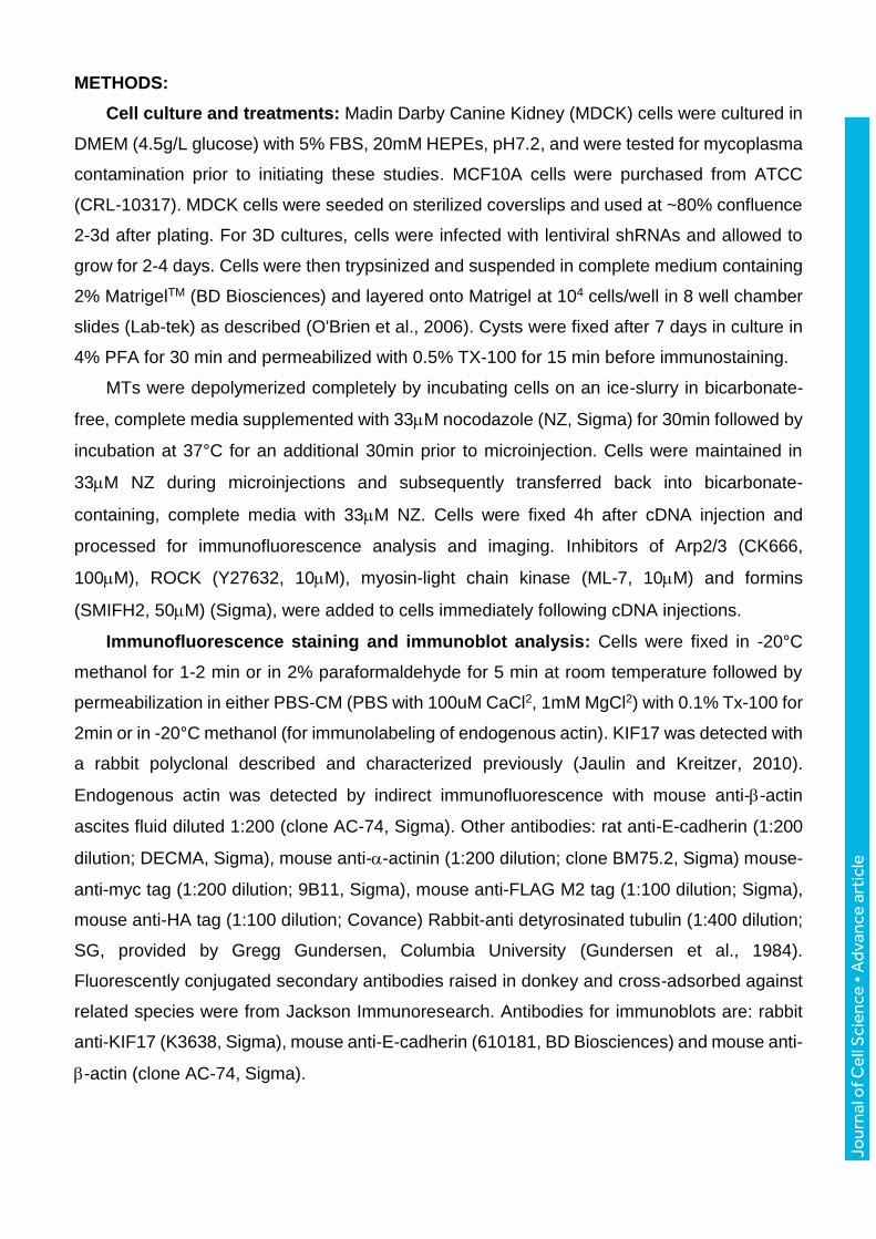
METHODS:
Cell culture and treatments: Madin Darby Canine Kidney (MDCK) cells were cultured in
DMEM (4.5g/L glucose) with 5% FBS, 20mM HEPEs, pH7.2, and were tested for mycoplasma
contamination prior to initiating these studies. MCF10A cells were purchased from ATCC
(CRL-10317). MDCK cells were seeded on sterilized coverslips and used at ~80% confluence
2-3d after plating. For 3D cultures, cells were infected with lentiviral shRNAs and allowed to
grow for 2-4 days. Cells were then trypsinized and suspended in complete medium containing
2% MatrigelTM (BD Biosciences) and layered onto Matrigel at 104 cells/well in 8 well chamber
slides (Lab-tek) as described (O'Brien et al., 2006). Cysts were fixed after 7 days in culture in
4% PFA for 30 min and permeabilized with 0.5% TX-100 for 15 min before immunostaining.
MTs were depolymerized completely by incubating cells on an ice-slurry in bicarbonate-
free, complete media supplemented with 33M nocodazole (NZ, Sigma) for 30min followed by
incubation at 37°C for an additional 30min prior to microinjection. Cells were maintained in
33M NZ during microinjections and subsequently transferred back into bicarbonate-
containing, complete media with 33M NZ. Cells were fixed 4h after cDNA injection and
processed for immunofluorescence analysis and imaging. Inhibitors of Arp2/3 (CK666,
100M), ROCK (Y27632, 10M), myosin-light chain kinase (ML-7, 10M) and formins
(SMIFH2, 50M) (Sigma), were added to cells immediately following cDNA injections.
Immunofluorescence staining and immunoblot analysis: Cells were fixed in -20°C
methanol for 1-2 min or in 2% paraformaldehyde for 5 min at room temperature followed by
permeabilization in either PBS-CM (PBS with 100uM CaCl2, 1mM MgCl2) with 0.1% Tx-100 for
2min or in -20°C methanol (for immunolabeling of endogenous actin). KIF17 was detected with
a rabbit polyclonal described and characterized previously (Jaulin and Kreitzer, 2010).
Endogenous actin was detected by indirect immunofluorescence with mouse anti--actin
ascites fluid diluted 1:200 (clone AC-74, Sigma). Other antibodies: rat anti-E-cadherin (1:200
dilution; DECMA, Sigma), mouse anti--actinin (1:200 dilution; clone BM75.2, Sigma) mouse-
anti-myc tag (1:200 dilution; 9B11, Sigma), mouse anti-FLAG M2 tag (1:100 dilution; Sigma),
mouse anti-HA tag (1:100 dilution; Covance) Rabbit-anti detyrosinated tubulin (1:400 dilution;
SG, provided by Gregg Gundersen, Columbia University (Gundersen et al., 1984).
Fluorescently conjugated secondary antibodies raised in donkey and cross-adsorbed against
related species were from Jackson Immunoresearch. Antibodies for immunoblots are: rabbit
anti-KIF17 (K3638, Sigma), mouse anti-E-cadherin (610181, BD Biosciences) and mouse anti-
-actin (clone AC-74, Sigma).
Jour
nal o
f Cel
l Sci
ence
• A
dvan
ce a
rtic
le
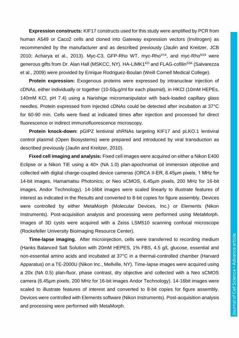
Expression constructs: KIF17 constructs used for this study were amplified by PCR from
human A549 or Caco2 cells and cloned into Gateway expression vectors (Invitrogen) as
recommended by the manufacturer and as described previously (Jaulin and Kreitzer, JCB
2010; Acharya et al., 2013). Myc-C3, GFP-Rho WT, myc-RhoV14, and myc-RhoN19 were
generous gifts from Dr. Alan Hall (MSKCC, NY). HA-LIMK1KD and FLAG-cofilinS3A (Salvarezza
et al., 2009) were provided by Enrique Rodriguez-Boulan (Weill Cornell Medical College).
Protein expression: Exogenous proteins were expressed by intranuclear injection of
cDNAs, either individually or together (10-50g/ml for each plasmid), in HKCl (10mM HEPEs,
140mM KCl, pH 7.4) using a Narishige micromanipulator with back-loaded capillary glass
needles. Protein expressed from injected cDNAs could be detected after incubation at 37°C
for 60-90 min. Cells were fixed at indicated times after injection and processed for direct
fluorescence or indirect immunofluorescence microscopy.
Protein knock-down: pGIPZ lentiviral shRNAs targeting KIF17 and pLKO.1 lentiviral
control plasmid (Open Biosystems) were prepared and introduced by viral transduction as
described previously (Jaulin and Kreitzer, 2010).
Fixed cell imaging and analysis: Fixed cell images were acquired on either a Nikon E400
Eclipse or a Nikon TiE using a 40× (NA 1.0) plan-apochromat oil immersion objective and
collected with digital charge-coupled device cameras (ORCA II-ER, 6.45μm pixels, 1 MHz for
14-bit images, Hamamatsu Photonics; or Neo sCMOS, 6.45µm pixels, 200 MHz for 16-bit
images, Andor Technology). 14-16bit images were scaled linearly to illustrate features of
interest as indicated in the Results and converted to 8-bit copies for figure assembly. Devices
were controlled by either MetaMorph (Molecular Devices, Inc.) or Elements (Nikon
Instruments). Post-acquisition analysis and processing were performed using MetaMorph.
Images of 3D cysts were acquired with a Zeiss LSM510 scanning confocal microscope
(Rockefeller University Bioimaging Resource Center).
Time-lapse imaging. After microinjection, cells were transferred to recording medium
(Hanks Balanced Salt Solution with 20mM HEPES, 1% FBS, 4.5 g/L glucose, essential and
non-essential amino acids and incubated at 37°C in a thermal-controlled chamber (Harvard
Apparatus) on a TE-2000U (Nikon Inc., Mellville, NY). Time-lapse images were acquired using
a 20x (NA 0.5) plan-fluor, phase contrast, dry objective and collected with a Neo sCMOS
camera (6.45µm pixels, 200 MHz for 16-bit images Andor Technology). 14-16bit images were
scaled to illustrate features of interest and converted to 8-bit copies for figure assembly.
Devices were controlled with Elements software (Nikon Instruments). Post-acquisition analysis
and processing were performed with MetaMorph.
Jour
nal o
f Cel
l Sci
ence
• A
dvan
ce a
rtic
le
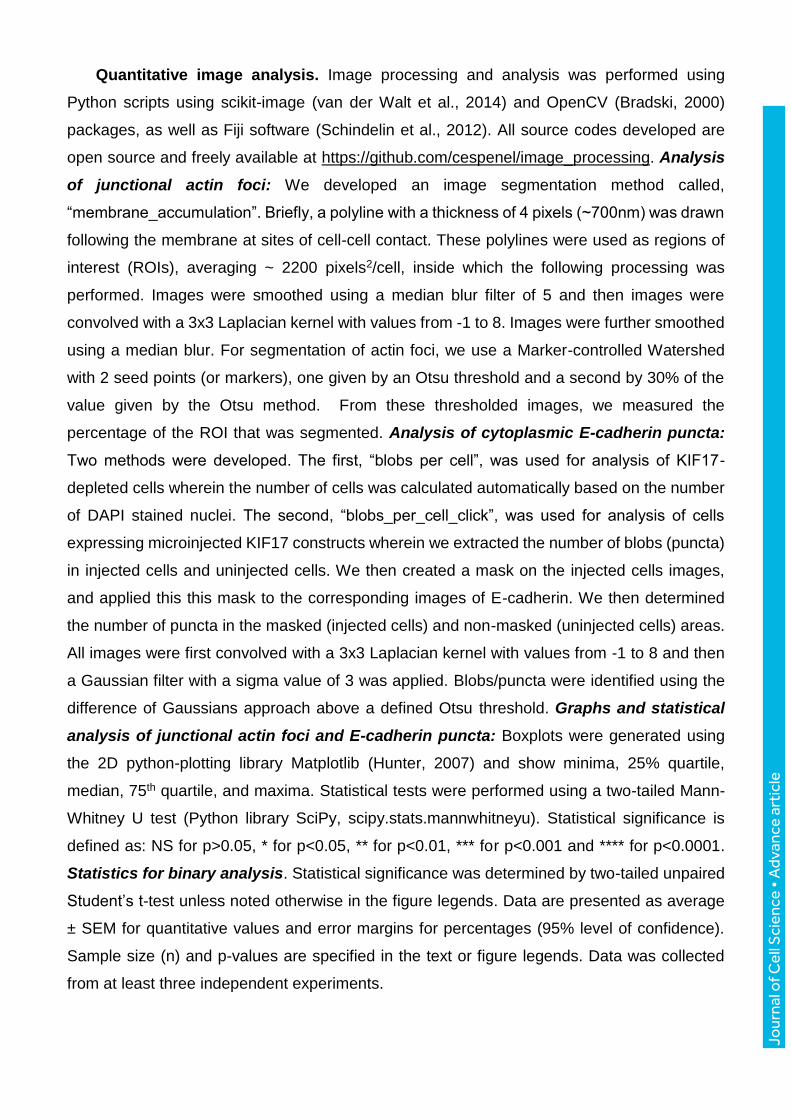
Quantitative image analysis. Image processing and analysis was performed using
Python scripts using scikit-image (van der Walt et al., 2014) and OpenCV (Bradski, 2000)
packages, as well as Fiji software (Schindelin et al., 2012). All source codes developed are
open source and freely available at https://github.com/cespenel/image_processing. Analysis
of junctional actin foci: We developed an image segmentation method called,
“membrane_accumulation”. Briefly, a polyline with a thickness of 4 pixels (~700nm) was drawn
following the membrane at sites of cell-cell contact. These polylines were used as regions of
interest (ROIs), averaging ~ 2200 pixels2/cell, inside which the following processing was
performed. Images were smoothed using a median blur filter of 5 and then images were
convolved with a 3x3 Laplacian kernel with values from -1 to 8. Images were further smoothed
using a median blur. For segmentation of actin foci, we use a Marker-controlled Watershed
with 2 seed points (or markers), one given by an Otsu threshold and a second by 30% of the
value given by the Otsu method. From these thresholded images, we measured the
percentage of the ROI that was segmented. Analysis of cytoplasmic E-cadherin puncta:
Two methods were developed. The first, “blobs per cell”, was used for analysis of KIF17-
depleted cells wherein the number of cells was calculated automatically based on the number
of DAPI stained nuclei. The second, “blobs_per_cell_click”, was used for analysis of cells
expressing microinjected KIF17 constructs wherein we extracted the number of blobs (puncta)
in injected cells and uninjected cells. We then created a mask on the injected cells images,
and applied this this mask to the corresponding images of E-cadherin. We then determined
the number of puncta in the masked (injected cells) and non-masked (uninjected cells) areas.
All images were first convolved with a 3x3 Laplacian kernel with values from -1 to 8 and then
a Gaussian filter with a sigma value of 3 was applied. Blobs/puncta were identified using the
difference of Gaussians approach above a defined Otsu threshold. Graphs and statistical
analysis of junctional actin foci and E-cadherin puncta: Boxplots were generated using
the 2D python-plotting library Matplotlib (Hunter, 2007) and show minima, 25% quartile,
median, 75th quartile, and maxima. Statistical tests were performed using a two-tailed Mann-
Whitney U test (Python library SciPy, scipy.stats.mannwhitneyu). Statistical significance is
defined as: NS for p>0.05, * for p<0.05, ** for p<0.01, *** for p<0.001 and **** for p<0.0001.
Statistics for binary analysis. Statistical significance was determined by two-tailed unpaired
Student’s t-test unless noted otherwise in the figure legends. Data are presented as average
± SEM for quantitative values and error margins for percentages (95% level of confidence).
Sample size (n) and p-values are specified in the text or figure legends. Data was collected
from at least three independent experiments.
Jour
nal o
f Cel
l Sci
ence
• A
dvan
ce a
rtic
le
Rho-GTP binding assay: Rhotekin-RBD was purchased from Cytoskeleton (Denver, CO)
and RhoA-GTP binding was performed as recommended by the manufacturer. Briefly, MDCK
were transfected with indicated constructs. After 24h, cells were washed in PBS and lysed in
25mM HEPES, pH 7.5, 150mM NaCl, 1% NP-40 (Igepal CA-630), 10mM MgCl2, 1mM EDTA
and 10% glycerol, 10 g/ml leupeptin, 10g/ml pepstatin, and 10 g/ml aprotinin. For assays
in KIF17-depleted cells, cells were also treated with RhoA activator, cytotoxic necrotizing factor
1 (CNF1, 55g/ml) as a positive control. Lysates were clarified by centrifugation at 13,000xg
at 4°C for 1min. Clarified lysates (200g) were divided in two; one to detect total RhoA, and
one for use in pull-downs. 100g of lysate was incubated with Rhotekin–RBD protein beads
(50 g) beads at 4°C for 90min. The beads were collected by centrifugation, washed
thoroughly and resuspended in 2X Laemmeli buffer. Input samples and collected beads were
analyzed by Western blot using a RhoA specific antibody. Densitometry was performed using
ImageJ (NIH). The amount of RBD-bound RhoA was normalized to total RhoA in cell lysates
for comparison of Rho activity (level of GTP-bound Rho) across samples.

Jour
nal o
f Cel
l Sci
ence
• A
dvan
ce a
rtic
le
ACKNOWLEDGMENTS:
This work was supported by grants from the National Institutes of Health (R01GM087575)
and the Irma T. Hirschl Trust to G. Kreitzer, and from the CNRS (ATIP-AVENIR program) and
the Gustave Roussy Foundation to F. Jaulin.
AUTHOR CONTRIBUTIONS:
BA, CE, FL, JR, JM, FJ and GK performed experiments. CE developed algorithms for
quantitative image analysis. FJ and GK conceived the project and wrote the manuscript.
Jour
nal o
f Cel
l Sci
ence
• A
dvan
ce a
rtic
le
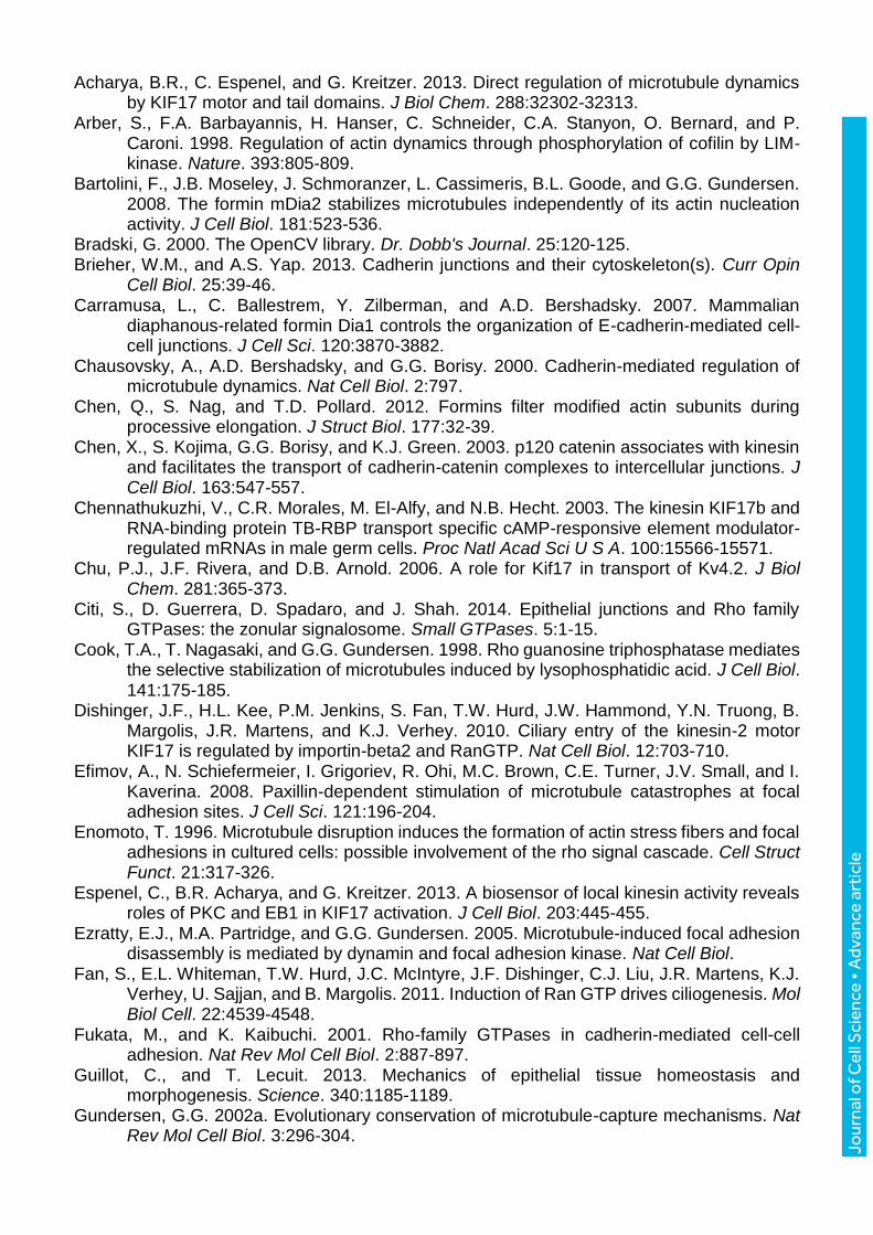
Acharya, B.R., C. Espenel, and G. Kreitzer. 2013. Direct regulation of microtubule dynamics by KIF17 motor and tail domains. J Biol Chem. 288:32302-32313.
Arber, S., F.A. Barbayannis, H. Hanser, C. Schneider, C.A. Stanyon, O. Bernard, and P. Caroni. 1998. Regulation of actin dynamics through phosphorylation of cofilin by LIM-kinase. Nature. 393:805-809.
Bartolini, F., J.B. Moseley, J. Schmoranzer, L. Cassimeris, B.L. Goode, and G.G. Gundersen. 2008. The formin mDia2 stabilizes microtubules independently of its actin nucleation activity. J Cell Biol. 181:523-536.
Bradski, G. 2000. The OpenCV library. Dr. Dobb's Journal. 25:120-125. Brieher, W.M., and A.S. Yap. 2013. Cadherin junctions and their cytoskeleton(s). Curr Opin
Cell Biol. 25:39-46. Carramusa, L., C. Ballestrem, Y. Zilberman, and A.D. Bershadsky. 2007. Mammalian
diaphanous-related formin Dia1 controls the organization of E-cadherin-mediated cell-cell junctions. J Cell Sci. 120:3870-3882.
Chausovsky, A., A.D. Bershadsky, and G.G. Borisy. 2000. Cadherin-mediated regulation of microtubule dynamics. Nat Cell Biol. 2:797.
Chen, Q., S. Nag, and T.D. Pollard. 2012. Formins filter modified actin subunits during processive elongation. J Struct Biol. 177:32-39.
Chen, X., S. Kojima, G.G. Borisy, and K.J. Green. 2003. p120 catenin associates with kinesin and facilitates the transport of cadherin-catenin complexes to intercellular junctions. J Cell Biol. 163:547-557.
Chennathukuzhi, V., C.R. Morales, M. El-Alfy, and N.B. Hecht. 2003. The kinesin KIF17b and RNA-binding protein TB-RBP transport specific cAMP-responsive element modulator-regulated mRNAs in male germ cells. Proc Natl Acad Sci U S A. 100:15566-15571.
Chu, P.J., J.F. Rivera, and D.B. Arnold. 2006. A role for Kif17 in transport of Kv4.2. J Biol Chem. 281:365-373.
Citi, S., D. Guerrera, D. Spadaro, and J. Shah. 2014. Epithelial junctions and Rho family GTPases: the zonular signalosome. Small GTPases. 5:1-15.
Cook, T.A., T. Nagasaki, and G.G. Gundersen. 1998. Rho guanosine triphosphatase mediates the selective stabilization of microtubules induced by lysophosphatidic acid. J Cell Biol. 141:175-185.
Dishinger, J.F., H.L. Kee, P.M. Jenkins, S. Fan, T.W. Hurd, J.W. Hammond, Y.N. Truong, B. Margolis, J.R. Martens, and K.J. Verhey. 2010. Ciliary entry of the kinesin-2 motor KIF17 is regulated by importin-beta2 and RanGTP. Nat Cell Biol. 12:703-710.
Efimov, A., N. Schiefermeier, I. Grigoriev, R. Ohi, M.C. Brown, C.E. Turner, J.V. Small, and I. Kaverina. 2008. Paxillin-dependent stimulation of microtubule catastrophes at focal adhesion sites. J Cell Sci. 121:196-204.
Enomoto, T. 1996. Microtubule disruption induces the formation of actin stress fibers and focal adhesions in cultured cells: possible involvement of the rho signal cascade. Cell Struct Funct. 21:317-326.
Espenel, C., B.R. Acharya, and G. Kreitzer. 2013. A biosensor of local kinesin activity reveals roles of PKC and EB1 in KIF17 activation. J Cell Biol. 203:445-455.
Ezratty, E.J., M.A. Partridge, and G.G. Gundersen. 2005. Microtubule-induced focal adhesion disassembly is mediated by dynamin and focal adhesion kinase. Nat Cell Biol.
Fan, S., E.L. Whiteman, T.W. Hurd, J.C. McIntyre, J.F. Dishinger, C.J. Liu, J.R. Martens, K.J. Verhey, U. Sajjan, and B. Margolis. 2011. Induction of Ran GTP drives ciliogenesis. Mol Biol Cell. 22:4539-4548.
Fukata, M., and K. Kaibuchi. 2001. Rho-family GTPases in cadherin-mediated cell-cell adhesion. Nat Rev Mol Cell Biol. 2:887-897.
Guillot, C., and T. Lecuit. 2013. Mechanics of epithelial tissue homeostasis and morphogenesis. Science. 340:1185-1189.
Gundersen, G.G. 2002a. Evolutionary conservation of microtubule-capture mechanisms. Nat Rev Mol Cell Biol. 3:296-304.
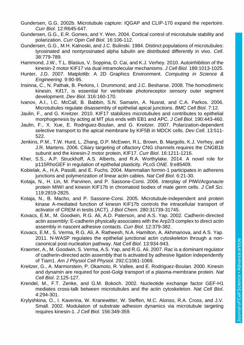
Jour
nal o
f Cel
l Sci
ence
• A
dvan
ce a
rtic
le
Gundersen, G.G. 2002b. Microtubule capture: IQGAP and CLIP-170 expand the repertoire. Curr Biol. 12:R645-647.
Gundersen, G.G., E.R. Gomes, and Y. Wen. 2004. Cortical control of microtubule stability and polarization. Curr Opin Cell Biol. 16:106-112.
Gundersen, G.G., M.H. Kalnoski, and J.C. Bulinski. 1984. Distinct populations of microtubules: tyrosinated and nontyrosinated alpha tubulin are distributed differently in vivo. Cell. 38:779-789.
Hammond, J.W., T.L. Blasius, V. Soppina, D. Cai, and K.J. Verhey. 2010. Autoinhibition of the kinesin-2 motor KIF17 via dual intramolecular mechanisms. J Cell Biol. 189:1013-1025.
Hunter, J.D. 2007. Matplotlib: A 2D Graphics Environment. Computing in Science & Engineering. 9:90-95.
Insinna, C., N. Pathak, B. Perkins, I. Drummond, and J.C. Besharse. 2008. The homodimeric kinesin, Kif17, is essential for vertebrate photoreceptor sensory outer segment development. Dev Biol. 316:160-170.
Ivanov, A.I., I.C. McCall, B. Babbin, S.N. Samarin, A. Nusrat, and C.A. Parkos. 2006. Microtubules regulate disassembly of epithelial apical junctions. BMC Cell Biol. 7:12.
Jaulin, F., and G. Kreitzer. 2010. KIF17 stabilizes microtubules and contributes to epithelial morphogenesis by acting at MT plus ends with EB1 and APC. J Cell Biol. 190:443-460.
Jaulin, F., X. Xue, E. Rodriguez-Boulan, and G. Kreitzer. 2007. Polarization-dependent selective transport to the apical membrane by KIF5B in MDCK cells. Dev Cell. 13:511-522.
Jenkins, P.M., T.W. Hurd, L. Zhang, D.P. McEwen, R.L. Brown, B. Margolis, K.J. Verhey, and J.R. Martens. 2006. Ciliary targeting of olfactory CNG channels requires the CNGB1b subunit and the kinesin-2 motor protein, KIF17. Curr Biol. 16:1211-1216.
Kher, S.S., A.P. Struckhoff, A.S. Alberts, and R.A. Worthylake. 2014. A novel role for p115RhoGEF in regulation of epithelial plasticity. PLoS ONE. 9:e85409.
Kobielak, A., H.A. Pasolli, and E. Fuchs. 2004. Mammalian formin-1 participates in adherens junctions and polymerization of linear actin cables. Nat Cell Biol. 6:21-30.
Kotaja, N., H. Lin, M. Parvinen, and P. Sassone-Corsi. 2006. Interplay of PIWI/Argonaute protein MIWI and kinesin KIF17b in chromatoid bodies of male germ cells. J Cell Sci. 119:2819-2825.
Kotaja, N., B. Macho, and P. Sassone-Corsi. 2005. Microtubule-independent and protein kinase A-mediated function of kinesin KIF17b controls the intracellular transport of activator of CREM in testis (ACT). J Biol Chem. 280:31739-31745.
Kovacs, E.M., M. Goodwin, R.G. Ali, A.D. Paterson, and A.S. Yap. 2002. Cadherin-directed actin assembly: E-cadherin physically associates with the Arp2/3 complex to direct actin assembly in nascent adhesive contacts. Curr Biol. 12:379-382.
Kovacs, E.M., S. Verma, R.G. Ali, A. Ratheesh, N.A. Hamilton, A. Akhmanova, and A.S. Yap. 2011. N-WASP regulates the epithelial junctional actin cytoskeleton through a non-canonical post-nucleation pathway. Nat Cell Biol. 13:934-943.
Kraemer, A., M. Goodwin, S. Verma, A.S. Yap, and R.G. Ali. 2007. Rac is a dominant regulator of cadherin-directed actin assembly that is activated by adhesive ligation independently of Tiam1. Am J Physiol Cell Physiol. 292:C1061-1069.
Kreitzer, G., A. Marmorstein, P. Okamoto, R. Vallee, and E. Rodriguez-Boulan. 2000. Kinesin and dynamin are required for post-Golgi transport of a plasma-membrane protein. Nat Cell Biol. 2:125-127.
Krendel, M., F.T. Zenke, and G.M. Bokoch. 2002. Nucleotide exchange factor GEF-H1 mediates cross-talk between microtubules and the actin cytoskeleton. Nat Cell Biol. 4:294-301.
Krylyshkina, O., I. Kaverina, W. Kranewitter, W. Steffen, M.C. Alonso, R.A. Cross, and J.V. Small. 2002. Modulation of substrate adhesion dynamics via microtubule targeting requires kinesin-1. J Cell Biol. 156:349-359.
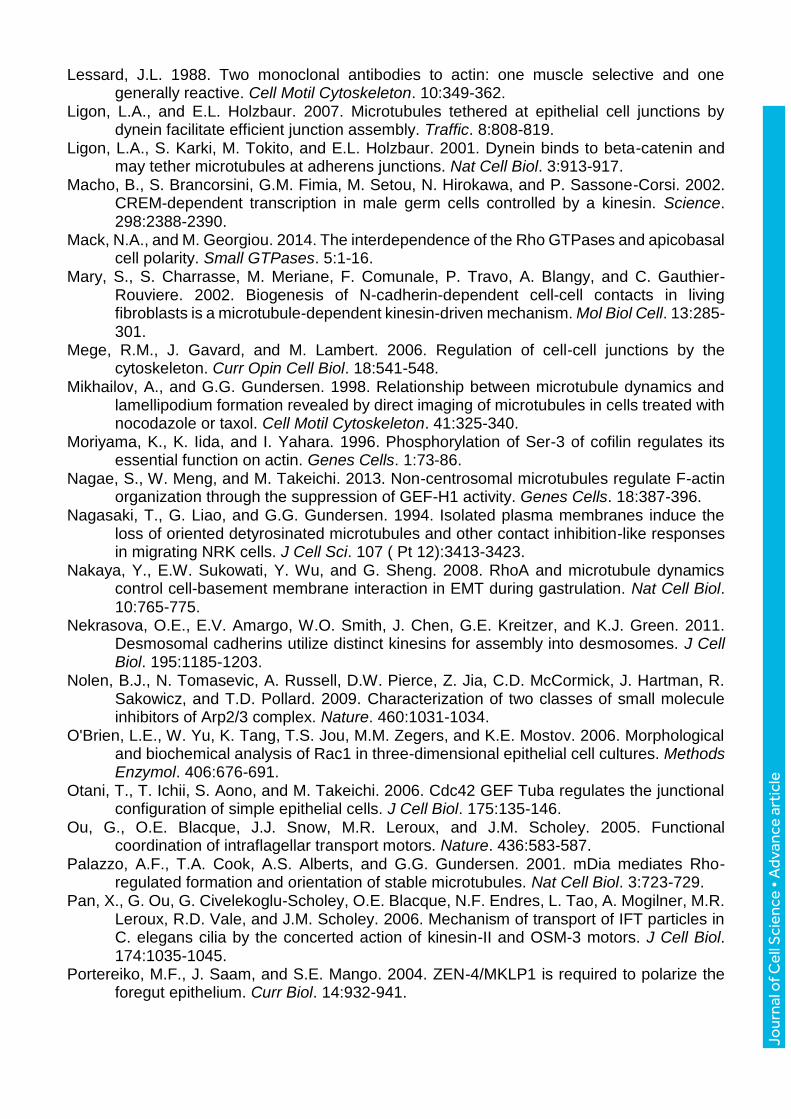
Jour
nal o
f Cel
l Sci
ence
• A
dvan
ce a
rtic
le
Lessard, J.L. 1988. Two monoclonal antibodies to actin: one muscle selective and one generally reactive. Cell Motil Cytoskeleton. 10:349-362.
Ligon, L.A., and E.L. Holzbaur. 2007. Microtubules tethered at epithelial cell junctions by dynein facilitate efficient junction assembly. Traffic. 8:808-819.
Ligon, L.A., S. Karki, M. Tokito, and E.L. Holzbaur. 2001. Dynein binds to beta-catenin and may tether microtubules at adherens junctions. Nat Cell Biol. 3:913-917.
Macho, B., S. Brancorsini, G.M. Fimia, M. Setou, N. Hirokawa, and P. Sassone-Corsi. 2002. CREM-dependent transcription in male germ cells controlled by a kinesin. Science. 298:2388-2390.
Mack, N.A., and M. Georgiou. 2014. The interdependence of the Rho GTPases and apicobasal cell polarity. Small GTPases. 5:1-16.
Mary, S., S. Charrasse, M. Meriane, F. Comunale, P. Travo, A. Blangy, and C. Gauthier-Rouviere. 2002. Biogenesis of N-cadherin-dependent cell-cell contacts in living fibroblasts is a microtubule-dependent kinesin-driven mechanism. Mol Biol Cell. 13:285-301.
Mege, R.M., J. Gavard, and M. Lambert. 2006. Regulation of cell-cell junctions by the cytoskeleton. Curr Opin Cell Biol. 18:541-548.
Mikhailov, A., and G.G. Gundersen. 1998. Relationship between microtubule dynamics and lamellipodium formation revealed by direct imaging of microtubules in cells treated with nocodazole or taxol. Cell Motil Cytoskeleton. 41:325-340.
Moriyama, K., K. Iida, and I. Yahara. 1996. Phosphorylation of Ser-3 of cofilin regulates its essential function on actin. Genes Cells. 1:73-86.
Nagae, S., W. Meng, and M. Takeichi. 2013. Non-centrosomal microtubules regulate F-actin organization through the suppression of GEF-H1 activity. Genes Cells. 18:387-396.
Nagasaki, T., G. Liao, and G.G. Gundersen. 1994. Isolated plasma membranes induce the loss of oriented detyrosinated microtubules and other contact inhibition-like responses in migrating NRK cells. J Cell Sci. 107 ( Pt 12):3413-3423.
Nakaya, Y., E.W. Sukowati, Y. Wu, and G. Sheng. 2008. RhoA and microtubule dynamics control cell-basement membrane interaction in EMT during gastrulation. Nat Cell Biol. 10:765-775.
Nekrasova, O.E., E.V. Amargo, W.O. Smith, J. Chen, G.E. Kreitzer, and K.J. Green. 2011. Desmosomal cadherins utilize distinct kinesins for assembly into desmosomes. J Cell Biol. 195:1185-1203.
Nolen, B.J., N. Tomasevic, A. Russell, D.W. Pierce, Z. Jia, C.D. McCormick, J. Hartman, R. Sakowicz, and T.D. Pollard. 2009. Characterization of two classes of small molecule inhibitors of Arp2/3 complex. Nature. 460:1031-1034.
O'Brien, L.E., W. Yu, K. Tang, T.S. Jou, M.M. Zegers, and K.E. Mostov. 2006. Morphological and biochemical analysis of Rac1 in three-dimensional epithelial cell cultures. Methods Enzymol. 406:676-691.
Otani, T., T. Ichii, S. Aono, and M. Takeichi. 2006. Cdc42 GEF Tuba regulates the junctional configuration of simple epithelial cells. J Cell Biol. 175:135-146.
Ou, G., O.E. Blacque, J.J. Snow, M.R. Leroux, and J.M. Scholey. 2005. Functional coordination of intraflagellar transport motors. Nature. 436:583-587.
Palazzo, A.F., T.A. Cook, A.S. Alberts, and G.G. Gundersen. 2001. mDia mediates Rho-regulated formation and orientation of stable microtubules. Nat Cell Biol. 3:723-729.
Pan, X., G. Ou, G. Civelekoglu-Scholey, O.E. Blacque, N.F. Endres, L. Tao, A. Mogilner, M.R. Leroux, R.D. Vale, and J.M. Scholey. 2006. Mechanism of transport of IFT particles in C. elegans cilia by the concerted action of kinesin-II and OSM-3 motors. J Cell Biol. 174:1035-1045.
Portereiko, M.F., J. Saam, and S.E. Mango. 2004. ZEN-4/MKLP1 is required to polarize the foregut epithelium. Curr Biol. 14:932-941.
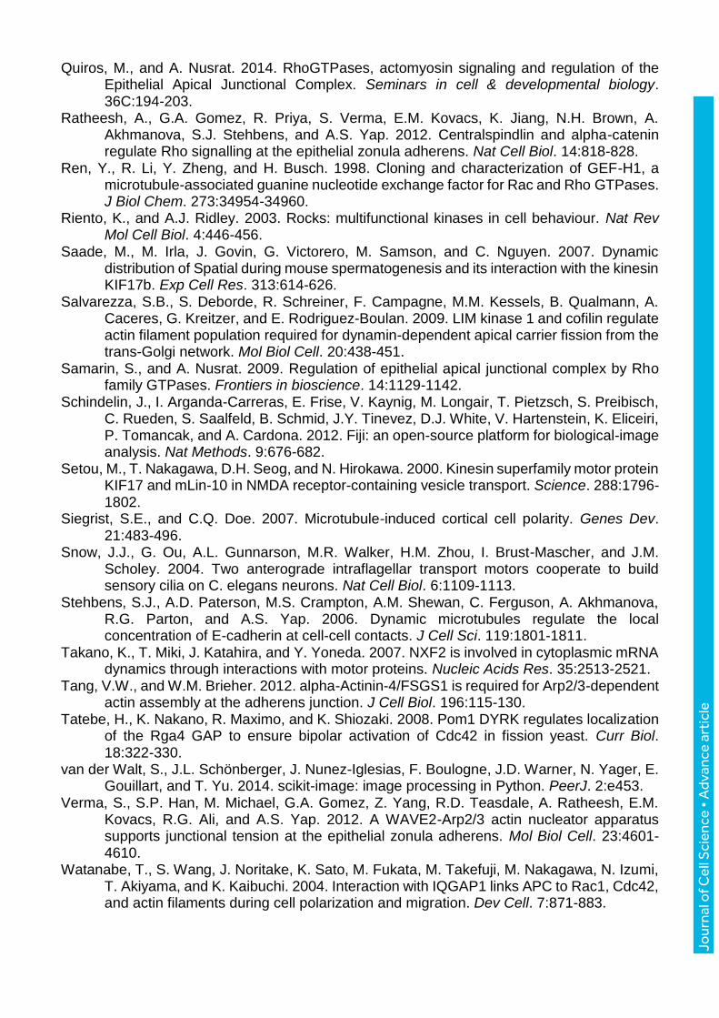
Jour
nal o
f Cel
l Sci
ence
• A
dvan
ce a
rtic
le
Quiros, M., and A. Nusrat. 2014. RhoGTPases, actomyosin signaling and regulation of the Epithelial Apical Junctional Complex. Seminars in cell & developmental biology. 36C:194-203.
Ratheesh, A., G.A. Gomez, R. Priya, S. Verma, E.M. Kovacs, K. Jiang, N.H. Brown, A. Akhmanova, S.J. Stehbens, and A.S. Yap. 2012. Centralspindlin and alpha-catenin regulate Rho signalling at the epithelial zonula adherens. Nat Cell Biol. 14:818-828.
Ren, Y., R. Li, Y. Zheng, and H. Busch. 1998. Cloning and characterization of GEF-H1, a microtubule-associated guanine nucleotide exchange factor for Rac and Rho GTPases. J Biol Chem. 273:34954-34960.
Riento, K., and A.J. Ridley. 2003. Rocks: multifunctional kinases in cell behaviour. Nat Rev Mol Cell Biol. 4:446-456.
Saade, M., M. Irla, J. Govin, G. Victorero, M. Samson, and C. Nguyen. 2007. Dynamic distribution of Spatial during mouse spermatogenesis and its interaction with the kinesin KIF17b. Exp Cell Res. 313:614-626.
Salvarezza, S.B., S. Deborde, R. Schreiner, F. Campagne, M.M. Kessels, B. Qualmann, A. Caceres, G. Kreitzer, and E. Rodriguez-Boulan. 2009. LIM kinase 1 and cofilin regulate actin filament population required for dynamin-dependent apical carrier fission from the trans-Golgi network. Mol Biol Cell. 20:438-451.
Samarin, S., and A. Nusrat. 2009. Regulation of epithelial apical junctional complex by Rho family GTPases. Frontiers in bioscience. 14:1129-1142.
Schindelin, J., I. Arganda-Carreras, E. Frise, V. Kaynig, M. Longair, T. Pietzsch, S. Preibisch, C. Rueden, S. Saalfeld, B. Schmid, J.Y. Tinevez, D.J. White, V. Hartenstein, K. Eliceiri, P. Tomancak, and A. Cardona. 2012. Fiji: an open-source platform for biological-image analysis. Nat Methods. 9:676-682.
Setou, M., T. Nakagawa, D.H. Seog, and N. Hirokawa. 2000. Kinesin superfamily motor protein KIF17 and mLin-10 in NMDA receptor-containing vesicle transport. Science. 288:1796-1802.
Siegrist, S.E., and C.Q. Doe. 2007. Microtubule-induced cortical cell polarity. Genes Dev. 21:483-496.
Snow, J.J., G. Ou, A.L. Gunnarson, M.R. Walker, H.M. Zhou, I. Brust-Mascher, and J.M. Scholey. 2004. Two anterograde intraflagellar transport motors cooperate to build sensory cilia on C. elegans neurons. Nat Cell Biol. 6:1109-1113.
Stehbens, S.J., A.D. Paterson, M.S. Crampton, A.M. Shewan, C. Ferguson, A. Akhmanova, R.G. Parton, and A.S. Yap. 2006. Dynamic microtubules regulate the local concentration of E-cadherin at cell-cell contacts. J Cell Sci. 119:1801-1811.
Takano, K., T. Miki, J. Katahira, and Y. Yoneda. 2007. NXF2 is involved in cytoplasmic mRNA dynamics through interactions with motor proteins. Nucleic Acids Res. 35:2513-2521.
Tang, V.W., and W.M. Brieher. 2012. alpha-Actinin-4/FSGS1 is required for Arp2/3-dependent actin assembly at the adherens junction. J Cell Biol. 196:115-130.
Tatebe, H., K. Nakano, R. Maximo, and K. Shiozaki. 2008. Pom1 DYRK regulates localization of the Rga4 GAP to ensure bipolar activation of Cdc42 in fission yeast. Curr Biol. 18:322-330.
van der Walt, S., J.L. Schönberger, J. Nunez-Iglesias, F. Boulogne, J.D. Warner, N. Yager, E. Gouillart, and T. Yu. 2014. scikit-image: image processing in Python. PeerJ. 2:e453.
Verma, S., S.P. Han, M. Michael, G.A. Gomez, Z. Yang, R.D. Teasdale, A. Ratheesh, E.M. Kovacs, R.G. Ali, and A.S. Yap. 2012. A WAVE2-Arp2/3 actin nucleator apparatus supports junctional tension at the epithelial zonula adherens. Mol Biol Cell. 23:4601-4610.
Watanabe, T., S. Wang, J. Noritake, K. Sato, M. Fukata, M. Takefuji, M. Nakagawa, N. Izumi, T. Akiyama, and K. Kaibuchi. 2004. Interaction with IQGAP1 links APC to Rac1, Cdc42, and actin filaments during cell polarization and migration. Dev Cell. 7:871-883.
Jour
nal o
f Cel
l Sci
ence
• A
dvan
ce a
rtic
le
Waterman-Storer, C.M., W.C. Salmon, and E.D. Salmon. 2000. Feedback interactions between cell-cell adherens junctions and cytoskeletal dynamics in newt lung epithelial cells. Mol Biol Cell. 11:2471-2483.
Wen, Y., C.H. Eng, J. Schmoranzer, N. Cabrera-Poch, E.J. Morris, M. Chen, B.J. Wallar, A.S. Alberts, and G.G. Gundersen. 2004. EB1 and APC bind to mDia to stabilize microtubules downstream of Rho and promote cell migration. Nat Cell Biol. 6:820-830.
Wojnacki, J., G. Quassollo, M.P. Marzolo, and A. Caceres. 2014. Rho GTPases at the crossroad of signaling networks in mammals: impact of Rho-GTPases on microtubule organization and dynamics. Small GTPases. 5:e28430.
Yamada, S., S. Pokutta, F. Drees, W.I. Weis, and W.J. Nelson. 2005. Deconstructing the cadherin-catenin-actin complex. Cell. 123:889-901.
Yamamoto, H., K. Imai, E. Kamegaya, Y. Takamatsu, M. Irago, Y. Hagino, S. Kasai, K. Shimada, T. Yamamoto, I. Sora, H. Koga, and K. Ikeda. 2006. Repeated methamphetamine administration alters expression of the NMDA receptor channel epsilon2 subunit and kinesins in the mouse brain. Ann N Y Acad Sci. 1074:97-103.
Yanagisawa, M., I.N. Kaverina, A. Wang, Y. Fujita, A.B. Reynolds, and P.Z. Anastasiadis. 2004. A novel interaction between kinesin and p120 modulates p120 localization and function. J Biol Chem. 279:9512-9521.
Yang, N., O. Higuchi, K. Ohashi, K. Nagata, A. Wada, K. Kangawa, E. Nishida, and K. Mizuno. 1998. Cofilin phosphorylation by LIM-kinase 1 and its role in Rac-mediated actin reorganization. Nature. 393:809-812.
Yoshimura, Y., and H. Miki. 2011. Dynamic regulation of GEF-H1 localization at microtubules by Par1b/MARK2. Biochem Biophys Res Commun. 408:322-328.
Jour
nal o
f Cel
l Sci
ence
• A
dvan
ce a
rtic
le
Figures
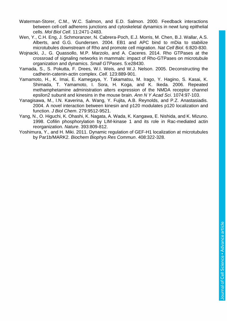
Figure 1. KIF17 localizes at cell-cell junctions and contributes to actin remodeling
during epithelial morphogenesis. (A) Co-localization of KIF17 with actin and E-cadherin at
cell-cell contacts in MDCK cells. Color overlay shows an enlarged region of KIF17 and E-
cadherin or -actin images; the KIF17 image was shifted by 5 pixels to highlight corresponding

Jour
nal o
f Cel
l Sci
ence
• A
dvan
ce a
rtic
le
staining patterns. (B) Co-localization of KIF17 and -actinin at cell-cell junctions. Color overlay
shows an enlarged view of the boxed region; the KIF17 image was shifted by 5 pixels. (C)
Immunoblot showing KIF17 in MDCK cells transduced with control (shNC) or KIF17-targeting
(shKIF17) shRNAs. Hsp90 was used as a loading control. (D) Localization of actin (phalloidin),
ZO-1 and nuclei (DAPI) in shNC and shKIF17 MDCK cysts grown in Matrigel for 7 days. Dotted
and dashed lines highlight apical and basal membranes respectively. Graph shows the ratio
of apical to basal actin fluorescence intensity determined by line-scan analysis. N=38 and 48
cells for shNC and shKIF17 respectively. Error bars = SEM, significance was determined with
a two-tailed, unpaired student’s t-test, **** p<0,0001.
Jour
nal o
f Cel
l Sci
ence
• A
dvan
ce a
rtic
le
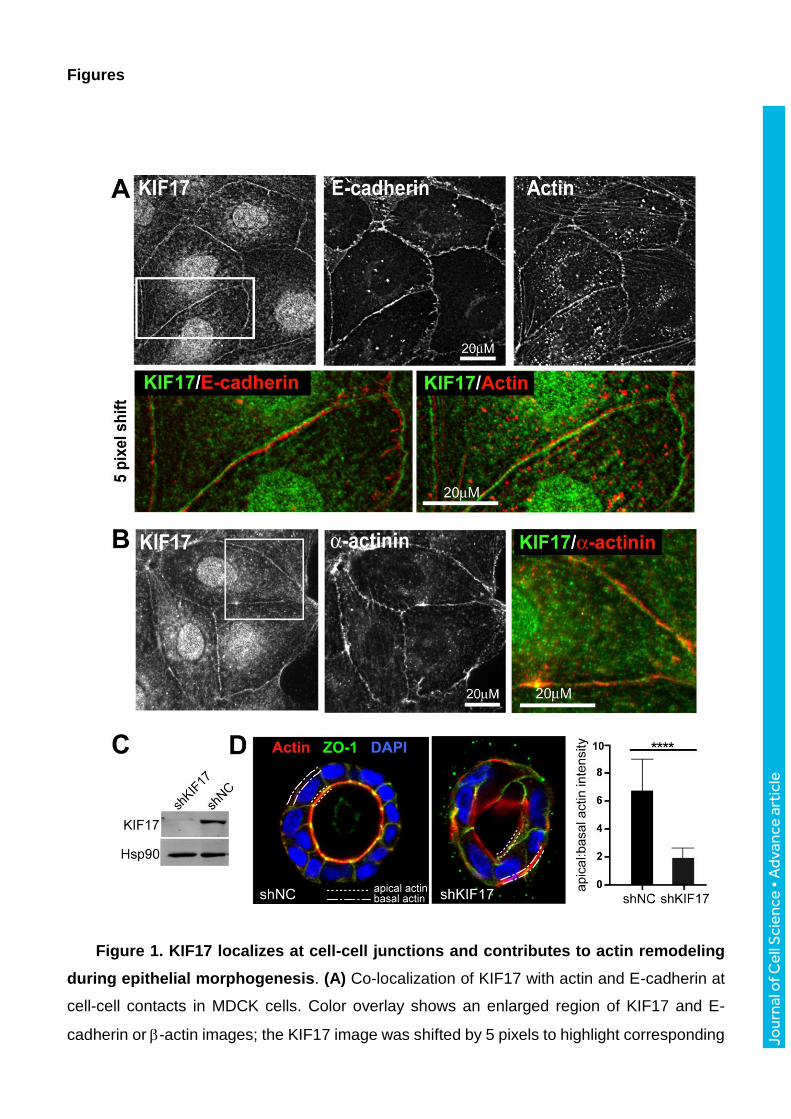
Figure 2. Localization of expressed, GFP-tagged KIF17 constructs. (A) Diagram
showing KIF17 constructs used for these studies. Images show localization of KIF17-FL, K339,
K370 and K490 in MDCK cells 3h after cDNA injection. Arrows indicate localization on MTs in
cell protrusions. Arrowheads indicate localization at cell-cell contacts. (B) Colocalization of
GFP-K370 with immunostained E-cadherin and -actin in MDCK cells. Color overlays show an
enlargement of GFP-K370 and -actin. In the lower overlay, the image of K370 was shifted by
Jour
nal o
f Cel
l Sci
ence
• A
dvan
ce a
rtic
le
7 pixels. (C) Quantification of the junctional localization of endogenous KIF17 and expressed
KIF17 constructs 3h after cDNA injection. Values were calculated as % total cells expressing
each construct. Results are from 3-6 independent experiments (endogenous KIF17, n=90
cells; GFP- KIF17-FL, n=60; GFP-K490, n=295; GFP-K370, n=664; GFP-K339, n=794). (D)
Quantification of the % cells with junctional GFP-K370 or GFP-K339 in the absence and
presence of co-expressed mCh-KIF17-Tail. Data are normalized to 100% in control conditions
(K370, n=162; K370+KIF17-Tail, n=121; K339, n=230; K339+KIF17-Tail, n=324 cells). Results
are from ≥2 independent experiments. Error bars = error margins with 95% confidence interval.
Significance was determined using a two-tailed unpaired student’s t-test, * p<0.05; ** p<0.01.
Jour
nal o
f Cel
l Sci
ence
• A
dvan
ce a
rtic
le
Figure 3. K370 expression stimulates accumulation of junctional actin. (A)
Localization of GFP-actin 4h after co-injection with mCherry control, empty vector (mCh-EV)
or mCh-K370 cDNAs. Insets in grayscale images show mCherry expression. Color overlay
shows magnified region of the boxed area of cells shown in inset. mCh-K370 image was shifted
by 5 pixels. (B) Localization of GFP-actin 4h after co-injection with mCh-K370 or mCh-KIF3A-
motor domain (mCh-KIF3A-M). Insets show mCherry expression. (C, D) Analysis of junctional
actin accumulation. (C) Sample images of GFP-actin 4h after cDNA injection. Dashed lines on
the left panel highlight regions of interest (ROIs) at cell-cell contact zones within which
segmentation was applied to identify junctional actin foci. The right panel shows the segmented
image. Yellow puncta highlight the segmented regions within the selected ROI analyzed. Inset
shows a magnified view of discrete junctional GFP-actin foci that are identified by
segmentation. (D) Box-whisker plots showing quantification of junctional actin foci identified by
Jour
nal o
f Cel
l Sci
ence
• A
dvan
ce a
rtic
le
segmentation as % of total ROI selected for measurement in each condition. Plots show
minimum, 25th quartile, median, 75th quartile, and maximum values. Diamond symbols indicate
outliers. Results are from images of injected cells in ≥3 independent experiments. Significance
was determined using a two-tailed Mann-Whitney U test. NS (not significant) for p>0.05, * for
p<0.05, ** for p<0.01, *** for p<0.001 and **** for p<0.0001.
Jour
nal o
f Cel
l Sci
ence
• A
dvan
ce a
rtic
le
Figure 4. Junctional actin accumulation induced by K370 is independent of MTs. (A)
Localization of mCh-actin, GFP-K370 and stable (detyrosinated) MTs in untreated and NZ-
treated (33M) cells. After injection, cells were maintained in NZ for 4h at 37°C before fixation.
(B) Localization of GFP-actin and the MT-binding mutant mCh-K370G288/294A 4h after cDNA
injection. (C) Box-whisker plots showing quantification of junctional actin foci identified by
Jour
nal o
f Cel
l Sci
ence
• A
dvan
ce a
rtic
le
segmentation as % of total ROI selected for measurement in each experimental condition.
Results are derived from images of injected cells in ≥2 independent experiments. Significance
was determined using a two-tailed Mann-Whitney U test. NS (not significant) for p>0.05.
Jour
nal o
f Cel
l Sci
ence
• A
dvan
ce a
rtic
le
Figure 5. RhoA signaling regulates junctional actin accumulation mediated by K370.
(A) MDCK cells expressing mCh-actin, GFP-K370 and either myc-C3 or myc-RhoN19 and
fixed 4h after cDNA injection. Insets show myc-immunostaining to detect expressed C3 and
RhoN19. (B) Localization of mCh-actin and GFP-K370 in untreated MDCK cells and in cells
treated with Y27632 (10M) or SMIFH2 (50M). Inhibitors were added immediately after cDNA
injection and cells were fixed after 4h. (C) Box-whisker plots showing quantification of
junctional actin foci identified by segmentation as a % of the total ROI selected for
measurement in each experimental condition. Results are from images of injected cells in 2-4
independent experiments. Significance was determined using a two-tailed Mann-Whitney U
test. NS (not significant) for p>0.05, * for p<0.05, ** for p<0.01, *** for p<0.001. (D)
Jour
nal o
f Cel
l Sci
ence
• A
dvan
ce a
rtic
le
Immunoblots showing pull-down of GTP-bound RhoA with the Rho binding domain of Rhotekin
(RBD) and total RhoA in cells expressing the indicated constructs. Graph shows relative
abundance of active GTP-RhoA in each condition. Error bars = SEM. Statistical significance
was determined using one-way Anova and Bonferroni’s multiple comparison test. * p< 0.05,
*** p<0.001. (E) Immunoblots showing pull-down of GTP-bound RhoA in cells treated with the
Rho-GTPase activator CNF1 (0.55µg/ml for 90 min) or transduced with shNC, shKIF17#1 or
shKIF17#2. Table shows relative abundance of GTP-RhoA pulled down under each condition.

Jour
nal o
f Cel
l Sci
ence
• A
dvan
ce a
rtic
le
Figure 6. K370 promotes junctional actin accumulation by inhibiting LIMK/cofilin
dependent actin-severing. (A) Localization of mCh-actin and GFP-K370 in MDCK cells co-
expressing HA-LIMK1 kinase dead (LIMK1KD) or FLAG-cofilinS3A 4h after cDNA injection.
Insets show HA and FLAG immunostaining to detect expressed LIMK1KD and cofilinS3A. (B)
Localization of mCh-actin and myc-K370 in cells co-expressing GFP-EV control or GFP-KIF17-
Tail 4h after cDNA injection. Inset shows GFP-KIF17-Tail in injected cells. GFP-EV was
detected in living cells before fixation but was frequently lost during processing for
Jour
nal o
f Cel
l Sci
ence
• A
dvan
ce a
rtic
le
immunofluorescence analysis; thus it is not shown here. (C) Graph showing % cells with
accumulated junctional actin foci for each experimental condition indicated normalized to GFP-
K370 or myc-K370 controls. Cells were injected with K370 and mCh-actin cDNAs alone or in
combination with HA-LIMK1KD (n=194), Flag-cofilinS3A (n=160) or GFP-KIF17-Tail (n=60).
CK666 (100M, n=92) and ML7 (10M, n=186) were added to cells immediately following
cDNA injection until fixation 4h later. Error bars = error margins with 95% confidence interval.
Significance was determined using a two-tailed unpaired student’s t-test. NS, not significant,
** p< 0.01; *** p<0.001.
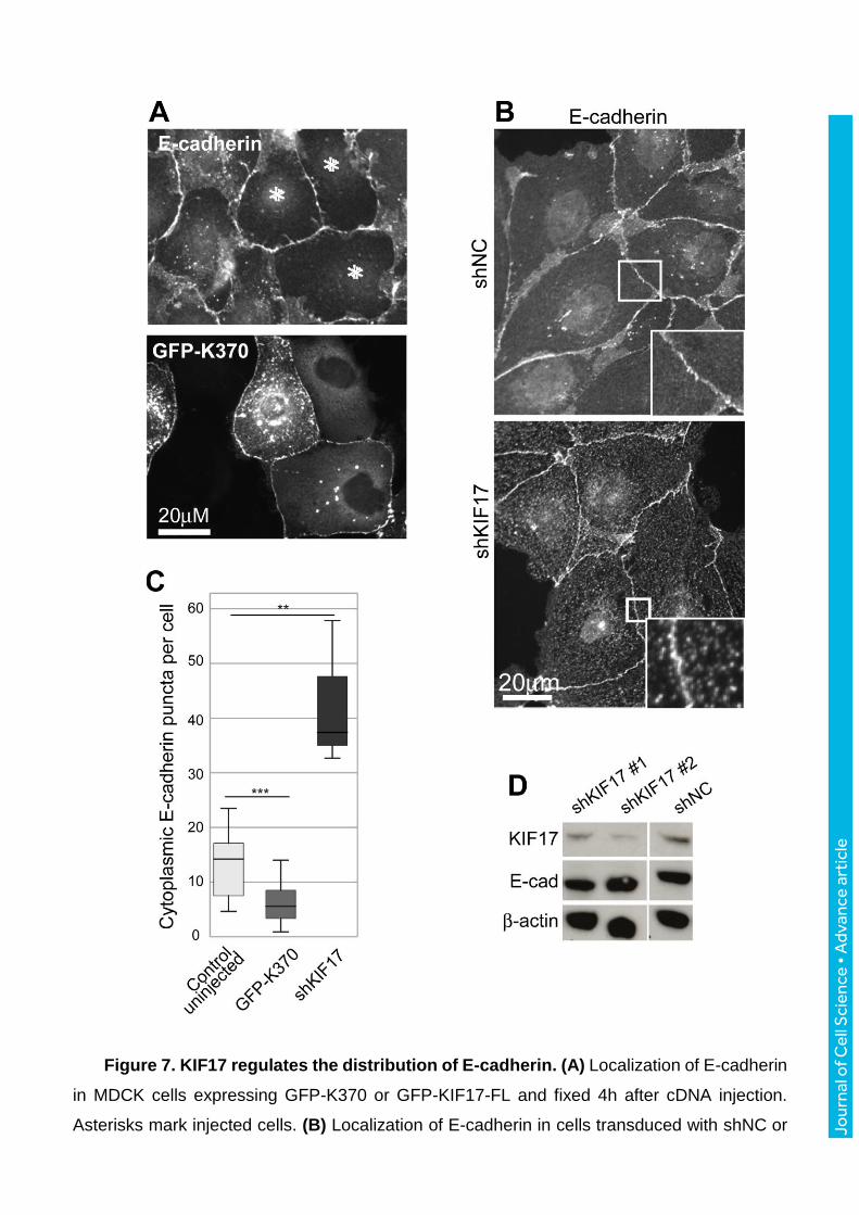
Jour
nal o
f Cel
l Sci
ence
• A
dvan
ce a
rtic
le
Figure 7. KIF17 regulates the distribution of E-cadherin. (A) Localization of E-cadherin
in MDCK cells expressing GFP-K370 or GFP-KIF17-FL and fixed 4h after cDNA injection.
Asterisks mark injected cells. (B) Localization of E-cadherin in cells transduced with shNC or
Jour
nal o
f Cel
l Sci
ence
• A
dvan
ce a
rtic
le
shKIF17. Boxed regions are magnified in insets. (C) Box-whisker plots showing quantification
of cytoplasmic E-cadherin puncta in uninjected controls and in cells expressing GFP-K370 and
in KIF17 depleted cells (shKIF17). Results are from images of injected cells in 2-4 independent
experiments. Significance was determined using a two-tailed Mann-Whitney U test. NS (not
significant, p>0.05), * for p<0.05, ** for p<0.01, *** for p<0.001. (D) Immunoblots showing
KIF17, E-cadherin and -actin in MDCK cells transduced with shNC, shKIF17#1 or shKIF17#2.
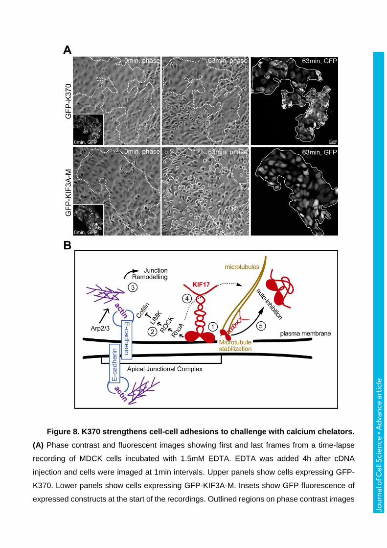
Jour
nal o
f Cel
l Sci
ence
• A
dvan
ce a
rtic
le
Figure 8. K370 strengthens cell-cell adhesions to challenge with calcium chelators.
(A) Phase contrast and fluorescent images showing first and last frames from a time-lapse
recording of MDCK cells incubated with 1.5mM EDTA. EDTA was added 4h after cDNA
injection and cells were imaged at 1min intervals. Upper panels show cells expressing GFP-
K370. Lower panels show cells expressing GFP-KIF3A-M. Insets show GFP fluorescence of
expressed constructs at the start of the recordings. Outlined regions on phase contrast images
Jour
nal o
f Cel
l Sci
ence
• A
dvan
ce a
rtic
le
show the area encompassed by GFP-KIF expressing cells. Fluorescent images on the far right
show GFP-KIFs at the last frame of the time-lapse. (B) Model for KIF17 function at the AJC of
epithelial cells: KIF17 associates with the AJC, where it colocalizes with E-cadherin and
junctional actin (1). The KIF17 motor domain is sufficient for junctional localization and triggers
activation of a RhoA signaling pathway leading to inhibition of cofilin (2). Decreased cofilin
severing activity allows for the accumulation of junctional actin, alters E-cadherin trafficking
and impairs junction dissociation in response to calcium depletion (3). KIF17 also regulates
microtubule stabilization (Jaulin et al. 2010) likely downstream of RhoA signaling (4). KIF17
functions at the AJC and on MTs are inhibited by intramolecular interactions between the motor
and tail domains (5).